
HHS Public Access J. M. Wendt Infection Through Solid...
13
Transmission of Methicillin-Resistant Staphylococcus aureus Infection Through Solid Organ: Transplantation: Confirmation Via Whole Genome Sequencing J. M. Wendt 1,2 , D. Kaul 3 , B. M. Limbago 1 , M. Ramesh 4 , S. Cohle 5 , A. M. Denison 1 , E. M. Driebe 6 , J. K. Rasheed 1 , S. R. Zaki 1 , D. M. Blau 1 , C. D. Paddock 1 , L. K. McDougal 1 , D. M. Engelthaler 6 , P. S. Keim 6 , C. C. Roe 6 , H. Akselrod 7 , M. J. Kuehnert 1,† , and S. V. Basavaraju 1,*,† 1 Centers for Disease Control and Prevention, National Center for Emerging and Zoonotic Infectious Diseases, Atlanta, GA 2 Centers for Disease Control and Prevention, Epidemic Intelligence Service, Office of Surveillance Epidemiology and Laboratory Services, Atlanta, GA 3 Division of Infectious Diseases, University of Michigan School of Medicine, Ann Arbor, MI 4 Henry Ford Health System, Detroit, MI 5 Kent County Office of the Medical Examiner, Grand Rapids, MI 6 The Translational Genomics Research Institute, TGen North, Flagstaff, AZ 7 Mount Sinai School of Medicine, New York, NY Abstract We describe two cases of donor-derived methicillin-resistant Staphylococcus aureus (MRSA) bacteremia that developed after transplantation of organs from a common donor who died from acute MRSA endocarditis. Both recipients developed recurrent MRSA infection despite appropriate antibiotic therapy, and required prolonged hospitalization and hospital readmission. Comparison of S. aureus whole genome sequence of DNA extracted from fixed donor tissue and recipients’ isolates confirmed donor-derived transmission. Current guidelines emphasize the risk posed by donors with bacteremia from multidrug-resistant organisms. This investigation suggests that, particularly in the setting of donor endocarditis, even a standard course of prophylactic antibiotics may not be sufficient to prevent donor-derived infection. * Corresponding author: Sridhar V. Basavaraju, [email protected]. † Co-senior authors. Disclaimer The findings and conclusions in this article are those of the authors and do not necessarily represent the official position of the U.S. Centers for Disease Control and Prevention. Use of trade names is for identification purposes only and does not constitute endorsement by the Department of Health and Human Services or the Centers for Disease Control and Prevention. Disclosure The authors of this manuscript have no conflicts of interest to disclose as described by the American Journal of Transplantation. HHS Public Access Author manuscript Am J Transplant. Author manuscript; available in PMC 2015 October 26. Published in final edited form as: Am J Transplant. 2014 November ; 14(11): 2633–2639. doi:10.1111/ajt.12898. Author Manuscript Author Manuscript Author Manuscript Author Manuscript
-
Upload
nguyenmien -
Category
Documents
-
view
218 -
download
2
Transcript of HHS Public Access J. M. Wendt Infection Through Solid...

Transmission of Methicillin-Resistant Staphylococcus aureus Infection Through Solid Organ:Transplantation: Confirmation Via Whole Genome Sequencing
J. M. Wendt1,2, D. Kaul3, B. M. Limbago1, M. Ramesh4, S. Cohle5, A. M. Denison1, E. M. Driebe6, J. K. Rasheed1, S. R. Zaki1, D. M. Blau1, C. D. Paddock1, L. K. McDougal1, D. M. Engelthaler6, P. S. Keim6, C. C. Roe6, H. Akselrod7, M. J. Kuehnert1,†, and S. V. Basavaraju1,*,†
1Centers for Disease Control and Prevention, National Center for Emerging and Zoonotic Infectious Diseases, Atlanta, GA
2Centers for Disease Control and Prevention, Epidemic Intelligence Service, Office of Surveillance Epidemiology and Laboratory Services, Atlanta, GA
3Division of Infectious Diseases, University of Michigan School of Medicine, Ann Arbor, MI
4Henry Ford Health System, Detroit, MI
5Kent County Office of the Medical Examiner, Grand Rapids, MI
6The Translational Genomics Research Institute, TGen North, Flagstaff, AZ
7Mount Sinai School of Medicine, New York, NY
Abstract
We describe two cases of donor-derived methicillin-resistant Staphylococcus aureus (MRSA)
bacteremia that developed after transplantation of organs from a common donor who died from
acute MRSA endocarditis. Both recipients developed recurrent MRSA infection despite
appropriate antibiotic therapy, and required prolonged hospitalization and hospital readmission.
Comparison of S. aureus whole genome sequence of DNA extracted from fixed donor tissue and
recipients’ isolates confirmed donor-derived transmission. Current guidelines emphasize the risk
posed by donors with bacteremia from multidrug-resistant organisms. This investigation suggests
that, particularly in the setting of donor endocarditis, even a standard course of prophylactic
antibiotics may not be sufficient to prevent donor-derived infection.
*Corresponding author: Sridhar V. Basavaraju, [email protected].†Co-senior authors.
DisclaimerThe findings and conclusions in this article are those of the authors and do not necessarily represent the official position of the U.S. Centers for Disease Control and Prevention. Use of trade names is for identification purposes only and does not constitute endorsement by the Department of Health and Human Services or the Centers for Disease Control and Prevention.
DisclosureThe authors of this manuscript have no conflicts of interest to disclose as described by the American Journal of Transplantation.
HHS Public AccessAuthor manuscriptAm J Transplant. Author manuscript; available in PMC 2015 October 26.
Published in final edited form as:Am J Transplant. 2014 November ; 14(11): 2633–2639. doi:10.1111/ajt.12898.
Author M
anuscriptA
uthor Manuscript
Author M
anuscriptA
uthor Manuscript

Introduction
Unexpected donor-derived infection transmission is confirmed in less than 1% of solid
organ transplants and may lead to allograft failure or recipient death (1). When organ donors
are deemed to be at high risk for infection with viral blood-borne pathogens, specific
recipient informed consent regarding the risk of viral infection transmission is advised (2).
However, available guidelines do not describe risk-stratification related to transmission of
bacterial infections from donors with endocarditis. Although successful use of organs from
donors with subacute bacterial endocarditis without evidence of distant septic emboli has
been reported (3,4), recent reports have described increased recipient morbidity and
mortality associated with transmission of multidrug-resistant organisms (MDROs) (5–7).
Potential donor-derived disease transmission events are reported to the Organ Procurement
and Transplantation Network (OPTN) per policy and reviewed by the ad hoc OPTN Disease
Transmission Advisory Committee (DTAC) which categorizes each as to the likelihood of
disease transmission. Through representation on DTAC, Centers for Disease Control and
Prevention (CDC) leads investigations of select cases of public health importance. We
investigated two cases of posttransplant methicillin-resistant Staphylococcus aureus
(MRSA) bacteremia in solid organ recipients whose common donor died of MRSA-related
endocarditis complications. These cases highlight the need for careful monitoring and
follow-up among recipients of organs from donors with acute MDRO endocarditis for
increased risk for donor-derived infections.
Case Reports
Epidemiologic review
Medical records of the donor and all organ recipients were reviewed to characterize clinical
courses, diagnostic evaluations, and laboratory and radiographic data. Organ donor autopsy
records were reviewed. The organ donor and both symptomatic recipients were hospitalized
at different facilities.
Organ donor
In December 2012, a male with history of nonmedical injection drug use (IDU) was
evaluated at an Emergency Department (ED) for 2 days of progressive confusion and
somnolence. He was minimally responsive and febrile (105.9°F). Broad-spectrum
antimicrobial therapy was initiated for presumed bacterial meningitis. Computed
tomography (CT) of the head revealed a large right parietal intracranial hemorrhage.
Peripheral blood cultures collected during the ED evaluation revealed the presence of
MRSA. The antimicrobial susceptibility test results are shown in Table 1. His neurologic
condition worsened and he was declared brain dead within 24 h.
Donor eligibility screening included review of premortem laboratory and radiographic data.
A standardized medical and social history questionnaire was administered to next-of-kin.
Given the history of active IDU, the donor was deemed to be at increased risk for window
period viral blood-borne pathogen infection; serology and nucleic acid testing for HIV,
hepatitis B virus and hepatitis C virus were nonreactive. Broad-spectrum antibiotics,
including vancomycin, were continued and no bacterial growth was noted on subsequent
Wendt et al. Page 2
Am J Transplant. Author manuscript; available in PMC 2015 October 26.
Author M
anuscriptA
uthor Manuscript
Author M
anuscriptA
uthor Manuscript
donor blood cultures. The last donor blood cultures were collected 3 days prior to organ
recovery. Pretransplant chest CT scan was remarkable for left upper lobe lung infiltrate and
transthoracic echocardiogram revealed a 1-cm mobile mitral valve vegetation.
The lungs, kidneys, pancreas and liver were recovered approximately 36 h after brain death
and transplanted into four recipients. By the time of organ recovery, the donor had received
antimicrobial therapy against MRSA infection, consisting of 2 g of vancomycin in the ED
and 1 g after transfer to the tertiary referral center, and had remained afebrile for >48 h.
Independent of organ donation, an autopsy was performed and cause of death was attributed
to cerebral hemorrhage resulting from septic emboli.
Standard informed consent along with informed consent for increased risk donors due to
IDU was obtained from recipients but did not specify donor MRSA endocarditis.
Lung recipient
The bilateral lung recipient had no previous history of MRSA infection or colonization,
including a negative screening nasal swab collected 7 days before transplantation for
hypersensitivity pneumonitis and interstitial lung disease related to common variable
immunodeficiency. A routine intraoperative lung biopsy culture was positive for MRSA.
Vancomycin therapy was initiated at the time of transplantation given donor MRSA
bacteremia. However, surveillance blood cultures collected 6 days after transplantation
revealed MRSA growth. Despite targeted antibiotic therapy continued at therapeutic doses,
MRSA growth was observed on four surveillance bronchoalveolar lavage (BAL) cultures
collected during the subsequent 6 weeks (Table 1). The patient did not develop signs or
symptoms consistent with MRSA infection during this time. After completion of 9 weeks of
antibiotic therapy, a negative BAL culture result was obtained 99 days after transplantation.
Nearly 6 months posttransplantation, the patient was readmitted with complaints of
increasing dyspnea on exertion. Admission vital signs were within normal limits. Chest
radiography revealed large right-sided pleural effusion and chest CT suggested extensive
right-sided multifocal consolidation. Diagnostic bronchoscopy was performed and BAL
culture revealed MRSA. Following 4 weeks of vancomycin therapy, symptoms resolved.
Throughout the clinical course, vancomycin dosing was appropriate (trough levels
maintained between 15 and 20µg/mL) and mean inhibitory concentration was ≤1µg/mL on
all isolates.
The blood isolate from day 6 and the BAL isolate from day 159 posttransplant were
submitted to CDC for characterization by spa-typing, pulsed-field gel electrophoresis
(PFGE) and whole-genome sequence analysis.
Liver recipient
The liver recipient had a history of ulcerative colitis and primary sclerosing cholangitis, and
was receiving empiric daptomycin at 4 mg/kg for lower extremity cellulitis at the time of
transplantation. The intraoperative liver biopsy culture was negative, but when MRSA
growth was observed on blood cultures collected from the recipient 3 h after transplantation,
daptomycin therapy was continued at 6 mg/kg dose for 14 days. Subsequent blood cultures
Wendt et al. Page 3
Am J Transplant. Author manuscript; available in PMC 2015 October 26.
Author M
anuscriptA
uthor Manuscript
Author M
anuscriptA
uthor Manuscript

were negative. The patient was discharged to a long-term care rehabilitation facility but was
re-admitted 58 days later with fever and chills. Blood cultures were positive for MRSA and
this isolate was submitted to CDC for characterization. No evidence of hepatic abscess was
observed on CT scan. The patient was unable to tolerate trans-esophageal echocardiogram,
but trans-thoracic echocardiogram did not identify valvular vegetation. A 6-week course of
vancomycin therapy was initiated with resolution of symptoms and subsequent negative
blood cultures.
Neither of the symptomatic recipients had chronic indwelling devices, both received
tacrolimus, mycophenolate mofetil, and prednisone for immunosuppression, and both
remained asymptomatic at 1 year follow-up.
Kidneys and pancreas recipients
Screening peripheral blood cultures collected from the left kidney and pancreas recipient
and the right kidney recipient revealed no growth. These recipients received transplants for
complications related to type 1 and type 2 diabetes mellitus, respectively. Both patients
received five doses of vancomycin prophylaxis following transplantation and had no signs
or symptoms consistent with MRSA infection.
Laboratory Methods
Histopathological analysis
Archived formalin-fixed paraffin-embedded (FFPE) autopsy tissue specimens of the mitral
valve and central nervous system tissue of the donor were evaluated using hematoxylin and
eosin and Lillie-Twort Gram stain. Tissue sections were also evaluated using an
immunohistochemical assay to detect specifically S. aureus with a hyperimmune mouse
anti-S. aureus ascitic fluid diluted at 1:100 (8).
Characterization of S. aureus
DNA was extracted and amplified from single 16-µmsections of FFPE mitral valve tissue as
previously described (9), from the lung recipient’s blood and BAL isolates, and from the
liver recipient’s blood isolate (10,11). Real-time polymerase chain reaction (PCR) was
performed to detect the nuc gene, an S. aureus-specific marker; mecA, which confers
methicillin resistance; and lukS-PV (Panton-Valentine) leukocidin (PVL) (10,12,13).
Assignment of SCCmec type and PFGE were performed as previously described (11).
The polymorphic X region of spa was amplified and sequenced as described (14). The spa
typing plug-in tool of BioNumerics v5.1 (Applied Maths, Austin, TX) synchronized with the
SeqNet/Ridom spa server (15) was used to assign spa types. A pulsed-field type of USA300
was inferred for S. aureus whose spa types correlated with multilocus sequence clonal
complex 8 and were PCR-positive for PVL and SCCmec IVa subtype.
Broth microdilution antimicrobial susceptibility was performed on all isolates submitted to
CDC (16).
Wendt et al. Page 4
Am J Transplant. Author manuscript; available in PMC 2015 October 26.
Author M
anuscriptA
uthor Manuscript
Author M
anuscriptA
uthor Manuscript

Whole genomic sequencing (WGS) was performed using DNA extracted from each
transplant-associated isolate and FFPE mitral valve tissue of the donor on the Illumina
MiSeq Benchtop sequencer, as similarly described (9,17). Reads were aligned against the
reference USA300-0114 genome, FPR3757, using Novoalign (http://www.novocraft.com),
and single nucleotide polymorphisms (SNPs) were determined using GATK (http://
www.broadinstitute.org/gatk/). Reads containing insertions or deletions, and those mapping
to multiple locations in the reference were removed from the final alignments. SNPs were
excluded if they did not meet a minimum coverage of 10× and if the variant was present in
less than 90% of the base calls for that position. Finally, loci that were not present in all
strains were removed and a matrix containing the remaining orthologous SNPs was
generated. Phylogenetic trees of recipient and background isolate genomes were constructed
using maximum parsimony analysis in MEGA5 (http://www.megasoftware.net).
Background genomes included 10 previously-sequenced epidemiologically unrelated
isolates recovered between years 2004 and 2012 from eight states with indistinguishable
PFGE patterns (Figure 1).
Laboratory Results
Histopathological analysis
Histological examination of the mitral valve of the donor revealed a large fibrinous
vegetation containing abundant colonies of Gram-positive and coccoid bacteria. Small
clusters of Gram-positive cocci were also identified with numerous septic emboli in the
central nervous system of the donor. An immunohistochemical assay specific for S. aureus
demonstrated intense staining specific in these same tissues (Figure 2).
Strain characterization
MRSA isolates from the lung and liver recipients were indistinguishable by PFGE and were
designated USA300-0114 (not shown), the dominant community-associated MRSA strain in
the United States (11). Like these isolates, S. aureus DNA extracted from the donor’s mitral
valve demonstrated spa type t008, mec type IVa, and was PVL-positive. All isolates had
identical antibiotic susceptibility test results (Table 1). Phylogenetic analysis using WGS
showed the donor and recipient isolates were more closely related to each other than the 10
background isolates. Only one SNP was identified among the recipient isolate genomes; 508
SNPs were identified among the recipient and background S. aureus genomes analyzed here.
These data support previous conclusions that the USA300-0114 strain type is highly clonal
(11), and provide empirical evidence that the recipients’ isolates originated from the same
source.
Discussion
This report describes MRSA transmission through solid organ transplantation from a
deceased donor with acute MRSA endocarditis and embolic stroke. These findings
underscore the challenges of transplant-transmitted MRSA infection management among
immunosuppressed solid organ recipients. Furthermore, they emphasize the need to balance
the risks of using organs from donors with acute MDRO endocarditis with the potential life-
Wendt et al. Page 5
Am J Transplant. Author manuscript; available in PMC 2015 October 26.
Author M
anuscriptA
uthor Manuscript
Author M
anuscriptA
uthor Manuscript

saving benefits of organ transplantation. Organs from donors with acute MDRO endocarditis
should be accepted only after a careful risk-benefit analysis of potential recipient adverse
outcomes, and clinicians should carefully monitor solid organ recipients for delayed
development of refractory, or recurrent donor-derived infections. This report further
highlights the importance of linking novel laboratory methods with epidemiologic
investigation when investigating donor-derived bacterial infections.
The risk of fatal neurologic complications from MRSA endocarditis among injection drugs
users has been described (18). U.S. Public Health Service guidelines define medical and
behavioral characteristics among organ donors associated with increased risk of blood-borne
viral infections (2), but currently there are no guidelines for infection risks associated with
using organs from donors with acute MDRO bacterial endocarditis. While organ
transplantation may be the optimal treatment for many patients with end-stage organ disease,
MDRO infection in the transplant setting is associated with increased mortality risk for the
recipient due to immunosuppression and limited therapeutic options (7). An increasing
number of potential organ donors are admitted to intensive care units where they are
exposed to MDROs, and organ recovery and transplantation may occur prior to knowledge
of final MDRO culture results (19). Given the growing number of patients awaiting
transplantation, shortage of available organs, and increasing numbers of donors with
potential multidrug-resistant infections, donor-derived transmission of MDRO bacterial
infections may increase in the future (20,21). Expansion of guidelines to include potential
risks of adverse recipient outcomes associated with using organs from donors with acute
MDRO endocarditis should be considered.
In this investigation, two organ recipients developed recurrent or persistent MRSA infection
following transplant. Occult septic embolization, particularly from mitral valve vegetation,
in the setting of MRSA bacteremia and endocarditis has been described (22,23). The median
time to clearance of MRSA bacteremia is typically between 7 and 9 days (24). In the liver
recipient, the MRSA-positive blood cultures collected 3 h after transplantation may reflect
persistence of the donor MRSA bloodstream infection, despite antibiotic therapy, at the time
of liver procurement. The recurrence of bacteremia in the liver recipient after 2 weeks of
vancomycin therapy may reflect incomplete treatment. High-grade donor bacteremia likely
resulted in extensive infection in transplanted organs. While clinical guidelines for MRSA
bacteremia recommend 2 weeks of antimicrobial therapy for uncomplicated infections,
extending therapy to 4 weeks may provide further benefit in resolving infection in
immunosuppressed solid organ recipients (24,25). While radiographic evidence did not
suggest abscesses in either recipient, it is also possible that the persistence of MRSA in the
lung recipient’s BAL cultures and recurrence of MRSA bacteremia in both the liver and
lung recipients may have resulted from occult, undetected micro-abscesses which did not
resolve despite prolonged antibiotic therapy.
This report also highlights the importance of linking novel laboratory methods with
epidemiologic investigation when investigating donor-derived bacterial infections. Donor
cultures are frequently performed at reference laboratories distant to the donor organ
procurement organization and communication of findings is often slower than for hospital-
based laboratories, particularly when highly resistant organisms are cultured. While donor
Wendt et al. Page 6
Am J Transplant. Author manuscript; available in PMC 2015 October 26.
Author M
anuscriptA
uthor Manuscript
Author M
anuscriptA
uthor Manuscript

sera must be archived for 10 years following organ procurement, there are no public health
recommendations or policy standards for archiving donor bacterial culture isolates. As a
result, these specimens may not be available for laboratory testing when donor-derived
transmission events are suspected.
In the present investigation, the novel application of molecular characterization of DNA
extracted from FFPE tissue, combined with characterization of isolates, permitted
comparison of MRSA from donor and recipient sources, all of which were characterized as
USA300, the most common cause of community-acquired MRSA infection in the United
States (11). The predominance of this clone among community-acquired MRSA infections
left open the possibility that the organ recipients had acquired this strain as a cause of
remote infection from a community source rather than an indolent transplant-associated
infection.WGS analysis was employed to investigate relatedness of strains infecting the
donor and recipient, and strongly supported organ donation as the common source of
infection in the lung and liver recipients by clearly showing that the donor and recipient
isolates were more closely related to each other than they were to the background isolates.
Furthermore, only one SNP among the transplant group indicates that these strains are from
a common source. Estimates of the mutation rate for S. aureus have been calculated
repeatedly at one SNP every 6–7 weeks (26). In instances where FFPE tissues are available,
novel laboratory tools can be used to confirm transplant transmission when suspected in
association with donors at increased risk.
The increased risk of morbidity and mortality associated with virulent MDROs underscores
the importance of recipient informed consent and vigilant posttransplant clinical surveillance
for timely and pathogen-appropriate treatment in the event of donor-derived infection. Solid
organ donors with acute MDRO endocarditis with risks of septic embolization may pose an
additional morbidity and mortality risk for organ recipients, and solid organ transplantation
should proceed only after an extensive risk-benefit evaluation. Recognition and reporting of
donor-derived bacterial transmissions events is crucial to improving recipient outcome and
tracking the magnitude of the public health burden. Communication is crucial concerning
donor infection such as antimicrobial susceptibility, virulence of the pathogen, and location
in sites refractory to treatment in gauging the risk of transmitted disease in the recipient.
Abbreviations
BAL bronchoalveolar lavage
CDC Centers for Disease Control and Prevention
CT computed tomography
DTAC Disease Transmission Advisory Committee
ED Emergency Department
FFPE formalin-fixed paraffin-embedded
HBV hepatitis B virus
HCV hepatitis C virus
Wendt et al. Page 7
Am J Transplant. Author manuscript; available in PMC 2015 October 26.
Author M
anuscriptA
uthor Manuscript
Author M
anuscriptA
uthor Manuscript
IDU nonmedical injection drug use
MDRO multidrug-resistant organism
MRSA methicillin-resistant Staphylococcus aureus
OPTN Organ Procurement and Transplantation Network
PCR polymerase chain reaction
PFGE pulsed-field gel electrophoresis
PHS U.S. Public Health Service
PVL Panton-Valentine leukocidin
SNP single nucleotide polymorphism
WGS whole genomic sequencing
References
1. Ison MG, Grossi P. AST Infectious Diseases Community of Practice. Donor-derived infections in solid organ transplantation. Am J Transplant. 2013; 13(Suppl 4):22–30. [PubMed: 23464995]
2. Seem DL, Lee I, Umscheid CA, Kuehnert M. PHS guideline for reducing human immunodeficiency virus, hepatitis B virus, and hepatitis C virus transmission through organ transplantation. Public Health Rep. 2013; 128:247–343. [PubMed: 23814319]
3. Caballero F, Lopez-Navidad A, Perea M, Cabrer C, Guirado L, Sola R. Successful liver and kidney transplantation from cadaveric donors with left-sided bacterial endocarditis. Am J Transplant. 2005; 5:781–787. [PubMed: 15760402]
4. Caballero F, Lopez-Navidad A, Domingo P, Sola R, Guirado L, Figueras J. Successful transplantation of organs retrieved from a donor with enterococcal endocarditis. Transpl Int. 1998; 11:387–389. [PubMed: 9787417]
5. Sifri CD, Ison MG. Highly resistant bacteria and donor-derived infections: Treading in uncharted territory. Transpl Infect Dis. 2012; 14:223–228. [PubMed: 22676635]
6. van Duin D, van Delden C. AST Infectious Diseases Community of Practice. Multidrug-resistant gram-negative bacteria infections in solid organ transplantation. Am J Transplant. 2013; 13(Suppl 4):31–41. [PubMed: 23464996]
7. Ariza-Heredia EJ, Patel R, Blumberg EA, et al. Outcomes of transplantation using organs from a donor infected with Klebsiella pneumoniae carbapenemase (KPC)-producing K. pneumoniae. Transpl Infect Dis. 2012; 14:229–236. [PubMed: 22624726]
8. Guarner J, Bartlett J, Reagan S, et al. Immunohistochemical evidence of Clostridium sp, Staphylococcus aureus, and group A Streptococcus in severe soft tissue infections related to injection drug use. Hum Pathol. 2006; 37:1482–1488. [PubMed: 16949918]
9. Ferdinands JM, Denison AM, Dowling NF, et al. A pilot study of host genetic variants associated with influenza-associated deaths among children and young adults. Emerg Infect Dis. 2011; 17:2294–2302. [PubMed: 22172537]
10. Fosheim GE, Nicholson AC, Albrecht VS, Limbago BM. Multiplex real-time PCR assay for detection of methicillin-resistant Staphylococcus aureus and associated toxin genes. J Clin Microbiol. 2011; 49:3071–3073. [PubMed: 21697325]
11. Limbago B, Fosheim GE, Schoonover V, et al. Characterization of methicillin-resistant Staphylococcus aureus isolates collected in 2005 and 2006 from patients with invasive disease: A population-based analysis. J Clin Microbiol. 2009; 47:1344–1351. [PubMed: 19321725]
12. Thomas LC, Gidding HF, Ginn AN, Olma T, Iredell J. Development of a real-time Staphylococcus aureus and MRSA (SAM-) PCR for routine blood culture. J Microbiol Methods. 2007; 68:296–302. [PubMed: 17046087]
Wendt et al. Page 8
Am J Transplant. Author manuscript; available in PMC 2015 October 26.
Author M
anuscriptA
uthor Manuscript
Author M
anuscriptA
uthor Manuscript

13. Moran GJ, Krishnadasan A, Gorwitz RJ, et al. Methicillin-resistant S. aureus infections among patients in the emergency department. N Engl J Med. 2006; 355:666–743. [PubMed: 16914702]
14. Harmsen D, Claus H, Witte W, et al. Typing of methicillin-resistant Staphylococcus aureus in a university hospital setting by using novel software for spa repeat determination and database management. J Clin Microbiol. 2003; 41:5442–5448. [PubMed: 14662923]
15. http://www.applied-maths.com/applications/staphylococcus-aureus-spa-typing.
16. Performance standards for antimicrobial susceptibility testing. CLSI approved standard M100-S23. 2013
17. Etienne KA, Gillece J, Hilsabeck R, et al. Whole genome sequence typing to investigate the Apophysomyces outbreak following a tornado in Joplin, Missouri. PLoS ONE. 2011; 7:e49989. [PubMed: 23209631]
18. Ruotsalainen E, Sammalkorpi K, Laine J, et al. Clinical manifestations and outcome in Staphylococcus aureus endocarditis among injection drug users and nonaddicts: A prospective study of 74 patients. BMC Infect Dis. 2006; 6:137. [PubMed: 16965625]
19. Bishara J, Goldberg E, Lev S, Singer P, Ashkenazi T, Cohen J. The utilization of solid organs for transplantation in the setting of infection with multidrug-resistant organisms: An expert opinion. Clin Transpl. 2012; 26:811–815.
20. CDC. Vital signs: Carbapenem-resistant enterobacteriaceae. MMWR Morb Mortal Wkly Rep. 2013; 62:165–170. [PubMed: 23466435]
21. Leichtman AB, Cohen D, Keith D, et al. Kidney and pancreas transplantation in the United States, 1997–2006: The HRSA breakthrough collaboratives and the 58 DSA challenge. Am J Transplant. 2008; 8:946–957. [PubMed: 18336698]
22. Hoen B, Duval X. Clinical practice. Infective endocarditis. N Engl J Med. 2013; 368:1425–1433. [PubMed: 23574121]
23. Van Riet J, Hill EE, Gheysens O, et al. (18)F-FDG PET/CT for early detection of embolism and metastatic infection in patients with infective endocarditis. Eur J Nucl Med Mol Imaging. 2010; 37:1189–1197. [PubMed: 20204357]
24. Liu C, Bayer A, Cosgrove SE, et al. Clinical practice guidelines by the Infectious Diseases Society of America for the treatment of methicillin-resistant Staphylococcus aureus infections in adults and children: Executive summary. Clin Infect Dis. 2011; 52:285–292. [PubMed: 21217178]
25. Fowler VG Jr, Boucher HW, Corey GR, et al. Daptomycin versus standard therapy for bacteremia and endocarditis caused by Staphylococcus aureus. N Engl J Med. 2006; 355:653–665. [PubMed: 16914701]
26. Young BC, Golubchik T, Batty EM, et al. Evolutionary dynamics of Staphylococcus aureus during progression from carriage to disease. Proc Natl Acad Sci USA. 2012; 109:4550–4555. [PubMed: 22393007]
Wendt et al. Page 9
Am J Transplant. Author manuscript; available in PMC 2015 October 26.
Author M
anuscriptA
uthor Manuscript
Author M
anuscriptA
uthor Manuscript
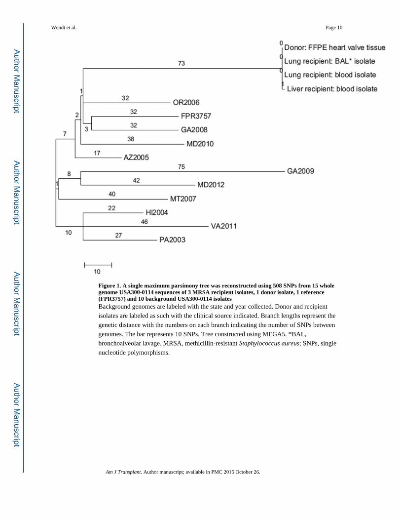
Figure 1. A single maximum parsimony tree was reconstructed using 508 SNPs from 15 whole genome USA300-0114 sequences of 3 MRSA recipient isolates, 1 donor isolate, 1 reference (FPR3757) and 10 background USA300-0114 isolatesBackground genomes are labeled with the state and year collected. Donor and recipient
isolates are labeled as such with the clinical source indicated. Branch lengths represent the
genetic distance with the numbers on each branch indicating the number of SNPs between
genomes. The bar represents 10 SNPs. Tree constructed using MEGA5. *BAL,
bronchoalveolar lavage. MRSA, methicillin-resistant Staphylococcus aureus; SNPs, single
nucleotide polymorphisms.
Wendt et al. Page 10
Am J Transplant. Author manuscript; available in PMC 2015 October 26.
Author M
anuscriptA
uthor Manuscript
Author M
anuscriptA
uthor Manuscript

Figure 2. (A) Gross appearance of a perforating bacterial vegetation on the posterior leaflet of the
mitral valve in the heart of the donor. (B) Gross appearance of a cystic lesion in the white
matter of the right cerebral hemisphere of the donor formed by a large intracerebral
hematoma. (C) Histological appearance of the mitral valve vegetation showing fibrin (pink)
admixed with innumerable clumps of bacteria (blue). Hematoxylin and eosin stain, original
magnification × 12.5. (D) Tissue Gram stain of mitral valve lesion showing coalescing
colonies of Gram-positive, coccoid bacteria. Lillie-Twort stain, original magnification ×
Wendt et al. Page 11
Am J Transplant. Author manuscript; available in PMC 2015 October 26.
Author M
anuscriptA
uthor Manuscript
Author M
anuscriptA
uthor Manuscript

100. (E) Occlusive septic embolus in a small vessel in the cerebral cortex, revealing
numerous coccoid bacteria enmeshed in fibrin. Hematoxylin and eosin stain, original
magnification × 158. (F) Same vessel as image (E), demonstrating immunohistochemical
evidence of infection with Staphylococcus aureus (red). Immunoalkaline phosphatase
technique with naphthol-fast red and hematoxylin counterstain, original magnification ×
158.
Wendt et al. Page 12
Am J Transplant. Author manuscript; available in PMC 2015 October 26.
Author M
anuscriptA
uthor Manuscript
Author M
anuscriptA
uthor Manuscript

Author M
anuscriptA
uthor Manuscript
Author M
anuscriptA
uthor Manuscript
Wendt et al. Page 13
Tab
le 1
Stap
hylo
cocc
us a
ureu
s an
timic
robi
al s
usce
ptib
ility
test
ing
resu
lts w
ith m
inim
um in
hibi
tory
con
cent
ratio
n (M
IC)
and
susc
eptib
le/in
term
edia
te/r
esis
tant
(SIR
) de
term
inat
ion
for
cultu
res
colle
cted
fro
m a
com
mon
org
an d
onor
and
lung
and
live
r re
cipi
ents
Org
an d
onor
Lun
g re
cipi
ent
Liv
er r
ecip
ient
Spec
imen
typ
e
Blo
odL
ung
biop
syB
lood
1B
AL
BA
LB
AL
BA
L1
Blo
odB
lood
1
Day
–3
Day
0D
ay 6
Day
15
Day
20
Day
43
Day
159
Day
0D
ay 7
2
Ant
ibio
tic
MIC
SIR
MIC
SIR
MIC
SIR
MIC
SIR
MIC
SIR
MIC
SIR
MIC
SIR
MIC
SIR
MIC
SIR
Clin
dam
ycin
≤0.5
S≤0
.25
S≤0
.25
S≤0
.25
S≤0
.25
S≤0
.25
S≤0
.25
S≤0
.25
S≤0
.25
S
Dap
tom
ycin
0.5
S≤0
.5S
0.5
S≤0
.5S
Gen
tam
icin
≤0.5
S≤0
.5S
≤2S
≤0.5
S≤0
.5S
≤0.5
S≤0
.5S
≤2S
Lev
oflo
xaci
n>
4R
>4
R8
R≥8
R≥8
R≥8
R≥8
R8
R
Lin
ezol
id≤2
S2
S2
S2
S2
S2
S2
S2
S2
S
Oxa
cilli
n>
2R
>2
R>
16R
≥4R
≥4R
≥4R
≥4R
>16
R
TM
P/SM
X≤2
/38
S≤1
0S
≤0.5
/9.5
S≤1
0S
≤10
S≤1
0S
≤10
S≤1
0S
≤0.5
/9.5
S
Van
com
ycin
≤2S
≤2S
≤0.5
S1
S1
S1
S1
S1.
5S
2S
BA
L, b
ronc
hoal
veol
ar la
vage
; Day
0, d
ay o
f tr
ansp
lant
; TM
P/SM
X, t
rim
etho
prim
/sul
fam
etho
xazo
le.
1 Tes
ted
at th
e C
ente
rs f
or D
isea
se C
ontr
ol a
nd P
reve
ntio
n us
ing
brot
h m
icro
dilu
tion.
Am J Transplant. Author manuscript; available in PMC 2015 October 26.
![Studies of Borate Glasses Doped with Transition Metal Ion ...shodhganga.inflibnet.ac.in/bitstream/10603/13455/14/14_chapter 4.pdftransmission [8,9], make these glasses potential candidates](https://static.fdocuments.us/doc/165x107/5f049dbd7e708231d40eda6b/studies-of-borate-glasses-doped-with-transition-metal-ion-4pdf-transmission.jpg)


