Heterotopic Ossification of the Elbow
14
Patient Presentation The formation of ectopic bone about the elbow commonly presents following elbow trauma, significant neural axis injury, or major burns with symptoms of stiffness or complete ankylosis. 1 By definition, any ossification that forms in tissues which do not typically make bone is considered ectopic or heterotopic. Patients who develop heterotopic ossification about the elbow can present with muscle, nerve, or joint related pain, but most often express frustration due to lack of passive and active motion. Loss of elbow motion leads to dra- matic impairment in upper extremity function. For example, a 50% loss of elbow motion will cause up to 80% impairment in upper extremity function. 2 The pathologic process behind ectopic bone formation has not been fully elucidated. This lack of understanding limits our ability to treat heterotopic bone formation both before and after it occurs. Certainly, the ideal management of any elbow contracture due to heterotopic ossification is prevention. Early and consistent active range of motion exercises, 3-5 medical intervention such as indomethacin and disphosphonates, 6,7 or radiation therapy 8 have all been proposed but remain unproven as potential treatment options for the prevention of heterotopic ossification about the elbow. If it does occur, surgical excision and elbow con- tracture release are viable options for regaining elbow motion. There are a few conditions that are associated with spontaneous formation of heterotopic bone about the elbow. Injury to bone, ligament, muscle, tendon, or joint capsule from the distal humerus to the proximal ulna or radius can incite local ectopic bone formation. 7,9-12 Iatrogenic trauma, such as lateral epicondylectomy or elbow arthroscopy, have been reported to cause elbow het- erotopic ossification. 13,14 Alternatively, trauma to the cen- tral nervous system (including spinal cord injury) and debilitating burns have also been identified as risk factors for spontaneous formation of heterotopic bone about the elbow. 4,5,7,15-17 Indications The primary indication for operative resection of heterotopic bone and elbow contracture release is lack of elbow motion. Once heterotopic ossification has developed and restricted elbow motion, it is nearly impossible to regain the lost motion with conservative measures, such as physical therapy, dynamic splinting, radiation therapy, or medication. Surgical resection of heterotopic bone about the elbow should be considered in patients who present with an unacceptable loss of flexion/exten- sion or pronation/supination. Restoration of a functional arc of motion is the primary goal in active individuals. From a bio- mechanical perspective, the functional arc of elbow motion has been demonstrated to be from 30° to 130° in the flexion/exten- sion plane and from 55° of pronation to 55° of supination in the plane of forearm rotation. 18 In an otherwise non-functional upper extremity, restoration of extension can even improve daily hygiene and nursing care. When considering the cosmetic appearance of the arm, one group of authors believed that achieving greater extension than 30° of flexion is needed for optimal patient satisfaction. 9 Historically, surgical excision was delayed until maturity of the heterotopic bone was proven. 19 Guides to assessing the maturity of ectopic bone included normalization of the alka- line phosphatase level, quiescent bone scan, and a solidified appearance on plain radiographs. However, recent reports of outcomes after surgical excision have used only radiographic appearance of the lesion as a determining measure of matura- tion. 11,20-23 Along with questioning the need for additional tests to ascertain the maturity of the heterotopic bone, there are case series which have investigated early excision of the immature ossification. 20,22-25 We do not believe there is suffi- cient scientific evidence to suggest that alkaline phosphatase chemistries or nuclear medicine scans are necessary to qualify the maturity (or lack thereof ) of elbow heterotopic ossification. The timing of surgical excision should be dependent on the patient’s presentation, the condition of the enveloping soft tis- sues, and the integrity of the crossing neurovascular structures. It is prudent to obtain sufficient radiographic imaging to under- American Society for Surgery of the Hand 425 CHAPTER 38 Heterotopic Ossification of the Elbow Seth D. Dodds, MD • Douglas P. Hanel, MD
-
Upload
phungkhanh -
Category
Documents
-
view
225 -
download
3
Transcript of Heterotopic Ossification of the Elbow

Patient PresentationThe formation of ectopic bone about the elbow commonlypresents following elbow trauma, significant neural axisinjury, or major burns with symptoms of stiffness or completeankylosis.1 By definition, any ossification that forms in tissueswhich do not typically make bone is considered ectopic orheterotopic. Patients who develop heterotopic ossificationabout the elbow can present with muscle, nerve, or jointrelated pain, but most often express frustration due to lack ofpassive and active motion. Loss of elbow motion leads to dra-matic impairment in upper extremity function. For example,a 50% loss of elbow motion will cause up to 80% impairmentin upper extremity function.2
The pathologic process behind ectopic bone formationhas not been fully elucidated. This lack of understandinglimits our ability to treat heterotopic bone formation bothbefore and after it occurs. Certainly, the ideal managementof any elbow contracture due to heterotopic ossification isprevention. Early and consistent active range of motionexercises,3-5 medical intervention such as indomethacinand disphosphonates,6,7 or radiation therapy8 have all beenproposed but remain unproven as potential treatmentoptions for the prevention of heterotopic ossification aboutthe elbow. If it does occur, surgical excision and elbow con-tracture release are viable options for regaining elbowmotion.
There are a few conditions that are associated withspontaneous formation of heterotopic bone about theelbow. Injury to bone, ligament, muscle, tendon, or jointcapsule from the distal humerus to the proximal ulna orradius can incite local ectopic bone formation.7,9-12
Iatrogenic trauma, such as lateral epicondylectomy orelbow arthroscopy, have been reported to cause elbow het-erotopic ossification.13,14 Alternatively, trauma to the cen-tral nervous system (including spinal cord injury) anddebilitating burns have also been identified as risk factorsfor spontaneous formation of heterotopic bone about theelbow.4,5,7,15-17
IndicationsThe primary indication for operative resection of heterotopicbone and elbow contracture release is lack of elbow motion.Once heterotopic ossification has developed and restrictedelbow motion, it is nearly impossible to regain the lost motionwith conservative measures, such as physical therapy, dynamicsplinting, radiation therapy, or medication. Surgical resection ofheterotopic bone about the elbow should be considered inpatients who present with an unacceptable loss of flexion/exten-sion or pronation/supination. Restoration of a functional arc ofmotion is the primary goal in active individuals. From a bio-mechanical perspective, the functional arc of elbow motion hasbeen demonstrated to be from 30° to 130° in the flexion/exten-sion plane and from 55° of pronation to 55° of supination inthe plane of forearm rotation.18 In an otherwise non-functionalupper extremity, restoration of extension can even improvedaily hygiene and nursing care. When considering the cosmeticappearance of the arm, one group of authors believed thatachieving greater extension than 30° of flexion is needed foroptimal patient satisfaction.9
Historically, surgical excision was delayed until maturity ofthe heterotopic bone was proven.19 Guides to assessing thematurity of ectopic bone included normalization of the alka-line phosphatase level, quiescent bone scan, and a solidifiedappearance on plain radiographs. However, recent reports ofoutcomes after surgical excision have used only radiographicappearance of the lesion as a determining measure of matura-tion.11,20-23 Along with questioning the need for additionaltests to ascertain the maturity of the heterotopic bone, thereare case series which have investigated early excision of theimmature ossification.20,22-25 We do not believe there is suffi-cient scientific evidence to suggest that alkaline phosphatasechemistries or nuclear medicine scans are necessary to qualifythe maturity (or lack thereof ) of elbow heterotopic ossification.
The timing of surgical excision should be dependent on thepatient’s presentation, the condition of the enveloping soft tis-sues, and the integrity of the crossing neurovascular structures.It is prudent to obtain sufficient radiographic imaging to under-
American Society for Surgery of the Hand 425
CHAPTER 38Heterotopic Ossification of the Elbow
Seth D. Dodds, MD • Douglas P. Hanel, MD

stand the extent of heterotopic ossification and elbow ankylosisprior to any definitive procedure. In patients with significantspinal cord or central nervous system trauma, surgical excisionshould be until maximal motor recovery occurs, which can beas long as 1 to 1.5 years after initial injury.19,26 If there are burnwounds present about the elbow, these should be entirely healedprior to creating a new incision.5,23 Neurologic, vascular, andlymphatic systems must be assessed and their status docu-mented and appropriately prioritized.27 For instance, the ulnarnerve can be encased in heterotopic bone causing cubital tun-nel symptoms which may dictate an earlier timing of sur-gery.22,27,28 The integrity of the skin and subcutaneous tissuesshould be evaluated. Lingering soft tissue swelling, edema, orerythema may all point to additional diagnoses that must bedealt with before deciding upon heterotopic bone resection. Ina majority of cases, we proceed with surgical resection when thesoft tissue envelope is quiescent.
Relevant AnatomyIt is critical to have a firm understanding of the neurovascularstructures that cross this joint, as well as their typical relation-ships to surrounding structures. Frequently, when ectopicbone is being excised about the elbow, the surgeon is workingin a previously scarred area where nerves may have been dis-sected, mobilized, or even transposed. The proximity of het-erotopic ossification to any nerve or blood vessel addsconsiderable risk to surgical excision. Patients must be awareof the potential for iatrogenic neurovascular injury prior tosigning informed consent.
The cutaneous nerves that require diligence are the poste-rior brachial cutaneous nerve and the medial and lateral ante-brachial cutaneous nerves to the forearm. The main peripheralnerves to identify are the median, ulnar, and radial, includingthe posterior interosseous nerve distally. Lastly, the operatingsurgeon must always be aware of the location of the brachialartery. The dissection and identification of these anatomicstructures will be reviewed within the description of the sur-gical technique.
Heterotopic bone can occur anywhere about the elbow,but it is typically posterior, deep to the triceps extending fromeither epicondyle to the olecranon, or anterior, associated withthe brachialis (Figures 1, 2). Evans5 believed that heterotopicbone forms and matures along a line from the olecranon tothe medial epicondylar ridge of the humerus at the medialborder of the triceps. This has been confirmed as a commonlocation by Tsionos et al.23 and McAuliffe and Wolfson22 in
more recent studies. Garland16 states the most common loca-tion for heterotopic ossification after a central neurologicinjury is either posterolateral (from the posterolateral humeralcondyle to the posterolateral olecranon, fixing the elbow at30° of flexion) or anterior within the brachialis. Another typ-ical location for ectopic bone formation about the elbow isbetween the radius and the ulna, causing a proximal radioul-nar synostosis (Figure 3).
Hastings and Graham29 proposed a classification systemof elbow ectopic bone formation. There are three groups orclasses based on functional range of motion which thoseauthors felt would be helpful in both assessment and treat-ment. Class I includes patients with positive radiographs forheterotopic ossification, but no functional limitations.Class II radiographs demonstrate heterotopic ossification,and there is a functional limitation—either in the flex-ion/extension axis (Class IIA), the pronation/supinationaxis (Class IIB), or both (Class IIC). Class III patients haveectopic bone with ankylosis either in flexion/extension(Class IIIA), pronation/supination (Class IIIB), or both(Class IIIC).1,29
Clinical Studies (Outcomes)The majority of outcome studies on surgical excision are ret-rospective reviews with small numbers of patients, as this is arelatively uncommon procedure in the realm of orthopaedicsurgery. Studies are varied in their operative approaches, aswell as in their post-operative protocols. Typically, most casesare treated on an individual basis according to each patient’sprior surgery, the condition of the overlying soft tissues, andthe surgeon’s preference for maintaining motion and prevent-ing the recurrence of heterotopic bone post-operatively.
Viola and Hanel20 reported on 15 cases of early release(24–78 weeks of initial injury) for post-traumatic elbow con-tractures with heterotopic ossification. These authors wereable to achieve a mean flexion/extension arc of 120° and amean pronation/supination arc of 152° at an average followup of 115 weeks. Here, indomethacin and continuous passivemotion were used post-operatively in most cases. Morimoto etal.24 studied nine consecutive patients who underwent earlyexcision (mean 7.7 months from initial injury) of medial-sided heterotopic ossification. Those authors found 112° offlexion and extension at an average follow up of 52 months.Recurrence of elbow stiffness did not occur despite radi-ographic evidence of spotty heterotopic ossification in five ofthe nine patients.
Wrist and Elbow Reconstruction & Arthroscopy
426 American Society for Surgery of the Hand

Ring and Jupiter21 found similar improvements in range ofmotion with their series of patients which included both post-traumatic and burn patients. However, six of their patientsrequired re-release of their elbow contractures (three post-traumatic and three post-burn). The burn patients demon-strated poorer functional outcomes and frequently wereinvolved with soft tissue coverage problems, requiring either
simultaneous or post-operative free flaps. Tsionos et al.23 had4 recurrences in their cohort of 35 post-burn patients that hadundergone surgical excision despite post-operativeindomethacin and continuous passive motion.
With regards to post-operative radiation therapy,McAuliffe and Wolfson22 used 5 doses of radiation therapy fora total of 1000 cGy and found an average of 103° of flexionand extension at an average follow up of 46 months. Thisgroup included eight patients treated with early excision(3–10 months initial injury). Multiple authors have success-fully employed single doses of peri-operative radiation (700cGy) for excision of elbow heterotopic ossification.9,30,31
None of the above studies noted a clinically significant recur-rence of the ectopic ossification.
Studies focused on the release of proximal radioulnar syn-ostosis have examined different types of interposition materialas well as the clinical outcomes of the takedown procedure.Failla et al.32 reported on 20 patients with an average followup of 40 months and found that approximately half of theachieved intra-operative motion was maintained over time.The interposition material varied in this report, but thoseauthors preferred a synthetic interposition material as out-comes were worse with biologic interpositions. Other authorshave refuted these results. Bell and Benger33 provided a reportof three successful cases of proximal radioulnar synostosistakedown followed by interposition of the anconeus. Theiroperative approach consisted of anterior and posterior capsu-
CHAPTER 38 • Heterotopic Ossification of the Elbow
American Society for Surgery of the Hand 427
Figure 1. Case 1: These anteroposterior and lateral radiographsshow complete ankylosis of the elbow at 130° of flexion after priorelbow injury.
Figure 2. Case 1: This is an intra-operative view of the ulnar nerve(marked by a penrose drain) encased in heterotopic ossification (HO) asit courses distally towards the cubital tunnel.
Figure 3. Case 3: An axial CT scan image demonstrates a proximalradioulnar synostosis after a two-incision distal biceps tendon repair.The heterotopic bone can be seen extending from the lateral aspect ofthe ulnar (U), circling around to the lateral aspect of the radius (R).

lar release as well as excision of the synostosis. Post-operatively, each patient was treated with single dose radiationtherapy (700 cGy), flexion/extension and pronation/supina-tion CPM, and indomethacin.33 Alternative soft tissue flapinterpositions include a pedicled brachioradialis flap,34 a pedi-cled adipofascial radial forearm flap,35 or a free adipofascialflap from the lateral arm.36
Jupiter and Ring11 described 18 cases of post-traumaticproximal radioulnar synostosis that underwent release with nopost-operative treatments other than physical therapy. Allpatients were treated with bone wax at the exposed osseousmargins, while eight patients had autologous fat interposition.The authors did not find the fat interposition to have anyeffect on final outcomes. At final follow up, average forearmrotation was 139° excluding one recurrence, which involved apatient with a head injury. While radiation therapy was notused in their study, Ring and Jupiter37 have advocated thistreatment modality in a more recent technique article.
Equipment and Implants for the SurgeryThe excision of ectopic bone from the elbow is a complex pro-cedure that requires pre-operative planning. For example,while it is not typical to require an implant for the surgery, atotal elbow arthroplasty, a radial head arthroplasty, and ahinged external fixator should be available. In addition, softtissue allografts, such as semitendonosis for ligamentousreconstruction or fascia lata for soft tissue interposition, maybe useful. Certain instruments not common to most handsurgery equipment sets can facilitate dissection, retraction,and heterotopic bone removal. Because of the proximity ofcritical neurovascular structures, instruments should be sharpand in good condition to allow for precision work.
In addition to operative equipment, consideration shouldbe given pre-operatively for planning a patient’s post-operativepain control and early rehabilitation. The anesthesia teamshould be consulted prior to the day of surgery so that theycan be prepared to place an axillary or infraclavicular catheterfor post-operative pain management while range of motionexercises are initiated. Though anesthesiologists frequentlyprefer placing a catheter pre-operatively, the nerve block canalso be done post-operatively to allow an assessment of neu-rologic function after the patient has awoken from anesthesia.A continuous or on-demand infusion catheter for post-operative pain control is very helpful in maintaining patientcomfort, especially if continuous passive motion is used. A
continuous passive motion machine for the elbow often needsto be ordered prior to the day of surgery so that it may beavailable in the recovery room after surgery. A thorough list-ing of the equipment and implants for elbow heterotopicbone excision surgery is shown in Table 1.
Operative ApproachA systematic approach to elbow contracture release withfocused goals (Table 2) facilitates excision of heterotopic bone.The specific surgical approach varies according to the under-lying pathology (such as the position of orthopaedic hard-ware, the location of heterotopic bone, or the presence ofcapsular contracture) and condition of the enveloping soft tis-sues (previous surgical incisions, muscle and tendon deficien-cies, nerve dysfunction, or burn contracture). Pre-operativecomputed tomography scans can be very helpful in under-standing the anatomic location of the heterotopic bone, espe-cially in cases of proximal radioulnar synostosis.
It is our preference to have the patient supine on theoperating table with the affected arm on a radiolucent handtable. Certainly if there is a stiff shoulder ipsilaterally, thepatient may also be in the lateral position with the affectedarm draped over a leg holder for posterior approaches.However, we find that draping the arm draped free on ahand table is simple and straightforward for anesthesia,nursing, and the operating surgeon. The shoulder may berotated to change the position of the elbow and transitioneasily from medial to lateral or anterior to posterior. Eitherposition is reasonable, but the chosen position must allowthe surgeon to have uncompromised access to both themedial and lateral aspects of the elbow. While it is not ourpractice to use a tourniquet during excision of heterotopicbone, a sterile tourniquet can be placed on the upper armprior to skin incision.
Typically, it is prudent to take advantage of prior skin inci-sions as long as they allow sufficient access to achieve the sur-gical goals. Fortunately, the skin overlying the elbow is richlyperfused, allowing both medial and lateral approaches to beperformed through a posterior skin incision, also termed the“global” approach to the elbow.38 The posterior incision alsoavoids the medial and lateral antebrachial cutaneous nerveswhich are susceptible to injury with medial or lateral skin inci-sions.39 Patients who have developed heterotopic ossificationsecondary to a burn injury frequently have a tenuous skin andsubcutaneous envelope about the elbow. These patients are atjeopardy for wound healing problems or skin flap necrosis
Wrist and Elbow Reconstruction & Arthroscopy
428 American Society for Surgery of the Hand

which may require free tissue transfer to cover remaining softtissue defects.21
A midline posterior incision begins the procedure. Theskin incision is typically straight, passing two centimetersmedial or lateral to the tip of the olecranon, but may be curvi-linear to incorporate previously placed surgical scars. To pre-serve the blood supply to the overlying soft tissues, fullthickness fasciocutaneous flaps are elevated from investing fas-cia of the triceps. If ulnar nerve dysfunction is present pre-operatively, a medial sided flap should be elevated to exposethe medial epicondyle of the distal humerus. From this posi-tion, the ulnar nerve can be located proximally as it passesthrough the medial intermuscular septum. The septumshould be released and excised, and an external neurolysis ofthe ulnar nerve should be performed. Free the nerve distally asit courses around the medial epicondyle and into the proximalaspect of the flexor carpi ulnaris (FCU). Once isolated, thenerve can be tagged with a vessel loop as a reminder of its loca-tion. It should be adequately freed to allow anterior transpo-sition after excision of the heterotopic bone (Figures 4, 5, 6).
Occasionally, the ulnar nerve can be found encased inectopic bone20 (Figures 1, 2). In these situations, the ulnarnerve should be identified in normal tissue proximally at thelevel of the intermuscular septum and distally between the
CHAPTER 38 • Heterotopic Ossification of the Elbow
American Society for Surgery of the Hand 429
Table 1. Soft Tissue Interpostion Flaps for Elbow HO
Operative Equipment
Standard Equipment
Preoperative radiographs and CT scan
Sterile tourniquet
Retractors, thyroid-type (long & narrow)
Lamina spreader
Vessel loops or Penrose drains
Rongeurs (including Kerrison and Pituitary)
Osteotomes and curettes
Screwdrivers for removal of retained implants
Suture anchors
Tensor fascia lata and semitendonosis allograft
Hinged elbow external fixation
Closed suction drain
Optional Equipment
Fluoroscopy
Fiber optic headlight
Total elbow arthroplasty
Radial head arthroplasty
Axillary or infraclavicular catheter
Lidocaine infusion pump
Continuous passive motion machine
Postoperative radiation therapy
Static progressive vs. dynamic splinting
Table 2. Key Principles to Operative Release of Elbow Heterotopic Ossification
• Preserve the integrity of the soft tissue envelope,use prior incisions when possible
• Identify crossing neurovascular structures• Decompress and transpose the ulnar nerve when
compressive symptoms exist• Excise heterotopic ossification from the elbow
until normal range of motion is restored• Release completely the anterior and posterior
elbow joint capsules• Preserve the collateral ligaments whenever possible• Assess intraoperative range of motion and stability

ulnar and humeral heads of the flexor carpi ulnaris. Overlyingectopic bone can then be gradually and carefully excised withrongeurs or osteotomes until the nerve is completely free.
If pre-operative radiographs and CT scan imaging demon-strate that the heterotopic bone is limited to the medial side ofthe elbow then one can proceed to medial anterior capsuleexposure and heterotopic bone resection (Figures 6–11). Onthe other hand, if the heterotopic bone is extensive and prima-
rily involving the lateral column, then we suggest identifyingand protecting the radial nerve before proceeding with the het-erotopic ossification resection. If hardware is retained about thehumeral shaft or distal humerus, then exposing and isolatingthe radial nerve is a prerequisite to safe hardware removal.
To expose the radial nerve, a lateral fasciocutaneous tissueflap is elevated in the same tissue plane as the medial dissec-tion. Frequently, the lateral branches of the posterior cuta-neous nerve of the arm are visible on the undersurface of thisflap. These branches can be followed proximally to their takeoff from the radial nerve proper. If this landmark is not read-ily available, then our second approach is to dissect the dis-tal portion of the flap towards the lateral epicondyle. Uponreaching this landmark the lateral intermuscular septum isidentified and the dissection is directed cephalad. The tri-ceps muscle belly is freed from posterior aspect of the inter-muscular septum. Small vessels and nerves seen entering thetriceps muscle should be followed proximally; they will leadto the radial nerve proper. If these markers fail to lead to thenerve, then cautiously proceed along the intermuscular sep-tum somewhere between 6 and 10 cm proximal to the tip ofthe lateral epicondyle; the nerve, surrounded by perineuralfat, will be encountered along the spiral groove as it passesthrough the lateral intermuscular septum to enter the ante-rior aspect of the arm (Figure 12). Once the radial nerve isidentified, the lateral intermuscular septum is removed and
Wrist and Elbow Reconstruction & Arthroscopy
430 American Society for Surgery of the Hand
Figure 4. Case 2: Anteroposterior and lateral radiographs demon-strate heterotopic ossification anterior to the elbow causing an anky-losed elbow with ulnar nerve dysfunction.
Figure 5. Cadaver dissection: The skin and subcutaneous layer hasbeen elevated to expose the medial musculature of the elbow. Blue ves-sel loops mark the median nerve proximal to the pronator teres and theulnar nerve distal to the medial epicondyle as it dives between thehumeral and ulnar heads of the flexor carpi ulnaris.
Figure 6. Cadaver dissection: The pronator teres is reflected todemonstrate the median nerve, brachialis, and anterior capsule (forceps).

the nerve followed distally. At the level of the elbow, thenerve can be more easily identified in the internervous planebetween the proximal aspect of the brachioradialis and thedistal aspect of the brachialis. Blunt dissection between thesetwo muscles, just superior to the joint line, will reveal theradial nerve before it dives under the supinator muscle moredistally (Figure 13). Having now identified and protected
the vulnerable ulnar and radial nerves, we then proceed withheterotopic ossification resection and release of the elbowcontracture.
Lateral ExposureThe “Column Procedure” described by Morrey40 for simplerelease of extraarticular elbow contracture affords exposure of
CHAPTER 38 • Heterotopic Ossification of the Elbow
American Society for Surgery of the Hand 431
Figure 7. Case 2: This intra-operative view shows the same medialapproach. The pronator teres and the brachialis are retracted to exposethe bridging heterotopic ossification (HO). A medial approach wasselected as this patient suffered ulnar nerve distribution paresthesiasand weakness. Lateral-sided ossification can be accessed through amedial approach.
Figure 9. Case 2: A close-up intra-operative view reveals an emptyspace after resection of the heterotopic bone. A full range of elbowmotion was achieved intra-operatively.
Figure 8. Case 2: This photograph shows the heterotopic ossifica-tion after resection.
Figure 10. Cadaver dissection: The medial triceps is retracted toexpose the posterior olecranon fossa.

both the posterior and anterior capsules of the elbow jointthrough either a limited lateral skin incision or an extensileposterior skin incision as previously described (Figures 12,13). The lateral epicondyle is localized; the lateral edge of thetriceps is freed from the lateral intermuscular septum; and thetriceps is elevated to expose posterior heterotopic bone andthe posterior capsule (Figure 14). Both the heterotopic boneand the capsule should be excised. Redundant synovium andsynovial fat within the olecranon fossa should also bedebrided. Additionally, any posterior osteophytes or redun-dant fracture callus that may be causing impingement or pre-venting full extension must be resected.
Exposure and release of the anterior capsule can be per-formed by elevating the distal or inferior origin of the bra-chioradialis and the entire origin of the extensor carpi radialislongus (ECRL) directly off the anterior aspect of the lateralepicondyle. Next, the brachialis can be elevated off of theanterior capsule and retracted anteriorly along with the bra-chioradialis, ECRL, and extensor carpi radialis brevis(ECRB), if needed. A long narrow thyroid-type retractor isvery useful in retracting these laterally-based muscles and thebrachialis off the anterior joint capsule to improve exposure.A head light may also improve visualization through this lat-eral window deep to the medial side of the elbow. This surgi-cal dissection affords access to the anterior elbow capsule(Figure 13) that can be excised under direct vision for con-tracture release. Anterior heterotopic bone typically formswithin the substance of the distal aspect of the brachialis mus-cle, just anterior to the anterior capsule of the elbow. This lat-
eral exposure allows straightforward excision of anterior het-erotopic ossification. The heterotopic bone can be carefullyelevated off the distal aspect of the anterior humerus using acombination of osteotomes and rongeurs.
Alternatively, the Kocher approach involves creating aninterval distal to the lateral epicondyle between the anconeusand the extensor carpi ulnaris (ECU), or one can exploit theinterval between the extensor carpi radialis longus (ECRL)and brevis (ECRB). Proximally, the ECRL is elevated off thelateral epicondyle along with the origin of the brachioradialis.These muscles are retracted superiorly and anteriorly to revealthe anterior capsule. This intermuscular approach betweenthe ECRL and ECRB provides exposure of the joint capsulewithout taking down the lateral collateral ligament.41
Medial ExposureIn severely contracted elbows this exposure affords uncom-promised visualization of the anterior capsule and crossingneurovascular bundles (ulnar nerve, median nerve, andbrachial artery). The medial approach begins with exposure ofthe posterior joint capsule and olecranon fossa from themedial side (Figures 5, 6, 7). The medial border of the tricepsis elevated off the intermuscular septum and medial epi-condylar ridge of the humerus. The undersurface of the mus-cle is then retracted to expose posterior heterotopicossification or the posterior capsule itself (Figure 10).
The anteromedial release takes advantage of the interner-vous plane between the ulnar innervated flexor carpi ulnaris(FCU) and the median innervated flexor-pronator muscle
Wrist and Elbow Reconstruction & Arthroscopy
432 American Society for Surgery of the Hand
Figure 11. Case 2: Intra-operative radiographs show resection ofthe bridging heterotopic bone.
Figure 12. Cadaver dissection: Elevation of the skin and subcuta-neous tissues reveals the pertinent musculature involved in the lateralapproach to the elbow. The radial nerve, coursing between the brachio-radialis and brachialis, is identified by a blue vessel loop.
group (palmaris longus, flexor carpi radialis [FCR], and prona-tor teres). Once the plane between the FCU and the flexor-pronator group has been developed and perforating vesselsligated, the common flexor-pronator origin can be elevated toimprove exposure to the brachialis and anterior joint capsule.An alternative, more limited intermuscular approach on themedial side is also possible. The pronator teres can be releasedfrom the medial epicondyle and dissected from the commonflexor origin. After the pronator teres has been elevated, theheterotopic bone can be resected along with the anterior jointcapsule (Figures 6, 7, 8, 9). Following contracture release, theflexor-pronator origin can be reattached to the medial epi-condyle with multiple suture anchors or sutured down to theepicondyle through bone tunnels. While the medial fascialborder of the triceps may be re-approximated to the fascia ofthe flexor-pronator origin, this step is not necessary. A tightmuscle layer closure often limits the motion gained in the con-tracture release. The ulnar nerve is typically transposed anteri-orly in a submuscular or subcutaneous fashion.
With a medial approach using the internervous planebetween the FCU and the flexor-pronator origin, care shouldbe taken to leave the origin of the FCU and the underlyinganterior band of the MCL undisturbed, maintaining post-operative elbow stability. Unfortunately, there are occasionswhere the heterotopic bone or elbow contracture is so severethat these structures, too, require release or excision. In suchcases, ligamentous reconstruction is necessary to avoid elbow
subluxation or dislocation while these patients are mobilizedpost-operatively. Autograft tendon (e.g., palmaris longus) maybe used for ligamentous reconstruction, but it is our prefer-ence to use allograft tendon to restore elbow stability eithermedially or laterally. Ligamentous reconstruction throughbone tunnels provides a stable anchor for early elbow motion.The restoration of stability can be augmented with sutureanchor reattachment of the flexor-pronator origin to themedial epicondyle, or of the mobile wad origin to the lateralepicondyle for lateral collateral reconstruction. If stability can-not be achieved with ligamentous reconstruction and muscleorigin repair, a dynamic external fixator should be applied.
Proximal Radioulnar SynostosisDistally, along the posterolateral edge of the proximal ulna,the entire anconeus and extensor carpi ulnaris complex can beelevated subperiosteally from the bone as needed for exposure(Figure 3). The anconeus and extensor carpi ulnaris complexare elevated off the ulna, while leaving the origin of these mus-cles from the lateral epicondyle (Figure 15). This will preventvascular injury to these muscles, especially the anconeus. Thesubperiosteal dissection is carried along the proximal ulnatowards the anterolateral surface of the radius and to the baseof the synostosis. If dissection continues in a subperiostealmanner, there should be no danger to the posteriorinterosseous nerve which is protected in the substance of thesupinator at the level of the proximal ulna. Consideration
CHAPTER 38 • Heterotopic Ossification of the Elbow
American Society for Surgery of the Hand 433
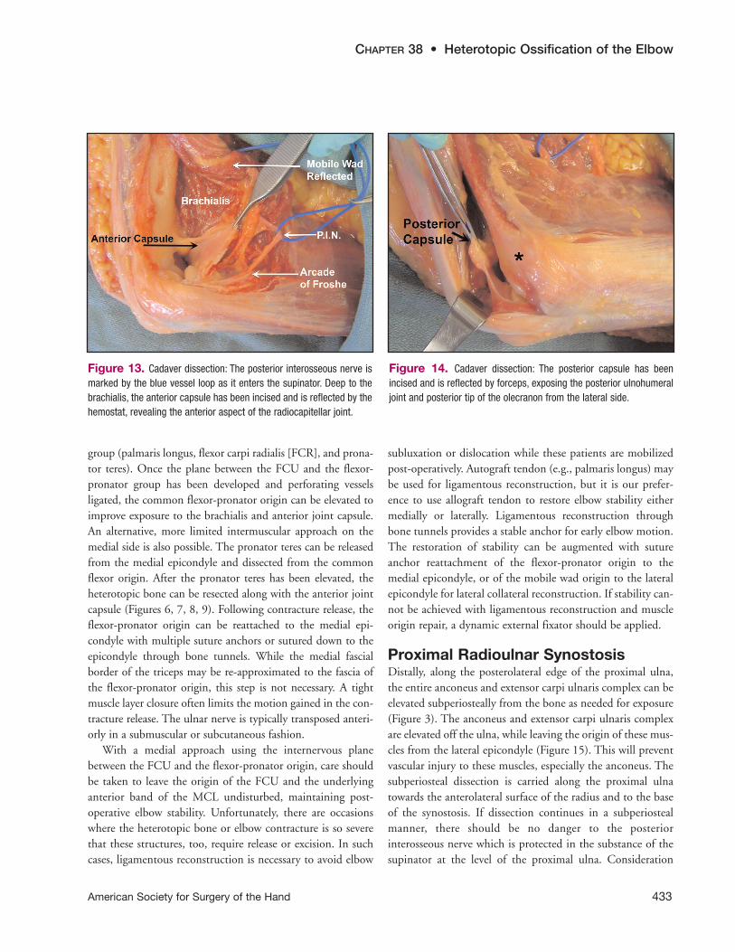
Figure 13. Cadaver dissection: The posterior interosseous nerve ismarked by the blue vessel loop as it enters the supinator. Deep to thebrachialis, the anterior capsule has been incised and is reflected by thehemostat, revealing the anterior aspect of the radiocapitellar joint.
Figure 14. Cadaver dissection: The posterior capsule has beenincised and is reflected by forceps, exposing the posterior ulnohumeraljoint and posterior tip of the olecranon from the lateral side.

should be made to locating this nerve in cases where the fore-arm is fixed in full supination. While pronation will sweep theposterior interosseous nerve anteriorly and medially awayfrom a posterolateral dissection, supination will bring thenerve closer to the field of dissection (Figures 16, 17).
Once localized, the synostosis is resected carefully with acombination of small rongeurs, osteotomes, and Kerrisonrongeurs. An appropriately-sized lamina spreader is helpful inretracting and opening up the space between the proximalradius and proximal ulna. Once the synostosis has beenreleased and there is freedom between the two bones, the lam-ina spreader offers greater direct visualization of any remain-ing ectopic bone. The synostosis should be excised down tothe native cortices of the proximal radius and proximal ulna(Figures 18, 19, 20).
Soft tissue interposition grafts or flaps are a useful adjunctin cases of complete radioulnar synostosis. Two optionsinclude a pedicled anconeus myofascial interposition flap ortensor fascia lata interposition. The pedicled anconeus flap iscreated from the posterolateral aspect of the anconeus fasciaand underlying muscle. The flap is elevated from distal toproximal off the proximal ulna up to the lateral epicondyleuntil there is sufficient freedom to mobilize the anconeus’sleading edge to the distal extent of the synostosis. By protect-ing the proximal origin and the lateral fascial attachments ofthe triceps, the primary blood supply—the medial collateralartery of the elbow—can be preserved.42 The distal end of the
flap is attached to the biceps tuberosity with the forearm infull pronation using suture anchors. Now, as the forearm issupinated the anconeus will be drawn into the proximalradioulnar space where the synostosis previously lay, creatinga thin interposition flap of viable muscle and fascia.
There are certainly cases of heterotopic ossification andradioulnar synostosis where transposing a local muscle flapmay be difficult or impossible. In such situations where analternative “bail-out” option is desired, a tensor fascia lataautograft or allograft may be interposed. The advantage toautograft is donor biocompatibility and minimal infectionrate. The disadvantages to autograft are not trivial: donor sitemorbidity, potential for early pain, hematoma, or superficialnerve injury, and additional site of scarring. Because of thesepotential complications and the reality that the interpositiongraft (whether autograft or allograft) is no longer viable afterit is harvested, the senior author has abandoned the use ofautograft tensor fascia lata in favor of allograft.
The technique is straightforward. Following synostosisexposure and resection, the tensor fascia lata allograft is shapedinto a 10–12 cm long and 4–5 cm wide graft. The allograft isthen carefully wrapped around either the ulna or the radiusand sutured into place using absorbable sutures. The choice ofwhich bone to encase with the graft depends largely on the sur-geon’s preoperative and intraoperative perceptions concerningthe origin of the synostosis. With a complete wrap the graft cansimply be sutured to itself or neighboring soft tissues to main-
Wrist and Elbow Reconstruction & Arthroscopy
434 American Society for Surgery of the Hand
Figure 15. Exposure of the proximal radioulnar joint by elevating theanconeus and the flexor carpi ulnaris (FCU) from the subcutaneous bor-der of the ulna (dotted line).
Figure 16. The anconeus and FCU are elevated as a unit and thebicipital tuberosity (B.T.) is exposed. In this cadaver dissection the fore-arm is supinated and the bicipital tuberosity is barely visible. The pos-terior interosseous nerve is at greater risk of injury in this position.

tain its position. If there is intact muscle about the proximalradius or ulna, preventing a circumferential wrap of the tensorfascia lata graft, suture anchors may be inserted at differentlocations to maximize the spread of the fascial graft. The goalsof this type of fascial interposition are twofold: 1) To providepainless forearm rotation, and 2) To act as a barrier to futurebridging heterotopic bone.
Final ConsiderationsThe excision of ectopic bone and contracture release is com-plete when normal elbow flexion and extension has beenrestored. The affected forearm should gain apronation/supination arc of at least 100°. Motion gainedshould be documented with intraoperative fluoroscopy orvideo and in the operative dictation. Documentation ofmotion is helpful after excision of the heterotopic bone; itserves as a baseline for clinical follow-up in the post-operativeperiod.
Once the heterotopic bone has been adequately excised, ajudgment must be made for the removal of indwelling hard-ware if it exists. Removal of hardware prior to excision of het-erotopic bone may result in an unintended intra-operative orpost-operative fracture. Internal fixation of the proximalradius is frequently placed through the interval describedabove for takedown of the synostosis and can be readilyremoved in those cases. Similarly, hardware about the proxi-mal ulna usually resides on its easily exposed posterior aspect.For removal of internal fixation about the distal humerus,
proximal dissection is required. A useful exposure to the distalhumerus is the bilateral tricipital or paratricipital approachdescribed by Alonso-Llames which involves opening the inter-muscular septi at the lateral and medial edges of the triceps.43
Through these paratricipital openings medial, lateral, andposterior distal humerus plates and can be removed withoutviolating the triceps insertion on the olecranon.
Upon completion, the surgical field is irrigated with sixliters of normal saline. Bleeding points in the soft tissues arecoagulated, and bleeding from exposed bone is sealed withbone wax. Routine use of closed suction drains (positionedanterior and posterior to the elbow) helps minimize soft tissueswelling and hematoma formation while the patient beginsimmediate post-operative range of motion exercises and/orcontinuous passive motion. Prior to closure of the deep soft
CHAPTER 38 • Heterotopic Ossification of the Elbow
American Society for Surgery of the Hand 435
Figure 17. With forearm pronation, the Bicipital Tuberosity (B.T.) andthe Biceps tendon become prominent. The posterior interosseous nerveis less prone to injury with forearm pronation.
Figure 18. Case 3: This intra-operative view reveals the proximalradioulnar synostosis with the heterotopic ossification (HO) marked.
Figure 19. Case 3: After the synostosis has been resected, the spacebetween the radius and ulna can be seen.
tissues, a lidocaine or bupivacaine continuous infusion pumpmay be placed to help maximize post-operative pain control(in cases where axillary catheters are not used). The catheterfor a bupivacaine infusion pump should be placed in a soft tis-sue layer or location separate from that of the closed suctiondrains. These pumps will affect the post-operative neurologicexamination as they will cause a local nerve block to neigh-boring peripheral nerves as the local anesthetic is distributedinto the soft tissues.
Divisions in the intermuscular septi are never repaired,but anterior muscle attachments to the distal humerus arealways repaired. For repairing the origins of these muscles,suture anchors or bone tunnels provide sufficient stability forbony reattachment with nonabsorbable sutures. If themedial, flexor-pronator muscle mass has been elevated, con-sideration can be made to submuscular transposition of theulnar nerve. Otherwise, a simple subcutaneous transpositionmaintained with a wide strip of flexor-pronator fascia fre-quently will suffice. If desired, the fascial layer may berepaired with an absorbable suture in a running or inter-rupted fashion. Subcutaneous and skin closure follows. Theskin closure should not be under tension, nor should it bewatertight such that it would prevent drainage when earlymotion is initiated.
Post-operative Management and RehabilitationA patient’s post-operative rehabilitation has significant influ-ence over the clinical outcome after contracture release andexcision of heterotopic bone. The affected upper extremity isimmobilized in a soft dressing that applies gentle compressionof the soft tissues. A regional anesthetic catheter placed by theanesthesia team can provide substantial pain relief in theimmediate post-operative setting. When such a catheter can-not be placed, we have used continuous bupivacaine infusionpumps to supplement intravenous narcotic pain control.With adequate analgesia, continuous passive motion of theelbow can be performed in flexion and extension or, alterna-tively, in pronation and supination for cases of isolatedradioulnar synostosis. It is our preference to use a bed-sidecontinuous passive motion machine for the elbow rather thanthe more mobile units that are able to provide motion duringambulation. The bedside units tend to maintain a constantposition, capitalizing on leverage to impart motion at thepatient’s elbow (Figure 21).
The passive motion is supplemented with frequent visits bya therapist. As soon as the first post-operative day, a supervised,daily therapy regimen that includes active, active-assisted, andpassive-assisted exercises flexion and extension exercises isimplemented. In order to prevent loss of forearm rotation,pronation, and supination, exercises are performed while flex-ing the elbow at 90° and grasping an object in the hand tominimize the contribution of radiocarpal pronation andsupination. Static progressive splinting at night is a usefuladjunct to preserve the motion gains achieved intraopera-tively.20 In cases of radioulnar synostosis, splints may be alter-nated nightly between static pronation and static supination.44
Radiation treatment of the resected bed of heterotopic ossifi-cation or synostosis has been touted to be successful in prevent-ing recurrence of ectopic bone.22,30,45 However, to ourknowledge no prospective randomized trial has shown thismodality to be better than resection alone. As this localized radi-ation procedure comes with some inherent risk of wound break-down, neuritis, lymphedema, and a remote risk of sarcoma, itshould be discussed with patients pre-operatively. A single doseof 700 cGy within the first 24 to 48 hours post-operatively istypically sufficient. In our practice, radiation therapy is limitedto patients with involvement of the proximal radioulnar joint.
Wrist and Elbow Reconstruction & Arthroscopy
436 American Society for Surgery of the Hand
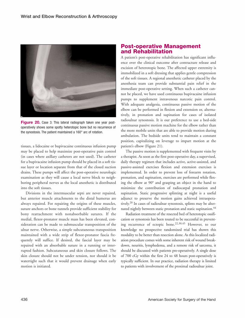
Figure 20. Case 3: This lateral radiograph taken one year post-operatively shows some spotty heterotopic bone but no recurrence ofthe synostosis. The patient maintained a 160° arc of rotation.

Indomethacin and other nonsteroidal anti-inflammatorymedicines are also considered useful adjuncts in the preven-tion of heterotopic ossification.46 Similar to radiation therapy,the specific agent, dosage, duration, and benefit of treatmentare not consistent among published studies.46 We do not pre-scribe indomethacin in our patients. We do prescribe intra-venous ketorolac during a patient’s hospital stay, primarily forpain control.
ConclusionExcision of heterotopic bone and release of contracture orankylosis about the elbow requires a sound knowledge ofanatomy. A posterior incision allows a “global” approach toboth the medial and lateral sides of the elbow. Anterior andposterior heterotopic bone can be resected using eitherapproach. Substantial improvement in elbow flexion andextension as well as pronation and supination can be obtainedwith this procedure. Every patient is treated with an aggressiverehabilitation protocol that includes continuous passivemotion. Adjuvant treatments such as indomethacin or radia-tion therapy are utilized on a case-by-case basis. While somemotion loss from the significant gains made intra-operativelycan be expected, a majority of patients who actively partici-pate in their daily motion exercises will achieve an optimaloutcome by preserving a functional arc of motion.
CHAPTER 38 • Heterotopic Ossification of the Elbow
American Society for Surgery of the Hand 437
Figure 21. Post-operatively, an elbow continuous passive motionmachine is used in conjunction with a regional anesthetic catheter.
References1. Viola RW, H Hastings, 2nd. Treatment of ectopic ossification about the
elbow. Clin Orthop Relat Res 2000 (370):65-86.
2. Sojbjerg JO. The stiff elbow. Acta Orthop Scand 1996; 67(6):626-631.
3. Crawford CM, G Varghese, MM Mani, JR Neff. Heterotopic ossifica-
tion: are range of motion exercises contraindicated? J Burn Care Rehabil
1986; 7(4):323-327.
4. Peterson SL, MM Mani, CM Crawford, JR Neff, JM Hiebert. Post-
burn heterotopic ossification: insights for management decision mak-
ing. J Trauma 1989; 29(3):365-369.
5. Evans EB. Heterotopic bone formation in thermal burns. Clin Orthop
Relat Res 1991 (263):94-101.
6. Nollen AJ. Effects of ethylhydroxydiphosphonate (EHDP) on hetero-
topic ossification. Acta Orthop Scand 1986; 57(4):358-361.
7. Hurvitz EA, BR Mandac, G Davidoff, JH Johnson, VS Nelson. Risk
factors for heterotopic ossification in children and adolescents with
severe traumatic brain injury. Arch Phys Med Rehabil 1992; 73(5):459-
462.
8. Stein DA, R Patel, KA Egol, FT Kaplan, NC Tejwani, KJ Koval.
Prevention of heterotopic ossification at the elbow following trauma
using radiation therapy. Bull Hosp Jt Dis 2003; 61(3-4):151-154.
9. Park MJ, HG Kim, JY Lee. Surgical treatment of post-traumatic stiff-
ness of the elbow. J Bone Joint Surg Br 2004; 86(8):1158-1162.
10. Ilahi OA, DW Strausser, GT Gabel. Post-traumatic heterotopic ossifi-
cation about the elbow. Orthopedics 1998; 21(3):265-268.
11. Jupiter JB, D Ring. Operative treatment of post-traumatic proximal
radioulnar synostosis. J Bone Joint Surg Am 1998; 80(2):248-257.
12. Jupiter JB. Complex fractures of the distal part of the humerus and
associated complications. Instr Course Lect 1995; 44:187-198.
13. Cushing M, GM Lourie, DV Miller, JA Hohn. Heterotopic ossification
after lateral epicondylectomy. J South Orthop Assoc 2001; 10(1):53-56.
14. Gofton WT, GJ King. Heterotopic ossification following elbow
arthroscopy. Arthroscopy 2001; 17(1):E2.
15. Garland DE, CE Blum, RL Waters. Periarticular heterotopic ossifica-
tion in head-injured adults. Incidence and location. J Bone Joint Surg
Am 1980; 62(7): 1143-1146.
16. Garland DE. Surgical approaches for resection of heterotopic ossifica-
tion in traumatic brain-injured adults. Clin Orthop Relat Res 1991
(263): 59-70.
17. Garland DE, JF Orwin. Resection of heterotopic ossification in patients
with spinal cord injuries. Clin Orthop Relat Res 1989 (242):169-176.
18. Morrey BF, LJ Askew, EY Chao. A biomechanical study of normal func-
tional elbow motion. J Bone Joint Surg Am 1981; 63(6):872-877.
19. Garland DE. A clinical perspective on common forms of acquired het-
erotopic ossification. Clin Orthop Relat Res 1991 (263):13-29.
20. Viola RW, DP Hanel. Early “simple” release of posttraumatic elbow
contracture associated with heterotopic ossification. J Hand Surg [Am]
1999; 24(2):370-380.
21. Ring D, JB Jupiter. Operative release of complete ankylosis of the elbow
due to heterotopic bone in patients without severe injury of the central
nervous system. J Bone Joint Surg Am 2003; 85-A(5):849-857.
22. McAuliffe JA, AH Wolfson. Early excision of heterotopic ossification
about the elbow followed by radiation therapy. J Bone Joint Surg Am
1997; 79(5):749-755.
23. Tsionos I, C Leclercq, JM Rochet. Heterotopic ossification of the elbow
in patients with burns. Results after early excision. J Bone Joint Surg Br
2004; 86(3):396-403.
24. Moritomo H, K Tada, T Yoshida. Early, wide excision of heterotopic
ossification in the medial elbow. J Shoulder Elbow Surg 2001;
10(2):164-168.
25. Peters WJ. Heterotopic ossification: can early surgery be performed,
with a positive bone scan? J Burn Care Rehabil 1990; 11(4):318-321.
26. Garland DE, DA Hanscom, MA Keenan, C Smith, T Moore. Resection
of heterotopic ossification in the adult with head trauma. J Bone Joint
Surg Am 1985; 67(8):1261-1269.
27. Varghese G, K Williams, A Desmet, JB Redford. Nonarticular compli-
cation of heterotopic ossification: a clinical review. Arch Phys Med
Rehabil 1991; 72(12):1009-1013.
28. Vorenkamp SE, TL Nelson. Ulnar nerve entrapment due to heterotopic
bone formation after a severe burn. J Hand Surg [Am] 1987; 12(3):378-
380.
29. Hastings H, 2nd, TJ Graham. The classification and treatment of het-
erotopic ossification about the elbow and forearm. Hand Clin 1994;
10(3):417-437.
30. Poggi MM, BE Thomas, PA Johnstone. Excision and radiotherapy for
heterotopic ossification of the elbow. Orthopedics 1999; 22(11):1059-
1061.
31. Heyd R, G Strassmann, B Schopohl,, N Zamboglou. Radiation therapy
for the prevention of heterotopic ossification at the elbow. J Bone Joint
Surg Br 2001; 83(3):332-334.
32. Failla JM, PC Amadio, BF Morrey. Post-traumatic proximal radio-ulnar
synostosis. Results of surgical treatment. J Bone Joint Surg Am 1989;
71(8):1208-1213.
33. Bell SN, D Benger. Management of radioulnar synostosis with mobi-
lization, anconeus interposition, and a forearm rotation assist splint.
J Shoulder Elbow Surg 1999; 8(6):621-624.
34. Fernandez DL, E Joneschild. “Wrap around” pedicled muscle flaps for
the treatment of recurrent forearm synostosis. Tech Hand Upper
Extremity Surg 2004; 8(2):102-109.
35. Jones NF, A Esmail, EK Shin. Treatment of radioulnar synostosis by
radical excision and interposition of a radial forearm adipofascial flap.
J Hand Surg [Am] 2004; 29(6):1143-1147.
36. Kanaya F, K Ibaraki. Mobilization of a congenital proximal radioulnar
synostosis with use of a free vascularized fascio-fat graft. J Bone Joint
Surg Am 1998; 80(8):1186-1192.
37. Ring D, JB Jupiter. Operative release of ankylosis of the elbow due to
heterotopic ossification. Surgical technique. J Bone Joint Surg Am 2004;
86-A Suppl 1:2-10.
38. Patterson SD, GI Bain, JA Mehta. Surgical approaches to the elbow.
Clin Orthop Relat Res 2000 (370):19-33.
39. Dowdy PA, GI Bain, GJ King, SD Patterson. The midline posterior
elbow incision. An anatomical appraisal. J Bone Joint Surg Br 1995;
77(5):696-699.
40. Morrey BF. Surgical treatment of extraarticular elbow contracture. Clin
Orthop Relat Res 2000 (370):57-64.
41. Cohen MS, H Hastings, 2nd. Post-traumatic contracture of the elbow.
Operative release using a lateral collateral ligament sparing approach.
J Bone Joint Surg Br 1998; 80(5):805-812.
42. Schmidt CC, GN Kohut, JA Greenberg, SE Kann, RS Idler, TR
Kiefhaber. The anconeus muscle flap: its anatomy and clinical applica-
tion. J Hand Surg [Am] 1999; 24(2):359-369.
43. Alonso-Llames M. Bilaterotricipital approach to the elbow. Its applica-
tion in the osteosynthesis of supracondylar fractures of the humerus in
children. Acta Orthop Scand 1972; 43(6):479-490.
44. Kamineni S, NG Maritz, BF Morrey. Proximal radial resection for post-
traumatic radioulnar synostosis: a new technique to improve forearm
rotation. J Bone Joint Surg Am 2002; 84-A(5):745-751.
45. Beingessner DM, SD Patterson, GJ King. Early excision of heterotopic
bone in the forearm. J Hand Surg [Am] 2000; 25(3):483-488.
46. Fransen M, B Neal. Non-steroidal anti-inflammatory drugs for pre-
venting heterotopic bone formation after hip arthroplasty. Cochrane
Database Syst Rev 2004 (3):CD001160.
Wrist and Elbow Reconstruction & Arthroscopy
438 American Society for Surgery of the Hand