Hiatus Hernia Treatment In Chennai | Hernia Treatment Center In India
Upload
avid-listenerCategory
view
42download
0
LECTURE FOR LECTURE FOR MBChB VMBChB V
HERNIA
Definition:
Protrusion of a viscus or part of it from thecavity which it is enclosed through an abnormality or point of weakness in the wall of the cavity.
It is the commonest surgical condition worldwide with a prevalence of 6%.
Commonest cause of intestinal obstruction in the developing world ≈ 75%.

Types of Hernia
1. Inguinal hernia – 80-90% - conformed in all communities.
2. Fermoral – 2-5% - More commoner in females
3. Umbilical Hernia – Common after neonatal sepsis. More in developing countries.
Developmental/genetic factors involved.
Seen more in blacks.
4. Paraumbilical Hernia
5. Epigastric Hernia – 1%
6. Incisional Hernia – 5%
Commonest in poor communities with poor facilities

Cont
7. Spigelian Hernia – Between muscle fibres and apaneurosis of of transverse abdominis.
8. Lumbar Hernia – At the superior or inferior lumbar triangle
9. Obturator herniae
10.Sciatic herniae

Aetiology:
Two main factors
A – Defect or weakness of the wall of the cavity
(i) Embryological/Anatomical Internal inguinal ring Femoral ring Obturator canal
(ii)Acquired factors Ageing – muscle weakness Infection of scar or poor surgical technique Frequent or multiple pregnancies Obesity Nerve injury at operation
Injury to the abdomen

B. Increased Intraabdominal pressure.Chronic coughChronic urinary obstructionChronic constipationHeavy manual workFrequent deliveries
GROIN HERNIAEInguinal/Femoral

Inguinal Canal:Contents Spermatic cord + vessels Ilionguinal nerve Genital bb of genitofemoral nerveIn female – Round ligament
It is a passage for gubernaculum testis to the scrotum in males and to labium majus in the Females.

It is about 4cm long and lies obliquely.
Its boundaries are:
1. Internal Ring: U-shaped and made of transversalis fascie.
Is about 2.5cm above mid-inguinal point width of 0.5 – 1cm.
Usually closes when there is increased abdominal pressure.
2. External Ring:
Is an opening in the ext. oblique apaneurosis

3. Anterior Wall
External oblique apaneurosis
Internal oblique fibres laterally
4. Posterior Wall
Laterally – Transversalis fascie + aparemosis of tranversus abdominis
Medially – Conjoint Tendon
5. Floor of the canal – Inguinal Ligament
6. Roof: Arched fibres of internal oblique
Hasselbach’s Triangle is formed with:
1.Inferior epigastric vessels laterally
2.Conjoint tendon – medially
3.Inguinal ligament – base
Inguinal hernia – Commonest in both sexes
Males - 95%
Females – 50%
M:F 20:1

Direct/Indirect hernia
Indirect – Affects all age groups
- Commonest 20-50 years (60%). Commonest on right than left 2.1 due to late descent of testis.
- About 10% are bilateral
Direct hernia – Right is as common as left. 10% of hernia of those >25 years.
Clinical Fxs:
1.Swelling in the groin – Reducible usually
2.Pain – can occur but is rare
Examination:
1.Visble swelling
2.Palpable cough impulse
3.Is reducible
Complete – Enters scrotum
Bubonocele – Limited to canal
Funicular – Above pubic tubercle
Complications
1.Irreducibility
2.Strengthening/Gangrene formation
3.Fistula formation
4.Rapture of the sac-enviscesation of hernia
Anatomical Complications
1.Ritchers hernia
2.Sliding hernia
3.Hernia en W
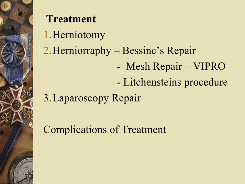
Treatment
1.Herniotomy
2.Herniorraphy – Bessinc’s Repair
- Mesh Repair – VIPRO
- Litchensteins procedure
3.Laparoscopy Repair
Complications of Treatment