Hereditary disorders of carbohydrate metabolism · metabolism (calories from fat/non-fat sources :...
60
Hereditary disorders of carbohydrate metabolism
Transcript of Hereditary disorders of carbohydrate metabolism · metabolism (calories from fat/non-fat sources :...
Hereditary disorders of carbohydrate metabolism
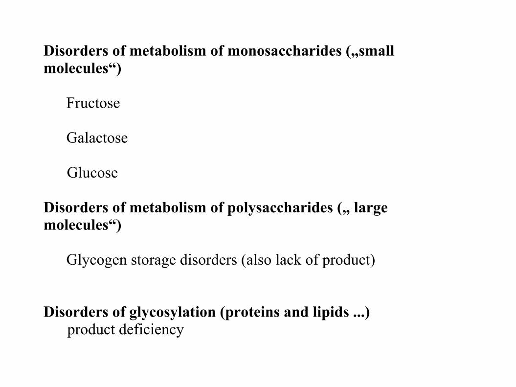
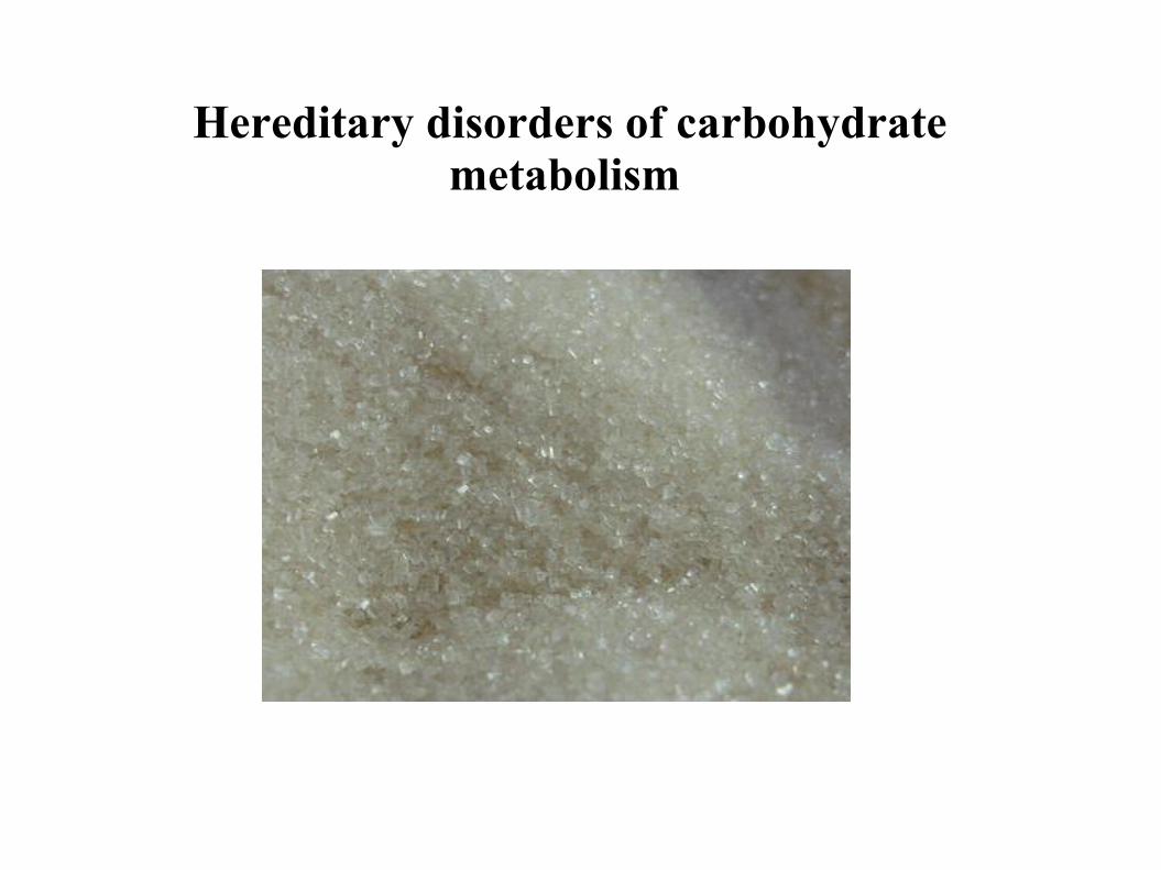
Disorders of metabolism of monosaccharides („small molecules“)
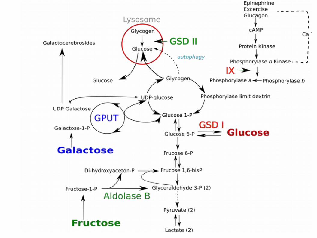
Fructose
Galactose Glucose
Disorders of metabolism of polysaccharides („ large molecules“)
Glycogen storage disorders (also lack of product)
Disorders of glycosylation (proteins and lipids ...)product deficiency
Inherited disorders of fructose metabolism
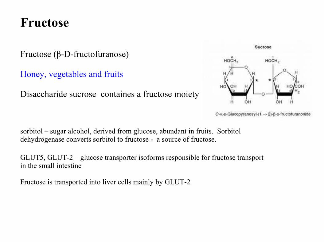
Fructose
Fructose (β-D-fructofuranose)
Honey, vegetables and fruits
Disaccharide sucrose containes a fructose moiety
sorbitol – sugar alcohol, derived from glucose, abundant in fruits. Sorbitol dehydrogenase converts sorbitol to fructose - a source of fructose.
GLUT5, GLUT-2 – glucose transporter isoforms responsible for fructose transport in the small intestine
Fructose is transported into liver cells mainly by GLUT-2
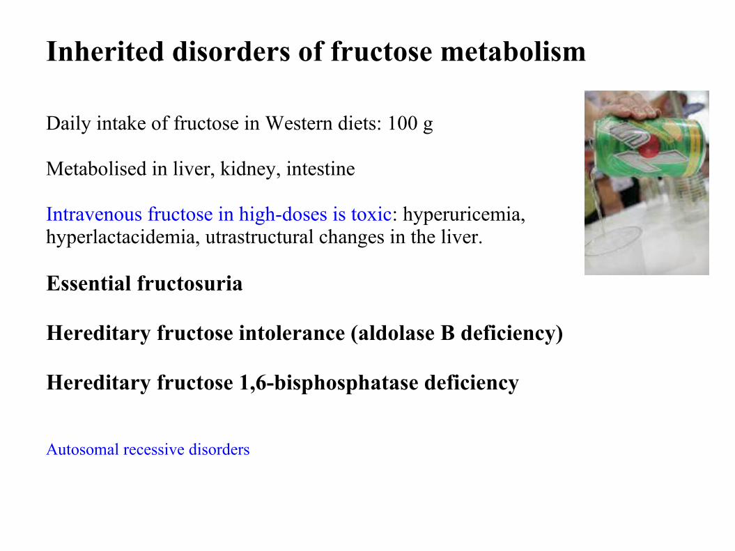
Inherited disorders of fructose metabolism
Daily intake of fructose in Western diets: 100 g
Metabolised in liver, kidney, intestine
Intravenous fructose in high-doses is toxic: hyperuricemia, hyperlactacidemia, utrastructural changes in the liver.
Essential fructosuria
Hereditary fructose intolerance (aldolase B deficiency)
Hereditary fructose 1,6-bisphosphatase deficiency
Autosomal recessive disorders
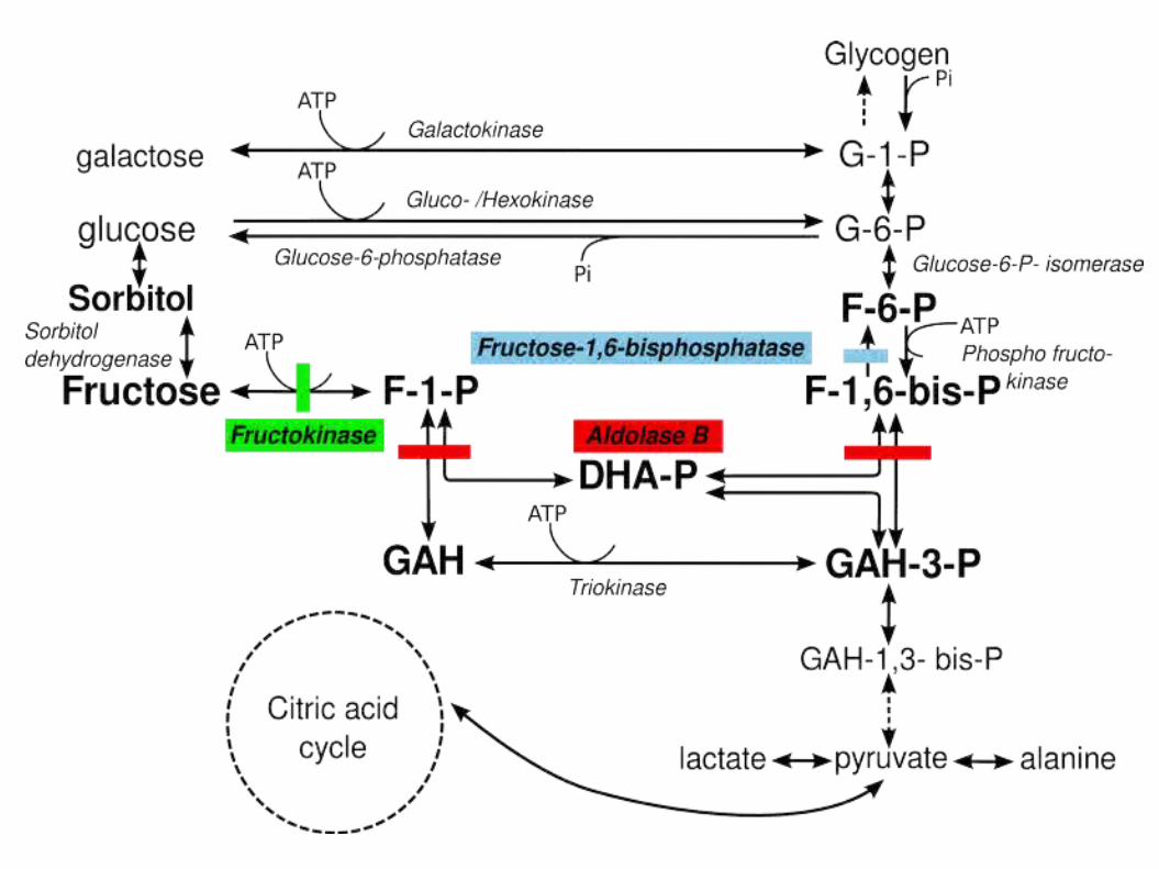
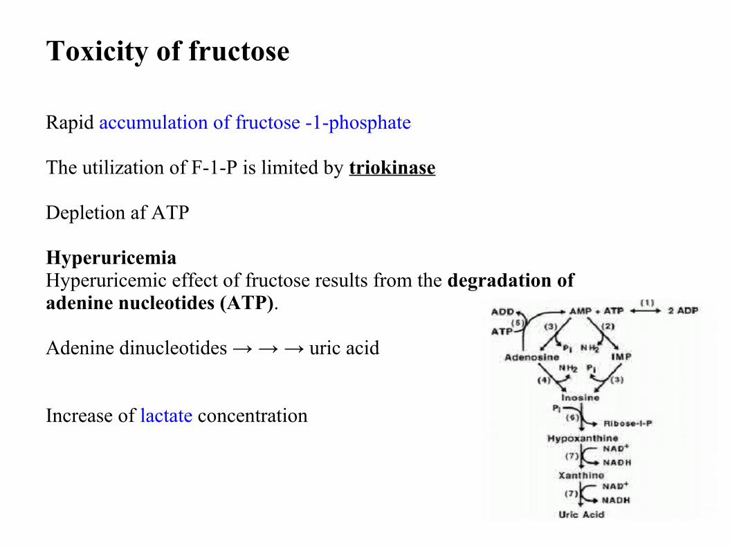
Toxicity of fructose
Rapid accumulation of fructose -1-phosphate
The utilization of F-1-P is limited by triokinase
Depletion af ATP
HyperuricemiaHyperuricemic effect of fructose results from the degradation of adenine nucleotides (ATP).
Adenine dinucleotides → → → uric acid
Increase of lactate concentration
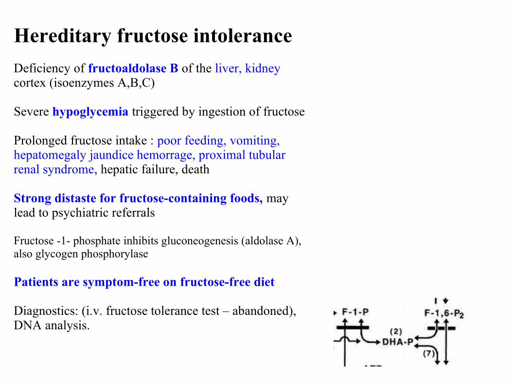
Hereditary fructose intolerance
Deficiency of fructoaldolase B of the liver, kidney cortex (isoenzymes A,B,C)
Severe hypoglycemia triggered by ingestion of fructose
Prolonged fructose intake : poor feeding, vomiting, hepatomegaly jaundice hemorrage, proximal tubular renal syndrome, hepatic failure, death
Strong distaste for fructose-containing foods, may lead to psychiatric referrals
Fructose -1- phosphate inhibits gluconeogenesis (aldolase A), also glycogen phosphorylase
Patients are symptom-free on fructose-free diet
Diagnostics: (i.v. fructose tolerance test – abandoned), DNA analysis.
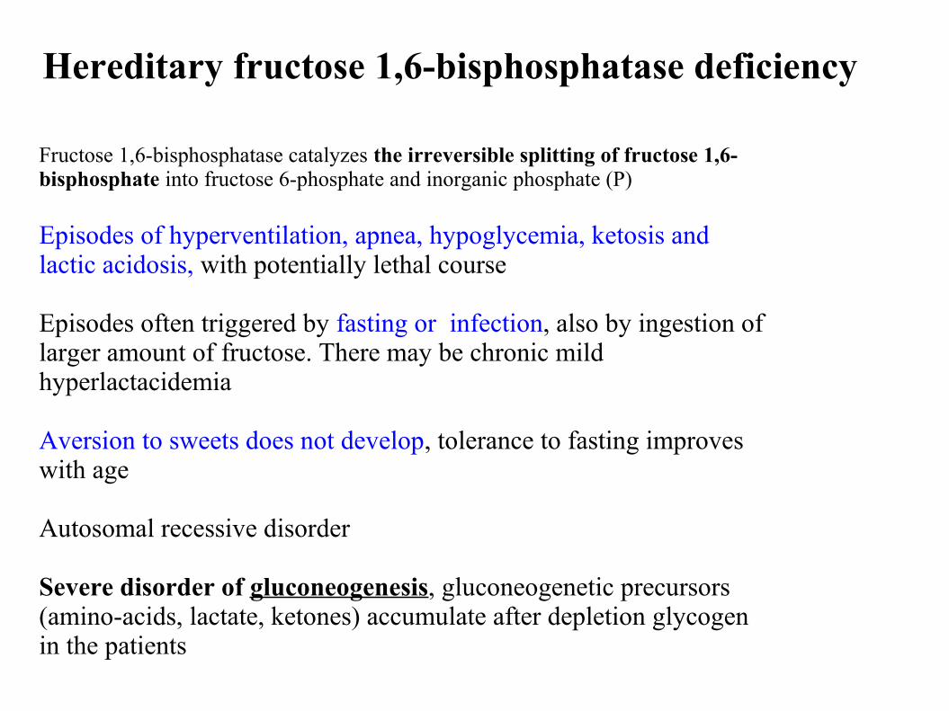
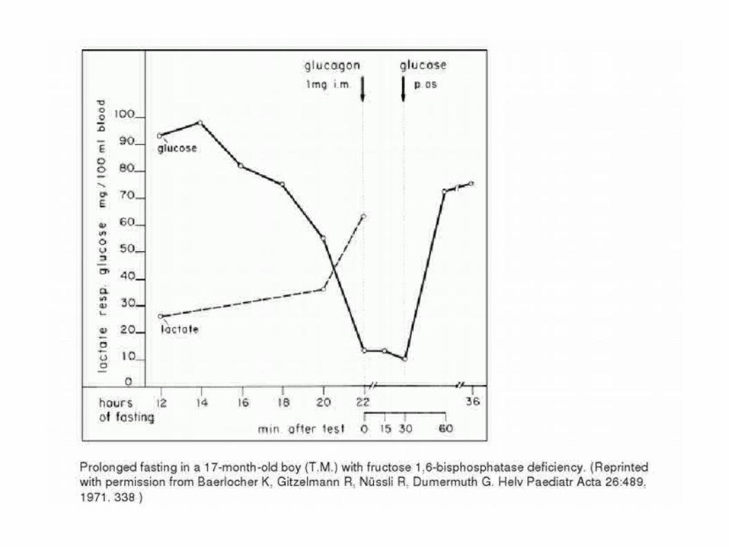
Hereditary fructose 1,6-bisphosphatase deficiency
Fructose 1,6-bisphosphatase catalyzes the irreversible splitting of fructose 1,6-bisphosphate into fructose 6-phosphate and inorganic phosphate (P)
Episodes of hyperventilation, apnea, hypoglycemia, ketosis and lactic acidosis, with potentially lethal course
Episodes often triggered by fasting or infection, also by ingestion of larger amount of fructose. There may be chronic mild hyperlactacidemia
Aversion to sweets does not develop, tolerance to fasting improves with age
Autosomal recessive disorder
Severe disorder of gluconeogenesis, gluconeogenetic precursors (amino-acids, lactate, ketones) accumulate after depletion glycogen in the patients
Essential fructosuria
Deficiency of liver fructokinase
Asymptomatic metabolic anomaly - benign
Hyperfructosemia and hyperfructosuria – important for differential diagnosis of other fructose metabolism disorders
Hereditary disorders of glucose transport
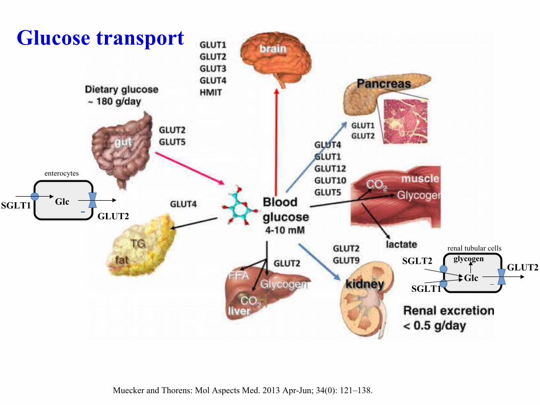
Hydrophillic glucose molecule does not cross easily lipohillic cellular membranes -> transporters are necessary to carry glucose across membranes
2 types of transporters:
a) Sodium-dependent glucose transporters (SGLTs, 'active' tranporters encoded by SLC5 family members) – couple sugar transport to sodium electrochemical gradient -> can transport against concentration gradient
b) Facilitative glucose transporters (GLUTs, uniporter systems, 'passive' transporters) – transport glucose only along an existing gradient.
Glucose transport
Glucose transport
Muecker and Thorens: Mol Aspects Med. 2013 Apr-Jun; 34(0): 121–138.
SGLT1GLUT2
Glc
enterocytes
renal tubular cells
GLUT2
SGLT1Glc
SGLT2 glycogen
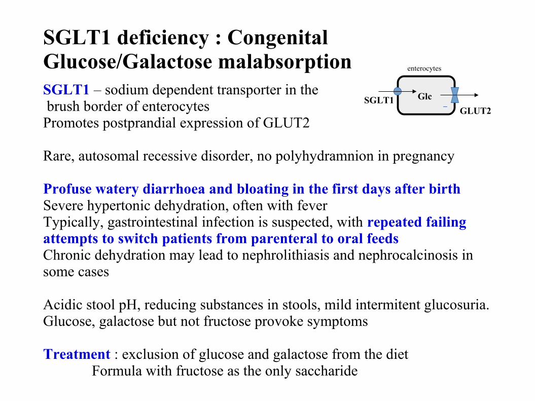
SGLT1 – sodium dependent transporter in the brush border of enterocytes Promotes postprandial expression of GLUT2
Rare, autosomal recessive disorder, no polyhydramnion in pregnancy
Profuse watery diarrhoea and bloating in the first days after birth Severe hypertonic dehydration, often with fever Typically, gastrointestinal infection is suspected, with repeated failing attempts to switch patients from parenteral to oral feedsChronic dehydration may lead to nephrolithiasis and nephrocalcinosis in some cases
Acidic stool pH, reducing substances in stools, mild intermitent glucosuria.Glucose, galactose but not fructose provoke symptoms
Treatment : exclusion of glucose and galactose from the dietFormula with fructose as the only saccharide
SGLT1 deficiency : Congenital Glucose/Galactose malabsorption
SGLT1GLUT2
Glc
enterocytes
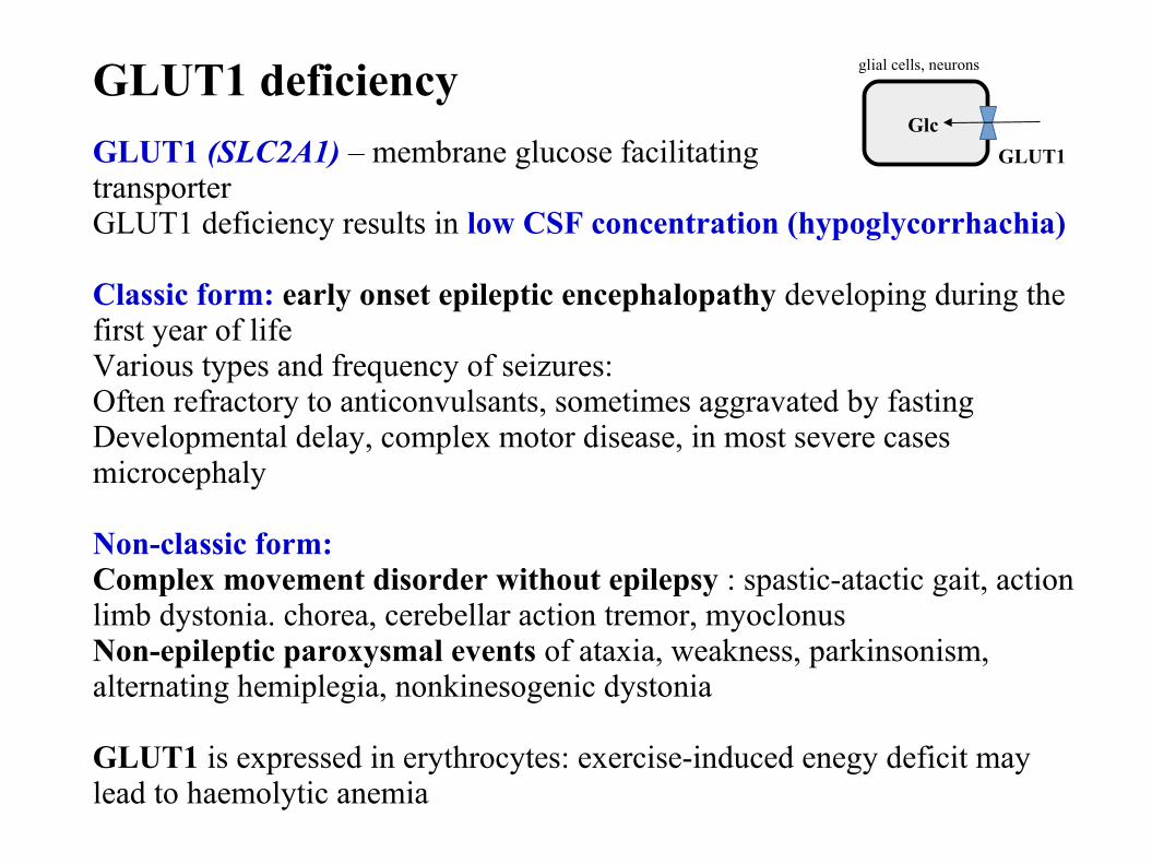
GLUT1 (SLC2A1) – membrane glucose facilitating transporter GLUT1 deficiency results in low CSF concentration (hypoglycorrhachia)
Classic form: early onset epileptic encephalopathy developing during the first year of life Various types and frequency of seizures: Often refractory to anticonvulsants, sometimes aggravated by fastingDevelopmental delay, complex motor disease, in most severe cases microcephaly
Non-classic form: Complex movement disorder without epilepsy : spastic-atactic gait, action limb dystonia. chorea, cerebellar action tremor, myoclonusNon-epileptic paroxysmal events of ataxia, weakness, parkinsonism, alternating hemiplegia, nonkinesogenic dystonia
GLUT1 is expressed in erythrocytes: exercise-induced enegy deficit may lead to haemolytic anemia
GLUT1 deficiency
GLUT1
Glc
glial cells, neurons
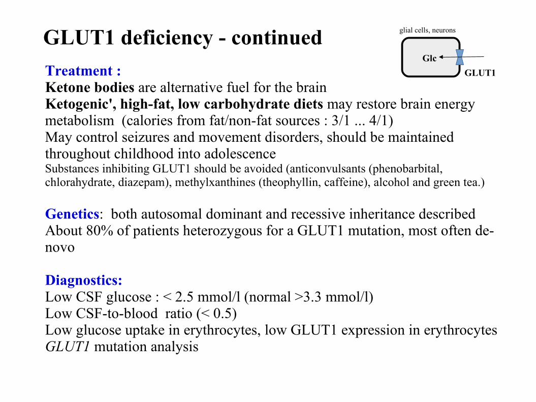
Treatment : Ketone bodies are alternative fuel for the brainKetogenic', high-fat, low carbohydrate diets may restore brain energy metabolism (calories from fat/non-fat sources : 3/1 ... 4/1)May control seizures and movement disorders, should be maintained throughout childhood into adolescence Substances inhibiting GLUT1 should be avoided (anticonvulsants (phenobarbital, chlorahydrate, diazepam), methylxanthines (theophyllin, caffeine), alcohol and green tea.) Genetics: both autosomal dominant and recessive inheritance describedAbout 80% of patients heterozygous for a GLUT1 mutation, most often de-novo
Diagnostics: Low CSF glucose : < 2.5 mmol/l (normal >3.3 mmol/l) Low CSF-to-blood ratio (< 0.5)Low glucose uptake in erythrocytes, low GLUT1 expression in erythrocytes GLUT1 mutation analysis
GLUT1 deficiency - continued
GLUT1
Glc
glial cells, neurons
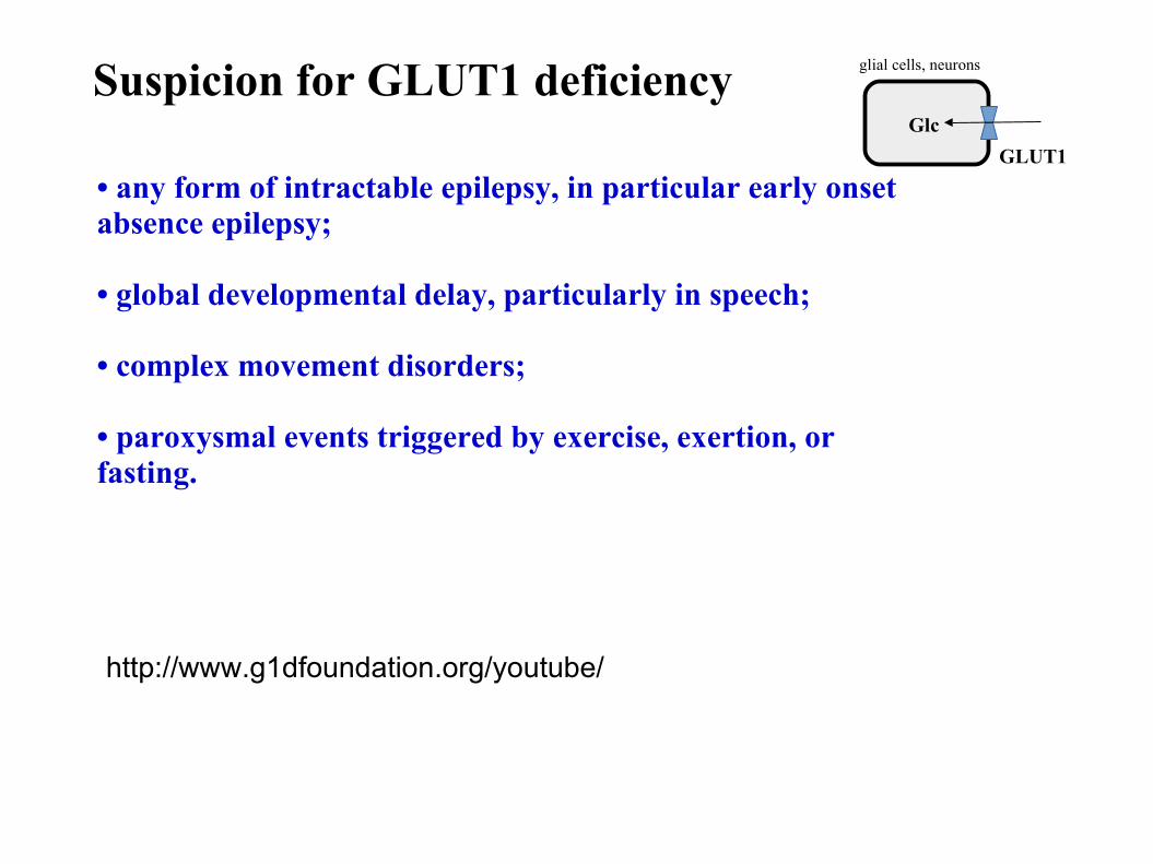
• any form of intractable epilepsy, in particular early onsetabsence epilepsy;
• global developmental delay, particularly in speech;
• complex movement disorders;
• paroxysmal events triggered by exercise, exertion, orfasting.
Suspicion for GLUT1 deficiency
GLUT1
Glc
glial cells, neurons
http://www.g1dfoundation.org/youtube/
Hereditary disorders of galactose metabolism
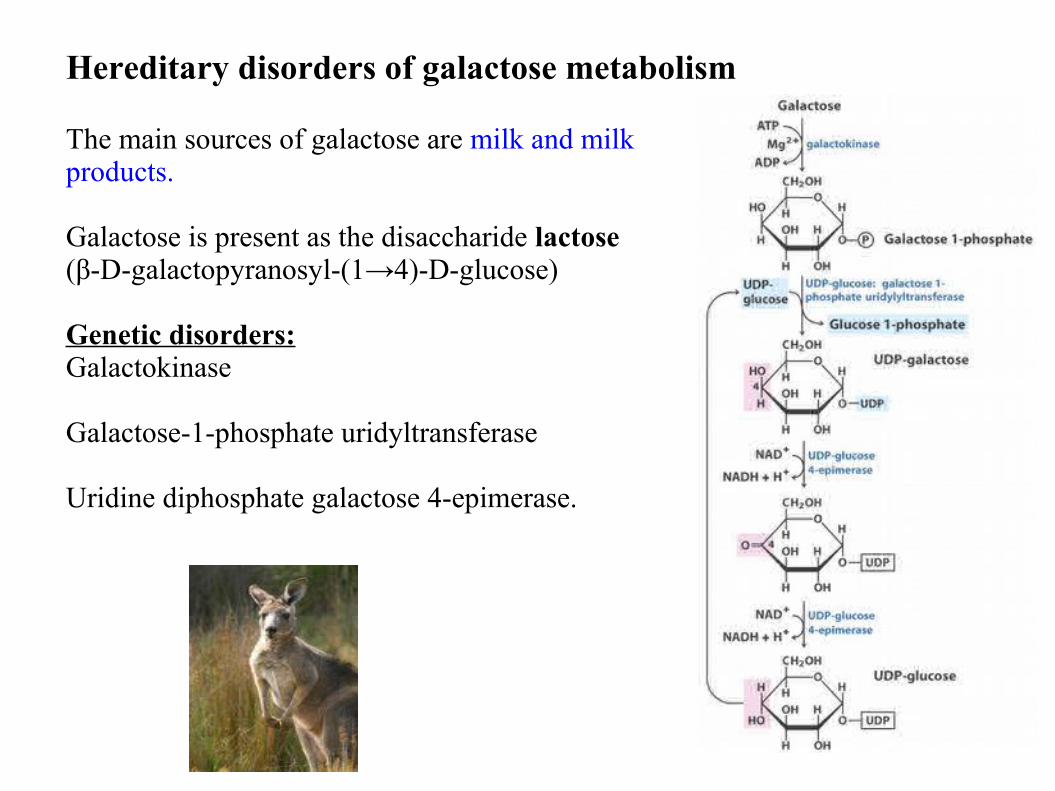
The main sources of galactose are milk and milk products.
Galactose is present as the disaccharide lactose (β-D-galactopyranosyl-(1→4)-D-glucose)
Genetic disorders:Galactokinase
Galactose-1-phosphate uridyltransferase
Uridine diphosphate galactose 4-epimerase.
Hereditary disorders of galactose metabolism
In the first weeks of life: poor feeding and weight loss, vomiting, diarrhea, lethargy,and hypotonia.
Severe liver dysfunction, hepatomegaly, icterus (often conjugated hyperbilirubinemia), bleeding diathesis, septicemia, renal tubular syndrome
Cataracts : osmotic oedema of the lens due to galactitol
Elevated galactose, galactitol, galactose-1-phosphate
Late complications Cognitive defectsOvarian failure in femalesAtaxia, low bone density.
AR, incidence 1:40 000- 60 000, Neonatal screening for galactose in some countries
Variants (Duarte)
Classical galactosemia: galactose-1-phosphate uridyltransferase deficiency
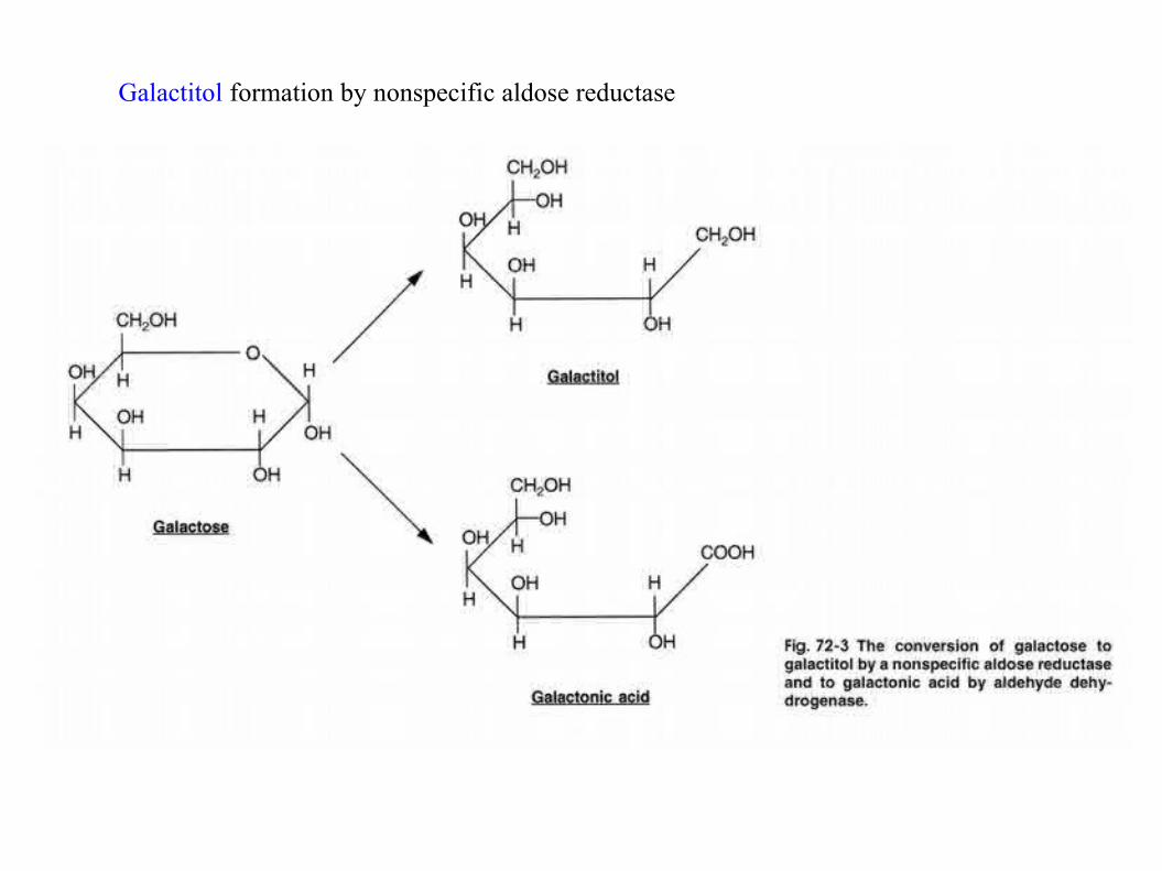
Galactitol formation by nonspecific aldose reductase
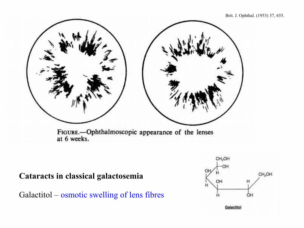
Cataracts in classical galactosemia
Galactitol – osmotic swelling of lens fibres
Brit. J. Ophthal. (1953) 37, 655.
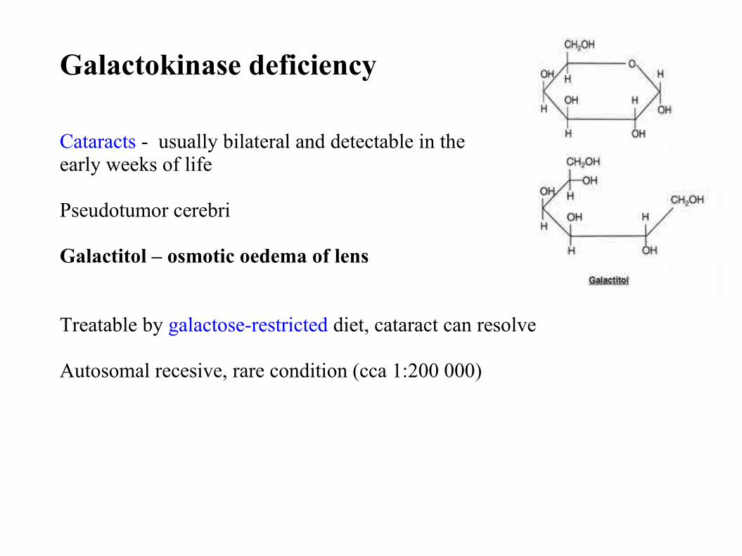
Cataracts - usually bilateral and detectable in theearly weeks of life
Pseudotumor cerebri
Galactitol – osmotic oedema of lens
Treatable by galactose-restricted diet, cataract can resolve
Autosomal recesive, rare condition (cca 1:200 000)
Galactokinase deficiency
Severe form:
Severe deficiency of epimerase activityExtremely rare
Newborns with vomiting, hepatopathy resembling classical galactosemia. Mental retardation
Mild form:
Partial deficiency of epimerase deficiencyIn most patients apparently benign condition
Intermediate forms
Autosomal recessive
Uridine diphosphate galactose 4-epimerase deficiency
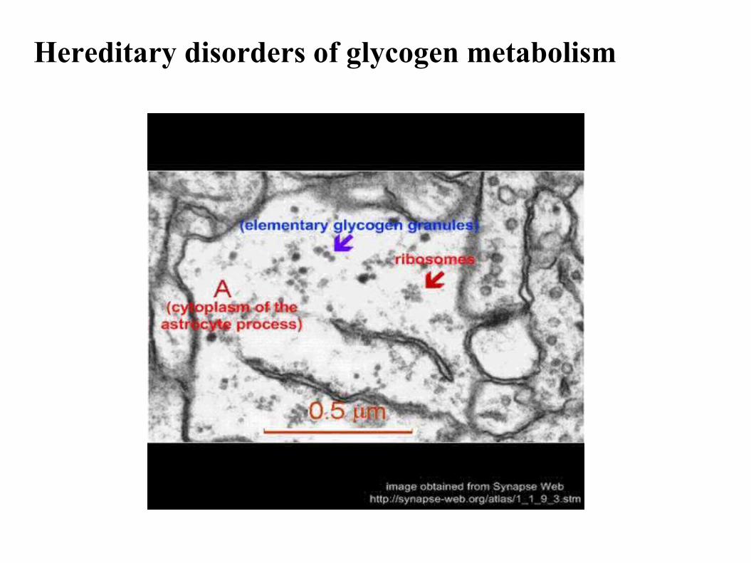
Hereditary disorders of glycogen metabolism
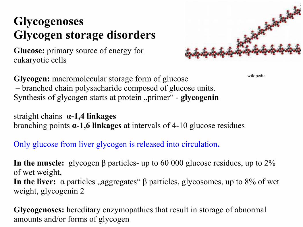
GlycogenosesGlycogen storage disordersGlucose: primary source of energy for eukaryotic cells
Glycogen: macromolecular storage form of glucose – branched chain polysacharide composed of glucose units. Synthesis of glycogen starts at protein „primer“ - glycogenin
straight chains α-1,4 linkagesbranching points α-1,6 linkages at intervals of 4-10 glucose residues
Only glucose from liver glycogen is released into circulation.
In the muscle: glycogen β particles- up to 60 000 glucose residues, up to 2% of wet weight, In the liver: α particles „aggregates“ β particles, glycosomes, up to 8% of wet weight, glycogenin 2
Glycogenoses: hereditary enzymopathies that result in storage of abnormal amounts and/or forms of glycogen
wikipedia
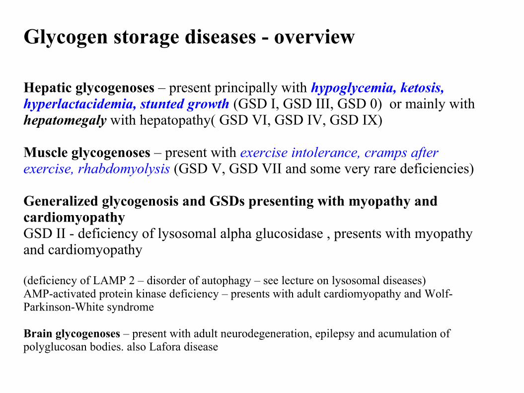
Glycogen storage diseases - overview
Hepatic glycogenoses – present principally with hypoglycemia, ketosis, hyperlactacidemia, stunted growth (GSD I, GSD III, GSD 0) or mainly with hepatomegaly with hepatopathy( GSD VI, GSD IV, GSD IX)
Muscle glycogenoses – present with exercise intolerance, cramps after exercise, rhabdomyolysis (GSD V, GSD VII and some very rare deficiencies)
Generalized glycogenosis and GSDs presenting with myopathy and cardiomyopathyGSD II - deficiency of lysosomal alpha glucosidase , presents with myopathy and cardiomyopathy
(deficiency of LAMP 2 – disorder of autophagy – see lecture on lysosomal diseases)AMP-activated protein kinase deficiency – presents with adult cardiomyopathy and Wolf-Parkinson-White syndrome
Brain glycogenoses – present with adult neurodegeneration, epilepsy and acumulation of polyglucosan bodies. also Lafora disease
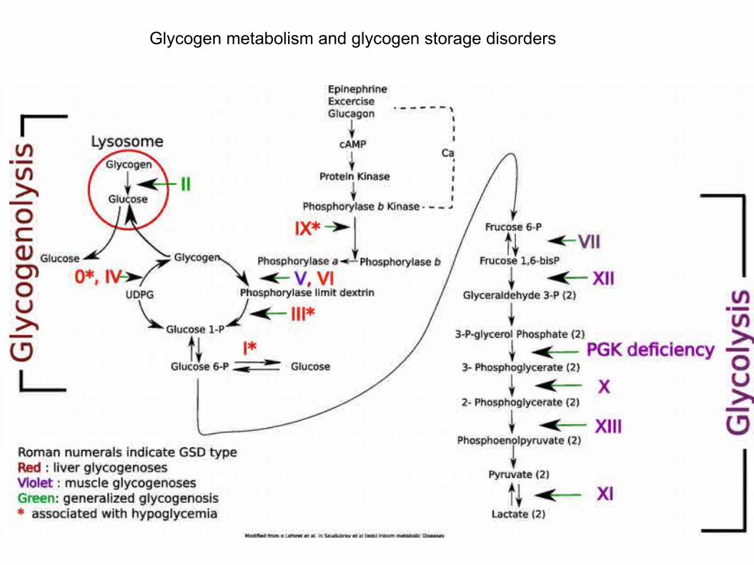
Glycogen metabolism and glycogen storage disorders
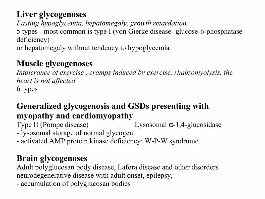
Liver glycogenosesFasting hypoglycemia, hepatomegaly, growth retardation5 types - most common is type I (von Gierke disease- glucose-6-phosphatase deficiency)or hepatomegaly without tendency to hypoglycemia
Muscle glycogenoses Intolerance of exercise , cramps induced by exercise, rhabromyolysis, the heart is not affected 6 types
Generalized glycogenosis and GSDs presenting with myopathy and cardiomyopathyType II (Pompe disease) Lysosomal α-1,4-glucosidase- lysosomal storage of normal glycogen- activated AMP protein kinase deficiency: W-P-W syndrome
Brain glycogenoses Adult polyglucosan body disease, Lafora disease and other disordersneurodegenerative disease with adult onset, epilepsy, - accumulation of polyglucosan bodies
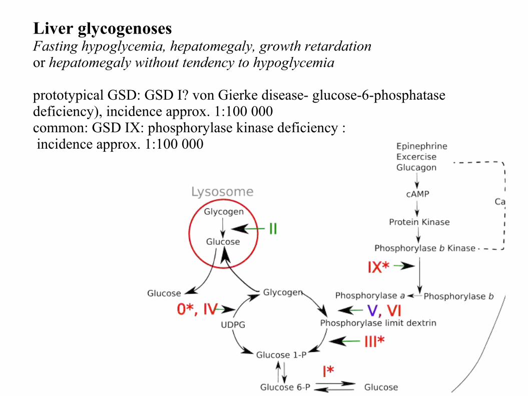
Liver glycogenosesFasting hypoglycemia, hepatomegaly, growth retardation or hepatomegaly without tendency to hypoglycemia
prototypical GSD: GSD I? von Gierke disease- glucose-6-phosphatase deficiency), incidence approx. 1:100 000common: GSD IX: phosphorylase kinase deficiency : incidence approx. 1:100 000
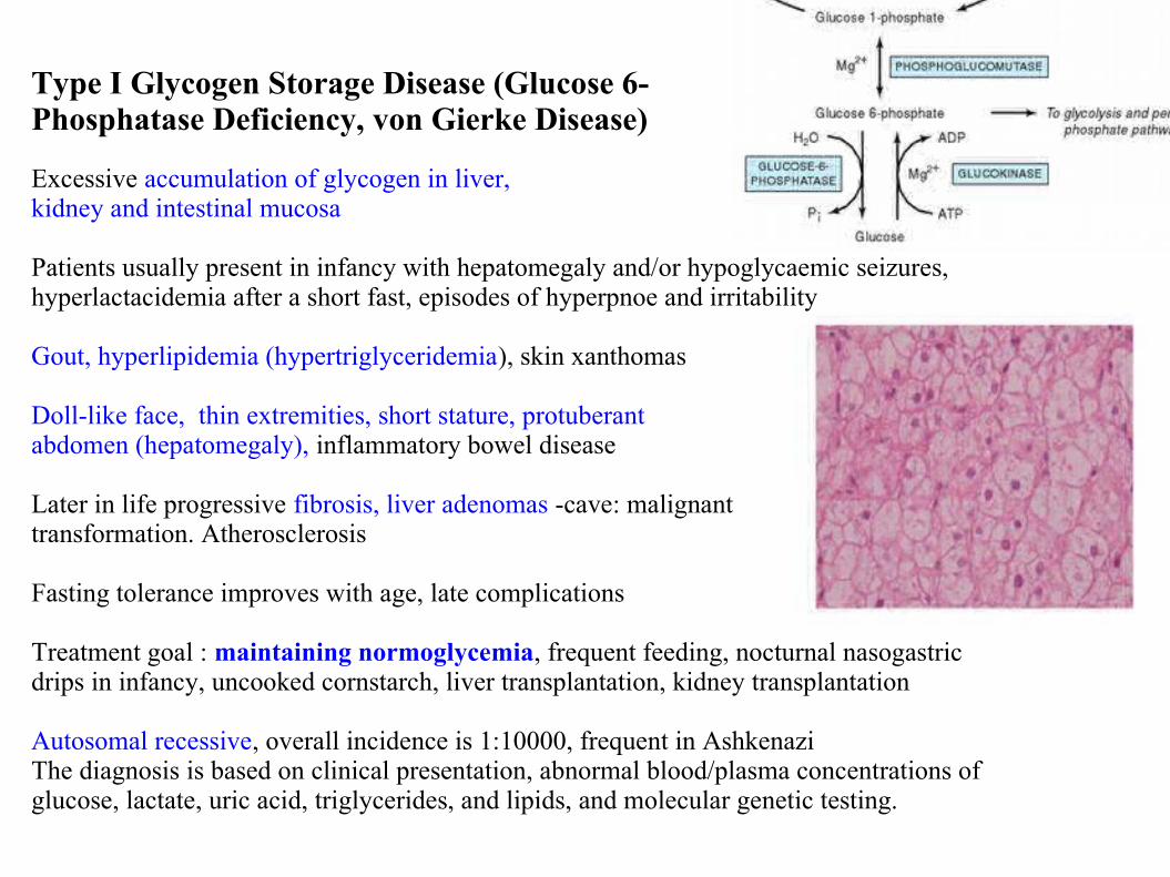
Type I Glycogen Storage Disease (Glucose 6-Phosphatase Deficiency, von Gierke Disease)
Excessive accumulation of glycogen in liver, kidney and intestinal mucosa
Patients usually present in infancy with hepatomegaly and/or hypoglycaemic seizures, hyperlactacidemia after a short fast, episodes of hyperpnoe and irritability
Gout, hyperlipidemia (hypertriglyceridemia), skin xanthomas
Doll-like face, thin extremities, short stature, protuberant abdomen (hepatomegaly), inflammatory bowel disease
Later in life progressive fibrosis, liver adenomas -cave: malignant transformation. Atherosclerosis
Fasting tolerance improves with age, late complications
Treatment goal : maintaining normoglycemia, frequent feeding, nocturnal nasogastric drips in infancy, uncooked cornstarch, liver transplantation, kidney transplantation
Autosomal recessive, overall incidence is 1:10000, frequent in AshkenaziThe diagnosis is based on clinical presentation, abnormal blood/plasma concentrations of glucose, lactate, uric acid, triglycerides, and lipids, and molecular genetic testing.
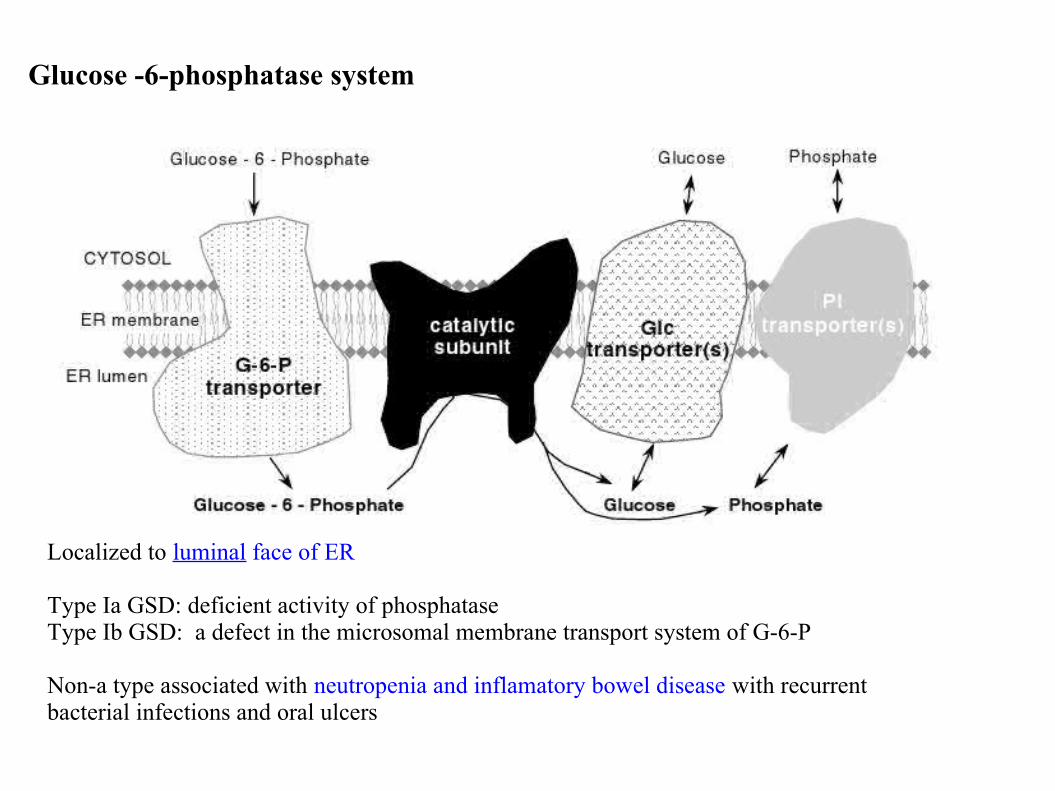
Glucose -6-phosphatase system
Localized to luminal face of ER
Type Ia GSD: deficient activity of phosphataseType Ib GSD: a defect in the microsomal membrane transport system of G-6-P
Non-a type associated with neutropenia and inflamatory bowel disease with recurrent bacterial infections and oral ulcers
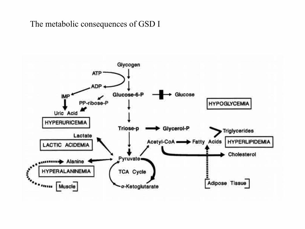
The metabolic consequences of GSD I
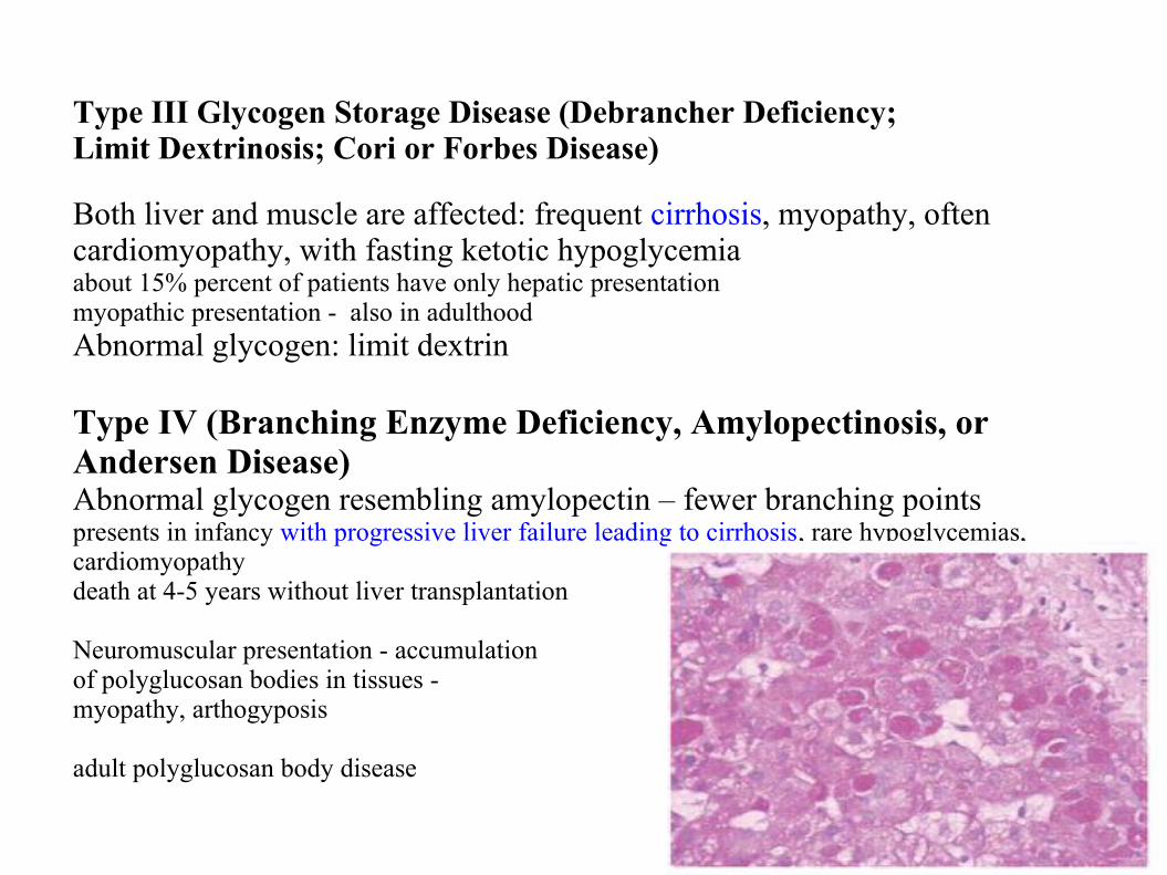
Type III Glycogen Storage Disease (Debrancher Deficiency; Limit Dextrinosis; Cori or Forbes Disease)
Both liver and muscle are affected: frequent cirrhosis, myopathy, often cardiomyopathy, with fasting ketotic hypoglycemia about 15% percent of patients have only hepatic presentationmyopathic presentation - also in adulthood Abnormal glycogen: limit dextrin
Type IV (Branching Enzyme Deficiency, Amylopectinosis, or Andersen Disease)Abnormal glycogen resembling amylopectin – fewer branching pointspresents in infancy with progressive liver failure leading to cirrhosis, rare hypoglycemias, cardiomyopathydeath at 4-5 years without liver transplantation
Neuromuscular presentation - accumulationof polyglucosan bodies in tissues - myopathy, arthogyposis
adult polyglucosan body disease
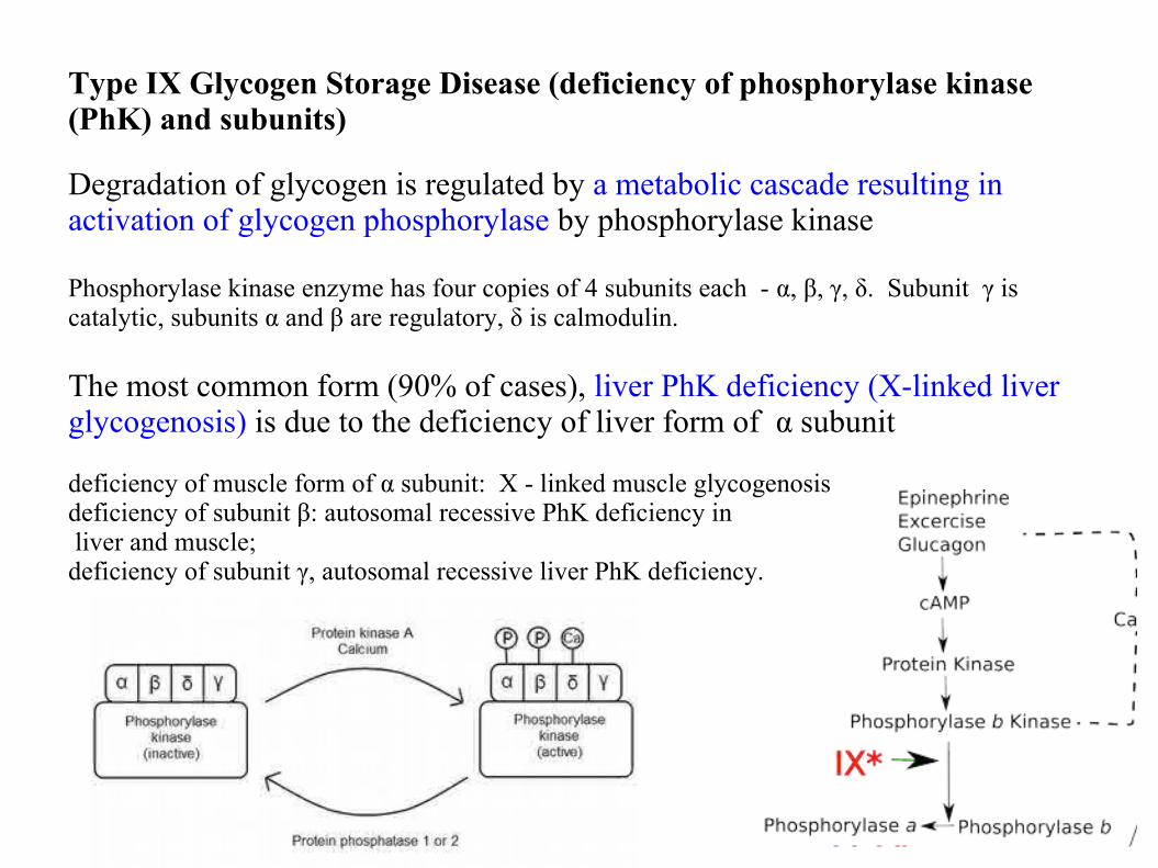
Type IX Glycogen Storage Disease (deficiency of phosphorylase kinase (PhK) and subunits)
Degradation of glycogen is regulated by a metabolic cascade resulting in activation of glycogen phosphorylase by phosphorylase kinase Phosphorylase kinase enzyme has four copies of 4 subunits each - α, β, γ, δ. Subunit γ is catalytic, subunits α and β are regulatory, δ is calmodulin.
The most common form (90% of cases), liver PhK deficiency (X-linked liver glycogenosis) is due to the deficiency of liver form of α subunit
deficiency of muscle form of α subunit: X - linked muscle glycogenosisdeficiency of subunit β: autosomal recessive PhK deficiency in liver and muscle; deficiency of subunit γ, autosomal recessive liver PhK deficiency.
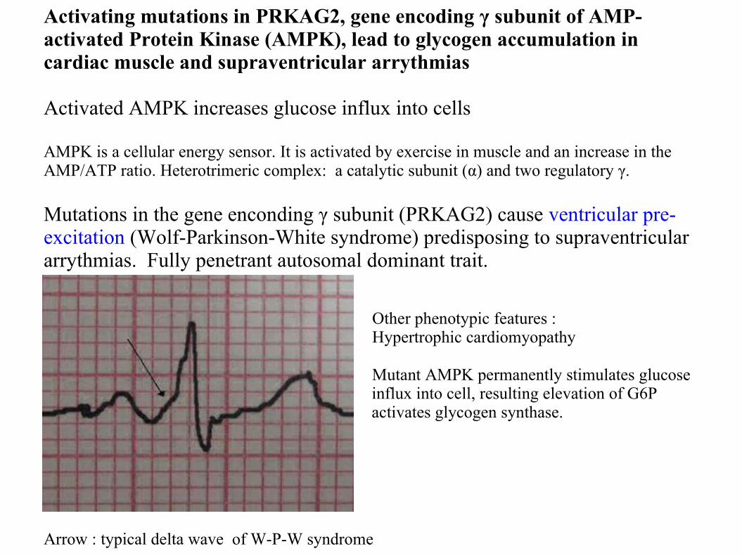
Activating mutations in PRKAG2, gene encoding γ subunit of AMP-activated Protein Kinase (AMPK), lead to glycogen accumulation in cardiac muscle and supraventricular arrythmias
Activated AMPK increases glucose influx into cells
AMPK is a cellular energy sensor. It is activated by exercise in muscle and an increase in the AMP/ATP ratio. Heterotrimeric complex: a catalytic subunit (α) and two regulatory γ.
Mutations in the gene enconding γ subunit (PRKAG2) cause ventricular pre-excitation (Wolf-Parkinson-White syndrome) predisposing to supraventricular arrythmias. Fully penetrant autosomal dominant trait.
Other phenotypic features : Hypertrophic cardiomyopathy
Mutant AMPK permanently stimulates glucose influx into cell, resulting elevation of G6P activates glycogen synthase.
Arrow : typical delta wave of W-P-W syndrome
Brain glycogenoses
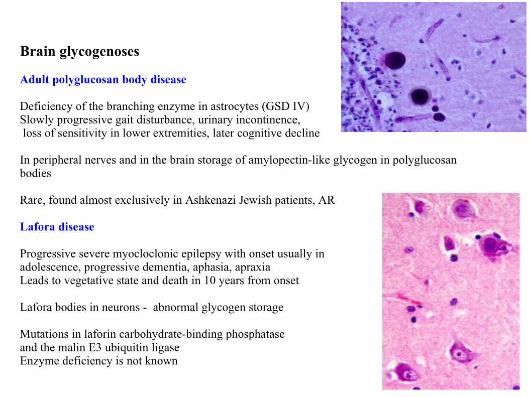
Adult polyglucosan body disease
Deficiency of the branching enzyme in astrocytes (GSD IV)Slowly progressive gait disturbance, urinary incontinence, loss of sensitivity in lower extremities, later cognitive decline
In peripheral nerves and in the brain storage of amylopectin-like glycogen in polyglucosan bodies
Rare, found almost exclusively in Ashkenazi Jewish patients, AR
Lafora disease
Progressive severe myocloclonic epilepsy with onset usually in adolescence, progressive dementia, aphasia, apraxiaLeads to vegetative state and death in 10 years from onset
Lafora bodies in neurons - abnormal glycogen storage
Mutations in laforin carbohydrate-binding phosphataseand the malin E3 ubiquitin ligaseEnzyme deficiency is not known
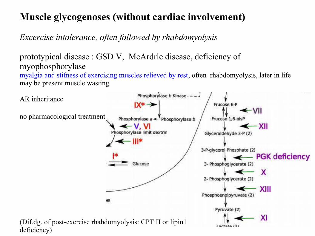
Muscle glycogenoses (without cardiac involvement)
Excercise intolerance, often followed by rhabdomyolysis
prototypical disease : GSD V, McArdrle disease, deficiency of myophosphorylase myalgia and stifness of exercising muscles relieved by rest, often rhabdomyolysis, later in life may be present muscle wasting
AR inheritance
no pharmacological treatment
(Dif.dg. of post-exercise rhabdomyolysis: CPT II or lipin1 deficiency)
Generalized glycogenosis: Morbus Pompe
M.Pompe
Deficiency of lysosomal acid alpha-glucosidase (acid maltase)Lysosomal storage of glycogen with normal structure
Infantile type:First symptoms in the first months of life: cardiomegaly, muscle weakness, macroglossiaProgressive course, death due to cardiopulmonary failure in the first two years of life
Adult typeSlowly progressive proximal myopathy and/or slowly progressive respiratory failureHeart is not affected
Intermediate typesMyopathy, heart can be affected
Diagnostics:Glycogen storage in tissuesmeasurement of enzyme activityMutation analysis Treatment monitoring: glucose tetrasaccharide Glc4
TreatmentEnzyme supplementation therapy (Myozyme)
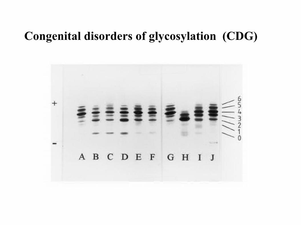
Congenital disorders of glycosylation (CDG)
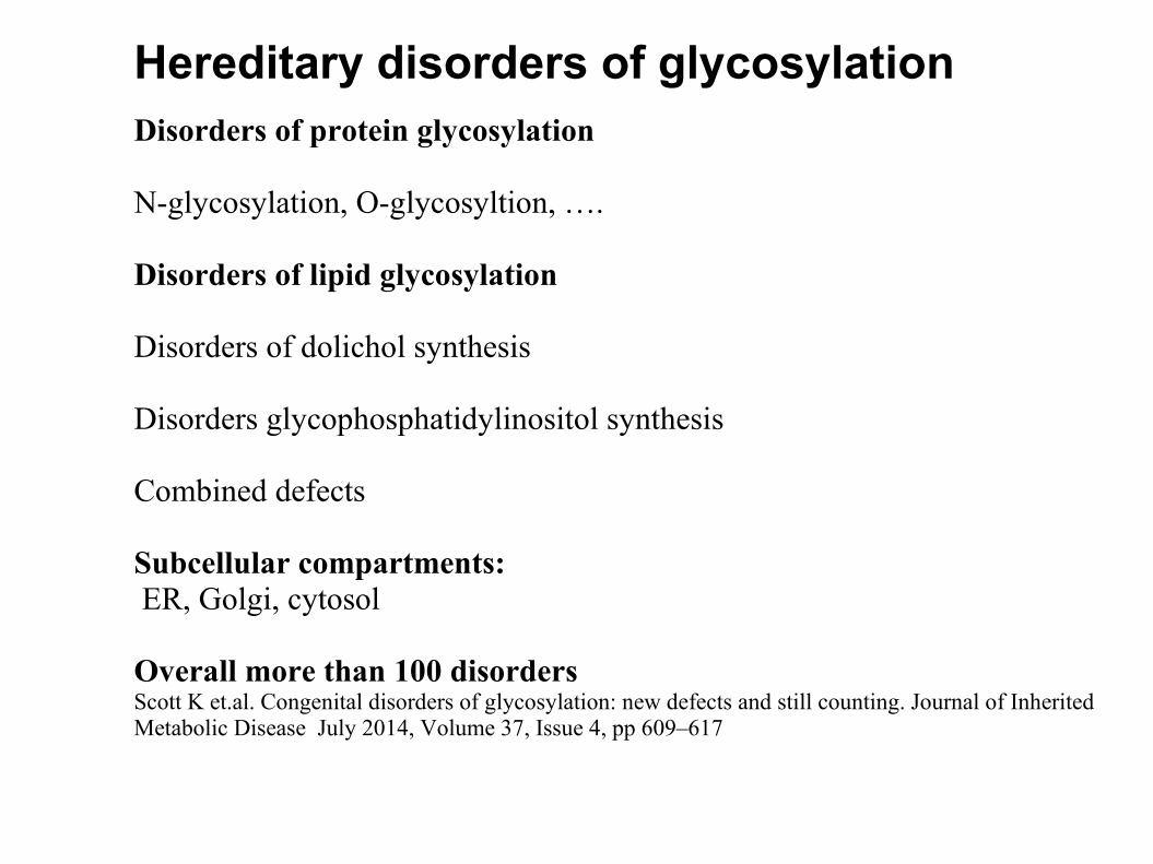
Disorders of protein glycosylation
N-glycosylation, O-glycosyltion, ….
Disorders of lipid glycosylation
Disorders of dolichol synthesis
Disorders glycophosphatidylinositol synthesis
Combined defects
Subcellular compartments: ER, Golgi, cytosol
Overall more than 100 disorders Scott K et.al. Congenital disorders of glycosylation: new defects and still counting. Journal of Inherited Metabolic Disease July 2014, Volume 37, Issue 4, pp 609–617
Hereditary disorders of glycosylation
N-glycosylation
O-glycosylation
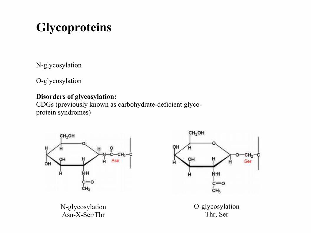
Disorders of glycosylation:CDGs (previously known as carbohydrate-deficient glyco-protein syndromes)
Glycoproteins
N-glycosylationAsn-X-Ser/Thr
O-glycosylationThr, Ser
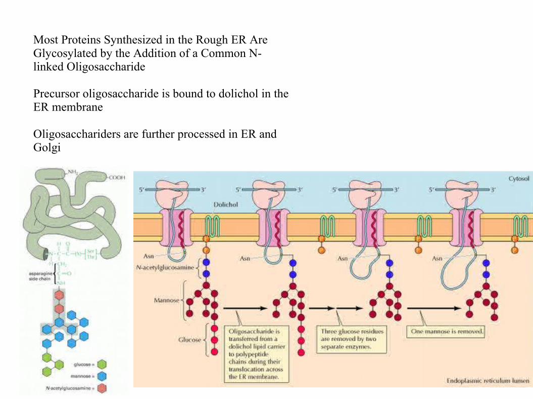
Most Proteins Synthesized in the Rough ER Are Glycosylated by the Addition of a Common N-linked Oligosaccharide
Precursor oligosaccharide is bound to dolichol in the ER membrane
Oligosacchariders are further processed in ER and Golgi
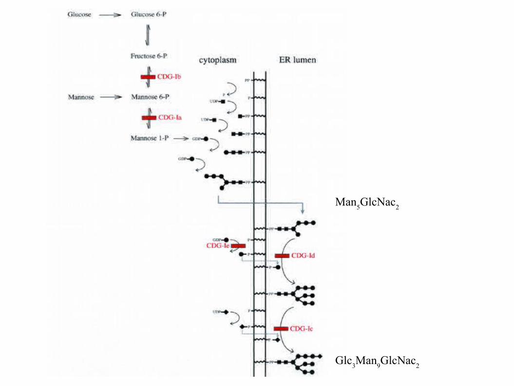
Man5GlcNac
2
Glc3Man
9GlcNac
2
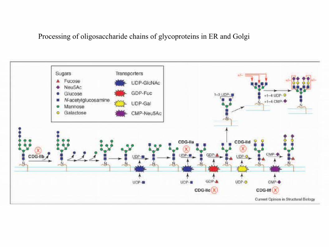
Processing of oligosaccharide chains of glycoproteins in ER and Golgi
O-glycosylation
Saccharide units are bound to Thr or Ser hydroxyl
ThreoninSerin
7 groups (classification after the first saccharide)
Glycosyltransferases add other saccharide unit in the Golgi apparatus
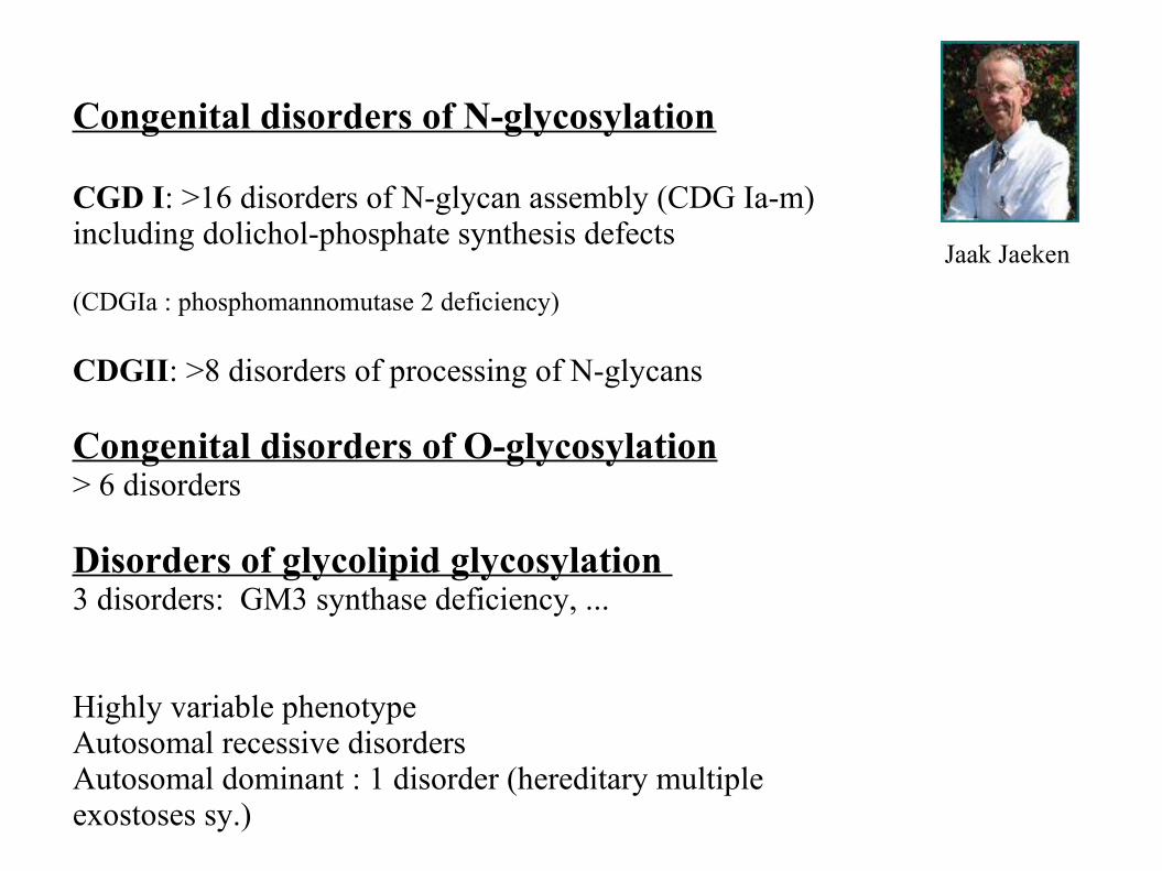
Congenital disorders of N-glycosylation
CGD I: >16 disorders of N-glycan assembly (CDG Ia-m) including dolichol-phosphate synthesis defects
(CDGIa : phosphomannomutase 2 deficiency)
CDGII: >8 disorders of processing of N-glycans
Congenital disorders of O-glycosylation> 6 disorders
Disorders of glycolipid glycosylation 3 disorders: GM3 synthase deficiency, ...
Highly variable phenotype Autosomal recessive disordersAutosomal dominant : 1 disorder (hereditary multiple exostoses sy.)
Jaak Jaeken
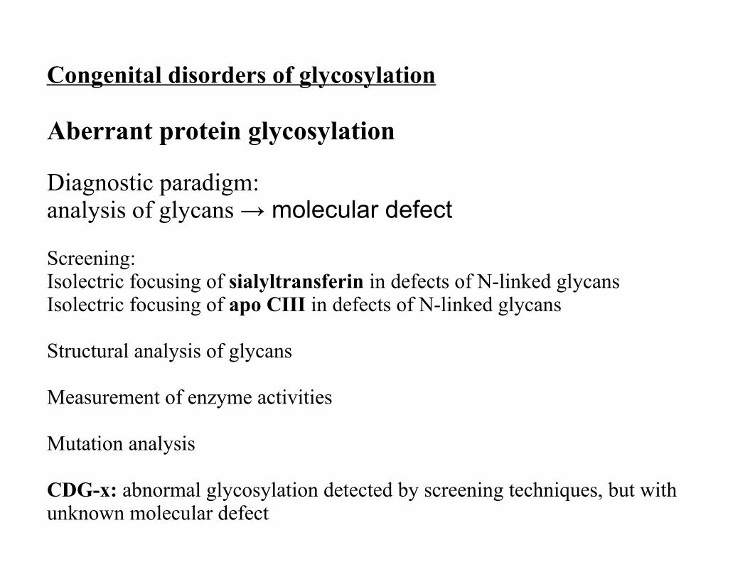
Congenital disorders of glycosylation
Aberrant protein glycosylation
Diagnostic paradigm:analysis of glycans → molecular defect
Screening: Isolectric focusing of sialyltransferin in defects of N-linked glycansIsolectric focusing of apo CIII in defects of N-linked glycans
Structural analysis of glycans
Measurement of enzyme activities
Mutation analysis
CDG-x: abnormal glycosylation detected by screening techniques, but with unknown molecular defect
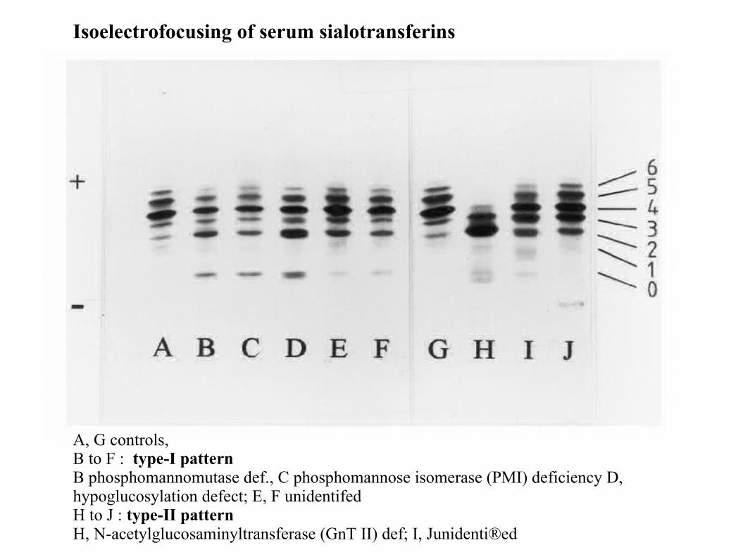
Isoelectrofocusing of serum sialotransferins
A, G controls, B to F : type-I patternB phosphomannomutase def., C phosphomannose isomerase (PMI) deficiency D, hypoglucosylation defect; E, F unidentifedH to J : type-II pattern H, N-acetylglucosaminyltransferase (GnT II) def; I, Junidenti®ed
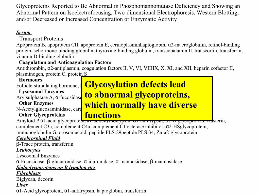
Glycoproteins Reported to Be Abnormal in Phosphomannomutase Deficiency and Showing an Abnormal Pattern on Isoelectrofocusing, Two-dimensional Electrophoresis, Western Blotting, and/or Decreased or Increased Concentration or Enzymatic Activity
Serum Transport ProteinsApoprotein B, apoprotein CII, apoprotein E, ceruloplasminhaptoglobin, α2-macroglobulin, retinol-binding protein, sehormone-binding globulin, thyroxine-binding globulin, transcobalamin II, transcortin, transferrin, vitamin D-binding globulin Coagulation and Anticoagulation FactorsAntithrombin, α2-antiplasmin, coagulation factors II, V, VI, VIIIIX, X, XI, and XII, heparin cofactor II, plasminogen, protein C, protein S Hormones Follicle-stimulating hormone, luteinizing hormone, prolactinthyroid-stimulating hormone Lysosomal EnzymesArylsulphatase A, α-fucosidase, β-galactosidase, β-glucuronidase, β-hexosaminidase Other EnzymesN-Acetylglucosaminidase, carboxypeptidase, cholinesterase Other GlycoproteinsAmyloid P α1-acid glycoprotein, a1-antichymotrypsin, α1-antitrypsin, α1-B glycoprotein, clusterin, complement C3a, complement C4a, complement C1 esterase inhibitor, α2-HSglycoprotein, immunoglobulin G, orosomucoid, peptide PLS:29peptide PLS:34, Zn-a2-glycoproteinCerebrospinal Fluidβ-Trace protein, transferrinLeukocytesLysosomal Enzymesα-Fucosidase, β-glucuronidase, α-iduronidase, α-mannosidase, β-mannosidaseSialoglycoproteins on B lymphocytesFibroblastsBiglycan, decorinLiverα1-Acid glycoprotein, α1-antitrypsin, haptoglobin, transferrin
Glycosylation defects leadto abnormal glycoproteins, which normally have diversefunctions
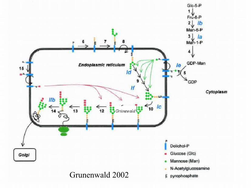
Grunenwald 2002

Grünewald 2007
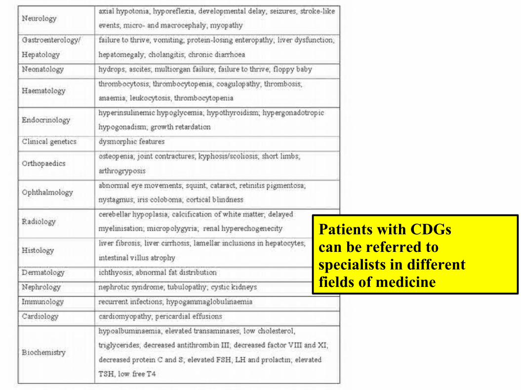
Patients with CDGs can be referred to specialists in different fields of medicine
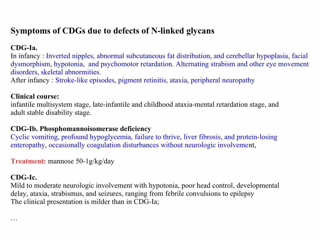
Symptoms of CDGs due to defects of N-linked glycans
CDG-Ia. In infancy : Inverted nipples, abnormal subcutaneous fat distribution, and cerebellar hypoplasia, facial dysmorphism, hypotonia, and psychomotor retardation. Alternating strabism and other eye movement disorders, skeletal abnormities. After infancy : Stroke-like episodes, pigment retinitis, ataxia, peripheral neuropathy
Clinical course:infantile multisystem stage, late-infantile and childhood ataxia-mental retardation stage, andadult stable disability stage.
CDG-Ib. Phosphomannoisomerase deficiency Cyclic vomiting, profound hypoglycemia, failure to thrive, liver fibrosis, and protein-losing enteropathy, occasionally coagulation disturbances without neurologic involvement,
Treatment: mannose 50-1g/kg/day
CDG-Ic. Mild to moderate neurologic involvement with hypotonia, poor head control, developmentaldelay, ataxia, strabismus, and seizures, ranging from febrile convulsions to epilepsyThe clinical presentation is milder than in CDG-Ia;
…

Recognizable clinical features in different N-linked glycosylation defects. a Abnormal fat distribution in phosphomannomutase 2 (PMM2)-CDG; CDG-Ia). b Liver cirrhosis in phosphomannose isomerase (MPI)-CDG (CDG-Ib). c Distal phalangeal aplasia in ALG6-CDG (CDG-Ic). d Ichthyosis and iridial and retinal coloboma are characteristic for SRD5A3-CDG. e Distal arthrogryposis in ALG8-CDG (CDG-Ih). f Myasthenic face and ptosis are common in DPAGT1-CDG (CDG-Ij). g Venous thrombosis leads to asymmetry in limb circumference in ALG1-CDG (CDG-Ik). Scott K et.al. JIMD 2014, Volume 37, Issue 4, pp 609–617
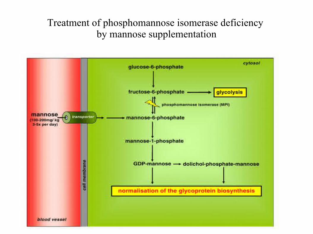
Treatment of phosphomannose isomerase deficiency by mannose supplementation
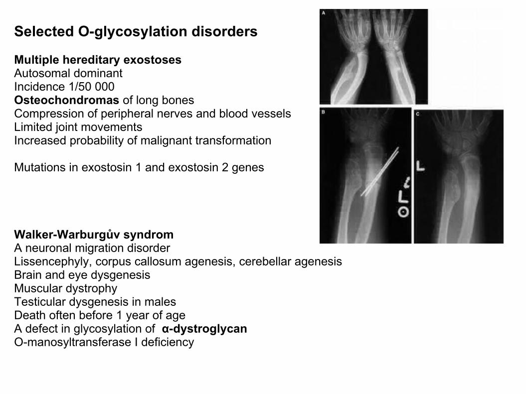
Selected O-glycosylation disorders
Multiple hereditary exostosesAutosomal dominantIncidence 1/50 000Osteochondromas of long bones Compression of peripheral nerves and blood vesselsLimited joint movementsIncreased probability of malignant transformation
Mutations in exostosin 1 and exostosin 2 genes
Walker-Warburgův syndromA neuronal migration disorderLissencephyly, corpus callosum agenesis, cerebellar agenesis Brain and eye dysgenesisMuscular dystrophyTesticular dysgenesis in malesDeath often before 1 year of age A defect in glycosylation of α-dystroglycanO-manosyltransferase I deficiency