HepatocyteGrowthFactorActsasaMotogenandGuidance ... · trol mice (C57B16/129sv) were provided by...
15
Development/Plasticity/Repair Hepatocyte Growth Factor Acts as a Motogen and Guidance Signal for Gonadotropin Hormone-Releasing Hormone-1 Neuronal Migration Paolo Giacobini, 1 Andrea Messina, 1 Susan Wray, 2 Costanza Giampietro, 3 Tiziana Crepaldi, 4 Peter Carmeliet, 5 and Aldo Fasolo 1 1 Department of Human and Animal Biology, University of Torino, 10123 Torino, Italy, 2 Cellular and Developmental Neurobiology Section, National Institute of Neurological Disorders and Stroke, National Institutes of Health, Bethesda, Maryland 20892, 3 Italian Foundation for Cancer Research Institute of Molecular Oncology, 20139 Milan, Italy, 4 Department of Anatomy, Pharmacology, and Forensic Medicine, University of Torino, 10125 Torino, Italy, and 5 Center for Transgene Technology and Gene Therapy, Flanders Interuniversity Institute for Biotechnology, University of Leuven, 3000 Leuven, Belgium Reproduction in mammals is under the control of the hypothalamic neuropeptide gonadotropin hormone-releasing hormone-1 (GnRH- 1). GnRH-1-secreting neurons originate during embryonic development in the nasal placode and migrate into the forebrain along olfactory nerves. Gradients of secreted molecules may play a role in this migratory process. In this context, hepatocyte growth factor (HGF) is a potential candidate, because it promotes cell motility in developing brain and has been shown previously to act as a motogen on immortalized GnRH-1 neurons (GN11). In this study, the role of HGF and its receptor Met during development of the GnRH-1 system was examined. GnRH-1 cells express Met during their migration and downregulate its expression once they complete this process. Tissue-type plasminogen activator (tPA), a known HGF activator, is also detected in migratory GnRH-1 neurons. Consistent with in vivo expression, HGF is present in nasal explants, and GnRH-1 neurons express Met. HGF-neutralizing antibody was applied to explants to examine the role of the endogenous growth factor. Migration of GnRH-1 cells and olfactory axon outgrowth were significantly reduced, in line with disruption of a guidance gradient. Exogenous application of HGF to explants increased the distance that GnRH-1 cells migrated, suggesting that HGF also acts as a motogen to GnRH-1 neurons. Functional experiments, performed on organotypic slice cultures, show that creation of an opposing HGF gradient inhibits GnRH-1 neuronal migration. Finally, tPA / :uPA / (urokinase-type plasminogen activator / ) knock-out mice exhibit strong reduction of the GnRH-1 cell population. Together, these data indicate that HGF signaling via Met receptor influences the development of GnRH-1. Key words: GnRH-1; LHRH; HGF; migration; olfactory system; development Introduction Gonadotropin hormone-releasing hormone-1 (GnRH-1) regu- lates anterior pituitary gonadotropes and, as such, is essential for reproduction. GnRH-1-secreting neurons originate from the na- sal placode (Wray, 2002) during embryonic development and migrate to the hypothalamus apposed to olfactory-vomeronasal nerves (Schwanzel-Fukuda et al., 1989; Wray et al., 1989). In humans, several monogenic disorders leading to isolated hy- pogonadotropic hypogonadism (IHH) are caused by disruption of GnRH-1 neuronal ontogeny/migration (Gonzalez-Martinez et al., 2004). However, mutations in these genes do not account for many individuals exhibiting IHH. This suggests that the full rep- ertoire of molecular cues regulating the GnRH-1 migratory pro- cess has not yet been identified. Factors already shown to influence GnRH-1 neuron migra- tion, either directly or indirectly via extension of olfactory axons (Wray, 2002; Wierman et al., 2004; Tobet and Schwarting, 2006), include neurotransmitters/neuropeptides (Fueshko et al., 1998; Bless et al., 2000; Simonian and Herbison, 2001; Pronina et al., 2003; Giacobini et al., 2004), surface molecules (Yoshida et al., 1999; Gamble et al., 2005), and growth factors (Cronin et al., 2004; Gill et al., 2004; Gill and Tsai, 2006). Guidance of the ax- onal/migratory pathway is also an important prerequisite for es- tablishment of the adult-like GnRH-1 cell distribution (Wray, 2002), and classical chemoattractants [(e.g., netrin-1 and stromal cell-derived factor-1 (SDF-1)] or chemorepellents (e.g., reelin) are distributed in gradients along the GnRH-1 migratory route and participate in directing appropriate migration (Schwarting et al., 2001, 2004, 2006; Cariboni et al., 2005). Hepatocyte growth factor (HGF) is a cytokine that, via its receptor Met, exhibits mitogenic, motogenic, and chemoattrac- tive activities in neuronal (Ebens et al., 1996; Maina et al., 1997; Streit and Stern, 1997; Yamamoto et al., 1997; Caton et al., 2000; Received Sept. 12, 2006; accepted Dec. 6, 2006. This work was supported by Compagnia di San Paolo (Neurotransplant Project 2004.2019), Ricerca Scientifica Applicata Comitato Interministeriale Programmazione Economica A23 Regione Piemonte, and Fondo per gli Inves- timenti della Ricerca di Base Grant RBNE01WY7P (Italy). We thank Andree Reuss for her help in generating nasal explants. Correspondence should be addressed to Dr. Paolo Giacobini, Department of Human and Animal Biology, Via Accademia Albertina 13, 10123 Torino, Italy. E-mail: [email protected]. DOI:10.1523/JNEUROSCI.4979-06.2007 Copyright © 2007 Society for Neuroscience 0270-6474/07/270431-15$15.00/0 The Journal of Neuroscience, January 10, 2007 • 27(2):431– 445 • 431
Transcript of HepatocyteGrowthFactorActsasaMotogenandGuidance ... · trol mice (C57B16/129sv) were provided by...

Development/Plasticity/Repair
Hepatocyte Growth Factor Acts as a Motogen and GuidanceSignal for Gonadotropin Hormone-Releasing Hormone-1Neuronal Migration
Paolo Giacobini,1 Andrea Messina,1 Susan Wray,2 Costanza Giampietro,3 Tiziana Crepaldi,4 Peter Carmeliet,5 andAldo Fasolo1
1Department of Human and Animal Biology, University of Torino, 10123 Torino, Italy, 2Cellular and Developmental Neurobiology Section, NationalInstitute of Neurological Disorders and Stroke, National Institutes of Health, Bethesda, Maryland 20892, 3Italian Foundation for Cancer Research Instituteof Molecular Oncology, 20139 Milan, Italy, 4Department of Anatomy, Pharmacology, and Forensic Medicine, University of Torino, 10125 Torino, Italy, and5Center for Transgene Technology and Gene Therapy, Flanders Interuniversity Institute for Biotechnology, University of Leuven, 3000 Leuven, Belgium
Reproduction in mammals is under the control of the hypothalamic neuropeptide gonadotropin hormone-releasing hormone-1 (GnRH-1). GnRH-1-secreting neurons originate during embryonic development in the nasal placode and migrate into the forebrain alongolfactory nerves. Gradients of secreted molecules may play a role in this migratory process. In this context, hepatocyte growth factor(HGF) is a potential candidate, because it promotes cell motility in developing brain and has been shown previously to act as a motogenon immortalized GnRH-1 neurons (GN11). In this study, the role of HGF and its receptor Met during development of the GnRH-1 systemwas examined. GnRH-1 cells express Met during their migration and downregulate its expression once they complete this process.Tissue-type plasminogen activator (tPA), a known HGF activator, is also detected in migratory GnRH-1 neurons. Consistent with in vivoexpression, HGF is present in nasal explants, and GnRH-1 neurons express Met. HGF-neutralizing antibody was applied to explants toexamine the role of the endogenous growth factor. Migration of GnRH-1 cells and olfactory axon outgrowth were significantly reduced, inline with disruption of a guidance gradient. Exogenous application of HGF to explants increased the distance that GnRH-1 cells migrated,suggesting that HGF also acts as a motogen to GnRH-1 neurons. Functional experiments, performed on organotypic slice cultures, showthat creation of an opposing HGF gradient inhibits GnRH-1 neuronal migration. Finally, tPA �/�:uPA �/� (urokinase-type plasminogenactivator �/�) knock-out mice exhibit strong reduction of the GnRH-1 cell population. Together, these data indicate that HGF signalingvia Met receptor influences the development of GnRH-1.
Key words: GnRH-1; LHRH; HGF; migration; olfactory system; development
IntroductionGonadotropin hormone-releasing hormone-1 (GnRH-1) regu-lates anterior pituitary gonadotropes and, as such, is essential forreproduction. GnRH-1-secreting neurons originate from the na-sal placode (Wray, 2002) during embryonic development andmigrate to the hypothalamus apposed to olfactory-vomeronasalnerves (Schwanzel-Fukuda et al., 1989; Wray et al., 1989). Inhumans, several monogenic disorders leading to isolated hy-pogonadotropic hypogonadism (IHH) are caused by disruptionof GnRH-1 neuronal ontogeny/migration (Gonzalez-Martinez etal., 2004). However, mutations in these genes do not account formany individuals exhibiting IHH. This suggests that the full rep-
ertoire of molecular cues regulating the GnRH-1 migratory pro-cess has not yet been identified.
Factors already shown to influence GnRH-1 neuron migra-tion, either directly or indirectly via extension of olfactory axons(Wray, 2002; Wierman et al., 2004; Tobet and Schwarting, 2006),include neurotransmitters/neuropeptides (Fueshko et al., 1998;Bless et al., 2000; Simonian and Herbison, 2001; Pronina et al.,2003; Giacobini et al., 2004), surface molecules (Yoshida etal., 1999; Gamble et al., 2005), and growth factors (Cronin et al.,2004; Gill et al., 2004; Gill and Tsai, 2006). Guidance of the ax-onal/migratory pathway is also an important prerequisite for es-tablishment of the adult-like GnRH-1 cell distribution (Wray,2002), and classical chemoattractants [(e.g., netrin-1 and stromalcell-derived factor-1 (SDF-1)] or chemorepellents (e.g., reelin)are distributed in gradients along the GnRH-1 migratory routeand participate in directing appropriate migration (Schwarting etal., 2001, 2004, 2006; Cariboni et al., 2005).
Hepatocyte growth factor (HGF) is a cytokine that, via itsreceptor Met, exhibits mitogenic, motogenic, and chemoattrac-tive activities in neuronal (Ebens et al., 1996; Maina et al., 1997;Streit and Stern, 1997; Yamamoto et al., 1997; Caton et al., 2000;
Received Sept. 12, 2006; accepted Dec. 6, 2006.This work was supported by Compagnia di San Paolo (Neurotransplant Project 2004.2019), Ricerca Scientifica
Applicata Comitato Interministeriale Programmazione Economica A23 Regione Piemonte, and Fondo per gli Inves-timenti della Ricerca di Base Grant RBNE01WY7P (Italy). We thank Andree Reuss for her help in generating nasalexplants.
Correspondence should be addressed to Dr. Paolo Giacobini, Department of Human and Animal Biology, ViaAccademia Albertina 13, 10123 Torino, Italy. E-mail: [email protected].
DOI:10.1523/JNEUROSCI.4979-06.2007Copyright © 2007 Society for Neuroscience 0270-6474/07/270431-15$15.00/0
The Journal of Neuroscience, January 10, 2007 • 27(2):431– 445 • 431

Ieraci et al., 2002; Gutierrez et al., 2004) and non-neuronal cells(Stella and Comoglio, 1999; Urbanek et al., 2005; Son et al.,2006). HGF and Met are widely distributed in developing brain(Jung et al., 1994; Thewke and Seeds, 1996; Achim et al., 1997;Thewke and Seeds, 1999; Korhonen et al., 2000); however, fewstudies address the function(s) of Met signaling during braindevelopment. To date, HGF has been shown to have motogeniceffects on migrating cortical neurons (Powell et al., 2001, 2003;Segarra et al., 2005). HGF is expressed in nasal embryonic mes-enchyme with an increasing gradient toward the border betweenthe nose and telencephalon (Sonnenberg et al., 1993; Thewke andSeeds, 1996). This pattern suggests that HGF/Met signaling mightimpact developmental events in the GnRH-1/olfactory system. Insupport of this, HGF exerts motogenic and chemotactic effects onthe GN11 immortalized GnRH-1 cell line (Giacobini et al., 2002).
To determine the role of HGF in the developing GnRH-1/olfactory system, this study (1) characterized Met expression innasal regions during the period of GnRH-1 neuronal migration,(2) perturbed HGF/Met signaling in two in vitro models (nasalexplants and slice cultures) in which primary GnRH-1 neuronsare maintained and cellular movement can be quantified, and (3)assessed the impact of the lack of HGF activators [plasminogenactivators (PAs)] on the GnRH-1 neuronal population in PAknock-out (KO) mice.
Materials and MethodsAnimalsExperiments were conducted in accordance with current EuropeanUnion and Italian law, under authorization of the Italian Ministry ofHealth, number 66/99-A.
CD-1 embryos (Charles River Laboratories, Milan, Italy) were har-vested at embryonic day 11.5 (E11.5), E12.5, E14.5, and E17.5 (plug day,E0.5) and used for RNA isolation, immediately frozen and stored(�80°C) until laser-capture microscopy, or postfixed [overnight; 4%paraformaldehyde (PFA) in 0.1 M phosphate buffer, pH 7.4] and cryo-protected and then frozen and stored (�80°C) until processing for im-munocytochemistry. Tissue-type PA �/� (tPA �/�):urokinase-typePA �/� (uPA �/�)-deficient mice and wild-type (WT) background con-trol mice (C57B16/129sv) were provided by Prof. P. Carmeliet [Centerfor Transgene Technology and Gene Therapy, Flanders InteruniversityInstitute for Biotechnology, University of Leuven, Leuven, Belgium)].CD-1 postnatal day 10 (PN10) mice and adult knock-out and WT ani-mals were anesthetized with an intraperitoneal injection of ketamine(200 mg/kg) and perfused with 4% paraformaldehyde. The brains weredissected and postfixed in the same fixative overnight at 4°C, cryopro-tected in sucrose solutions, and then frozen and stored (�80°C) untilprocessing for immunohistochemistry.
Nasal explantsNasal regions were cultured as described previously (Fueshko and Wray,1994). Briefly, embryos were obtained from timed pregnant animals inaccordance with National Institutes of Health (NIH)/National Instituteof Neurological Disorders and Stroke guidelines and Animal Care andUse Committee approval and with current European Union and Italianlaw. Nasal pits of E11.5 staged NIH-Swiss embryos were isolated underaseptic conditions in Gey’s balanced salt solution (Invitrogen Grand Is-land, NY) enriched with glucose (Sigma-Aldrich, St. Louis, MO). Nasalexplants were adhered onto coverslips by a plasma (Cocalico Biologicals,Reamstown, PA)/thrombin (Sigma-Aldrich) clot. The explants weremaintained in defined serum-free medium (SFM) (Fueshko and Wray,1994) at 37°C with 5% CO2. From culture day 3 to day 6, fresh mediumcontaining fluorodeoxyuridine (8 � 10 �5
M; Sigma-Aldrich) was givento inhibit proliferation of dividing olfactory neurons and non-neuronalexplant tissue. The medium was changed to fresh SFM twice a week.
Transcript analysesAll primers were designed from published GenBank sequences andscreened using BLAST (basic local alignment search tool) to ensure spec-
ificity of binding. Primers were pretested on brain cDNA and thereafterused throughout the described protocols at a concentration of 250 nM.Amplified products were run on a 1.5% agarose gel.
Reverse transcription-PCR analysisTotal RNA was isolated from noses and brains obtained from E11.5 miceusing RNA STAT-60 (Tel-Test, Friendswood, TX) following the manu-facturer’s protocol. Briefly, the tissue was homogenized (1 ml of RNASTAT-60 per 50 –100 mg of tissue), chloroform was added (0.2 ml/mlhomogenate), and the mixture was spun. To the aqueous layer, isopro-panol was added (0.5 ml) to precipitate RNA. RNA pellet was washed(75% ethanol), air dried, and resuspended (DEPC-treated water). TotalRNA from adult mouse brain served as positive control tissue. For thereverse transcription (RT)-PCR, 0.5 �g of each sample was used. First-strand cDNA was synthesized using the SuperScript III First-Strand Syn-thesis System for RT-PCR (Invitrogen) following the manufacturer’s in-structions. PCR was performed using 4 �l of cDNA and the appropriateoligonucleotides in 30 �l PCRs using standard reaction buffer [(in mM)10 Tris-HCl, pH 8.3, 50 KCl, and 1.5 MgCl2], 0.8 mM deoxynucleotidetriphosphate (Invitrogen) and 0.025 U/�l REDTaq DNA polymerase(Sigma-Aldrich). The following primers were used: 5�-GGGA-CTGCAGCAGCAAAGC-3� and 5�-GTCTGAGCATCTAGAGTTT-CC-3� for c-met amplification (Chan et al., 1988). For HGF, 5�-GGGGAATGAGAAATGCAGTCAG-3� and 5�-CCTGTATCCATGGA-TGCTTC-3� were used (Tashiro et al., 1990). The number of cycles andthe annealing temperature used for each primer pair were as follows: 25cycles and 59°C for c-met; 30 cycles and 55°C for HGF. No products wereamplified in water or brain RNA not reverse transcribed.
Laser capture microdissection and RT-PCR ontissue-specific regionsLaser capture microdissection (LCM) permits cells to be isolated (“cap-tured”) from tissue sections for molecular analyses. In this study, olfac-tory epithelium (OE), vomeronasal organ (VNO) epithelium, and lowerjaw were captured from E14.5 and E17.5 mouse frozen sections (see Fig.1C,D) using a PALM LCM system (Zeiss, Thornwood, NY). The laser-microdissected tissues were popped into a sterile Microfuge cap contain-ing 1 �l of 0.1% Triton X-100 and subsequently centrifuged for 1 min at7500 � g (maximum) to relocate material to the bottom of a sterile tube.Prime RNase inhibitor (7 �l diluted 1:100 in DEPC-treated water; Ep-pendorf, Hamburg, Germany) was added. Captured tissue was used tosynthesize first-strand cDNA using the SuperScript III First-Strand Syn-thesis System for RT-PCR (Invitrogen) following the manufacturer’s in-structions. Controls without reverse transcriptase were performed todemonstrate the absence of contaminating genomic DNA. Brain totalRNA was also reverse transcribed and used as a positive control.
PCR was performed for �III-tubulin (a general neuronal marker),early B-cell factor 2 (EBF-2) [an olfactory transcription factor and thusmarker of olfactory/vomeronasal receptor neurons (Wang et al., 1997)],c-met, and HGF at 40 cycles on a thermocycler (30 s denaturation at94°C, 30 s annealing at 55– 65°C, and 2 min elongation at 72°C). PCRprimer pairs were as follows: �III-tubulin forward primer, 5�-GAGGA-CAGAGCCAAGTGGAC-3�; �III-tubulin reverse primer, 5�-CAGGGC-CAAGACAAGCAG-3�; EBF-2 forward primer, 5�-TGCAGTAGTT-GCTAACAGTGG-3�; EBF-2 reverse primer, 5�-TTTCCAATGCTAG-AAGCCTAAC-3�.
Cell isolation and PCR analysisNasal explants were washed twice with 1� PBS (without Mg � or Ca 2�)and placed in 2 ml of the same solution. GnRH-1-like neurons wereidentified by their bipolar morphology, association with outgrowing ax-ons, and location within the explant (see Fig. 4 B). At two time points [4.5and 28 d in vitro (div)], single GnRH-1 cells (n � 5 for each in vitro stage)were isolated from nasal explants using a micropipette (see Fig. 4 A–C)controlled by a micromanipulator (Narishige, Tokyo, Japan) connectedto an inverted microscope (IX51; Olympus Optical, New Hyde Park,NY), cDNA was produced, and PCR amplification was performed asdescribed previously (Kramer et al., 2000; Giacobini et al., 2004). Briefly,a single cell was lysed and reverse transcribed [AMV (avian myeloblasto-sis virus) and MMLV (Moloney murine leukemia virus)-reverse tran-
432 • J. Neurosci., January 10, 2007 • 27(2):431– 445 Giacobini et al. • HGF Regulates GnRH-1 Neuronal Migration
scriptases; 37°C for 15 min; 65°C for 10 min] using an oligo-dT primer(50 OD/ml; pd(T)19 –24). The cDNA was end labeled with terminal trans-ferase (37°C for 15 min; 65°C for 10 min). Subsequent PCR amplificationwas performed using AL1 primers [ATT GGA TCC AGG CCG CTC TGGACA AAA TAT GAA TTC (T)24] (Dulac and Axel, 1995) for 25 cycles ina DNA Thermal Cycler (94°C for 1 min, 42°C for 2 min, and 72°C for 6min, with 10 s of extension time at each cycle; PerkinElmer, Wellesley,MA). After the first 25 cycles, fresh Taq was added, and 25 more cycles ofPCR were performed minus the 10 s extensions. The resulting productwas phenol-chloroform extracted and then ethanol precipitated, and analiquot was run on a 1.5% agarose gel. Total brain RNA (1 �g) served asa positive control. All cDNA pools were initially screened for GnRH-1(correct cell phenotype), �-tubulin, and L19 (two housekeeping genes,microtubule and ribosomal) using PCR. All cells used in this study werepositive for all three transcripts. Primers sequences used were as follows:GnRH-1, 5�-GCTAGGCAGACAGAAACTTGC-3� and 5�-GCATCTA-CATCTTCTTCTGCC-3�; �-tubulin, described above; and L19, 5�-CCTGAAGGTCAAAGGGAATGTGTTC-3� and 5�-GGACAGAGTCT-TGATGATCTCCTCC-3�. Each reaction mixture was generated asdescribed above, and 2 �l of each primer and 1 �l of template cDNA wereadded. The PCR program was as follows: 10 min at 94°C prerun; 30 s at94°C, 30 s at 55°C or 65°C (depending on primers), and 2 min at 72°C, for35 cycles; and 10 min at 72°C postrun. The same PCR profile was used forsubsequent screening with the following primers: tPA forward primer(5�-AAGTTTGCACTGGGGACAAG-3�), tPA reverse primer (5�-TCCCAAGAGTTGAGGAGTGTG-3�), uPA forward primer (5�-GTC-TTCCATGTGATGCTCCA-3�), and uPA reverse primers (5�-AC-CCAGTGAGGATTGGATGA-3�). Specific bands were observed in totalE17.5 brain lanes, whereas no bands were seen in water lanes.
ImmunocytochemistryPrimary antisera used were against GnRH-1 [SW-1, rabbit (Rb) poly-clonal (Wray et al., 1988), kindly provided by Dr. S. Wray; LR-5, Rbpolyclonal, kindly provided by Dr. R. Benoit, Montreal General Hospital,Montreal, Quebec, Canada; SMI41, mouse monoclonal antibody (Stern-berger Monoclonals, Baltimore, MD)], HGF (#AF294-NA, goat poly-clonal; R & D Systems, Minneapolis, MN) Met (#SP260 and #H-190,rabbit polyclonal, and #B-2, mouse monoclonal IgG; Santa Cruz Bio-technology, Santa Cruz, CA), peripherin (#AB1530, rabbit polyclonal;Millipore, Billerica, MA), neural cell adhesion molecule (NCAM;#C9672, mouse monoclonal IgG; Sigma-Aldrich), and tPA (#ASMTPA,rabbit polyclonal; Molecular Innovations, Southfield, MI).
Mouse tissue sections or nasal explants were immunocytochemicallystained as described previously (Fueshko and Wray, 1994; Wray et al.,1994). Mouse embryos and postnatal and adult brains were cryosec-tioned respectively at 16 �m (embryos) and free-floating at 30 �m (post-natal or adult brains). These sections and explants were fixed with 4%formaldehyde for 1 h before immunocytochemistry. Briefly, sections ornasal explants were washed in PBS, incubated in 10% NGS/0.3% TritonX-100 (NGS/Tx-100; 1 h), washed several times in PBS, and placed inprimary antibody (overnight at 4°C). The next day, tissues were washedin PBS, incubated in biotinylated secondary antibody [1 h; 1:500 in PBS/0.3% Triton X-100; goat anti-rabbit biotinylated (GAR-Bt; Vector Lab-oratories, Burlingame, CA); goat anti-mouse biotinylated (GAM-Bt;Millipore)], and processed using a standard avidin– biotin– horseradishperoxidase/3�, 3-diaminobenzidine (DAB) protocol. For double immu-noperoxidase staining, the chromogen for the first antigen–antibodycomplex was DAB [brown precipitate (Kramer and Wray, 2000)],whereas the chromogen for the second antigen–antibody complex wasSG substrate (blue precipitate; Vector Laboratories). Primary antiseradilutions were as follows: anti-GnRH-1 (SW-1; 1:3000), anti-peripherin(1:2000), anti-Met (#SP260 and # H-190; 1:200). For double-immunofluorescence experiments, primary antisera were diluted as fol-lows: anti-GnRH-1 (SW-1, 1:1000; SMI41, 1:3000), anti-NCAM (1:60),anti-HGF (1:10), anti-Met (#SP260 and # H-190; 1:100), and anti-tPA(1:500). Sections or nasal explants were incubated overnight (4°C) in amixture of primary antibodies diluted in NGS/Tx-100 and visualizedusing Alexa Fluor 488 and Alexa Fluor 568 conjugated secondary anti-
bodies (1:500; Invitrogen). Anti-Met and anti-HGF antisera were incu-bated for two nights at 4°C.
When two polyclonal primary antibodies were used (anti-Met/SW-1and anti-tPA/SW-1), staining of the first antigen–antibody complex wasperformed with goat anti-rabbit Alexa Fluor 488 (1:500; Invitrogen) sec-ondary antibody. This step was followed by a blocking reaction with ananti-rabbit Fab fragment (Jackson ImmunoResearch Laboratories, WestGrove, PA) (Giacobini et al., 2004) for 1 h (RT), followed by PBS washes,fixation (4% formalin for 30 min), and more PBS washes before applica-tion of the second primary antibody, which was visualized with conju-gated fluorescent goat anti-rabbit cyanine 3 (Cy3; 1:800; Jackson Immu-noResearch Laboratories).
E12.5 slice cultures were fixed with 4% PFA for 1 h and processed forimmunohistochemistry. To detect GnRH-1 immunoreactivity, sliceswere incubated at 4°C for five nights with LR-5 antibody diluted 1:4000in PBS containing 10% NGS and 1% Tx-100. For secondary antibodyprocessing, slices were washed several times with PBS for 1 h beforeincubation overnight at 4°C with goat anti-rabbit Cy3 (1:500; JacksonImmunoResearch Laboratories).
Specimen were mounted in DABCO (1,4-diazabicyclo[2.2.2]octane;Sigma-Aldrich) and observed with a laser-scanning Olympus Fluoviewconfocal system (Olympus Optical).
Functional assaysNasal explant. To determine the function of HGF in the developingolfactory/GnRH-1 systems, pharmacological perturbations were per-formed, and olfactory axon outgrowth and GnRH-1 cell migration werequantified. Explants in experimental groups were maintained in SFMcontaining either a blocking HGF antibody (5 �g/ml; #AF294-NA; R & DSystems) or 25 ng/ml human recombinant HGF (Sigma-Aldrich). Drugconcentrations were based on data from previous migrational studies(Powell et al., 2001; Giacobini et al., 2002). Nasal explants were treated at3 div with pharmacological agents for 72 h. Control explants were main-tained in SFM that was changed, as in the treatment groups, at 3 and 6div. At 7 div, explants were processed for double-label immunocyto-chemistry for GnRH-1/peripherin (see above), and then the GnRH-1 cellmigration as well as the maximum olfactory axon outgrowth were quan-tified as described below. Treatments performed at 1 div (for 72 h) didnot induce any significant change in the parameters examined comparedwith control cultures (data not shown).
Quantification of GnRH-1 cell number, migration, and olfactory fiberoutgrowth. For each explant, the number of GnRH-1-immunopositivecells was counted on the main tissue mass, as well as in the periphery ofthe explant (Fueshko et al., 1998; Giacobini et al., 2004). The main tissuemass contained the nasal pit/olfactory epithelial region, surroundingmesenchyme, and nasal midline cartilage (see Fig. 3, schematic). Theperiphery refers to the area surrounding the main tissue mass into whichcells had spread and/or migrated. Data are presented as mean � SEM.Quantification of GnRH-1 cell migration and olfactory axon outgrowthwas performed digitizing the stained explant. Images were taken underan Olympus IX50 inverted microscope (Olympus Optical) equippedwith a CCD camera CoolSNAP-Pro (Media Cybernetics, Silver Spring)and images edited with Image-Pro Plus software (Media Cybernetics).For cell migrational measurements, a caliber with a series of concentricarcs separated by a uniform distance (200 �m) was overlaid on the dig-itized image. The total number of cells in each zone of the periphery ofthe explant was recorded via computer-assisted analysis (Image-Pro Plussoftware; Media Cybernetics) (see Fig. 6). In general, under all treatmentconditions, the number of GnRH-1 cells decreases as a function of dis-tance from the main tissue mass. GnRH-1 cell migration was calculatedas the distance from the main tissue mass edge to the outer sector of theperiphery.
For fiber outgrowth measurements, the distance from the border ofthe explant at which the majority of peripherin-positive fibers ended wasrecorded; this method was chosen because the complex nature of thefiber network prevented quantification of individual fiber lengths. Amean distance for fiber outgrowth was obtained for each treatmentgroup, and values were reported as the mean � SEM. All experiments
Giacobini et al. • HGF Regulates GnRH-1 Neuronal Migration J. Neurosci., January 10, 2007 • 27(2):431– 445 • 433

used explants generated by different individuals on multiple culturedates.
Statistical analysis of fiber outgrowth was performed with a one-wayANOVA followed by Fisher’s least significant difference (LSD) post hocanalysis ( p � 0.05) using the statistical software SPSS 12.0 (SPSS, Chi-cago, IL). A mean total GnRH-1 cell number (inside the explant and/or inthe periphery) was obtained for each treatment group and analyzed usinga one-way ANOVA. These values were taken as an indication of GnRH-1cell survival. Data for cell movement were compared for SFM and exper-imental groups by constructing contingency tables and applying the � 2
test for independence. This nonparametric analysis was chosen becausezonal distance (200 �m grouping) was used instead of continuous mea-surements, and the number of observations per treatment (culture num-ber) was not identical. A stringent p value of 0.001 was chosen for signif-icance in these experiments.
HGF-expressing cellsTo obtain HGF clones in which expression of murine HGF can be spe-cifically induced by doxycycline (Dox), we adopted the tetracycline(Tet)-Off technology described by Gossen and Bujard (1992). The mouseHGF cDNA was inserted in the bidirectional pBI response plasmid con-taining enhanced green fluorescent protein (EGFP) as a reporter gene(Baron et al., 1995), which makes it possible to trace cells expressing thetransgene by EGFP imaging. The plasmid was introduced into stableMadin-Darby canine kidney (MDCK) Tet-Off cells, and clones resistantto blasticidin were selected and expanded. The HGF cDNA construct wasamplified from murine HGF plasmid (a kind gift from Dr. W. J. LaRo-chelle) using the Platinum Pfx polymerase (Invitrogen) and the followingoligonucleotides (Sigma-Aldrich): 5�-TTGCACGCGTCCACCAT-GATGTGGGGGACCAAAC-3� and 5�-TTACACGCGTGTTAACTTA-CTTTCCAAGTCGGTTCATCTCTATGTCTGTATACAACTTGTATG-TCAAAA-3�. The obtained cDNA encoded the full-length HGF se-quence, flanked with MluI sites and fused to a Kozak consensus ribosomebinding site at the N terminus and to oligonucleotides encoding the 11 aavesicular stomatitis virus glycoprotein G (VSVG) tag at the C terminus.The fidelity of the HGF insert was verified by sequencing (MWG Biotech,Ebersberg, Germany). The insert was then ligated into the MluI site ofpBI-EGFP (Clontech, Mountain View, CA) to generate the HGF-TRE-EGFP responder plasmid. MDCK cells expressing the tetracycline trans-activator (tTA) under cytomegalovirus promoter (MDCK-Tet-Off cellline; Clontech) were transfected with pBI-HGF-TRE-EGFP plasmid anda blasticidin selection plasmid. Exponentially growing MDCK-tTA cellswere seeded 24 h before DNA transfer on 10 cm tissue culture dishes andtransfected using Lipofectamine 2000 (Invitrogen). Cells were selectivelygrown in growth media containing 5 �g/ml blasticidin (Sigma-Aldrich)for 2 weeks. Different resistant clones were picked by selective trypsiniza-tion and checked for inducible expression of EGFP reporter gene andHGF by in vivo imaging of EGFP and scatter assay of conditioned me-dium, respectively (see Fig. 8). Clone n.1, which expresses high levels ofEGFP- and HGF-transfected protein, was selected for this study.
Tet-Off cells were grown in monolayer at 37°C in 5% CO2, in DMEM(Invitrogen) containing 4500 mg of glucose, 1 mM sodium pyruvate, 2mM glutamine, 100 �g/ml streptomycin, and 100 U/ml penicillin, andsupplemented with 10% fetal bovine serum (FBS; Invitrogen). Toturn off EGFP and HGF expression, Dox (1 �g/ml) was added to theculture medium. Cells within six passages were used throughout theexperiments.
Western blotFor Western blotting analysis, equal amounts of proteins (100 �g)were boiled in sample buffer (33% bromphenol blue, 33% �-mercapto-ethanol, and 33% glycerol) and subjected to 7% SDS-PAGE. Proteinswere blotted onto Hybond-C Extra membrane (GE Healthcare, Piscat-away, NJ). Filters were probed with specific primary antibodies: 1:500anti-mouse met #B-2 (Santa Cruz Biotechnology), 1:500 HGF goat anti-serum (R & D Systems), 1:1000 P5D4 Mab (monoclonal antibody)mouse antiserum raised against the 11 aa C terminus of VSVG (Crepaldiet al., 1997) to detect HGF protein in lysates from MDCK cells expressingtagged HGF. In the latter case, total extracts were run under nonreducing
conditions. Immunoblots were developed with an enhanced chemilumi-nescence kit, ECL (GE Healthcare).
Cell scattering assayA classic scatter assay, using Met-expressing MDCK epithelial cells(Stoker et al., 1987; Montesano et al., 1991; Powell et al., 2001), was usedto determine whether culture medium conditioned (CM) by nasal ex-plants or Tet-Off cells contained functional HGF. MDCK cells were cul-tured in the same medium used for transfected cells, supplemented with5% FBS (Invitrogen). MDCK cells (8000) were plated onto glass cover-slips. The following day, discrete colonies were formed and then treatedwith known concentrations of HGF or CM (diluted 1:1 in fresh culturemedium) collected from nasal explants at 3 div (medium conditioned for3 d) or from Tet-Off cells cultured in the absence or presence of Dox(medium conditioned for 5–7 d). In a subset of experiments, a blocking-function HGF antibody was added (5 �g/ml). Twenty-four hours later,the MDCK cells were washed in PBS, fixed with 4% formaldehyde, andstained either by the nuclear cell dye 4,6-diamidino-2-phenylindole(DAPI) or by crystal violet. Images were taken under an Olympus IX50inverted microscope (Olympus Optical) equipped with a CCD cameraCoolSNAP-Pro (Media Cybernetics). Quantitative analysis of the scatterresponse was performed on digitized images that were overlaid on circleswith a diameter of 80 �m (see Fig. 5E). MDCK cells were counted withineach circle superimposed on areas in which cells were detectable. Ingeneral, the number of cells within these counting frames decreases as afunction of cell scatter. Values were reported as the mean � SEM.ANOVA followed by Fisher’s LSD post hoc analysis was used to comparegroups ( p � 0.001).
Tet-Off cell aggregatesTet-Off cells were collected by trypsinization, resuspended in 20 �l ofgrowth-factor free Matrigel (BD Biosciences, San Jose, CA) diluted 1:1with the culture medium and seeded in 20 �l drops of this solution(200,000 cells for both cell lines, with or without Dox) on the lid of aculture dish. The lid was then turned upside down and incubated at 37°Cfor 10 –20 min. As the droplets of cell aggregates were set, they were cutinto four pieces (each one containing �50,000 cells) with a sterile blade.
Embryonic slice culturesTimed pregnant CD-1 mice (Charles River Laboratories) were harvestedat E12.5 to generate whole-head organotypic slice cultures following theprocedures described previously (Tobet et al., 1996; Bless et al., 2000).Briefly, embryonic heads were embedded in 8% low-gelling-temperatureagarose (type VIIa; Sigma-Aldrich), and parasagittal sections were cut at300 �m using a vibratome and placed into Petri dishes containing ice-cold dissection medium (Leibovitz’s L-15; pH 7.4; Invitrogen). Theseslices were moved carefully to avoid any torsion, stretch, or compressiontrauma, which may compromise the migration of GnRH-1 neurons invitro.
Day 0. E12.5 organotypic slices went through all of the steps describedabove until the point of plating. At this point, the tissue was fixed with 4%PFA and stained for GnRH-1 as described above.
Day 1. All slices that were subjected to functional treatments weremaintained in culture for 24 h. Organotypic slices were plated onto 30mm Millicell inserts (Millipore) coated with a thin layer of growth factor-free Matrigel (BD Biosciences). The inserts were placed into dishes con-taining 2 ml of culture medium. This medium was partially removedfrom the wells, such that only a thin layer of liquid remained coveringeach slice. The slices were maintained in a humidified incubator (37°C)for 1 d. The slice culture medium consisted of Neurobasal medium (In-vitrogen) supplemented with B27 supplement (Invitrogen), 0.5 mM glu-tamine (Invitrogen), and 25 �g/ml gentamycin (Invitrogen).
Aggregates of Tet-Off cells (50,000 cells) expressing EGFP–HGF (withor without Dox) were placed at the rostral tip of the nasal region at 0 div.After 24 h, organotypic cultures were fixed with 4% PFA and stained forGnRH-1 as described above. Slices were used for the quantitative analy-ses if they contained at least 250 GnRH-1-immunoreactive (GnRH-1-ir)neurons and if the connection between the nasal compartment and thebrain appeared intact.
Data analysis. Quantitative analysis of GnRH-1 neurons was per-
434 • J. Neurosci., January 10, 2007 • 27(2):431– 445 Giacobini et al. • HGF Regulates GnRH-1 Neuronal Migration

formed as a function of location with GnRH-1 cells assigned to one oftwo regions (nasal region and CNS). Total number of cells was calculatedfor each slice and combined to give group means � SEM. Given that thetotal number of GnRH-1 neurons per slice did not change between treat-ment groups (see Results), GnRH-1 cell distribution is presented as theaverage percentage of labeled cells located in the nose or in the CNSunder different treatment conditions. Where a significant overall one-way ANOVA was found ( p � 0.05), post hoc comparisons using Fisher’s
LSD test were performed to further clarify sig-nificant differences between individual treat-ment groups ( p � 0.05).
Quantitative analysis of GnRH-1 neuronsin tPA�/�:uPA�/� mutant animalsSerial sagittal sections (30 �m; four series) fromtPA �/�:uPA �/� and WT mice (n � 3 for eachgroup) were cut and labeled for GnRH-1 as de-scribed above (GnRH-1 immunoreactivity vi-sualized using DAB substrate). Total numbersof GnRH-1 cells were calculated in each brainand combined to give group means � SEM.Data for GnRH-1 cell number between WT andknock-out animals were compared by one-wayANOVA followed by a Fisher’s LSD post hoctest. ANOVA data were considered significantlydifferent if p � 0.05.
ResultsHGF/Met expression in the developingnasal regionsTo identify whether Met and HGF wereexpressed prenatally in nasal regions, nosetissue was removed at E11.5, whenGnRH-1 neurons are in the presumptiveVNO, and RT-PCR experiments were per-formed (Fig. 1A,B). Both c-met and HGFtranscripts were detected in all samples(E11.5 head, E11.5 nose, and adult brain)but water. Western blot analysis was usedto document protein expression in embry-onic nose tissues (Fig. 1B, right). HGF isinitially biosynthesized and secreted in abiologically inactive single-chain form(pro-HGF; �100 kDa) and is subse-quently activated by specific serine pro-teases into an �-chain (69 kDa) and a�-chain (34 kDa) form containing a totalof five glycosylation sites (Nakamura et al.,1989). E12 nose tissues and E16 head ex-tracts (positive control) showed distinctbands corresponding to the �-chain (Fig.1B, right). Protein bands of �-chain werenot single, showing that these proteinswere glycosylated heterogeneously. Thetop band corresponds to the glycosylatedform (69 kDa), whereas the bottom bandrepresents the nonglycosylated �-chain(53 kDa). E12 nose protein extracts con-tained a barely detectable band of inactivesingle-chain HGF (100 kDa), indicatingthat most HGF in these tissues is theactivated form. A 145 kDa Met-immunoreactive band was also evident inthe same protein extracts (Fig. 1B, right).These results indicate that at E12, both ac-tive HGF and its receptor are expressed in
the nasal compartment.To determine the expression of HGF and c-met transcripts in
more defined regions of the nasal compartment, LCM was used.Single punches from olfactory epithelium (OE) and VNO wereremoved at E14.5, and RT-PCR experiments were performed(Fig. 1C–E). This embryonic age corresponds to a stage of robustaxonal outgrowth from the OE to the developing olfactory bulb.
Figure 1. Nasal regions express HGF and its receptor Met during embryonic development. Schematic of an E11.5–E12.5 head[fb, OE, presumptive VNO, tongue (t), and third ventricle (III) are depicted]. The dashed line indicates the boundary between noseand brain and represents the region taken for nasal RNA isolation in B. B, Gel documentation of products produced by RT-PCRamplification using specific primers for c-met and HGF. Total RNA was isolated from nose and whole head at E11.5. Adult brain wasused as positive control tissue. Transcripts for both c-met and HGF were detected in all samples but water. Western blot analysisshowed that HGF is expressed in its active form in protein extracts of E12 noses as well in whole heads of E16, used as positivecontrol tissue. Western blot was run under reducing conditions. Met expression was also detected in the same samples. C, D,Photographs of E14.5 sagittal sections after LCM. Representative pictures show examples of microdissected OE (C) and VNO (D). Noother tissue was removed from the nasal section, and the remaining tissue was intact after the capture procedure. E, Total RNAisolated from dissected regions was subjected to RT-PCR. A fragment of the expected size (519 bp) was detected for c-met in theOE and in VNO. Expression of the olfactory marker EBF-2 (165 bp) confirmed the morphology of the dissected tissue. PCR using HGFprimers showed the expected amplicon (314 bp) in the positive control lane (CNTR; E17.5 whole-embryo extracts) but not in OE orVNO. No PCR product was observed in reactions that omitted either reverse transcriptase or starting material (water). F, G, Sagittalsection of an E14.5 mouse nose double stained for NCAM (expressed by olfactory/vomeronasal axons; F ) and Met (G). Metimmunoreactivity is distributed in the developing OE and VNO structures and along the olfactory/vomeronasal fibers. G, Inset,Colocalization between the two antigens. MW, Molecular weight; OB, olfactory bulb. Scale bars: (in C) C, D, 30 �m; (in F ) F, G, 100�m; G, inset, 22 �m. Asterisks indicate laser-captured areas.
Giacobini et al. • HGF Regulates GnRH-1 Neuronal Migration J. Neurosci., January 10, 2007 • 27(2):431– 445 • 435
After reverse transcription of the mRNA,PCR with specific primers for �-tubulin(positive control) and the olfactorymarker Olf/EBF-2 was performed (Fig.1E). A 158 bp band corresponding to�-tubulin (data not shown) and a 165 bpband corresponding to EBF-2 productwere detected in both OE and VNO, sup-porting the olfactory nature of the laser-captured tissues (Wang et al., 1997). Prod-ucts for c-met were found in the OE as wellas in the VNO section, with a stronger ex-pression in the latter tissue (Fig. 1E). HGFtranscript was not detected in the OE andVNO regions, whereas a specific band ofcorrect size was found in the control lane(E17.5 whole-embryo extracts) (Fig. 1E).These LCM RT-PCR data are in agreementwith previous in situ hybridization studiesshowing c-met and HGF mRNAs in thedeveloping murine OE and in the sur-rounding nasal mesenchyme, respectively(Thewke and Seeds, 1996).
To determine whether Met protein wasexpressed by the developing olfactory ax-ons, double-label immunofluorescencewas performed for Met and NCAM, amarker of the olfactory/vomeronasal sys-tem (Calof and Chikaraishi, 1989; Miragallet al., 1989). Met and NCAM expressionsoverlapped on fibers emerging from theVNO at E14.5, as shown by single confocalplanes (Fig. 1F,G, inset), and were coex-pressed in olfactory/vomeronasal axonbundles from the nasal tract to the medialsurface of the forebrain throughout theanalyzed stages (E12.5–E17.5). Because oflow signal-to-noise levels in brain, we wereunable to detect specific immunoreactivity for Met along thecaudal nerve that GnRH-1 cells follow into the ventral forebrain(Yoshida et al., 1995).
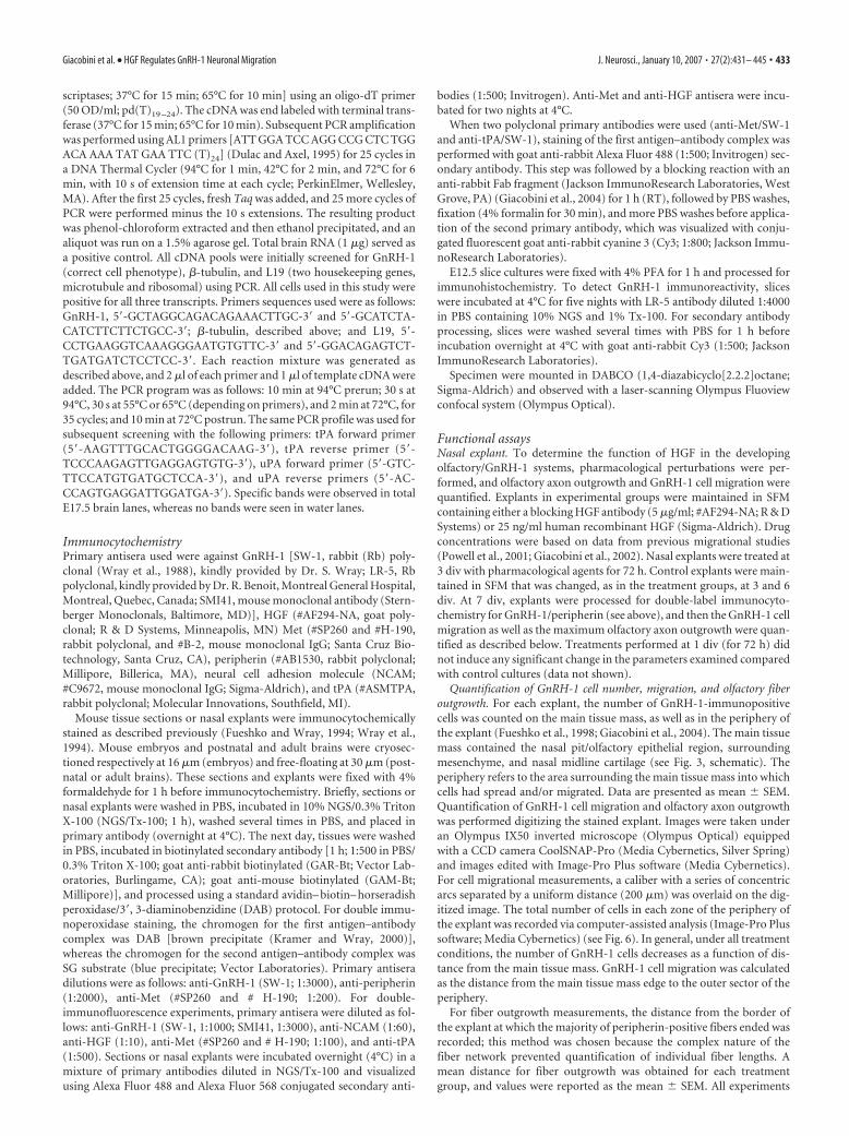
Migrating GnRH-1 neurons express MetImmunohistochemistry indicated Met protein expression in thepresumptive VNO epithelium as well as in cells migrating out ofthis structure into the nasal mesenchyme (Fig. 2A, arrows) andalong vomeronasal fibers of E12.5 embryos (Fig. 2A, arrow-heads). To establish whether Met-positive cells were GnRH-1-migrating neurons, double immunohistochemical stainings wereperformed. Double-labeling experiments for GnRH-1 and Metindicated that, at E12.5 (Fig. 2B,C) and E14.5 (data not shown),the majority of GnRH-1 neurons were Met immunopositive (Fig.2B,C, arrows), as revealed by merged single confocal planes (Fig.2C, inset, arrows). Once within the brain, it was difficult to de-termine whether GnRH-1 neurons maintained Met expression,because of the high level of expression of this receptor in otherCNS cells (data not shown). However, at postnatal day 10, Metexpression was broadly downregulated throughout the brain, al-though there was evidence of discrete Met staining within thehypothalamus (Fig. 2D, arrowheads). This corresponds to a stagewhen the GnRH-1 migratory process is over. Double labeling forGnRH-1 (Fig. 2D–F, arrows) and Met (arrowheads) at PN10revealed no coexpression between the two antigens, as shown by
single confocal planes (Fig. 2E,F). Thus, Met immunoreactivityis associated with migrating GnRH-1 neurons, being downregu-lated once these cells complete their migration.
HGF/Met expression in nasal explantsThe HGF/Met expression pattern observed in nasal regions dur-ing development together with results from previous studies(Sonnenberg et al., 1993; Thewke and Seeds, 1996; Powell et al.,2001) suggest that HGF may have a role in regulating the GnRH-1migratory process. In this context, nasal explants represent avaluable tool to separate spatial from temporal cues and focus onthe properties of GnRH-1 neurons by controlling extracellularinfluences (Fueshko and Wray, 1994; Giacobini et al., 2004). Ithas been shown previously that the migrational pattern ofGnRH-1 neurons observed in vivo reproducibly occurs in nasalexplants in vitro; with a shift in location of the GnRH-1 cell pop-ulation from the olfactory pit epithelia (OPEs) to the edge of themain tissue mass occurring from 1 to 3 div and continuing tomore distant sites from 3 to 7 div (Fueshko and Wray, 1994).
To use nasal explants for functional studies, we first verifiedthat this system retained expression of HGF and its receptor sim-ilar to the in vivo expression pattern. At 3 div, the majority ofGnRH-1 neurons are located in the inner tissue mass of the ex-plant, but some have started to migrate out into the periphery ofthe explant (Fig. 3B, arrows). At this stage, HGF immunoreactiv-
Figure 2. Met receptor expression in GnRH-1 neurons correlates with migration. A–C, Sagittal sections of E12.5 mouse immu-nostained with indicated antibodies. A, Met-immunoreactive cells emerged from the developing VNO and migrated through theolfactory mesenchyme (arrows) toward the forebrain. Met staining is also evident along the vomeronasal fibers coming out of theVNO. Arrowheads indicate Met-ir vomeronasal fibers. B, C, Double-label immunofluorescence for Met (red; B) and GnRH-1 (green;C) indicates that migrating GnRH-1 neurons spanning across the nasal regions coexpress Met (C, inset, arrows). D–F, Coronalsection of hypothalamic area of a PN10 mouse double labeled for GnRH-1 (red; arrows) and Met (green; arrowheads). E, F,High-power confocal analysis showed that GnRH-1-immunoreactive cells and fibers do not colocalize with Met-immunopositive elements at this stage. III, Third ventricle. Scale bars: A, 30 �m; (in B) B, C, 10 �m; (in B) D, 15 �m; C,inset, 10 �m; (in E) E, F, 8 �m.
436 • J. Neurosci., January 10, 2007 • 27(2):431– 445 Giacobini et al. • HGF Regulates GnRH-1 Neuronal Migration

ity was robustly expressed in the submucosa adjacent to the OPE,in the midline cartilage, and in mesenchymal cells located at theborder between the inner tissue mass and the periphery, coincid-ing with the site at which GnRH-1 neurons and olfactory axonsexit (Fig. 3C, asterisks). This latter region corresponds to thefrontonasal mesenchyme, also known as nasal/forebrain junction
(n/fb J) in vivo. The expression pattern ofMet receptor was then examined at thesame in vitro stage. Met coexpressed withmigrating GnRH-1 neurons as well as withthe olfactory neurons in the OPE (Fig. 3D,arrowhead). At 7 div, GnRH-1 neurons arelocated in the periphery of nasal explantsin close association with the olfactory fi-bers. At this stage, the majority of GnRH-1neurons expressed Met (Fig. 3E,F, arrow-heads), although some Met-positive/GnRH-1-negative cells were detected aswell (Fig. 3E, arrows). In addition, olfactoryaxons, along which GnRH-1 neurons mi-grated, exhibited Met staining (Fig. 3E).Hence, consistent with in vivo results, Metreceptor demarcated the olfactory systemand the migrating GnRH-1 cell population.
tPA is expressed in migrating GnRH-1neurons in vitrotPA and uPA are serine proteases that, inaddition to other proteases related toblood coagulation factor XII, have beenshown to cleave and activate pro-HGF(Mars et al., 1993). Moreover, PAs expres-sion is most pronounced during cell mi-gration and axonal outgrowth processes inthe developing nervous system (Seeds etal., 1997). Thus, tPA and uPA expressionby GnRH-1 neurons was evaluated invitro.
Single GnRH-1 cells were removedfrom nasal explants at 4.5 and 28 div (Fig.4A–C, arrows), two in vitro stages repre-sentative of GnRH-1 cells during migra-tory and postmigratory phases, respec-tively (Fueshko and Wray, 1994). cDNApools were examined for tPA and uPAtranscripts by single-cell RT-PCR. At 4.5div, the majority of GnRH-1 cells (four offive) expressed tPA but not uPA tran-scripts. By 28 div, all GnRH-1 neurons(n � 5) were negative for both transcripts(Fig. 4D). Double immunofluorescencefor GnRH-1 and tPA was performed in na-sal explants at 4.5 div (Fig. 4E). These ex-periments revealed coexpression of the an-tigens (Fig. 4E, arrows, inset) as well asexpression of tPA along olfactory axons,confirming previous in situ hybridizationstudies (Thewke and Seeds, 1996). Thus,immunocytochemical experiments con-firmed single-cell PCR results showingthat tPA is expressed in GnRH-1 cells in atemporal window limited to the neuronalmigratory process.
Functional analyses
Nasal explants release bioactive HGF in the culture mediumThe expression analyses demonstrate that HGF protein is presentin nasal regions both in vivo and in vitro with a temporal and
Figure 3. HGF and Met expression in nasal explants mimics expression in vivo. A, Schematic of a nasal explant removed from anE11.5 mouse and maintained in serum-free media for 7 d. Ovals represent OPEs; in the center is the nasal midline cartilage (NMC)and surrounding mesenchyme (M). GnRH-1 neurons (dots) migrate from OPE following olfactory axons to the midline and off theexplant into the periphery. B, C, Double immunofluorescence was performed using antibodies to GnRH-1 (green; B, C) and HGF(red; C) at 3 div. Note that GnRH-1 neurons at this stage migrate off the OPE through the nasal mesenchyme and emerge into theperiphery of the explant. Dashed lines indicate the border between the inner tissue mass and the periphery (B). C, HGF is expressedin the submucosa lining the OPE structures, in the nasal midline cartilage, and in the n/fb J mesenchyme (asterisks). D, OPE in innertissue mass of a 3 div nasal explant stained for GnRH-1 (green) and Met (red). Met was robustly expressed in the olfactoryepithelium. In addition, a GnRH-1 neuron migrating out of the OPE clearly expressed Met (arrowhead). E, At 7 div, a largepopulation of GnRH-1 neurons is located in the periphery of the explant. The majority of GnRH-1 neurons coexpressed Metreceptor (bottom inset, arrowheads). Few GnRH-1-positive/Met-negative cells were also detected (top inset, arrow), as well asmigrating cells, which were positive for Met but not for GnRH-1 (bottom inset, arrow). Met immunoreactivity was also evidentalong the olfactory axon network. F, Nasal explant at 7 div triple stained for the amidated form of GnRH-1 (antibody SMI41; green),Met (red), and DAPI (nuclear dye; blue). Three-dimensionally reconstructed GnRH-1-positive cells colabeled with Met are shown.Reconstructed orthogonal projections are presented as viewed in the x–z (bottom) and y–z (right) planes. Scale bars: (in B) B, C,100 �m; D, 20 �m; E, 30 �m; E, insets, 10 �m; F, 4 �m.
Giacobini et al. • HGF Regulates GnRH-1 Neuronal Migration J. Neurosci., January 10, 2007 • 27(2):431– 445 • 437

spatial pattern to impact GnRH-1/olfac-tory system development. The form ofHGF observed in E12 noses by Westernblots is indicative of a biologically activeprotein. To test whether the embryonicnasal region is able to release functionalHGF, nasal explant CM was collected, anda typical scatter assay was performed, tak-ing advantage of the Met-expressingMDCK cell line. In the absence of HGF,these cells grow in compact colonies (Fig.5A, inset, arrows). The addition of 10ng/ml HGF for 48 h to MDCK culturesinduced a typical change in morphology ofMDCK cells and a scatter response result-ing in cell dispersion (Fig. 5B, inset, ar-rows). CM from 3 div nasal explants alsoenhanced migration capacity (Fig. 5C),which was blocked by the addition ofHGF-neutralizing antibody (5 �g/ml)(Fig. 5D). The scatter response was quan-tified by measuring the number of MDCKcells contained within each countingframe (see Materials and Methods) (Fig.5E, arrows). This number decreases as afunction of cell dispersion after increasedmigratory activity. Quantification of thescatter response showed more than a 50%reduction in the cell number containedwithin each counting frame in HGF- andCM-treated groups compared with con-trol conditions (Fig. 5E).
Anti-HGF disrupts GnRH-1 neuronalmigration and olfactoryaxon outgrowthTo determine the role of endogenous HGFon GnRH-1/olfactory system develop-ment, the explants were treated with anti-HGF (5 �g/ml). The same concentrationof this antibody has been used in previousstudies to neutralize the activity of HGF(Powell et al., 2001; Giacobini et al., 2002).Nasal explants were treated from 3 to 6 div,a temporal window characterized by mas-sive olfactory axonal growth and GnRH-1neuronal migration from the inner tissuemass to the periphery of the explant(Fueshko and Wray, 1994). Nasal explantswere fixed at 7 div and stained for GnRH-1and peripherin, which stains the olfactorysystem (Fig. 6A). No significant differ-ences were found in total number ofGnRH-1 cells inside the inner tissue mass(control, 64 � 9; n � 18; anti-HGF treat-ment, 74 � 9; n � 20) or in the peripheryof the explant (control, 176 � 22; n � 20;anti-HGF treatment, 150 � 18; n � 21).No changes in GnRH-1 cell number aftertreatment suggests that mitogenic and sur-vival effects of HGF on GnRH-1 neuronsare unlikely. However, application of anti-HGF severely stunted the migration of
Figure 5. Nasal explants release functional HGF. A–D, Images show MDCK cells that were plated at identical densities andstained with nuclear dye DAPI (white). A, In SFM conditions, MDCK cells organized in typical colonies (inset; DAPI and bright field;arrows point to individual cells in a cluster). B, In the presence of 10 ng/ml HGF, MDCK cells dispersed (scatter) and moved awayfrom each other (inset). Conditioned medium from 3 div nasal explants induced scatter response of MDCK cells (C), which wasprevented by the addition of HGF-neutralizing antibody (D). E, Quantitative analysis of the scatter response was performed ondigitized images that were overlaid on circles (counting frames) with a diameter of 80 �m (see Materials and Methods). Thenumber (No) of cells within the counting frames decreases as a function of cell-scatter response [n � 4 wells counted for SFM- andHGF-treated group; n � 3 wells counted for CM and CM plus antibody (CM�Ab) groups; asterisks indicate statistical differencesversus SFM and CM�Ab conditions; p � 0.001]. Scale bars: (in A) A–D, 80 �m; (in A, inset) A, B, insets, 24 �m.
Figure 4. Primary GnRH-1 neurons express tPA during their migration. A, Photomicrograph of a nasal explant maintained for4.5 div. Numerous GnRH-1-like neurons (phase-bright cells) can be seen in the periphery of the explant. The dashed line delineatesthe main nasal tissue from the periphery of the explant. Bipolar GnRH-1-like cells in the periphery of the explant are identified insitu (B, arrow) and removed (C, arrow) with a microcapillary pipette. D, Representative gel of PCR products from single-cell RT-PCRperformed on GnRH-1 cells (4.5 and 28 div) extracted from the explant periphery. Products produced by PCR amplification usingL19-, GnRH-1-, tPA-, and uPA-specific primers. tPA transcript was detected in primary GnRH-1 neurons at 4.5 div (80%) but not at28 div. uPA transcript was not detected in GnRH-1 neurons at either 4.5 or 28 div. No specific band was detected in water(W). B, E17.5 brain, positive control. E, Nasal explant at 4.5 div double stained for GnRH-1 (red; arrows) and tPA (green).Inset, A single confocal plane showing a GnRH-1-positive cell colabeled with tPA. Scale bars: A, 100 �m; (in B) B, C, 10�m; E, 50 �m; E, inset, 5 �m.
438 • J. Neurosci., January 10, 2007 • 27(2):431– 445 Giacobini et al. • HGF Regulates GnRH-1 Neuronal Migration

GnRH-1 cells (Fig. 6B–D, arrowheads, insets) and theperipherin-fiber network in the periphery of the explant (Fig.6C,D, arrows, insets). Quantitative assessment of olfactory fiberoutgrowth revealed a significant reduction in the maximum dis-tance from the border of the explant after treatment with anti-HGF ( p � 0.001; control, 1188 � 47 �m; n � 17; anti-HGFtreatment, 996 � 95 �m; n � 16). The complex nature of the fibernetwork prevented quantification of individual fiber lengths.Therefore, the observed reduced distance of the peripherin-positive fiber bundles could be the result of a defect in olfactoryaxon elongation or of an altered orientation of the olfactoryaxons.
For GnRH-1 cell migrational measurements, a caliber with aseries of concentric arcs separated by a uniform distance (200�m) was overlaid on the digitized image, and the number of cellsin each zone was counted (Fig. 6A). In addition to changes inolfactory axons, GnRH-1 neurons were closer to the border of theexplant in the anti-HGF treated group compared with controls(Fig. 6B–D) ( p � 0.005). In control explants, 14% of the entireGnRH-1 cell population was dispersed beyond zone 4 (600 �mfrom the border of the explant tissue mass), whereas when ex-plants received anti-HGF, only 6% of GnRH-1 neurons migrated600 �m into the periphery. In treated explants, GnRH-1 neu-rons also displayed an abnormal migratory behavior. In controlconditions, the majority of migrating cells were uniformly ori-
ented in a proximal-to-distal direction(Fig. 6C, inset), whereas in the presence ofHGF antibody, such polarized directionwas lost, and GnRH-1 neurons orientationappeared more random (Fig. 6D, inset).Although “randomly” oriented, the mi-grating neurons maintained contact withperipherin-positive fibers, which also ap-peared more entwined and less direction-ally oriented after anti-HGF treatment(Fig. 6C,D). Thus, we were unable to de-termine whether the loss of orientation ofGnRH-1 neurons was attributable to cell-autonomous mechanisms or instead di-rectly dependent on altered outgrowth ofolfactory axons.
Exogenous HGF increases GnRH-1 cellmigration in nasal explantsWe next evaluated the effect of exogenousHGF on GnRH-1 cell migration and/or onolfactory axon outgrowth. HGF treatmentdid not affect the total number of GnRH-1neurons compared with controls (control,221 � 46; n � 11; HGF-treated, 204 � 19;n � 10). However, a significant shift in thelocation of GnRH-1 neurons was noted inHGF-treated cultures (Fig. 7) ( p � 0.001).In this group, 21% of GnRH-1 neurons inthe periphery of the explant migrated be-yond zone 5 ( 800 �m from the edge ofthe main tissue mass), compared with con-trols, which displayed only 8% of theGnRH-1 population in this same com-partment. To determine whether HGF hadan effect on olfactory axon outgrowth aswell, the mean maximum network out-growth of peripherin fibers was analyzed.
Quantitative analysis revealed that the extent of fiber outgrowthwas similar among HGF-treated and control explants (control,olfactory axon outgrowth: 1215 � 57 �m; n � 11; HGF-treated,olfactory axon outgrowth: 1292 � 48; n � 10), and directionalitywas maintained in all explant groups (data not shown).
HGF is a guidance signal for migrating GnRH-1 neuronsThe HGF expression pattern suggests that GnRH-1 neurons mayfollow this diffusible molecule as they move from the VNO to-ward the nasal/forebrain junction. If this were the case, then ad-dition of an exogenous source of HGF in a direction opposite tothe normal migratory pathway (i.e., the rostral tip of the nose)should disrupt or delay GnRH-1 neuronal migration. To test thishypothesis, functional experiments were performed by cocultur-ing for 24 h cell aggregates of HGF-transfected cells, together withparasagittal slices of whole heads of E12.5 mice. This embryonicage corresponds to a stage in which GnRH-1 neurons span fromthe VNO to the nasal/forebrain junction, on their way into theCNS (Wray, 2002).
MDCK EGFP–HGF stable transfectants were generated usingthe Tet-Off expression system (Gossen and Bujard, 1992). Cloneswere screened through EGFP imaging (Fig. 8A, top left). Whencells were cultured in the presence of 1 �g/ml Dox, EGFP andHGF expressions were turned off (Fig. 8A, top right).
To verify HGF biosynthesis in these cells, total extracts were
Figure 6. Neutralization of endogenous HGF alters GnRH-1 cell motility and olfactory axon outgrowth. A, Photomicrograph ofnasal explant immunocytochemically labeled for GnRH-1 (brown) and peripherin (blue) at 7 div. Images were digitized andoverlaid on a calibration meter composed of concentric arcs. B, Quantitative analysis of GnRH-1 cell distribution in the peripheryof the explant after anti-HGF treatment. Fewer GnRH-1 neurons were located in the farthest zones of the anti-HGF-treatedexplants compared with controls (0.6 –1 mm away from the border of the explant; *p � 0.001; n � 20 and 21 for control andanti-HGF-treated groups, respectively), whereas there was a concomitant accumulation of GnRH-1 cells closer to the explanttissue mass. C, D, Photomicrograph of nasal explants immunocytochemically labeled for GnRH-1 (arrowheads) and peripherin(arrows) at 7 div in control conditions (C) and after anti-HGF treatment (D). Anti-HGF treatment prevented GnRH-1 cells andolfactory fibers from moving into the periphery but stayed closer to the border of the explant tissue mass. After treatment, GnRH-1cells displayed an atypical migratory behavior, losing the proximal (P)-to-distal (D) orientation detected in control explants, andthe olfactory fiber network also appeared highly disorganized (C and D, insets and schematics). The dashed lines indicate theborder of the explant tissue mass. Scale bar (in A): A, C, D, 200 �m; C, D, insets, 20 �m.
Giacobini et al. • HGF Regulates GnRH-1 Neuronal Migration J. Neurosci., January 10, 2007 • 27(2):431– 445 • 439

run under nonreducing conditions and immunoblotted withanti-VSVG (Fig. 8B). Tet-Off MDCK cells expressing EGFP andHGF were grown in the absence (lane 1) or presence (lane 2) ofDox. Tet-Off MDCK cells expressing only EGFP were used asnegative control (lane 3). Transfected HGF was identified as pro-HGF inside the cells and was expressed only in the absence ofDox. In addition, the ability to produce and release active HGF inthe culture medium was tested by using the scatter assay. CM wascollected from Tet-Off cells grown with or without Dox for sev-eral days, diluted 1:1 with fresh culture medium, and appliedonto MDCK cells (Fig. 8A, bottom).
HGF-releasing cell aggregates were placed at the tip of thenose (Fig. 9C,D). The mean numbers of cells that were GnRH-1immunoreactive per slice (� SEM) were 273 � 21 (control, 0 div;n � 6), 301 � 16 (control, 1 div, with Dox; n � 6), 285 � 19(HGF, 1 div, without Dox; n � 4), with no significant differenceamong groups ( p 0.05) (Fig. 9B). The changing positions ofGnRH-1-ir neurons from nasal compartment to the CNS be-tween day 0 and day 1 control slices provided evidence of migra-tion in vitro (Fig. 9B). Slices grown for 1 div in the presence ofEGFP–HGF cell aggregates silenced with Dox showed a broaddistribution of GnRH-1 cells from the nasal compartment (Fig.9E, arrowheads) to the basal forebrain (Fig. 9E, arrows). GnRH-1neurons migrating through the nasal mesenchyme displayed abipolar morphology and were visible as streams of neurons di-rected toward the cribriform plate (Fig. 9E, inset). At this stage,32% of the total GnRH-1 neurons entered the brain (Fig. 9B,E,arrows). In contrast, GnRH-1 cells displayed an atypical migra-tory behavior when embryonic slices werecocultured with EGFP–HGF-expressingcell aggregates cultured in the absence ofDox (Fig. 9D,F). The vast majority ofGnRH-1 neurons (90%) did not reach theforebrain (Fig. 9B,F, arrowheads), as a re-sult of accumulation in the nasal compart-ment (Fig. 9F, inset). Significant differ-ences in GnRH-1 cell distribution betweengroups treated with and without Dox wereobserved both in the nose and in the CNS( p � 0.05).
Intense peripherin immunoreactivitywas found on olfactory/vomeronasal ax-ons that crossed the nasal mesenchymeand projected into the forebrain in cul-tures both with and without Dox (Fig.9G,H). The olfactory fiber network in thenose as well as in the forebrain did not ap-pear to be disrupted when HGF-releasingcell aggregates were cocultured with em-bryonic slice cultures (Fig. 9G,H).
Fewer GnRH-1 neurons are present inthe tPA/uPA deficient miceEarly embryonic lethality of met mutantshas prevented in vivo studies on these miceto determine the functional role of HGF inGnRH-1 neuron development (Bladt et al., 1995; Schmidt et al.,1995; Uehara et al., 1995). Therefore, we investigated the effect ofdeletion of tPA and uPA genes on the number of GnRH-1 neu-rons in adult brains. These serine proteases have been shown toactivate the progrowth factor HGF (Mars et al., 1993). Moreover,previous studies showed that mice with combined deficiencies oftPA and uPA are subfertile and display reduced gonadotropin-
induced ovulation efficiency (Carmeliet et al., 1994; Leonardssonet al., 1995). The total number of GnRH-1 neurons was com-pared in 60- to 90-d-old WT male mice (n � 3) and age andsex-matched double-KO mice for tPA and uPA genes (n � 3).Analysis revealed a significant reduction in GnRH-1 cell numberin the brains of mutants compared with WT (WT, 642 � 16;tPA�/�:uPA�/�, 422 � 24; p � 0.001). A reduction of �35% was
Figure 7. Exogenous HGF increases GnRH-1 cell motility in nasal explants. Quantitative anal-ysis of GnRH-1 cell migration after exogenous application of HGF. The same analysis described inFigure 4 was used in these experiments. HGF (25 ng/ml) applied from 3 to 6 div significantlyincreased, at 7 div, the number of GnRH-1 cells reaching the farthest zones compared withcontrols (n � 11 control; n � 10 HGF-treated group; *p � 0.001).
Figure 8. Characterization of the Tet-Off MDCK cell line. A, Photomicrograph of the Tet-Off MDCK cell line expressing EGFP andHGF. Note that EGFP is highly expressed by 70 – 80% of cells (top left). Cells were cultured in the absence or presence of 1 �g/mlDox. When cells are shifted to Dox-containing medium, EGFP expression is turned off within 24 h after the shift (top right). Bottomleft, MDCK scatter after a 24 h incubation with CM collected from Tet-Off MDCK EGFP–HGF stable clone, grown in the absence ofDox. Bottom right, MDCK cells are organized in discrete, compact colonies after exposure of CM collected from with Dox Tet-Offcells. B, Western blot analysis for HGF in total extracts of MDCK cells expressing tagged HGF. Total extracts were run undernonreducing conditions and immunoblotted with anti-VSVG antibody. Tet-Off MDCK cells expressing EGFP and HGF were grownin the absence (lane 1) or presence (lane 2) of Dox. Tet-Off MDCK cells expressing only EGFP were used as a negative control (lane3). Transfected HGF was identified as pro-HGF inside the cells (100 kDa) and was expressed only in the absence of Dox. Scale bars:A, top, 5 �m; A, bottom, 20 �m.
440 • J. Neurosci., January 10, 2007 • 27(2):431– 445 Giacobini et al. • HGF Regulates GnRH-1 Neuronal Migration

found in the KO animals. Figure 10, A andB, shows representative stainings forGnRH-1 neurons (arrows, single GnRH-1cells; arrowhead, cluster of GnRH-1 cells)in the diagonal band of Broca (dbb) of thehypothalamus of WT and double-KOmice. At this level, numerous GnRH-1neurons are normally detected (Fig. 10A).In contrast, few GnRH-1 neurons are de-tected at this level in KO mice (Fig. 10B).The median eminence of tPA�/�:uPA�/�
brains was also sparsely innervated byGnRH-1-immunoreactive terminals (Fig.10D, arrowhead, inset) compared withWT (Fig. 10C, arrowhead, inset).
DiscussionDevelopment of the olfactory andGnRH-1 neuroendocrine systems is inti-mately entwined in early embryogenesis(Wray, 2002). The mechanisms directingthe initiation of cell migration and olfac-tory axon extension from nose to fore-brain are unclear but likely require specificmotogenic and guidance cues. In this re-port, we show that HGF and Met areexpressed in a spatiotemporal pattern toimpact GnRH-1/olfactory system devel-opment. Functional analysis supports thenotion that HGF plays an important rolein regulating GnRH-1 neuronal migrationacross nasal regions toward the CNS dur-ing embryogenesis.
Previous studies have shown that c-metand tPA mRNAs are expressed in olfactoryepithelium, whereas HGF transcript local-ized to the surrounding nasal mesenchymestarting at E11 in mouse (Sonnenberg etal., 1993; Thewke and Seeds, 1996), when
Figure 9. HGF acts as a guidance signal for GnRH-1 neuronal migration during embryogenesis. A, Schematic of an E12.5 headslice culture [fb, presumptive VNO, and tongue (t) are depicted]. A dashed line indicates the boundary between nose and brain.Aggregates of Tet-Off cells (black oval), cultured in the presence or absence of Dox, were placed at the rostral tip of the nose ofE12.5 slice cultures. B, Quantitative analysis of GnRH-1 cell distribution in the nasal compartment and in the CNS of E12.5 slicecultures at 0 div [control (cntr); n � 6] and grown in vitro for 24 h with EGFP–HGF cell aggregates (with Dox, n � 6; without Dox,
4
n � 4). Analysis of GnRH-1 neurons location revealed an ac-cumulation of cells in the nasal region when organotypicslices were cocultured with HGF-releasing cell aggregates. C,E, Normal migrating GnRH-1 neurons in slices cocultured withTet-Off EGFP–HGF cell line in the presence of Dox. Note thatEGFP is turned off in the cell aggregate. GnRH-1-positive cellsmigrate in chains through the nasal compartment (arrow-heads) and enter the brain (arrows). E, Inset, A high-powerview of typical migratory GnRH-1 neurons crossing the nasalmesenchyme characterized by a bipolar morphology and by achain-like organization. D, When cocultures were performedwith transfected cells shifted to a medium without Dox,GnRH-1 neurons accumulate in the nasal region and fail toenter the brain (B, D, F ). The inset in F shows the abnormalphenotype of GnRH-1 neurons in these cultures. Many cellsappear round and lack a leading and a trailing process, typicalof migrating cells. G, H, Antibodies to peripherin react withvomeronasal and olfactory axons that extend along the nasalmesenchyme to the forebrain. No difference in the organiza-tion of the fiber network was evident among the treatmentconditions (G, with Dox; H, without Dox). bfb, Basal forebrain.Scale bars: (in C) C, D, 300 �m; (in E) E–H, 150 �m; (in E,inset) E, F, insets, 20 �m.
Giacobini et al. • HGF Regulates GnRH-1 Neuronal Migration J. Neurosci., January 10, 2007 • 27(2):431– 445 • 441

the GnRH-1/olfactory systems are in theirinitial stages of development. Here, bio-logically active HGF and Met protein ex-pression was documented in nasal regionsas early as E12. The majority of GnRH-1neurons located in the nasal compartmentwere found to be Met immunopositivewith expression correlated with migration.Using RT-PCR, c-met mRNA was de-tected in laser-captured tissues of OE andVNO at E14.5, confirming previous in situhybridization studies (Sonnenberg et al.,1993; Thewke and Seeds, 1996). More-over, immunohistochemistry coupledwith confocal microscopy revealed thatMet protein is expressed along NCAM-positive olfactory fibers during embryonicdevelopment. Thus, the spatiotemporalexpression of HGF and Met receptor innasal regions correlates with migration ofGnRH-1 neurons toward the CNS and de-velopment of the olfactory sensory system.
To determine the functional role ofHGF in the development of the GnRH-1system, we took advantage of an in vitromodel, nasal explants, which has been suc-cessfully used for other functional studies(Fueshko et al., 1998; Kramer and Wray,2000; Giacobini et al., 2004). These ex-plants maintain large numbers of GnRH-1neurons, migrating in a manner similar tothat observed in vivo, as well as directedolfactory axon outgrowth (Fueshko andWray, 1994). Expression of Met in the ol-factory system and in primary GnRH-1neurons in nasal explants was similar tothat observed in vivo. HGF was expressedas early as 3 div, a stage of active cell mi-gration and olfactory axon outgrowth, and the expression paral-leled HGF transcript distribution described previously in vivo(Sonnenberg et al., 1993; Thewke and Seeds, 1996). HGF immu-noreactivity was observed in the olfactory mucosa surroundingthe OE, in the nasal midline cartilage, and in the frontonasalmesenchyme, which is the region apposed to the ventromedialforebrain before dissection. Olfactory pathway development de-pends on induction between the frontonasal mesenchyme andadjacent olfactory epithelia (LaMantia et al., 2000). Interestingly,HGF mRNA has been shown to be unevenly distributed in thenasal mesenchyme during embryonic development, being ex-pressed in a gradient with higher levels toward the forebrain(Sonnenberg et al., 1993). A similar expression pattern has beenshown for SDF-1 in these regions (Schwarting et al., 2006). Thistranscript is expressed in a steep gradient in the developing nasalmesenchyme, being lower proximal to the VNO and greater atthe nasal/forebrain junction. SDF-1 was shown to be importantfor the migration of GnRH-1 neurons (Toba et al., 2004;Schwarting et al., 2006). Other studies demonstrated that SDF-1and HGF regulate recruitment of mesenchymal stem cells towarddamaged tissues (Ji et al., 2004; Urbanek et al., 2005) and thatHGF chemotactic response could be potentiated by SDF-1 (Sonet al., 2006). Similarly, muscle progenitor cells migrate towardSDF-1-expressing targets with CXCR4 (CXC receptor 4), theSDF-1 receptor, and Gab1 [Grb2 (growth factor receptor-bound
protein 2)-associated binding protein 1], the adaptor moleculethat transmits Met signaling, cooperating to control this process(Vasyutina et al., 2005). Thus, we cannot rule out cross talk be-tween the HGF and SDF-1 signaling pathways in regulating di-rected migration of GnRH-1 neurons from the VNO to the brain.
In addition to Met, primary GnRH-1 neurons express tPA.Interestingly, this expression pattern correlated with migration ofthese cells, being downregulated in postmigratory GnRH-1 neu-rons. Previous studies showed that tPA transcript is expressed inmigrating neurons crossing the nasal mesenchyme during earlystages of embryonic development (Friedman and Seeds, 1994).Here, we suggest that at least part of these cells are GnRH-1neurons and that these cells display the molecular machinery toactivate HGF in the immediate vicinity of its c-met receptor, thusinitiating a cell signaling cascade that influences cell movement.
Our in vitro experiments demonstrated that biologically activeHGF was released into the medium of nasal explants. In the mo-togenic assay, this medium induced a scatter response in MDCKcells, which was blocked by HGF-neutralizing antibody. HGF wasreleased into the nasal explant medium in the initial period ofGnRH-1 neuronal migration and olfactory axon elongation.Anti-HGF treatment significantly stunted GnRH-1 migratorybehavior and olfactory axon outgrowth (elongation or orienta-tion), supporting endogenous HGF acting as a motogen onGnRH-1 neurons and a growth promoter for olfactory axons.
Figure 10. GnRH-1 neuronal population is reduced in tPA �/�:uPA �/� mice. A, GnRH-1-immunoreactive cell bodies arelocated principally within the preoptic area and dbb of the WT mouse brain (arrows, single GnRH-1 neurons; arrowhead, cluster ofGnRH-1 neurons in the dbb). B, A major loss of GnRH-1 neurons was found in the brain of adult double-KO mice (arrows and inset).Note that the level of the section represented in B is comparable with A. The principal fiber projections of GnRH-1 neurons are tothe median eminence (me; C, arrowhead), and this region showed a dramatic loss of GnRH-1 fibers in mutant mice (D, arrowhead).Insets, High-power views of the median eminence in WT and KO brains. Scale bars: (in A) A–D, 100 �m; (in A, inset) insets, 20 �m.
442 • J. Neurosci., January 10, 2007 • 27(2):431– 445 Giacobini et al. • HGF Regulates GnRH-1 Neuronal Migration

When GnRH-1 cells were subject to exogenous uniform HGFconcentrations, the cells did migrate farther and kept their spatialorientation. No effect was observed in terms of olfactory axongrowth, in contrast with olfactory axon changes detected in theblocking-function experiments. This apparent discrepancy mayresult because olfactory axon extension cannot be stimulatedabove an intrinsic limit or a limit imposed by general fibroblastoutgrowth. Therefore, exogenous HGF might be insufficient topromote additional elongation of olfactory axons.
After HGF exposure, parallel changes were not detected in theGnRH-1 and olfactory system. Thus, the changes observed inGnRH-1 cell migration do not appear to be an effect dependenton alterations in olfactory axon outgrowth and suggest that theHGF effect on the motility of GnRH-1 neurons is in fact cellautonomous. It is important to note that, although anti-HGFtreatment induced a striking accumulation of cells and olfactoryfibers closer to the nasal explant tissue mass, it did not preventGnRH-1 cells and peripherin-positive fibers from moving/ex-tending into the periphery of the explant. Likely, residual HGFand perhaps other factors present in the nasal explants contributeto the initiation of cell movement and olfactory axon outgrowth.
We showed previously that HGF acts as a chemoattractant forimmortalized GnRH-1 cells (Giacobini et al., 2002). Organotypicslices have been shown previously to contain the conditions nec-essary to allow GnRH-1 migration from the developing olfactorysystem to the basal forebrain while keeping intact their migratoryroute (Tobet et al., 1996). Thus, retained in these slices are theguidance cues for GnRH-1/olfactory system development thatare clearly lacking in immortalized cells. In addition, some phe-notypic/behavioral traits of the cell lines could be a consequenceof the immortalization procedure which may alter the expressionpattern and the activity of such cells (Martinez de la Escalera andClapp, 2001). Thus, to evaluate whether HGF acts as a guidancecue during the migration of primary GnRH-1 neurons, we usedtissue slices prepared from mouse embryos (Tobet et al., 1996;Bless et al., 2000). Tissue slices that maintain connection betweenforebrain and nasal compartment were generated from E12.5whole heads. Embryonic slices were cocultured with aggregates ofEGFP–HGF-transfected cells. In these experiments, after 1 d invitro, GnRH-1 neurons accumulated in the nasal region whenslices were cocultured with HGF-releasing transfected cell aggre-gates placed in an opposite direction with respect to the normalGnRH-1 migratory pathway.
The disruption of GnRH-1 cell motility observed in these ex-periments may result from their exposure to two attractive HGFgradients (one endogenous and one exogenous) acting from twoopposite sites or from threshold concentrations of exogenousHGF, which fail to generate a motogenic response. In these invitro coculture experiments, no alterations were observed inperipherin-positive olfactory/vomeronasal nerve axons amongtreatment groups, suggesting that the observed HGF-inducedGnRH-1 neuronal migration defect is cell autonomous. How-ever, it should also be noted that, at E12.5, olfactory fibers couldno longer be sensitive to HGF stimulation, as they have started tocontact their tissue target (i.e., the presumptive olfactory bulbs).In fact, it is known that at E11 in the mouse, axons begin to extendfrom the OE, forming small fascicles which pierce the basal lam-ina (Marin-Padilla and Amieva, 1989).
HGF-induced activity requires proteolytic processing, whichis, at least in part, operated through enzymatic cleavage by PAs(Naldini et al., 1992; Mars et al., 1993). Mice carrying combineddeficiencies for tPA and uPA genes are subfertile (Carmeliet et al.,1994), consistent with the hypothesis that the lack of tPA and uPA
genes prevents active HGF from promoting GnRH-1 neuronalmigration into the forebrain. To examine this possibility, thenumber of GnRH-1 neurons in adult brains of tPA�/�:uPA�/�
mice was compared with those of WT animals. The lack of tPAand uPA clearly affected the size of the GnRH-1 neuronal popu-lation, decreasing the number of GnRH-1 cells detected in brain.Whether this effect is directly related to loss of active HGF onGnRH-1 neurons or to additional mechanisms affecting GnRH-1differentiation or cell survival will require additional investigations.
Very little is known about early interactions between migrat-ing GnRH-1 neurons and the nasal mesenchyme, which lead toproper initial movement from the presumptive VNO toward therostral forebrain. Our results are consistent with a role for HGF asa motogen as well as a chemotactic signal for developing GnRH-1neurons, acting to positively modulate cell– cell and cell–ECMinteractions during the early aspects of their migratory process.
ReferencesAchim CL, Katyal S, Wiley CA, Shiratori M, Wang G, Oshika E, Petersen BE,
Li JM, Michalopoulos GK (1997) Expression of HGF and cMet in thedeveloping and adult brain. Brain Res Dev Brain Res 102:299 –303.
Baron U, Freundlieb S, Gossen M, Bujard H (1995) Co-regulation of twogene activities by tetracycline via a bidirectional promoter. Nucleic AcidsRes 23:3605–3606.
Bladt F, Riethmacher D, Isenmann S, Aguzzi A, Birchmeier C (1995) Essen-tial role for the c-met receptor in the migration of myogenic precursorcells into the limb bud. Nature 376:768 –771.
Bless EP, Westaway WA, Schwarting GA, Tobet SA (2000) Effects ofgamma-aminobutyric acid(A) receptor manipulation on migratinggonadotropin-releasing hormone neurons through the entire migratoryroute in vivo and in vitro. Endocrinology 141:1254 –1262.
Calof AL, Chikaraishi DM (1989) Analysis of neurogenesis in a mammalianneuroepithelium: proliferation and differentiation of an olfactory neuronprecursor in vitro. Neuron 3:115–127.
Cariboni A, Rakic S, Liapi A, Maggi R, Goffinet A, Parnavelas JG (2005)Reelin provides an inhibitory signal in the migration of gonadotropin-releasing hormone neurons. Development 132:4709 – 4718.
Carmeliet P, Schoonjans L, Kieckens L, Ream B, Degen J, Bronson R, De VosR, van den Oord JJ, Collen D, Mulligan RC (1994) Physiological conse-quences of loss of plasminogen activator gene function in mice. Nature368:419 – 424.
Caton A, Hacker A, Naeem A, Livet J, Maina F, Bladt F, Klein R, Birchmeier C,Guthrie S (2000) The branchial arches and HGF are growth-promoting andchemoattractant for cranial motor axons. Development 127:1751–1766.
Chan AM, King HW, Deakin EA, Tempest PR, Hilkens J, Kroezen V, EdwardsDR, Wills AJ, Brookes P, Cooper CS (1988) Characterization of themouse met proto-oncogene. Oncogene 2:593–599.
Crepaldi T, Gautreau A, Comoglio PM, Louvard D, Arpin M (1997) Ezrin isan effector of hepatocyte growth factor-mediated migration and morpho-genesis in epithelial cells. J Cell Biol, 138:423– 434.
Cronin AS, Horan TL, Spergel DJ, Brooks AN, Hastings MH, Ebling FJ(2004) Neurotrophic effects of BDNF on embryonic gonadotropin-releasing hormone (GnRH) neurons. Eur J Neurosci 20:338 –344.
Dulac C, Axel R (1995) A novel family of genes encoding putative phero-mone receptors in mammals. Cell 83:195–206.
Ebens A, Brose K, Leonardo ED, Hanson Jr MG, Bladt F, Birchmeier C, BarresBA, Tessier-Lavigne M (1996) Hepatocyte growth factor/scatter factor isan axonal chemoattractant and a neurotrophic factor for spinal motorneurons. Neuron 17:1157–1172.
Friedman GC, Seeds NW (1994) Tissue plasminogen activator expression inthe embryonic nervous system. Brain Res Dev Brain Res 81:41– 49.
Fueshko S, Wray S (1994) LHRH cells migrate on peripherin fibers in em-bryonic olfactory explant cultures: an in vitro model for neurophilic neu-ronal migration. Dev Biol 166:331–348.
Fueshko SM, Key S, Wray S (1998) GABA inhibits migration of luteinizinghormone-releasing hormone neurons in embryonic olfactory explants.J Neurosci 18:2560 –2569.
Gamble JA, Karunadasa DK, Pape JR, Skynner MJ, Todman MG, Bicknell RJ,Allen JP, Herbison AE (2005) Disruption of ephrin signaling associates
Giacobini et al. • HGF Regulates GnRH-1 Neuronal Migration J. Neurosci., January 10, 2007 • 27(2):431– 445 • 443

with disordered axophilic migration of the gonadotropin-releasing hor-mone neurons. J Neurosci 25:3142–3150.
Giacobini P, Giampietro C, Fioretto M, Maggi R, Cariboni A, Perroteau I,Fasolo A (2002) Hepatocyte growth factor/scatter factor facilitates mi-gration of GN-11 immortalized LHRH neurons. Endocrinology143:3306 –3315.
Giacobini P, Kopin AS, Beart PM, Mercer LD, Fasolo A, Wray S (2004)Cholecystokinin modulates migration of gonadotropin-releasinghormone-1 neurons. J Neurosci 24:4737– 4748.
Gill JC, Tsai PS (2006) Expression of a dominant negative FGF receptor indeveloping GNRH1 neurons disrupts axon outgrowth and targeting tothe median eminence. Biol Reprod 74:463– 472.
Gill JC, Moenter SM, Tsai PS (2004) Developmental regulation ofgonadotropin-releasing hormone neurons by fibroblast growth factorsignaling. Endocrinology 145:3830 –3839.
Gonzalez-Martinez D, Hu Y, Bouloux PM (2004) Ontogeny of GnRH andolfactory neuronal systems in man: novel insights from the investigationof inherited forms of Kallmann’s syndrome. Front Neuroendocrinol25:108 –130.
Gossen M, Bujard H (1992) Tight control of gene expression in mammaliancells by tetracycline-responsive promoters. Proc Natl Acad Sci USA89:5547–5551.
Gutierrez H, Dolcet X, Tolcos M, Davies A (2004) HGF regulates the devel-opment of cortical pyramidal dendrites. Development 131:3717–3726.
Ieraci A, Forni PE, Ponzetto C (2002) Viable hypomorphic signaling mu-tant of the Met receptor reveals a role for hepatocyte growth factor inpostnatal cerebellar development. Proc Natl Acad Sci USA99:15200 –15205.
Ji JF, He BP, Dheen ST, Tay SS (2004) Interactions of chemokines and che-mokine receptors mediate the migration of mesenchymal stem cells to theimpaired site in the brain after hypoglossal nerve injury. Stem Cells22:415– 427.
Jung W, Castren E, Odenthal M, Vande Woude GF, Ishii T, Dienes HP,Lindholm D, Schirmacher P (1994) Expression and functional interac-tion of hepatocyte growth factor-scatter factor and its receptor c-met inmammalian brain. J Cell Biol 126:485– 494.
Korhonen L, Sjoholm U, Takei N, Kern MA, Schirmacher P, Castren E, Lind-holm D (2000) Expression of c-Met in developing rat hippocampus:evidence for HGF as a neurotrophic factor for calbindin D-expressingneurons. Eur J Neurosci 12:3453–3461.
Kramer PR, Wray S (2000) Novel gene expressed in nasal region influencesoutgrowth of olfactory axons and migration of luteinizing hormone-releasing hormone (LHRH) neurons. Genes Dev 14:1824 –1834.
Kramer PR, Krishnamurthy R, Mitchell PJ, Wray S (2000) Transcriptionfactor activator protein-2 is required for continued luteinizing hormone-releasing hormone expression in the forebrain of developing mice. Endo-crinology 141:1823–1838.
LaMantia AS, Bhasin N, Rhodes K, Heemskerk J (2000) Mesenchymal/epi-thelial induction mediates olfactory pathway formation. Neuron28:411– 425.
Leonardsson G, Peng XR, Liu K, Nordstrom L, Carmeliet P, Mulligan R,Collen D, Ny T (1995) Ovulation efficiency is reduced in mice that lackplasminogen activator gene function: functional redundancy amongphysiological plasminogen activators. Proc Natl Acad Sci USA92:12446 –12450.
Maina F, Hilton MC, Ponzetto C, Davies AM, Klein R (1997) Met receptorsignaling is required for sensory nerve development and HGF promotesaxonal growth and survival of sensory neurons. Genes Dev 11:3341–3350.
Marin-Padilla M, Amieva MR (1989) Early neurogenesis of the mouse ol-factory nerve: Golgi and electron microscopic studies. J Comp Neurol288:339 –352.
Mars WM, Zarnegar R, Michalopoulos GK (1993) Activation of hepatocytegrowth factor by the plasminogen activators uPA and tPA. Am J Pathol143:949 –958.
Martinez de la Escalera G, Clapp C (2001) Regulation of gonadotropin-releasing hormone secretion: insights from GT1 immortal GnRH neu-rons. Arch Med Res 32:486 – 498.
Miragall F, Kadmon G, Schachner M (1989) Expression of L1 and N-CAMcell adhesion molecules during development of the mouse olfactory sys-tem. Dev Biol 135:272–286.
Montesano R, Matsumoto K, Nakamura T, Orci L (1991) Identification of a
fibroblast-derived epithelial morphogen as hepatocyte growth factor. Cell67:901–908.
Nakamura T, Nishizawa T, Hagiya M, Seki T, Shimonishi M, Sugimura A,Tashiro K, Shimizu S (1989) Molecular cloning and expression of hu-man hepatocyte growth factor. Nature 342:440 – 443.
Naldini L, Tamagnone L, Vigna E, Sachs M, Hartmann G, Birchmeier W,Daikuhara Y, Tsubouchi H, Blasi F, Comoglio PM (1992) Extracellularproteolytic cleavage by urokinase is required for activation of hepatocytegrowth factor/scatter factor. EMBO J 11:4825– 4833.
Powell EM, Mars WM, Levitt P (2001) Hepatocyte growth factor/scatterfactor is a motogen for interneurons migrating from the ventral to dorsaltelencephalon. Neuron 30:79 – 89.
Powell EM, Campbell DB, Stanwood GD, Davis C, Noebels JL, Levitt P(2003) Genetic disruption of cortical interneuron development causesregion- and GABA cell type-specific deficits, epilepsy, and behavioral dys-function. J Neurosci 23:622– 631.
Pronina T, Ugrumov M, Adamskaya E, Kuznetsova T, Shishkina I, BabichevV, Calas A, Tramu G, Mailly P, Makarenko I (2003) Influence of sero-tonin on the development and migration of gonadotropin-releasing hor-mone neurones in rat foetuses. J Neuroendocrinol 15:549 –558.
Schmidt C, Bladt F, Goedecke S, Brinkmann V, Zschiesche W, Sharpe M,Gherardi E, Birchmeier C (1995) Scatter factor/hepatocyte growth fac-tor is essential for liver development. Nature 373:699 –702.
Schwanzel-Fukuda M, Bick D, Pfaff DW (1989) Luteinizing hormone-releasing hormone (LHRH)-expressing cells do not migrate normally inan inherited hypogonadal (Kallmann) syndrome. Brain Res Mol BrainRes 6:311–326.
Schwarting GA, Kostek C, Bless EP, Ahmad N, Tobet SA (2001) Deleted incolorectal cancer (DCC) regulates the migration of luteinizing hormone-releasing hormone neurons to the basal forebrain. J Neurosci 21:911–919.
Schwarting GA, Raitcheva D, Bless EP, Ackerman SL, Tobet S (2004) Netrin1-mediated chemoattraction regulates the migratory pathway of LHRHneurons. Eur J Neurosci 19:11–20.
Schwarting GA, Henion TR, Nugent JD, Caplan B, Tobet S (2006) Stromalcell-derived factor-1 (chemokine C-X-C motif ligand 12) and chemokineC-X-C motif receptor 4 are required for migration of gonadotropin-releasing hormone neurons to the forebrain. J Neurosci 26:6834 – 6840.
Seeds NW, Siconolfi LB, Haffke SP (1997) Neuronal extracellular proteasesfacilitate cell migration, axonal growth, and pathfinding. Cell Tissue Res290:367–370.
Segarra J, Balenci L, Drenth T, Maina F, Lamballe F (2005) Combined sig-naling through ERK, PI3K/AKT, and RAC1/p38 is required for met-triggered cortical neuron migration. J Biol Chem 281:4771– 4778.
Simonian SX, Herbison AE (2001) Differing, spatially restricted roles ofionotropic glutamate receptors in regulating the migration of GnRH neu-rons during embryogenesis. J Neurosci 21:934 –943.
Son B-R, Marquez-Curtis LA, Kucia M, Wysoczynski M, Turner AR, Ratajc-zak J, Ratajczak MZ, Janowska-Wieczorek A (2006) Migration of bonemarrow and cord blood mesenchymal stem cells in vitro is regulated bystromal-derived factor-1-CXCR4 and hepatocyte growth factor-c-metaxes and involves matrix metalloproteinases. Stem Cells 24:1254 –1264.
Sonnenberg E, Meyer D, Weidner KM, Birchmeier C (1993) Scatter factor/hepatocyte growth factor and its receptor, the c-met tyrosine kinase, canmediate a signal exchange between mesenchyme and epithelia duringmouse development. J Cell Biol 123:223–235.
Stella MC, Comoglio PM (1999) HGF: a multifunctional growth factor con-trolling cell scattering. Int J Biochem Cell Biol 31:1357–1362.
Stoker M, Gherardi E, Perryman M, Gray J (1987) Scatter factor is afibroblast-derived modulator of epithelial cell mobility. Nature327:239 –242.
Streit AC, Stern CD (1997) Competence for neural induction: HGF/SF,HGF1/MSP and the c-Met receptor. Ciba Found Symp 212:155–165; dis-cussion 165–168.
Tashiro K, Hagiya M, Nishizawa T, Seki T, Shimonishi M, Shimizu S, Naka-mura T (1990) Deduced primary structure of rat hepatocyte growth fac-tor and expression of the mRNA in rat tissues. Proc Natl Acad Sci USA87:3200 –3204.
Thewke DP, Seeds NW (1996) Expression of hepatocyte growth factor/scat-ter factor, its receptor, c-met, and tissue-type plasminogen activator dur-ing development of the murine olfactory system. J Neurosci16:6933– 6944.
Thewke DP, Seeds NW (1999) The expression of mRNAs for hepatocyte
444 • J. Neurosci., January 10, 2007 • 27(2):431– 445 Giacobini et al. • HGF Regulates GnRH-1 Neuronal Migration

growth factor/scatter factor, its receptor c-met, and one of its activatorstissue-type plasminogen activator show a systematic relationship in thedeveloping and adult cerebral cortex and hippocampus. Brain Res821:356 –367.
Toba Y, Temple JL, Wray S (2004) The stromal cell-derived factor-1/CXCR4 ligand-receptor modulates development of the GnRH-1 neuro-nal system in mouse. Soc Neurosci Abstr 30:491.11.
Tobet SA, Schwarting GA (2006) Minireview: recent progress ingonadotropin-releasing hormone neuronal migration. Endocrinology147:1159 –1165.
Tobet SA, Hanna IK, Schwarting GA (1996) Migration of neurons contain-ing gonadotropin releasing hormone (GnRH) in slices from embryonicnasal compartment and forebrain. Brain Res Dev Brain Res 97:287–292.
Uehara Y, Minowa O, Mori C, Shiota K, Kuno J, Noda T, Kitamura N (1995)Placental defect and embryonic lethality in mice lacking hepatocytegrowth factor/scatter factor. Nature 373:702–705.
Urbanek K, Rota M, Cascapera S, Bearzi C, Nascimbene A, De Angelis A,Hosoda T, Chimenti S, Baker M, Limana F, Nurzynska D, Torella D,Rotatori F, Rastaldo R, Musso E, Quaini F, Leri A, Kajstura J, Anversa P(2005) Cardiac stem cells possess growth factor-receptor systems thatafter activation regenerate the infarcted myocardium, improving ventric-ular function and long-term survival. Circ Res 97:663– 673.
Vasyutina E, Stebler J, Brand-Saberi B, Schulz S, Raz E, Birchmeier C (2005)CXCR4 and Gab1 cooperate to control the development of migratingmuscle progenitor cells. Genes Dev 19:2187–2198.
Wang SS, Tsai RY, Reed RR (1997) The characterization of the Olf-1/EBF-like HLH transcription factor family: implications in olfactory gene reg-ulation and neuronal development. J Neurosci 17:4149 – 4158.
Wierman ME, Pawlowski JE, Allen MP, Xu M, Linseman DA, Nielsen-PreissS (2004) Molecular mechanisms of gonadotropin-releasing hormoneneuronal migration. Trends Endocrinol Metab 15:96 –102.
Wray S (2002) Development of gonadotropin-releasing hormone-1 neu-rons. Front Neuroendocrinol 23:292–316.
Wray S, Gahwiler BH, Gainer H (1988) Slice cultures of LHRH neurons inthe presence and absence of brainstem and pituitary. Peptides9:1151–1175.
Wray S, Grant P, Gainer H (1989) Evidence that cells expressing luteinizinghormone-releasing hormone mRNA in the mouse are derived from pro-genitor cells in the olfactory placode. Proc Natl Acad Sci USA86:8132– 8136.
Wray S, Key S, Qualls R, Fueshko SM (1994) A subset of peripherin positiveolfactory axons delineates the luteinizing hormone releasing hormoneneuronal migratory pathway in developing mouse. Dev Biol166:349 –354.
Yamamoto Y, Livet J, Pollock RA, Garces A, Arce V, deLapeyriere O, Hender-son CE (1997) Hepatocyte growth factor (HGF/SF) is a muscle-derivedsurvival factor for a subpopulation of embryonic motoneurons. Develop-ment 124:2903–2913.
Yoshida K, Tobet SA, Crandall JE, Jimenez TP, Schwarting GA (1995) Themigration of luteinizing hormone-releasing hormone neurons in the de-veloping rat is associated with a transient, caudal projection of the vome-ronasal nerve. J Neurosci 15:7769 –7777.
Yoshida K, Rutishauser U, Crandall JE, Schwarting GA (1999) Polysialicacid facilitates migration of luteinizing hormone-releasing hormone neu-rons on vomeronasal axons. J Neurosci 19:794 – 801.
Giacobini et al. • HGF Regulates GnRH-1 Neuronal Migration J. Neurosci., January 10, 2007 • 27(2):431– 445 • 445