Hepatocellular Carcinoma: retrospective cohort study of ... · Hepatopatia alcoólica foi...
33
Hepatocellular Carcinoma: retrospective cohort study of clinical characteristics, treatment and outcome Dissertation for application to the Master’s degree in Medicine, submitted to the Institute of Biomedical Sciences Abel Salazar of University of Porto. Tiago Almeida Valadas de Castro Affiliation: Institute of Biomedical Sciences Abel Salazar Student nr. 071001155 Address: Rua de Jorge Viterbo Ferreira, nr. 228, 4050-313 Porto Contact: +49 152 37157697 | e-mail address: [email protected] Mentor: Isabel Maria Teixeira de Carvalho Pedroto - Head of service of Gastroenterology - CHP Hospital Santo António - Invited assistant Professor at the Institute of Biomedical Sciences Abel Salazar University of Porto Academic year 2012/2013
Transcript of Hepatocellular Carcinoma: retrospective cohort study of ... · Hepatopatia alcoólica foi...

Hepatocellular Carcinoma:
retrospective cohort study of clinical characteristics, treatment
and outcome
Dissertation for application to the Master’s degree in Medicine, submitted to the
Institute of Biomedical Sciences Abel Salazar of University of Porto.
Tiago Almeida Valadas de Castro
Affiliation: Institute of Biomedical Sciences Abel Salazar
Student nr. 071001155
Address: Rua de Jorge Viterbo Ferreira, nr. 228, 4050-313 Porto
Contact: +49 152 37157697 | e-mail address: [email protected]
Mentor: Isabel Maria Teixeira de Carvalho Pedroto
- Head of service of Gastroenterology - CHP Hospital Santo António
- Invited assistant Professor at the Institute of Biomedical Sciences Abel Salazar
University of Porto
Academic year 2012/2013

Abstract
Aim: The aims of this cohort study are, first, to describe the clinical and etiological profile and,
second, to report treatment outcomes of patients with hepatocellular carcinoma presenting to
a tertiary care hospital in the North of Portugal.
Methods: Retrospective analysis of the electronic charts of 139 patients admitted in our
institution between July 2008 and September 2012.
Results: The final cohort sample included 116 cases. Alcoholic liver disease was identified
in 77%, hepatits C infection was seen in 38% and hepatitis B infection in 18% of patients.
Cirrhosis was present in 94% of patients. According to Barcelona Clinic Liver Cancer
classification, cancer stage at diagnosis was found as follows: 2% very early, 34% early,
40% intermediate, 7.4% advanced and 15.6% at terminal stage. Treatment allocation was
decided in a multidisciplinary team and intention-to-cure treatments included 4 hepatic
resections, 22 liver transplantations and 18 ablations. Chemoembolization was primarily
allocated as palliative treatment in 46 patients and 6 patients were treated exclusively with
sorafenib. Fourteen patients received supportive care only. The Kaplan-Meier estimate for
median survival for the entire cohort was 36 months. Treatment was associated with
prolonged survival and the best outcomes were seen in transplant patients (median not
reached, 1 and 3-year survival rate of 85%). Median survival estimates and survival rates at
1- and 3-year for radiofrequency ablation patients and chemoembolized patients were 13
months, 62% and 15% and 26 months, 75%, and 43%, respectively.
Conclusion: Chronic harmful alcohol consumption was found to be uncommonly high in this
cohort. The majority of patients were diagnosed at late stages of disease which carries a
poor prognosis. Comparing with currently available literature, patients undergoing
chemoembolization presented with higher long-term survival rates whereas, surprisingly,
ablation patients had poorer outcomes. Further studies are recommended to identify
additional prognostic factors.
Key-words: hepatocellular, Barcelona Clinic Liver Cancer staging, alcoholic liver disease,
hepatitis C, hepatitis B, liver transplantation, radiofrequency ablation, chemoembolization,
northern Portugal.

Resumo
Objectivo: O presente estudo coorte tem como objetivos a caracterização clínica, descrição
do perfil etiológico e resultados de sobrevivência de doentes com carcinoma hepatocelular
que foram referidos a um hospital central no Norte de Portugal.
Métodos: Análise retrospetiva dos processos eletrónicos de 139 doentes que foram
referenciados a esta instituição durante o período de Julho 2008 até Setembro 2012.
Resultados: A amostra final incluiu 116 casos. Hepatopatia alcoólica foi identificada em
77%, infeção pelo vírus da hepatite C em 38% e infeção pelo vírus da hepatite B em 18%.
Presença de cirrose verificou-se em 94% dos casos. De acordo com a classificação
Barcelona Clinic Liver Cancer, doentes foram diagnosticados no seguinte estádio: 2%
estádio 0, 34% estádio A, 40% estádio B, 7.4% estádio C e 15.6% em estádio D. Após
decisão em equipa multidisciplinar, foram alocados 4 resseções hepáticas, 22 transplantes
de fígado e 18 ablações por radiofrequência. Quimioembolização foi realizada com intenção
paliativa em 46 casos, 6 doentes foram tratados exclusivamente com Sorafenib e 14
receberam apenas tratamento sintomático. Utilizando curvas de Kaplan-Meier, a
sobrevivência dos doentes neste coorte foi estimada em 36 meses. A realização de
tratamento foi associada a sobrevida prolongada, sendo superior nos doentes
transplantados (mediana não atingida, taxa de sobrevivência aos 12 e 36 meses de 85%.
Estimativas da mediana e taxas de sobrevivência aos 12 e 36 meses em doentes tratados
com radiofrequência ou com quimioembolização foram, respectivamente, 13 meses, 62%,
15% e 26 meses, 75%, 43%.
Conclusões: O consumo crónico e excessivo de álcool apresentou uma prevalência
invulgarmente alta neste coorte. A maioria dos doentes foi diagnosticada em estádios
avançados de doença, associado a um pior prognóstico. Comparando com a literatura
atualmente disponível, os doentes tratados com quimioembolização apresentaram uma
sobrevida superior num longo termo enquanto doentes tratados por radiofrequência
apresentaram resultados inferiores aos expectáveis.
Palavras-chave: hepatocelular, classificação Barcelona Clinic Liver Cancer, hepatopatia
alcóolica, hepatite C, hepatite B, transplant hepático, ablação por radiofrequência,
quimioembolização, norte de Portugal.

Introduction
Liver cancer ranks as the sixth most common cancer worldwide and is considered a
global health problem (1). Hepatocellular carcinoma (HCC), which represents more than 90%
of primary liver cancers (2), has a strong male preponderance with a male-to-female ratio
estimated at 2.4 (1).
Chronic viral hepatitis, caused by hepatitis B virus (HBV) and hepatitis C virus (HCV)
are the most important risk factors being responsible for up to 54% and 31%, respectively, of
global HCC cases (2). The occurrence of HCC shows a clear geographic distribution, as high
incidence and prevalence rates occur in Africa and East Asia, which concentrate 85% of
HCC cases in the world, and where the endemic nature of HBV infection accounts for the
important burden of this risk factor in HCC epidemiology. In Western countries, lower rates of
HCC are found and HBV infection accounts for only 10-15% of the attributable risk factor for
HCC (2) (3), whereas chronic HCV infection and harmful alcohol consumption are the more
frequent etiological causes (4). For the past two decades, growing information on patients with
nonalcoholic steatohepatitis (NASH) has established this hepatic inflammatory process as
important cause of liver cirrhosis and HCC (5). Incidence of HCC associated with NASH is
expected to rise due to better recognition of such etiology in HCC cases previously
considered cryptogenic (6), and more importantly, due to the growing epidemics of metabolic
syndromes related to obesity and diabetes mellitus (7) (8) (9).
Cirrhosis represents an end stage of liver disease and itself carries a high oncogenic
risk (10). All etiologic forms of cirrhosis may be complicated by tumor formation, but the risk is
higher in patients with chronic viral infection (2). Although HCC can occur in the non-cirrhotic
liver (11) (12), presence of a cirrhotic background accounts for up to 90% of HCC cases in the
Western countries (13) and the resulting degree of liver dysfunction may hinder cancer
treatment (4). Hence, prognostic prediction and treatment recommendation needs a
multidimensional evaluation, considering tumor burden and its invasive pattern, liver function
and cancer related symptoms (14) (15). Although several multidimensional staging systems for
HCC are available (Okuda (16), Barcelona Clinic Liver Cancer (BCLC) (17), Cancer of the Liver
Italian Program (CLIP) (18) and Japan Integrated Staging (JPS) (19)), the only system currently
linking staging with treatment decision is the BCLC system (20). The BCLC classification is
currently endorsed in the European Association for the Study of Liver diseases (EASL (2)) and
the American Association for the Study of Liver Diseases (AASLD) (21) most recent reviews of
guidelines for management of HCC. Both guidelines currently advocate a less invasive
diagnostic approach, as advances in imaging techniques have enabled diagnosis based on

identification of radiological hallmarks of HCC (contrast uptake in arterial phase and washout
in the venous/late phase) (22).
Alfa-fetoprotein, a known tumor biomarker widely used in the HCC subset, is no
longer recommended for diagnosis or screening (2). Screening for HCC should be performed
in populations at high risk every 6 months using ultrasound, targeting patients with cirrhosis,
HBV carriers with active hepatitis or family history of HCC and chronic HCV-infected patients
that present with advanced liver fibrosis F3 (2).
Surveillance aims for cancer diagnosis at earlier stages, where treatments such as
liver transplantation, resection and local ablation are performed with intention to cure, and
can achieve an excellent outcome of 5-year survival rates reaching 70-90% in optimally
selected patients (23) (24) (25) (26) (27).
Despite more widespread surveillance and advances in diagnosis and treatment
availability, HCC is still more frequently diagnosed at non-early stages of disease even in
Western countries (15). Even though palliative treatments such as chemoembolization and
oral chemotherapy (sorafenib) are proven to improve survival (28) (29), patients with advanced
disease have a poor prognosis. Moreover, terminal stages bear a very poor prognosis with
less than a 6-month life expectancy and no survival benefit from treatment (17). Overall, liver
cancer is one of the most fatal cancers and ranks worldwide as third cause of cancer-related
death (1).

Methods
Study design and Selection of Participants
Between July 2008 and September 2012, all patients referred to a multidisciplinary
consultation in our institution in the North of Portugal with a presumptive diagnosis of HCC
were eligible for the present study. Patients were identified as having code 155.0 from the
International Classification of Diseases, 9th revision (ICD-9). Being an academic tertiary care
hospital and a center for liver transplantation, the majority of patients were referrals from
other health care institutions for either confirmation of diagnosis and/or treatment with
posterior follow-up.
A retrospective analysis was performed by review of electronic medical records, which
included admission and discharge notes and medical reports from auxiliary imaging
techniques and treatments. Exclusion criteria included: very significant unavailability of
information in the electronic charts; lack of reference to HCC etiology and absence of clinical
and laboratory information in medical charts that would allow exclusion of chronic viral
hepatitis and alcoholic liver disease; patients referred from external institutions exclusively for
treatment and patients lost to follow-up. Study methodology was approved by institutional
board review.
HCC Diagnosis
The diagnosis of HCC was histologically proven with biopsy or established according to
non-invasive criteria as defined by EASL and AASL guidelines (2) (21), using imaging
techniques of computerized tomography (CT) and magnetic resonance imaging (MRI). Non-
invasive diagnosis of HCC was assumed in cirrhotic patients with nodules of more than 2 cm
and upon identification of HCC radiological hallmarks on at least one imaging technique.
Patients with nodules of 1-2 cm had diagnosis preferably supported by two positive image
findings. Biopsy was performed in cases of uncertain or atypical radiological findings and in
non-cirrhotic patients. Additionally, diagnosis of HCC during specific screening was noted.
Date of diagnosis was assumed when a nodule with high suspicion of HCC was first
identified with posterior confirmation of diagnosis. Such methods aimed to prevent
confounding date of HCC diagnosis with the identification of regenerative hepatic nodules.
Since this study also analyzed patients referred from other health care services, in some
cases this time may possibly have been underestimated.

Clinical characterization
HCC patient profiles were characterized with laboratory data, identification of etiology
of liver disease, assessment of liver status and tumor characterization.
Laboratory data
Laboratory data included full blood count and liver function tests including aspartate
transaminase (AST), alanine transaminase (ALT), bilirubin, gamma glutamyl transpeptidase
(GGT), alkaline phosphatase (ALP), albumin, international normalized ratio (INR) and
prolonged prothrombin time (pPT). HCV infection was screened with anti-HCV (COBAS
EleCsys, Roche, Switzerland) and confirmatory test of HCV infection was performed using
western blotting. HBV infection was identified by the presence of HBsAg and infection status
was characterized with anti-HBs, anti-HBc, anti-HBe, HBeAg using ECLIA (COBAS, Roche,
Switzerland). Serum alfa-fetoprotein (aFP) was measured using ECLIA (COBAS, Roche,
Switzerland).
Etiology
Chronic viral hepatitis was considered in patients seropositive for HCV (antibody)
and/or HBV (detectable HBsAg). Alcoholic liver disease had been considered by physicians
when excessive and chronic consumption of alcohol was identified during patient anamnesis
and additional information from laboratory data commonly used in clinical practice when such
pattern of consumption is suspected (increased GGT, MCV, AST/ALT ratio). If information in
medical cases was not clear about previous exclusion of chronic viral hepatitis or alcoholic
liver disease, then HCC was classified as cryptogenic. NASH was suspected in patients
presenting with previous diagnosis of non-alcoholic fatty liver disease (NAFLD) on ultrasound
and/or its risk factors (obesity, diabetes or insulin resistance, hyperlipidemia). Biopsy was
necessary for confirmation of HCC-related NASH. Expected less commonly seen etiologies
such as exposure to aflatoxin, inherited metabolic diseases and autoimmune hepatitis were
grouped in one category (“other etiologies”). Presence of other less established risk factors
for HCC and co-morbidities such as smoking, obesity (Body Mass Index ≥30kg/m2), diabetes
and hyperlipidemia was retrieved from medical records. The latter two were identified as
having been previously diagnosed elsewhere or by identification of specific chronic
medication (oral anti-diabetics/insulin therapy or hypolipidemic agents, respectively).

Assessment of liver status
Cirrhosis was assumed upon ultrasonographic findings and CT imaging and clinical
signs that are known to correlate with advanced hepatic fibrosis and portal hypertension such
as presence of thrombocytopenia (<100.000), splenomegaly, esophageal varices and ascites
(30), (31). Biopsy was performed on patients with atypical presentation or absence of such
findings. Child-Pugh score (32) was used for indirect evaluation of liver function (2).
Tumor characterization
Data necessary for tumor characterization were retrieved from image reports of
abdominal ultrasound, CT or MRI studies. In similar fashion to the BCLC classification, the
number of lesions were grouped into three categories: 1 lesion, 2 to 3 lesions and 4 or more
lesions. The size of the biggest lesion was categorized as follows: up to 2 cm, between 2 and
3 cm and more than 3 cm. Invasive pattern was evaluated as presence of portal venous
invasion of the main trunk or a first-order branch (right or left main trunk) and presence of
extrahepatic spread.
Staging and Treatment
The BCLC staging system (17) was used for classification of HCC stage and orientation
for treatment allocation, consisting of the following stages: very early (0), early (A),
intermediate (B), advanced (C) and terminal (D). When appropriate for statistical analysis,
BCLC stages were grouped into 2 categories: early HCC stage, comprising stages 0 and A
and non-early HCC (stages B, C and D). Final treatment decision for all patients was made
by a multidisciplinary team comprising internists, gastroenterologists or hepatologists, liver
transplant and hepatobiliary-pancreatic surgeons, medical oncologists and radiologists.
All treatment options were equally available from the beginning of the study. Potentially
curative treatments included orthotopic liver transplantation, hepatic resection (including
lobectomy and segmental resection) and ablation with radiofrequency. Palliative treatments
included transarterial therapies and systemic oral therapy with sorafenib. Two types of
transarterial therapies were offered at our institution: bland catheter embolization (TAE) using
Lipiodol was performed until 2010; thereafter, chemoembolization (TACE) using drug-eluting
beads containing a solution of doxorubicin was the standard transarterial therapy. Some
patients performed multiple sessions of the same treatment (multiple sessions of TACE or
ablation with radiofrequency) or combined different treatment modalities. Multiple therapies
were offered to selected patients in one of the following scenarios: those who presented with

tumor de novo or tumor recurrence with or without evolution to subsequent BCLC stage; as
palliative treatment for control of symptoms and increase of survival; as downstaging
intervention for tumors without Milan criteria for liver transplantation; or as neoadjuvant
therapy to locoregional therapies or those on the waiting list for transplant. Symptomatic and
supportive care was offered to patients in terminal stages or with contraindications or refusal
of any other therapy.
Follow up and evaluation of outcomes
Follow-up consultation included complete clinical examination, full blood count, liver
function tests, serum alpha-protein and CT image. All patients were offered follow-up in an
outpatient regimen and frequency of consultation was dependent on type of treatment. For
patients performing invasive treatments, follow-up was performed one month after procedure
and 3 months thereafter during the first year. If there were signs of residual tumor or tumor
recurrence, patients were re-evaluated for additional treatment sessions; if not, control image,
was made every 3 to 6 months. Patients starting oral chemotherapy (sorafenib) were
followed for management of side-effects every 1 to 3 months, according to treatment
tolerability and evolution of liver function.
Death was the only primary end-point analyzed and was classified as either HCC-
related (when due to tumor progression or liver failure) or HCC-unrelated. Overall survival
estimates were calculated from date of diagnosis until death occurred. When analyzing
treatment outcomes, time 0 was assumed as the day when treatment was performed, or day
of first prescription for patients taking sorafenib. Specifically, for patients undergoing adjuvant
therapies or downstaging for potentially curative treatment, time 0 was assumed when the
latter was performed.
Statistical Analysis
The results are expressed as mean and standard deviation values or median and
range for interval variables and as proportions for categorical and ordinal variables.
Correlation of interval variables was analyzed using Pearson’s correlation test. If normal
distribution could be assumed, comparison of interval variables between two or more
independent samples was analyzed using the independent t-test or one-way ANOVA,
respectively. Interval variables without normal distribution, ordinal and categorical variables
were analyzed using the Wilcoxon-Mann-Whitney or Kruskall-Wallis tests when appropriate.
Proportions were analyzed using Pearson's chi-square test; Fischer’s exact test was used as

an alternative in contingency tables of 2x2 variables. Survival rates were analyzed using the
Kaplan-Meier method and differences between groups were evaluated using the log-rank
test. When complete follow-up data was not available, data on survival was censored at the
time of the last documented contact with the patient. A p-value of <0.05 was considered
significant; when using sequential Mann-Whitney tests, p-value significance was calculated
using the Bonferroni correction. Statistical analyses were performed with SPSS for Windows,
version 21.0 (IBM, Chicago, IL, USA).

Results
A total of 139 patients had their medical cases reviewed for the present study. Twenty
three patients were excluded from further analysis: 15 patients had significant lack of clinical
data in their electronic records; 4 patients had other primary liver tumors
(cholangiocarcinoma) and 1 had secondary liver tumor, but had been incorrectly coded with
ICD 155.0; one patient with haemochromatosis and two with NASH etiologies were excluded
due lack of representativity. The final cohort sample included 116 patients. For 93 patients it
was possible to record previous attendance in a HCC screening program, of whom 42 (45%)
had HCC diagnosed during surveillance consultation. A single lesion measuring 1 to 2 cm
was diagnosed in 10 cases, of which 75% had been diagnosed during screening. In the
majority of patients, HCC was confirmed with lesions of >2 cm, of which 50% had been
diagnosed during screening surveillance.
Clinical characterization
The mean age at presentation was 61.2 ± 10.5 years (range 28–89 years) and 102
subjects (88%) were male (male-to-female ratio 7.3:1). Tables I and II summarize clinical
characterization for this cohort.
HCC etiology
The etiologies of HCC were alcoholic liver disease (44%), combined etiology (32.7%),
HCV infection (13.8%) and HBV infection (9.5%). Combined etiology was seen only as
alcoholic liver disease (ALD) alongside only one type of viral infection. No co-infection of
HCV and HBV was found in this cohort. Mean age at time of HCC diagnosis of patients with
combined etiology was lower than those seen in any single etiology. Patients with alcohol as
single risk factor presented with the highest mean age (65.6 ± 9.8 years) in this cohort.
However, statistical significance was only seen between this group and those with combined
HCV infection and ALD (55.4 ± 5.5 years; p<0,001).
As only half of medical records included the necessary information for BMI calculation,
analysis of obesity as co-morbidity was not performed. Concerning the remaining risk factors
and co-morbidities, shown in table I, diabetes was more consistently seen in patients with
ALD (p<0.008) and smoking habits were significantly higher in patients with combined
etiologies (p<0.002).
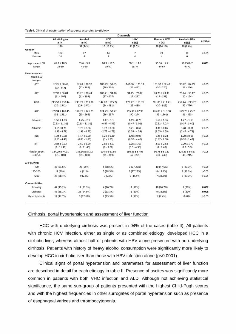
Table I. Clinical characterization of patients according to etiology
Diagnosis
All etiologies
n (%) Alcohol
n (%) HCV n (%)
HBV n (%)
Alcohol + HCV n (%)
Alcohol + HBV n (%)
p-value
116 51 (44%) 16 (13.8%) 11 (9.5%) 28 (24.1%) 10 (8.6%) Gender
Male 102 47 14 7 24 10 >0.05 Female 14 4 2 4 4 0
Age mean ± SD 61.9 ± 10.5 65.6 ± 9.8 60.3 ± 11.5 60.1 ± 14.8 55.36 ± 5.5 58.25±8.7 0.001
range 28-89 46-89 39-77 28-74 44-67 46-72
Liver analytics mean ± SD
(range)
AST 87.25 ± 68.48
(22 - 412)
57.61 ± 30.97 (22 - 182)
108.29 ± 59.55 (26 - 234)
143.36 ± 121.13 (25 - 412)
101.32 ± 62.48 (30 - 270)
93.22 ± 87.49 (29 - 256)
>0.05
ALT 67.93 ± 56.84 (11 - 407)
43.26 ± 30.44 (11 - 193)
108.71 ± 94.16 (27 - 407)
94.45 ± 73.42 (17 - 237)
74.73 ± 43.39 (19 - 158)
73.44 ± 36.17 (20 - 154)
>0.05
GGT 213.52 ± 238.44 (20 - 1542)
243.79 ± 293.36 (29 - 1542)
142.07 ± 115.72 (24 - 491)
179.27 ± 151.76 (25 - 480)
201.05 ± 211.41 (20 - 925)
252.44 ± 243.26 (52 - 776)
>0.05
ALP 159.94 ± 169.45 (52 - 1561)
170.77 ± 121.29 (65 - 666)
124.29 ± 53.77 (56 - 237)
155.36 ± 67.96 (90 - 274)
176.09 ± 310.88 (52 - 1561)
129.78 ± 75.7 (81 - 323)
>0.05
Bilirubin 1.59 ± 1.63 (0.33 - 11.31)
1.75 ± 2.1 (0.33 - 11.31)
1.47 ± 1.1 (0.47 - 4.56)
1.19 ± 0.76 (0.47 - 3.02)
1.68 ± 1.35 (0.52 - 7.03)
1.27 ± 1.13 (0.37 - 3.40)
>0.05
Albumin 3.65 ±0.71 (1.93 - 4.78)
3.70 ± 0.66 (1.93 - 4.72)
3.77 ± 0.60 (2.77 - 4.73)
3.71 ± 0.62 (2.59 - 4.59)
3.36 ± 0.85 (2.05 - 4.59)
3.78 ± 0.81 (2.44 - 4.78)
>0.05
INR 1.24 ± 0.38 (0.85 - 4.40)
1.17 ± 0.20 (0.85 - 1.85)
1.29 ± 0.30 (1 - 1.95)
1.48 ± 0.98 (0.97 - 4.40)
1.25 ± 0.23 (0.87 - 1.60)
1.20 ± 0.15 (0.99 - 1.42)
>0.05
pPT 2.84 ± 2.32 (0 - 11.40)
2.60 ± 2.29 (0 - 11.40)
2.88 ± 2.67 (0 - 9.00)
2.28 ± 1.67 (0.5 - 4.90)
3.69 ± 2.58 (0 - 8.40)
2.29 ± 1.77 (0.2 - 5.9)
>0.05
Platelet count (x109/L
124.29 ± 74.91 (31 - 409)
135.16 ± 87.72 (31 - 409)
104.5 ± 67.66 (31 - 269)
160.36 ± 57.93 (67 - 251)
96.78 ± 51.29 (31 - 249)
129.33 ± 69.67 (45 - 215)
>0.05
aFP (ng/ml)
<20 48 (51.6%) 28 (65%) 5 (38.5%) 3 (27.25%) 10 (47.6%) 3 (33.3%) >0.05
20-200 19 (20%) 4 (11%) 5 (38.5%) 3 (27.25%) 4 (19.1%) 3 (33.3%) >0.05
>200 28 (28.4%) 9 (24%) 3 (23%) 5 (45.5%) 7 (33.3%) 3 (33.3%) >0.05
Co-morbidities
Smoking 47 (45.2%) 17 (33.3%) 4 (26.7%) 1 (10%) 18 (66.7%) 7 (70%) 0.002
Diabetes 43 (38.1%) 28 (54.9%) 2 (13.3%) 1 (10%) 9 (33.3%) 3 (30%) 0.008
Hyperlipidemia 14 (12.7%) 9 (17.6%) 2 (13.3%) 1 (10%) 2 (7.4%) 0 (0%) >0.05
Cirrhosis, portal hypertension and assessment of liver function
HCC with underlying cirrhosis was present in 94% of the cases (table II). All patients
with chronic HCV infection, either as single or as combined etiology, developed HCC in a
cirrhotic liver, whereas almost half of patients with HBV alone presented with no underlying
cirrhosis. Patients with history of heavy alcohol consumption were significantly more likely to
develop HCC in cirrhotic liver than those with HBV infection alone (p<0.0001).
Clinical signs of portal hypertension and parameters for assessment of liver function
are described in detail for each etiology in table II. Presence of ascites was significantly more
common in patients with both VHC infection and ALD. Although not achieving statistical
significance, the same sub-group of patients presented with the highest Child-Pugh scores
and with the highest frequencies in other surrogates of portal hypertension such as presence
of esophageal varices and thrombocytopenia.

Table II. Characterization of cirrhosis, liver function and tumor burden stratified by underlying liver disease.
Diagnosis
All etiologies
n (%) Alcohol
n (%) HCV n (%)
HBV n (%)
Alcohol + HCV n (%)
Alcohol + HBV n (%)
p-value
Cirrhosis present 102 (94.4%) 45 (97.8%) 15 (100%) 5 (55.6%) 28 (100%) 9 (90%) <0.000 absent 6 (5.6%) 1 (2.2%) 0 (0%) 4 (44.4%) 0 (0%) 1 (10%)
Child-Pugh >0.05
A 42 (43%) 19 (45.5%) 7 (53.8%) 6 (54.5%) 6 (27.3%) 4 (44.4%) B 40 (41%) 17 (40.5%) 5 (38.5%) 4 (36.4%) 10 (45.4%) 4 (44.4%) C 15 (16%) 6 (14.3%) 1 (7.7%) 1 (9.1%) 6 (27.3%) 1 (11.2%)
Platelet count (x10
6/L) >0.05
<100.000 48 (48%) 20 (46.5%) 8 (57.1%) 2 (18.2%) 14 (60.9%) 4 (44.4%) >100.000 52 (52%) 23 (53.5%) 6 (42.9%) 9 (81.8%) 9 (39.1%) 5 (55.6%)
Esophageal varices >0.05 present 46 (52.3%) 21(56.8%) 5(45.5%) 2 (22.2%) 15 (68.2%) 3 (33.4%) absent 42 (47.7%) 16 (43.2%) 6 (54.5%) 7 (77.8%) 7 (31.8%) 6 (66.7%)
Ascites 0.017
present 28 (26.2%) 12 (26.1%) 0 (0%) 1 (9.1%) 12 (46.2%) 3 (30%) absent 79 (73.8%) 34 (73.9%) 14 (100%) 10 (90.9%) 14 (53.8%) 7 (70%)
Number of lesions >0.05
1 59 (55%) 27 (57.4%) 9 (60%) 4 (40%) 13 (52%) 6 (60%) 2-3 15 (14%) 8 (17%) 3 (20%) 0 (0%) 3 (12%) 1 (10%) >3 33 (31%) 12 (25.5%) 3 (20.%) 6 (60%) 9 (36%) 3 (30%)
Size of solitary lesions >0.05
<2 cm 5 (9.6%) 1 (4.1%) 2 (28.6%) 1 (33.3%) 1 (8.3%) 0 (0%) 2 - 5 cm 31 (59.6%) 13 (54.2%) 4 (57.1%) 1 (33.3%) 8 (66.7%) 5 (83.3%) >5 cm 16 (30.8%) 10 (41.7%) 1 (14.3%) 1 (33.3%) 3 (25%) 1 (16.7%)
Size of multinodular lesions
n 32 13 6 2 7 4 mean size (mm) ± SD
57.47 ± 40.6
65.54 ± 49.9 76.67 ± 43.9 42 ± 11.3 38 ± 17.9 50.75 ± 34.0
range (20 - 198) (21 - 198) (25 - 132) (34 – 50) (20 - 70) (23 - 100)
Portal vein invasion >0.05 present 13 (11.7%) 4 (8.2%) 1 (6.7%) 2 (18.2%) 5 (19.2%) 1 (10.0%) absent 98 (88.3%) 45 (91.8%) 14 (93.3%) 9 (81.8%) 21 (80.8%) 9 (90%)
Tumor characterization
Tumor burden and vascular invasion are described in table II. No association was seen
between higher tumor burden or presence of portal vein invasion and underlying etiology.
Multinodular tumors were more likely to present with portal vein thrombosis than solitary
tumors (20.8% versus 3.4%, respectively; p<0.004). Tumor pattern (solitary or multinodular
lesions) was not associated with the serum level of alpha-fetoprotein (using both cut-offs of
20 and 200ng/ml). No dependent correlation was found between this tumor marker and
nodule size. Patients diagnosed during screening were more likely to be seen with solitary
tumor than unscreened patients (70% versus 49%; p<0.05) and with smaller mean size of
solitary tumors (33.9 mm versus 59.8 mm; p<0.003). However, such patients were not more
commonly diagnosed with tumors smaller than 2 cm.

Staging
BCLC stage could be determined for 96 patients (table III). Patients at stage A had
mainly solitary tumors ≤5cm (91%) and where seen with approximately equal proportion of
Child A (45.5%) and Child B (54.5%). Child-Pugh score distribution was not significantly
different in patients diagnosed at intermediate stage (59% in Child-Pugh A and 41% in Child-
Pugh B). Only three (20%) patients at terminal stage had portal invasion at time of diagnosis.
Patients diagnosed during HCC screening were more likely to be found at earlier HCC
stages (BCLC classification 0 or A) (p<0.003).
Table III. Clinical characterization according to BCLC classification.
Early HCC stage
n (%) Not Early HCC stage
n (%) p-value
Diagnosis
Screened 20 (52.6%) 18 (47.4%) <0.003
Not Screened 11(22.4%) 38 (77.6%)
Etiology >0.05
Alcohol 33.3% 66.7%
HCV 42.9% 57.1%
HBV 30% 70%
HCV + Alcohol 25% 75%
HBV + Alcohol 44.4% 55.6%
BCLC 0 n (%)
BCLC A n (%)
BCLC B n (%)
BCLC C n (%)
BCLC D n (%)
All patients 2 (2%) 33 (34%) 39 (40%) 7 (7.4%) 15 (15.6%)
Child-Pugh >0.05*
A 2 (100%) 15 (45.5%) 23 (59%) 1 (14.3%) 0 (0%)
B 0 (0%) 18 (54.5%) 16 (41%) 6 (85.7%) 0 (0%)
C 0 (0%) 0 (0%) 0 (0%) 0 (0%) 15 (100%) -
Tumor burden >0.05†
Solitary 2 (100%) 30 (91%) 11 (28%) 1 (14.3%) 8 (53.3%)
Multinodular (>2 lesions)
0 3 (9%) 28 (72%) 6 (85.7%) 7 (46.7%)
* Comparison between patients classified with stage A versus patients at stage B † Comparison between patients classified with stage B versus patients at stage C
Treatment
Information on treatment performed (table IV) was available in 110 patients and three
groups were defined: those treated with a potentially curative treatment; those who only had
palliative treatment; and those who only received supportive treatment. Potential curative
treatment was undertaken by 44 (40%) patients as follows: 4 resections, 22 liver transplants
and 18 ablations by radiofrequency. Neo-adjuvant treatments were performed in 12 patients
(10 of the liver transplanted, one of resection and one of RFA) and 5 of the transplanted
patients underwent downstaging using TACE/TAE. The second group of patients consisted
of 52 (47%) patients performing exclusively palliative treatments, of which 46 had multiple
sessions of TACE/TAE (a total of 127 sessions, with a range of 1 to 6 consecutive sessions
per patient) and 6 patients received only sorafenib. For 14 (13%) patients only supportive
care was offered. Comparing patients from the two groups of treatment, potentially curative
treatment and palliative treatment, the former ones were more likely to have been classified
with earlier stages of HCC (p<0.001) and to be seen within Milan criteria (p<0.000). No
differences were seen for Child score or for frequency of portal vein invasion. All patients
receiving only symptomatic care had been classified as non-early HCC. Comparing these
with treated patients, such patients were more likely to exceed Milan criteria (p<0.05), to
present with portal vein invasion (p<0.000) and with higher Child-Pugh scores (p<0.000).
Table IV. Staging, liver function and tumor burden according to intention of treatment.
Intention-to-cure Palliative p-value Treated patients Supportive p-value
BCLC <0.001 - Early HCC 23 (60.5%) 12 (24%) 35 (39.8%) 0 (0%) Non-Early HCC 15 (39.5%) 38 (76%) 53 (60.2%) 14 (100%)
Child-Pugh >0.05 <0.000
A 20 (57.1%) 20 (43.3%) 40 (49.4%) 2 (14.3%) B 11 (31.4%) 22 (47.8%) 33 (40.7%) 5 (35.7%) C 4 (11.4%) 4 (8.7%) 8 (9.9%) 7 (50%)
Milan Criteria <0.000 <0.05
within 30 (76.9%) 14 (28%) 44 (49.4%) 3 (21.4%) without 9 (23.1%) 36 (72%) 45 (50.6%) 11 (78.6%)
Portal vein invasion >0.05 <0.000
Present 2 (4.8%) 3 (5.8%) 5 (5.3%) 6 (42.9%) Absent 40 (95.2%) 49 (94.2%) 89 (94.7% 8 (57.1%)
Outcome and survival analysis
Forty-three patients died during follow-up, of which 39 had death caused by hepatic
failure or HCC progression and for the remaining 4 information on death cause was
unavailable. Required information for survival analysis was available in a total of 105 patients,
of which 39 (37%) died, 41 (39%) were confirmed to be alive at the end of follow-up and 25
(24%) were lost to follow-up. The median follow-up period was 15 months (range: 0–67).
Median overall survival (OS) estimates using Kaplan-Meier method for the entire cohort was
35 months and the 1- and 3-year survival rate was 80% and 48%, respectively (fig. 1). The
median, 1-year and 3-years OS estimates for treated patients were 46 months, 90% and
55%, respectively (fig. 2). Median and 1-year OS for patients that just received supportive
care were 5 months and 25% (fig. 2); patient survival in this group did not exceed 15 months.
Treated patients had significantly higher OS than those in the later group (p<0.0001).

surv
ival
rat
e (
% )
survival period (months)
Fig. 1. Kaplan-Meier overall survival (OS) estimation for the entire cohort (n=105
patients).
surv
ival
rat
e (
% )
survival period (months)
Fig. 2. Kaplan-Meier overall survival (OS) estimation stratified by patients
receiving any treatment (n=88 patients) and patients receiving only supportive
care (n=14 patients). Log-rank test of survival rate: treated versus supportive
care, p<0.0001).
OS treated patients
OS supportive care
OS Cohort
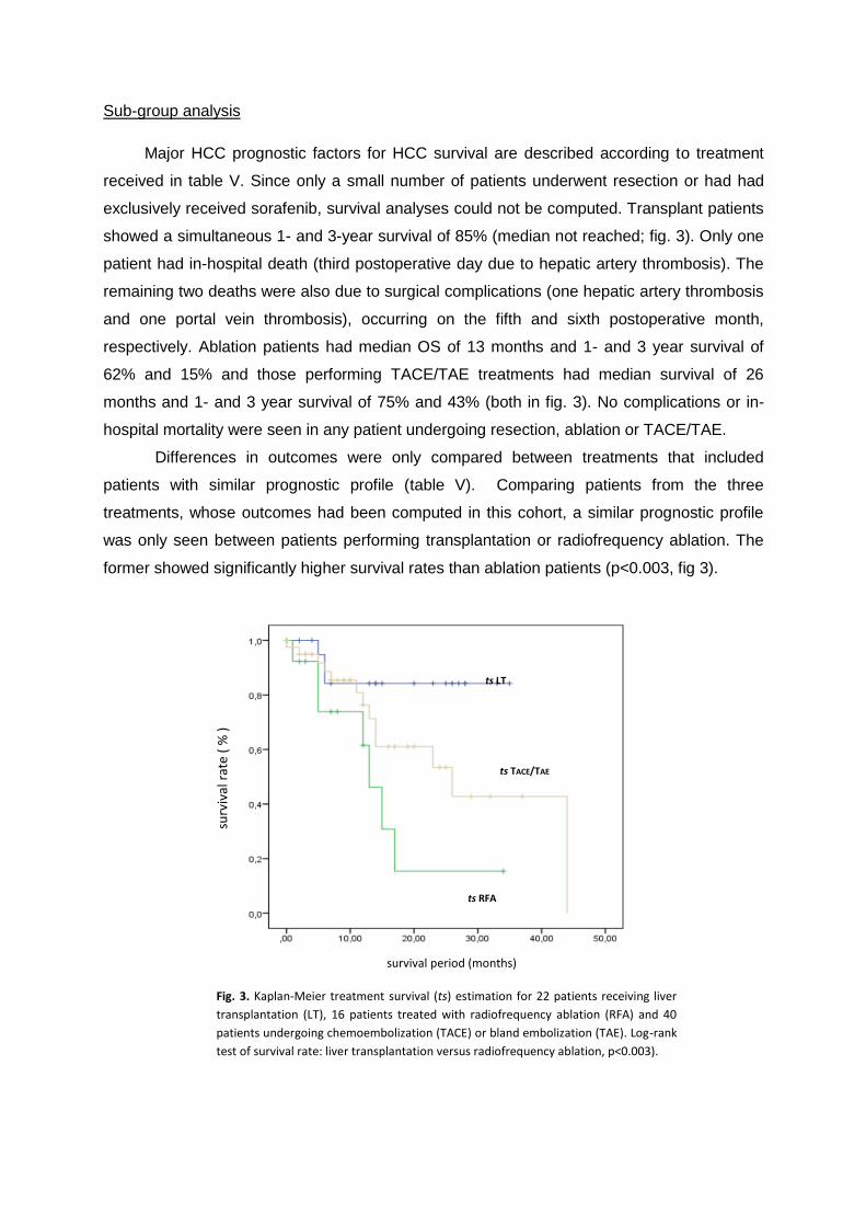
Sub-group analysis
Major HCC prognostic factors for HCC survival are described according to treatment
received in table V. Since only a small number of patients underwent resection or had had
exclusively received sorafenib, survival analyses could not be computed. Transplant patients
showed a simultaneous 1- and 3-year survival of 85% (median not reached; fig. 3). Only one
patient had in-hospital death (third postoperative day due to hepatic artery thrombosis). The
remaining two deaths were also due to surgical complications (one hepatic artery thrombosis
and one portal vein thrombosis), occurring on the fifth and sixth postoperative month,
respectively. Ablation patients had median OS of 13 months and 1- and 3 year survival of
62% and 15% and those performing TACE/TAE treatments had median survival of 26
months and 1- and 3 year survival of 75% and 43% (both in fig. 3). No complications or in-
hospital mortality were seen in any patient undergoing resection, ablation or TACE/TAE.
Differences in outcomes were only compared between treatments that included
patients with similar prognostic profile (table V). Comparing patients from the three
treatments, whose outcomes had been computed in this cohort, a similar prognostic profile
was only seen between patients performing transplantation or radiofrequency ablation. The
former showed significantly higher survival rates than ablation patients (p<0.003, fig 3).
surv
ival
rat
e (
% )
survival period (months)
Fig. 3. Kaplan-Meier treatment survival (ts) estimation for 22 patients receiving liver
transplantation (LT), 16 patients treated with radiofrequency ablation (RFA) and 40
patients undergoing chemoembolization (TACE) or bland embolization (TAE). Log-rank
test of survival rate: liver transplantation versus radiofrequency ablation, p<0.003).
ts LT
ts TACE/TAE
ts RFA

Table V. Major prognostic factors for survival of patients with hepatocellular carcinoma, stratified by treatment received.
Resection Transplant RFA TACE/TAE Sorafenib p-value 1* p-value 2 † p-value 3 ‡
Age (mean ± SD) 73.67 ± 6.66 55.76 ± 5.16 62.18 ± 11.56 65.12±8.99 55.0± 8.75 >0.05 <0.0001 >0.05 Child-Pugh >0.05 >0.05 >0.05
A 3 (100%) 8 (47.1%) 9 (60%) 19 (47.5%) 1 (16.7%) B 0 7 (41.2%) 4 (26.7%) 18 (45%) 4 (66.7%) C 0 2 (11.8%) 2 (13.3%) 3 (7.5%) 1 (16.7%)
Number of lesions >0.05 >0.05 <0.003
1 3 (75%) 13 (68.4%) 15 (88.2%) 21 (47.7%) 1 (16.7%) 2-3 0 (0%) 2 (10.5%) 2 (11.8%) 10 (22.7%) 0 (0%) > 3 1 (25%) 4 (21.1%) 0 (0%) 13 (29.5%) 5 (83.3%)
Size of solitary lesion >0.05 <0.002 <0.0001
< 2 cm 0 1 (7.7%) 2 (13.3%) 1 (4.8%) 0 (0%) 2-5 cm 0 11 (84.6%) 13 (86.7%) 10 (47.6%) 1 (100%) > 5cm 3 (100%) 1 (7.7%) 0 10 (47.8%) 0 (0%)
Milan criteria >0.05 <0.002 <0.0001
Within 0 13 (72.2%) 17 (100%) 13 (29.5%) 1 (16.7%) without 4 (100%) 5 (27.8%) 0 31 (70.5%) 5 (83.3%)
Portal Vein >0.05 >0.05 >0.05
Present 0 1 (4.8%) 1 (5.9%) 1 (2.2%) 2 (33.3%) Absent 4 (100%) 20 (95.2%) 16 (94.1%) 45 (97.8%) 4 (66.7%)
Ascites >0.05 >0.05 >0.05
present 0 3 (14.3%) 5 (31.3%) 8 (18.6%) 3 (50%) Absent 4 (100%) 18 (85.7%) 11 (68.8%) 35 (81.4%) 3 (50%)
Bilirubin >0.05 >0.05 >0.05
≤2.0 3 (100%) 14 (87.5%) 12 (85.7%) 34 (81%) 3 (60%) >2.0 0 (0%) 2 (12.5%) 2 (14.3%) 8 (19%) 2 (14.3%)
aFP (ng/ml) >0.05 <0.005 <0.05
<200 3 (100%) 16 (100%) 14 (93.3) 25 (62.5%) 3 (50%) ≥200 0 (0%) 0 (0%) 1 (6.7%) 15 (37.5%) 3 (50%)
* Comparison of prognostic factors between patients receiving liver transplantation with patients undergoing radiofrequency ablation
† Comparison of prognostic factors between patients receiving liver transplantation and those performing palliative chemoembolization
‡ Comparison of prognostic factors between patients undergoing radiofrequency ablation and those performing palliative chemoembolization

Discussion
The aim of this retrospective cohort study is to increase the availability of
epidemiological data concerning HCC patients in Portugal, in particular clinicopathological
characterization, etiological profile and survival outcomes in patients treated at a tertiary care
center in the North of Portugal.
The majority of patients were male (88%) and the mean age at presentation of all
patients was 61.3 ± 10.5 years. These findings are in line with several European studies that
also reported HCV infection and harmful alcohol consumption as leading etiologic causes for
HCC (33) (34), (35) (36) (20) (10). In our cohort study alcohol consumption was the most frequent risk
factor, presenting either as a single etiology (44%) or in combination with HCV or HBV
infection (32.7%). The total proportion of patients HCV infected (35%) or HBV infected (17%)
was similar to the range of reported infection rates reported in some European HCC series (37)
(38) (39) (33). However, presence of important alcoholic consumption history in 72% of our
patients with confirmed HCC represents an unusually high proportion compared to those
described in current literature. Assessing alcohol consumption poses several difficulties that
may lead to overdiagnosis (40). However the probability of such hypothesis may be hinder by
three facts. Firstly, a local study addressing the clinical characterization of HCC patients
(n=84) at a different institution also reported a considerably high prevalence (63.2%) of
alcohol as risk factor for HCC (41). Secondly, according to the OECD – Health Data (2008)
report Portugal has the 8th higher mean of annual alcohol consumption per capita in the world
(11.4l) (42). Thirdly, our institution is located in the North of Portugal, a region with higher
prevalence of alcohol consumption than in other regions in mainland (43). Additionally, results
from a Portuguese national study evaluating the burden of disease attributable to alcohol
drinking (44) reported significant male gender predominance in those with excessive drinking
(>60g ethanol/d). Considering these facts and adding together the high proportion of reported
alcohol history in our patients may explain the very high male-to-female ratio in our study
(7.3:1).
HCC predominantly arose in clinically evident or histologically proven liver cirrhosis
(94%), which has been also seen in large cohort European studies (34) (39) (20) (14). Irrespective
of the underlying liver disease, cirrhosis is a major factor able to modulate HCC risk and the
annual risk of developing HCC in cirrhotic liver is estimated between 1% to 6% (11). Although
the risk of cirrhosis is higher for those with hepatitis infection (2), alcoholic cirrhosis is probably
the most important risk factor for HCC in populations with low prevalence of HBV and HCV
infection and low exposure to aflatoxins (45), such as developed Western countries (9). Once

cirrhosis is established, a higher risk of HCC correlates with the severity of portal
hypertension (46) and liver stiffness as measured by transient elastography (47) (48). In the
present study, chronic HCV infection and alcohol abuse were etiologic factors very likely to
develop HCC in a cirrhotic liver, whereas the occurrence of HCC in non-cirrhotic liver was
more likely to happen in HBV-infected patients. Of note, patients with comorbid HCV
infection and alcoholic liver disease were seen with more severe portal hypertension. These
associations are in agreement with those described in the literature that we briefly review
below.
Alcohol drinking is associated with an increased risk of developing cirrhosis and liver
cancer (49). Although HCC can develop in patients with alcohol-induced liver disease who do
not have cirrhosis (40), it occurs remarkably more frequently in a cirrhotic liver (45). This finding
is consistent with the concept that the hepatocarcinogenicity of alcohol abuse is most likely
due to the development of cirrhosis (12). Recently, a meta-analysis estimated the relative risk
(RR), according to specific doses of alcohol, for developing cirrhosis and liver cancer with
adjustment for gender, age and hepatitis viral infection (50). Estimated RR of developing liver
cirrhosis for doses of 50g/day and for 100g/day were 7.13 (95%CI 6.36–8.00) and 26.52
(95%CI 22.26–31.59), respectively; the RR of liver cancer in absence of cirrhosis for an
alcohol intake of 50g/day and for 100g/day were 1.40 (95%CI 1.25–1.56) and 1.81 (95% CI
1.50–2.81), respectively. In a large Swedish cohort (51), the relative risk for HCC in patients
having cirrhosis due to alcoholic liver disease was 22.4 (95% CI 16.8–29.2) times higher than
the risk in the general population.
In HCV-infected patients alone, HCC occurs predominately in those with cirrhosis (52),
(53), (54), (55), (56), although it can occur, less commonly, in its absence (37), (57), (3). The risk for
developing cirrhosis 20 years after initial HCV infection among those chronically infected
varies between studies, but is estimated at around 10-15% for mean and 1-5% for women (58).
The annual incidence of HCC in subjects with established HCV-related cirrhosis is estimated
to be 0.5%-5% in Western countries (46) (59) (60). Recently, the HALT-C trial (Hepatitis C Long-
Term Treatment against Cirrhosis) (46) proposed a score (61) for risk stratification of HCC in
patients with chronic HCV infection, comprising older age, black race, lower platelet count,
higher alkaline phosphatase, esophageal varices and smoking as predictors of higher risk.
Such risk stratification is important to optimize and improve the cost-effectiveness of
surveillance programs.
Cirrhosis occurs in 20 to 30% of HBV-infected patients (62), (63). In contrast with HCV
infection and heavy alcohol consumption, around 40% of chronic HBV infected patients
develop HCC in non-cirrhotic liver (64). In this cohort, a similar proportion of non-cirrhotic
patients were found in those solely infected with HBV. Long-term follow-up studies have

demonstrated that approximately 2% per year of patients with cirrhosis due to HBV infection
develop HCC (65).
Additionally, a considerable amount of patients had combined etiologies (32%), in
whom the mean age at diagnosis of HCC was lower than those with a single etiology. In
patients with chronic viral hepatitis induced either by HCV infection or HBV infection, the
effect of alcohol abuse has been well described in other studies as having a synergic effect
(66) (67) and that progression of liver disease severity is very rapid and aggressive (68). The co-
existence of these etiological HCC causes (alcohol and HBV or alcohol and HCV infection)
are associated with increased risk of developing both cirrhosis and HCC and with HCC
occurring at an earlier age (69) (70) (71). Some studies advocate some explanations such as
alcohol’s ability to enhance both HBV and HCV replication and also through its oxidative
stress mechanisms, among others more thoroughly described elsewhere (72) (40), (73), (52) (74).
Diagnosis of HCC
Regarding diagnosis, only 11% of patients had HCC confirmed on a nodule less than 2
cm, of which half had to perform biopsy for histological confirmation. The majority of patients
were diagnosed with tumor size bigger than 2 cm. The risk of satellites and microscopic
vascular invasion greatly increases beyond this size cut-off and levels of tumor recurrence
and treatment failure rise exponentially as the tumor burden increases (75) (76). HCC diagnosis
in small tumors represents a major clinical challenge. Firstly, the radiological hallmark in such
tumors only occurs in a small proportion of patients (77) and secondly, even with dynamic
contrast-enhanced MRI and 4-phase multidetector CT, up to 25-30% of small tumors are
expected to be undernoted (78). Biopsy is still required in most instances (2), but nonetheless,
sensitivity of liver biopsy depends itself upon nodule size and pathological diagnosis is
particularly complex for nodules between 1 and 2 cm (79). Currently, up to 30% of new HCC
diagnoses in Japan correspond to small tumors of less than 2 cm, due to widespread
surveillance programs (80). Although only 5-10% of patients in the West are presently
diagnosed with very-early HCC, but this is expected to rise in parallel with the wider
implementation of surveillance policies (23). Surveillance for HCC aims to reduce disease-
related mortality by increasing the number of patients diagnosed within early stages of
disease and therefore enhancing the applicability and cost–effectiveness of curative
therapies (2). Although HCC surveillance is generally accepted, its implementation is
suboptimal in real-life practice (81). Early cirrhosis is usually clinically unapparent, and a
substantial proportion of patients develop hepatocellular carcinoma before cirrhosis is
recognized (82) (83).
In this cohort only 45% of the patients had been diagnosed during surveillance
programs, which is a suboptimal adherence (<60%) (84). Though diagnosis of tumors smaller

than 2 cm was not significantly more common in screened patients, it is noteworthy that such
patients were more frequently diagnosed with tumors within Milan criteria and inferior mean
size of solitary tumor, therefore achieving significant migration of HCC diagnosis to earlier
stages (BCLC stage 0 and A). As result of implementation of surveillance in Western
countries among cirrhotic patients, the trend of diagnosing patients in earlier HCC stages is
expected to rise from 5-10% (until 1990) to up to 40-60% in 2010-2020 (23). According to
BCLC classification, 36% of patients in this cohort were classified as early HCC stages, 47%
were found in intermediate or advanced stage and 16% were in terminal stage. Such
distribution between early and non-early stages was similar to those reported in other studies
using BCLC classification (85) (86) (87) (88).
Treatment outcomes
Consistent with current HCC-treatment state-of-art, treatment decision had impact on
patient prognosis. The twenty-two patients receiving liver transplantation showed the best
prognosis among all patients: a 1 and 3-year survival rate up to 85%, which was similar to
that reported in recent HCC surgical series (89) (90) (91) (92) (93). However, outcome evaluation in
liver transplantation should also include the 5-year survival rate and recurrence-free rate
which could not be computed due to short cohort period and because information relating to
recurrence was not systematically retrieved from medical records. Recurrence of tumor is a
major factor in evaluation of long-term outcomes of liver transplantation in HCC patients.
Optimal selection of patients according to Milan criteria has significantly improved rates in
terms of 5-year survival (from less than 40% up to 70%) and recurrence (from 32-54% (94) to
below 15% (95)). Tumor recurrence might result from previously undetected extrahepatic
metastasis or from release of tumor cells during surgical manipulation, which includes
transplantation itself and also previous interventional therapy such as ablation or
chemoembolization (96).
Compared with other studies with patients performing radiofrequency ablation with
tumor within Milan criteria (97) (98) (99) (100) (101) (102), survival in this cohort was found to be inferior
for both time cut-offs (reported 1-year survival rate ranged from 87% to 98% and 3-year
survival from 57% to 82%). Known independent predictors of survival in radiofrequency
ablation are initial complete response, Child-Pugh score, number or size of nodules and
baseline alpha-fetoprotein levels (2). Except for tumor response, which was not assessed,
patients in this cohort were seen with similar clinicopathological characteristics as for Child-
Pugh score, serum bilirubin level, tumor number and size, baseline alpha-fetoprotein and
additionally age. Approximately 60% of our ablation patients had tumor <3 cm, which is an
important size cut-off for the expected rate of complete tumor necrosis (103). When reviewing

individually ablation patients, a significant number of patients presented with clinical signs of
portal hypertension such as platelet count <100.000 (47%), ascites (31%) and esophageal
varices (56%). In the above studies, presence of esophageal varices was not systematically
described. For the remaining two, only presence of ascites (range: 0 to 9%) was
considerably lower than the proportion found in ablation patients in this cohort. Additionally,
with exception of one cohort study (102), etiology distribution was also a obvious difference:
where most of these studies had mainly patients with viral etiology (range from 82% to 92%
whereas in this study only 68%), alcoholic liver disease alone was considerably more
frequent (range from 8% to 14% compared to 34% in this sub-group of patients). However, in
this study alcoholic etiology did not seem to be associated with worst liver function or severity
of portal hypertension.
Overall, the median survival for intermediate HCC cases is expected to be about 20
months after chemoembolization (2). In this study, no distinction was made between TACE
and TAE. Although bland embolization is no longer recommended by the EASL guidelines (2),
a meta-analysis of randomized controlled trials found no differences in survival outcomes
between these two sub-types of treatment (104). Considering the two randomized trials (105) (106)
where survival benefit from chemoembolization was first identified as reference for
comparison, patients in this cohort have shown similar median overall survival estimates
(range: 17 – 28.7 months) and 1-year survival rate (range: 57 – 82%). However, estimate for
3-year survival was considerably higher in this cohort (43% versus a range from 26 up to
29%). Comparing with the study were best outcomes were achieved (105), patients had
matching Child-Pugh score, tumor size, absence of portal vein invasion, serum bilirubin level
and proportion of patients with ascites. In this study, a slightly higher preponderance of
solitary tumor (47.7% versus 32%) and fewer patients with <100 ng/ml of alpha-fetoprotein
(55% versus 82.5%) were seen. As in the sub-group of patients treated with radiofrequency
ablation, alcoholic liver disease as the sole etiology was much more frequent (56.5% versus
11%).
Limitations of this study
The quality of data reported was to some extent impaired due unavailability of complete
clinical, analytical and radiological information in the electronic charts. Additionally, as this
cohort analyzed data in a referral hospital and because both digital and physical records
exist, a considerable amount of patients had essential information for optimal HCC
characterization only attached in the latter. A more thorough sub-group data analysis in this
cohort was also limited to small sample size of some sub-groups of patients, precluding
outcome analysis in resection patients and those treated exclusively with sorafenib. Also,

treatment outcome analysis might be biased due to necessary grouping to overcome
statistical limitations of small sized samples. In this cohort, a significant proportion of patients
underwent multiple therapies such as neo-adjuvant treatments and downstaging in patients
receiving potentially curative treatments. Nevertheless, current state-of-art is controversial
about the benefit or disadvantage in such cases as robust data from randomized controlled
trials are lacking at the time of writing of this report.

References
1. International Agency for Resarch on Cancer. Cancer Fact Sheet. GLOBOCAN 2008 - Section
of Cancer Information. [Online] Whorld Health Organization, 2010. [Cited: 13 May 2013.]
http://globocan.iarc.fr/factsheet.asp.
2. European Association for the study of the liver, European Organisation for research and
treatment of cancer. EASL_EORTC Clinical practice guidelines: management of hepatocellular
carcinoma. Journal of Hepatology, 2012. 56; 908-943.
3. Hiotis S, Rahbari N, Villanueva G, Klegar E, Luan W, Wang Q, Yee H. Hepatitis B vs. hepatitis
C infection on viral hepatitis-associated hepatocellular carcinoma. Gastroenterology. 2012,
12:64.
4. European Association for the study of the Liver. EASL Clinical practical guidelines:
management of alcoholic liver disease. Journal of Hepatology, 2012. 57 j 399–420.
5. Powell EE, Cooksley WG, Hanson R, Searle J, Halliday JW, Powell LW. The natural history of
nonalcoholic steatohepatitis: a follow-up study of forty-two patients for up to 21 years.
Hepatology. 1990. 11(1): 74-80.
6. JM, Clark. The epidemiology of nonalcoholic fatty liver disease in adults. Journal Clinical
Gastroenterology. 2006: 40(3) 1: S5-10.
7. Danaei G, Finucane M, Lu Y, Singh G, Cowan M, Paciorek C, Lin J, Farzadfar F, Khang Y,
Stevens G, Rao M, Ali M, Riley L, Robinson C, Ezzati M,. National, regional, and global trends
in fasting plasma glucose and diabetes prevalence since 1980: systematic analysis of health
examination surveys and epidemiological studies with 370 country-years and 2·7 million
participants. The Lancet. 2011. 378: 31-40.
8. Takuma Y, Nouso K. Nonalcoholic steatohepatitis-associated hepatocellular carcinoma: Our
case series and literature review. World Journal of Gastroenterology. 2010; 16(12): 1436–1441.
9. Blachier M, Leleu H, Peck-Radosavljevic M, Valla DC, Roudot-Thoraval F. The burden of liver
disease in Europe: a review of available epidemiological data. Switzerland : European
Association for the study of the liver, 2013. ISBN 978-2-8399-1176-4.
10. Marinho RT, Giria J, Moura M. Rising costs and hospital admissions for hepatocellular
carcinoma in Portugal (1993-2005). World Journal of Gastroenterology . 2007; 13(10): 1522-
1527.
11. Roncalli M, Terracciano L, Tommaso L, Davi E, Colombo M. Liver precancerous lesions and
hepatocellular carcinoma: The histology report. Digestive and Liver Disease. 2011. (43): 361–
372.
12. Trevisiani F, Frigerio M, Santi V, Grignaschi A, Bernardi M. Hepatocellular carcinoma in
non-cirrhotic liver: a reappraisal. Digestive and Liver Disease. 2010. 42(5): 341-347.
13. El-Serag HB. Hepatocellular carcinoma. New England Journal Medical. 2011;365:1118–
1127.
14. Fattovich G, Stroffrolini T, Zagni I, Donato F. Hepatocellular Carcinoma in Cirrhosis:
Incidence and Risk Factors. Gastroenterology . 2004;127: 35–50.
15. Llovet JM, Fuster J, Bruix J. The Barcelona Approach: Diagnosis, Staging and Treatment of
Hepatocellular Carcinoma. Liver Transplantation. 2004. 10: 115-120.
16. patients., Natural history of hepatocellular carcinoma and prognosis in relation to
treatment. Study of 850. Okuda K, Ohtsuki T, Obata H, Tomimatsu M, Okazaki N, Hasegawa H,
Nakajima Y, Ohnishi K. Cancer. 1985 Aug 15; 56(4):918-28.
17. Llovet JM, Brú C, Bruix J. Prognosis of hepatocellular carcinoma: the BCLC staging
classification. Seminars in Liver Disease. 1999; 19(3):329-38.
18. A new prognostic system for hepatocellular carcinoma: a retrospective study of 435
patients: the Cancer of the Liver Italian Program (CLIP) investigators. Hepatology. 1998 Sep;
28(3):751-5.
19. Kudo M, Chung H, Osaki Y. Review Prognostic staging system for hepatocellular carcinoma
(CLIP score): its value and limitations, and a proposal for a new staging system, the Japan
Integrated Staging Score (JIS score). Journal of Gastroenterology. 2003; 38(3):207-15.
20. op den Winkel M, Nagel D, Sappl J, op den Winkel P, Lamerz R, Zech CJ, Straub G, Nickel T,
Rentsch M, Stieber P, Göke B, Kolligs FT. Prognosis of Patients with Hepatocellular Carcinoma.
Validation and Ranking of Established Staging-Systems in a Large Western HCC-Cohort.
Pubmed. [Online] 5 de October de 2012. [Citação: 2 de May de 2013.]
http://www.ncbi.nlm.nih.gov/pmc/articles/PMC3465308/. 10.1371/journal.pone.0045066.
21. Bruix J, Sherman M. Management of Hepatocellular Carcinoma. Hepatology. 2005, 42(5):
1208–1236.
22. Song DS, Bae SH. Changes of guidelines diagnosing hepatocellular carcinoma during the
last ten-year period. Clincal and MolecularHepatology. 2012;18:258-267.
23. Llovet J., Bruix J. Novel advancements in the management of hepatocellular carcinoma in
2008. Journal of Hepatology. 2008. 48(1): S20-S37.
24. Takayama T, Makuuchi M, Hirohashi S, Sakamoto M, Yamamoto J, Shimada K. Early
hepatocellular carcinoma as an entity with a high rate of surgical cure. Hepatology. 1998. 28:
1241-1246.
25. Royaie S, Blume IN, Thung SN, Guido M, Fiel MI, Hiotis S. A system of classifying
microvascular invasion to predict outcome after resection in patients with hepatocellular
carcinoma. Gastroenterology. 2009. 137: 850-855.
26. Livraghi T, Meloni F, Di Stasi M, Rolle E, Solbiati L, Tinelli C,. Sustained complete response
and complications rates after radiofrequency ablation of very early hepatocellular carcinoma in
cirrhosis: is resection still the treatment of choice? Hepatology . 2008.47: 82–89.

27. Arii S, Yamaoka Y, Futagawa S, Inoue K, Kobayashi K, Kojiro M, et al. Results of surgical
and nonsurgical treatment for small-sized hepatocellular carcinomas: a retrospective and
nationwide survey in Japan. The Liver Cancer Study Group of Japan. Hepatology. 2000.
32:1224-1229.
28. Llovet JM, Bruix J. Systematic review of randomized trials for unresectable hepatocellular
carcinoma: chemoembolization improves survival. Hepatology. 2003;37:429–442.
29. Llovet JM, Ricci S, Mazzaferro V, Hilgard P, Gane E, Blanc JF, de Oliveira AC, Santoro A,
Raoul JL, Forner A, Schwartz M, Porta C, Zeuzem S, Bolondi L, Greten TF, Galle PR, Seitz JF,
Borbath I, Häussinger D et al. SHARP Investigators Study Group. Sorafenib in advanced
hepatocellular carcinoma. New England Journal of Medicine. 2008. 359: 378–390.
30. J., Wiegand e T., Berg. The etiology, diagnosis and prevention of liver cirrhosis. Part 1 of a
series of liver chirrosis. Deutsches Aerzteblatt International. 2013; 110(6): 85-91.
31. Lee SS, Byoun Y-S, Jeong S-H, Kim Y-M, Gil H, in B-Y,Seong M, Jang ES, Kim J-W. Type and
causes of liver disease in Korea: single center experience, 2005-2010. Clinical and Molecular
Hepatology. 2012; 18:309-315.
32. Pugh RN, Murray-Lyon IM, Dawson JL, Pietroni MC, Williams R. Transection of the
oesophagus for bleeding oesophageal varices. British Journal of Surgery. 1973. 60(8): 646-649.
33. Schöniger-Hekeke M, Müller C, Kutilek M, Oesterreicher C, Ferenci P, Gangl A.
Hepatocellular carcinoma in Central Europe: prognostic features and survival. Gut. 2001; 48(1):
103-109.
34. Borie F, Trètarre B, Bouvier A-M, Faivre J, Binder F, Launoy G, Delafosse P, Tissot J, Peng J,
Grousclaude P, Guizard A-V, Gras-Aygon C. Primitive liver cancers: epidemiology and
geographical study in France. European Journal of Gastroenterology & Hepatology. 2009.21(9):
984-989.
35. Van Roey G, Fevery J, Van Steenbergen W. Hepatocellular carcinoma in Belgium: clinical
and virological characteristics of 154 consecutive cirrhotic and non-cirrhotic patients. European
Journal of Gastroenterology and Hepatology. 2000;12:61–66.
36. Donato F, Tagger A, Chiesa R, Ribero ML, Tomasoni V, Fasola M, Gellatti U, Portera G,
Boffetta P, Nardi G. Hepatitis B and C virus infection, alcohol drinking, and hepatocellular
carcinoma: a case-control study in Italy. Bresica HCC study. Hepatology. 1997; 26:579-584.
37. Bralet MP, Règimbeau JM, Pineau P, Dubois S, Loas G, Degos F, Valla D, Belghiti J, Degott
C, Terris B. Hepatocellular carcinoma occurring in nonfibrotic liver: epidemiologic and
histopathologic analysis of 80 French cases. Hepatology. 2000;32:200-204.
38. Chiesa R, Donato F, Tagger A, Favret M, Ribero ML, Nardi G, Gelatti U, Bucella E, Tomasi E,
Portolani N, Bonetti M, Bettini L, Pelizzari G, Salmi A, Savio A, Garatti M, Callea F. Etiology of
hepatocellular carcinoma in Italian patients with and without cirrhosis. Cancer Epidemiology
Biomarkers & Prevention. 2000;9:213–216.
39. Schütte K, Kipper M, Kahl S, Bornschein J, Götze T, Adolf D, Arend J, Seidensticker R,
Lippert H, Ricke J, Maltfertheiner P. Clinical characteristics and time trends in etiology of
cancer in Germany. Digestion. 2013. 87:147-159.
40. Morgan T, Mandayam S, Jammal M. Alcohol and Hepatocellular Carcinoma.
Gastroenterology . 2004;127:S87–S96.
41. Leitão, Daniel Basílio. Caracterização Clínico-Patológica do Carcinoma Hepatocelular em
doentes diagnosticados e tratados no IPO-Porto e avaliação de sobrevivência dos doentes
registados no Registo Oncológico da Região Norte (RORENO). Porto : Instituto de Ciências
Biomédicas Abel Salazar. Universidade do Porto., 2010.
42. Organisation for Economic Co-Operation and Development. Health Status.
OECD.StatExtracts. [Online] 2012. [Cited: 18 May 2013.]
http://stats.oecd.org/index.aspx?DataSetCode=HEALTH_STAT.
43. Marques-Vidal P, Dias C. Trends and Determinants of Alcohol Consumption in Portugal:
Results From the National Health Surveys 1995 to 1996 and 1998 to 1999. Alcoholism: Clinical
and Experimental Research. 2005, Vol 29; No 1; pp 89–97.
44. Cortez-Pinto H, Gouveia M, Pinheiro L, Costa J, Borges M, Carneiro A. The Burden of
Disease and the Cost of Illness Attributable to Alcohol Drinking—Results of a National Study.
Alcoholism: Clinical and Experimental Research. Vol 34, No 8, 2010: pp 1442–1449.
45. Chuang SC, La Vecchia C, Boffetta P. Liver cancer: Descriptive epidemiology and risk
factors other than HBV and HCV infection. Cancer Letters. 2009. 286: 9-14.
46. Lok AS, Seeff LB, Morgan TR, Bisceglie AM, Sterling RK, Curto TM, Everson GT, Lindsay KL,
Lee WM, Bonkovsky HL, Dienstag JL, GhanyMG, Morishima C, Goodman ZD, HALT-C Trial
Group. Incidence of Hepatocellular Carcinoma and Associated Risk Factors in Hepatitis C-
Related Advanced Liver Disease . Gastroenterology. 2009. 136 (1): 138-148.
47. Masuzaki R, Tateishi R, Yoshida H, Goto E, Sato T, Ohki T. Prospective risk assessment for
hepatocellular carcinoma development in patients with chronic hepatitis C by transient
elastography. Heptaology. 2009. 49: 1954-1961.
48. Jung KS, Kim SU, Ahn SH, Park YN, Kim do Y, Park JY, et al. Risk assessment of hepatitis B
virus-related hepatocellular carcinoma development using liver stiffness measurement
(FibroScan). . Hepatology . 2011. 53:885–894.
49. P. Boffetta, M. Hashibe. Alcohol and cancer. Lancet Oncology. 2006. 7: 146-156.
50. Corrao G, Bagnardi V, Zambon A, La Vecchia C. A meta-analysis of alcohol consumption
and the risk of 15 diseases. Preventive Medicine. 2004. 38: 613-619.
51. Kuper H, Ye W, Broomé U, Romelsjo A, Mucci LA, Ekbom A, Adami HO, Trichopoulos D,
Nyrén O. The risk of liver and bile duct cancer in patients with chronic viral hepatitis,
alcoholism or cirrhosis. Hepatology. 2001. 34: 714-718.

52. Roudot-Thoraval F, Bastie A, Pawlotsky JM, Dhumeaux D. Epidemiological factors
affecting the severity of hepatitis C virus-related liver disease: a French survey of 6,664
patients. The Study Group for the Prevalence and the Epidemiology of Hepatitis C Virus.
Hepatology. 1997. 26(2): 485-90.
53. Simonetti RG, Camma C, Fiorello F, Cottone M, Rapicetta M, Marino L, Fiorentino G, Craxì
A, Ciccaglione A, Giuseppetti R, Stroffolini T, Pagliaro L. Hepatitis C virus infection as a risk
factor for hepatocellular carcinoma in patients with cirrhosis. A case control study. Annals of
Internal Medicine. 1992. 116: 97-102.
54. Fattovich G, Giustina G, Degos F, Tremolada F, Diodati G, Almasio P, Nevens F, Solinas A,
Mura D, Brouwer J, Thomas H, Njapoum C, Casarin C, Bonetti P, Fuschi P, Basho J, Tocco A,
Bhalla A, Galassini R, Noventa F, Schlam SW, Realdi G. Morbidity and mortality in
compensated cirrhosis type C: a retrospective follow-up sutdy of 384 patients.
Gastroenterology. 1997. 112: 463-472.
55. Benvegnu L, Gios M, Boccatto S, et al. Natural history of compensated viral cirrhosis: a
prospective study on the incidence and the hirarchy of major complications. Gut. 2004.
53:744-749.
56. Degos F, Christidis C, Ganne-Carrie N, Farmachidi J-P, Degott C, Guettier C, Trinchet J-C,
Beaugrand M, Chevret S. Hepatitis C virus related cirrhosis: time to occurence of
hepatocellular carcinoma and death. Gut. 2000. 47:131-136.
57. Le Treut YP, Pons J, Hardwigsen J, et al. Hepatocellular carcinoma in a non-cirrhotic liver.
Presentation of series of 77 operated patients. Chirurgie. 1999. 124: 485-493.
58. Yu ML, Chuang WL. Treatment of chronic hepatitis C in Asia: when East meets West.
Journal of Gastroenterology and Hepatology. 2009. 24(3): 336-345.
59. Seef LB, Hoofnagle JH. Epidemiology of hepatocellular carcinoma in areas of low hepatitis
B and hepatitis C endemicity. . Oncogene. 2006. 25: 3771-3777.
60. El-Serag HB, Rudolph KL. Hepatocellular carcinoma: epidemiology and molecular
carcinogenesis. Gastroenterology. 2007. 132: 2557-2576.
61. HALT-C Formula for predicting risk of HCC. Hepatitis C Long-Term Treatment aginst
Cirrhosis Trial. [Online] [Cited: 25 May 2013.] http://www.haltctrial.org/hccform.html.
62. Mota A, Areias J, Cardoso MF. Chronic liver disease and cirrhosis among patients with
hepatitis B virus infection in nothern Portugal with reference to the viral genotypes. Journal of
Medical Virology. 2010; 83(1): 71-77.
63. Cadranel JF, Lahmek P, Causse X, Bellaiche G, Bettan L, Fontanges T, Medini A, Henrion J,
Chousterman M, Condat B, Hervio P, Periac P, Eugène C, Moindrot H, Grasset D, Nouel O,
Pilette C, Szostak-Talbodec N, Cayla JM, Si-Ahmed SN, Dumouchel P, et al. Epidemiology of
chronic hepatitis B infection in France: risk factors for significant fibrosis--results of a
nationwide survey. Alimentary Pharmacology and Therapeutics. 2007. 26: 565-576.

64. M., Sherman. Hepatocellular Carcinoma: Epidemiology, Surveillance, and Diagnosis.
Seminars of Liver Disease. 2010. 30:3–16.
65. Ioannou G, Splan M, Weiss N, McDonald G, Beretta L, Lee S. Incidence and predictors of
hepatocellular carcinoma in patients with cirrhosis. Clinical Gastroenterology and Heptaology.
2007. 5(8): 938-945.
66. Donato F, Tagger A, Gelatti U, Parrinello G, Boffetta P, Albertini A, Decarli A, Trevisi P,
Ribero M, Martelli C, Porru S, Nardi G. Alcohol and Hepatocellular Carcinoma: The effect of
life time intake and hepatitits virus infection in men and women. American Journal of
Epidemiology. 2002. 155 (4): 323-331.
67. B., Gao. Interaction of alcoholic and hepatits viral proteins: implication in synergistic effect
of alcohol drinking and viral hepatitis on liver injury. Alcohol. 2002. 27: 69-72.
68. Mota A, Guedes F, Areias J, Pinho L, Cardos MF. Alcohol consumption among patients with
hepatitis B infection in nothern Portugal considering gender and hepatitis B virus genotype
differences. Alcohol. 2010. 44(2): 149-156.
69. Noda K, Yoshihara H, Suzuki K, Yamada Y, Kasahar A, Hayashi N, Fusamoto H, Kamada T.
Progression of type C chronic hepatitis to liver cirrhosis and hepatocellular carcinoma - its
relationship to alcohol drinking and the agr of transfusion. Alcoholism: Clinical and
Experimental Research. 1996. 20: 95a-100a.
70. Ohnishi K, Iida S, Iwama S, Goto N, Noumora F, Takashi M, Mishima A, Kono K, Kimura K,
Musha H, Kotota K, Okuda K. The effect of chronic habitual alcohol intake on the development
of liver cirrhosis and hepatocellular carcinoma: relation to hepatitis B surface antigen carriage.
Cancer. 1983; 49:672-677.
71. Pereira FE, Goncalves CS, Zago Mda P. The effect of ethanol intake on the development of
hepatocellular carcinoma in HBsAg carriers. Arquivo de Gastroenterologia. 1994. 126:460-468.
72. Larkin J, Clayton MM, Liu J, Feitelson MA. Chronic ethanol consumption stimulates
hepatitis B virus gene expression and replication in transgenic mice. Hepatology. 2001. 34:
792-797.
73. Aghemo A, Colombo M. Hepatocellular carcinoma in chronic hepatitis C: from bench to
bedside. Semminars in Immunopathology. 2013. 35 (1): 111-120.
74. Schiff E, Ozden N. Hepatitis C and Alcohol. National Institute on Alcohol Abuse and
Alcoholism - National Institutes of Health. [Online] 29 September 2004. [Cited: 18 May 2013.]
http://pubs.niaaa.nih.gov/publications/arh27-3/232-239.htm.
75. T., Roskams. Anatomic pathology impact on prognosis and response to therapy. Clinical
Liver Disease. 2011. 15: 245-259.
76. Llovet JM, Schwartz M, Mazzaferro V. Resection and liver transplantation for
hepatocellular carcinoma. Seminars of Liver Disease. 2005. 25: 181-200.
77. Bolondi L, Gaiani S, Celli N, Golfieri R, Grigioni WF, Leoni S, Venturi AM, Piscaglia F.
Characterization of small nodules in cirrhosis by assessment of vascularity: the problem of
hypovascular hepatocellular carcinoma. Hepatology. 2005. 42: 27–34.
78. Colli A, Fraquelli M, Casazza G, Massironi S, Colucci A, Conte D, Duca P. Accuracy of
ultrasonography, spiral CT, magnetic resonance, and alpha-fetoprotein in diagnosing
hepatocellular carcinoma: a systematic review. American Journal of Gastroenterology. 2006.
101(3): 513-523.
79. Roskmas T, Kojiro M. Pathology of early hepatocellular carcinoma: conventional and
molecular diagnosis. Seminars of Liver Disease. 2010. 30: 17-25.
80. M., Kudo. Review of 4th Single Topic Conference on HCC. Hepatocellular carcinoma:
intrenational consensus and controversies. . Hepatology Research. 2007. 37(2): S83-S87.
81. Yang JD, Kim WR. Surveillance for Hepatocellular Carcinoma in Patients With Cirrhosis.
Clinical Gastroenterology and Hepatology. 2012. 10: 16-21.
82. Stravitz RT, Heuman DM, Chand N, Sterling RK, Shiffman ML, Luketic VA, Sanyal AJ, Habib
A, Mihas AA, Giles HC, Maluf DG, Cotterell AH, Posner MP, Fisher RA. Surveillance for
hepatocellular carcinoma in patients with cirrhosis improves outcome. American Journal of
Medicine. 2008. 121 (2):119-126.
83. Toyoda H, Kumada T, Kiriyama S, Sone Y, Tanikawa M, Hisanaga Y, Yamaguchi A, Isogai M,
Kaneoka Y, Washizu J. Impact of surveillance on survival of patients with initial hepatocellular
carcinoma: a study from Japan. Clinical Gastroenterology and Hepatology. 2006. 4(9): 1170-
1176.
84. Forner A, Llovet JM, Bruix J. Hepatocellular carcinoma. Lancet. 2012. 379: 1245-1255.
85. Vitale A, Morales RR, Zanus G, Farinati F, Burra P, Angeli P, Frigo AC, Del Poggio P,
Rapaccini G, Di Nolfo MA, Benvegnù L, Zoli M, Borzio F, Gianni EG, Caturelli E, Chiaramonte
M, Trevisiani F, Cillo U. Barcelona Clinic Liver Cancer staging and transplant survival benefit for
patients with hepatocellular carcinoma: a multicentre, cohort study. The Lancet - Oncology.
2011. 12(7): 654-662.
86. Marrero JA, Fontana RJ, Barrat A, Askari F, Conjeevaram HS, Su GL, Lok AS. Prognosis of
Hepatocellular Carcinoma: comparison of 7 staging systems in an American Cohort.
Hepatology. 2005. 41: 707-716.
87. Cillo U, Bassanello M, Vitale A, Grigoletto FA, Burra P, Fagiuoli S, D'Amico F, Ciarleglio FA,
Boccagni P, Brolese A, Zanus G, D'Amico DF. The critical issue of hepatocellular carcinoma
prognostic classification: which is the best tool available? Journal of Hepatology. 2004.
40(1):124-131.
88. Sirivatanauksorn Y, Tovikkai C. Comparison of Staging Systems of Hepatocellular
Carcinoma. HPB Surgery. Hindawi Publishing Corporation, Volume 2011, Article ID 818217, 7
pages.
89. Bozorgzadeh A, Orloff M, Abt P, Tsoulfas G, Younan D, Kashyap R, Jain A, Mantry P,
Maliakkal B, Khorana A, Schwartz S. Survival outcomes in liver transplantation for
hepatocellular carcinoma, comparing impact of hepatitis C versus other etiology of cirrhosis.
Liver Transplantation. 2007. (13(6): 807-813.
90. Harnois DM, Steers J, Andrews JC, Rubin JC, Pitot HC, Burgart L, Wiesner RH, Gores GJ.
Preoperative hepatic artery chemoembolization followed by orthotopic liver transplantation
for hepatocellular carcinoma. Liver Transplantation and Surgery. 1999. 5(3): 192-199.
91. Figueras J, Jaurrieta E, Valls C, Benasco C, Rafecas A, Xiol X, Fabregat J, Casanovas T,
Torras J, Baliellas C, Ibañez L, Moreno P, Casais L. Survival after liver transplantation in
cirrhotic patients with and without hepatocellular carcinoma: a comparative study. Hepatology.
1997. 25(6): 1485-1489.
92. Kanwal F, Befeler A, Chari RS, Marrero J, Kahn J, Afdhal N, Morgan T, Roberts L, Mohanty
SR, Schwartz J, Van Thiel D, Li J, Zeringue A, Di'Biscglie A. Potentially curative treatment in
patients with hepatocellular cancer—results from the liver cancer research network.
Alimentary Pharmacology and Therapeutics. 2012. 36:257-265.
93. Facciuto ME, Rochon C, Pandey M, Rodriguez-Davalos M, Samaniego S, Wolf DC, Kim-
Schluger L, Rozenblit G, Scheiner P. Surgical dilemma: liver resection or liver transplantation
for hepatocellular carcinoma and cirrhosis. Intention-to-treat analysis in patients within and
outwith Milan criteria. HPB. 2009. 11: 398-404.
94. Kurtovic J, Riordan SM, Williams R. Liver transplantation for hepatocellular carcinoma.
Best Practice and Research Clinical Gasroenterology. 2005. 19:147-160.
95. Welker M-W. Bechstein W-O, Zeuzem S, Trojan J. Recurrent hepatocellular carcinoma
after liver transplantation – an emerging clinical challenge. Transplant international. 2013.
26(2): 109-118.
96. Kim YS, Lim him H, Lee WJ, Joh JW, Park CK. Recurrence of Hepatocellular Carcinoma after
Liver Transplantation: Patterns and Prognostic Factors Based on Clinical and Radiological
Features. American Journal of Radiology. 2007. 189(2): 352-358.
97. Huang J, Yan L, Chen Z, Wu H, Du L, Wang J, Xu Y, Zenq Y. A randomized trial comparing
radiofrequency ablation and surgical resection for HCC conforming to the Milan criteria. Annals
of Surgery. 2010. 252(6): 903-912.
98. Lupo L, Panzera P, Gianenelli G, Memeo M, Gentile A, Memeo V. Single hepatocellular
carcinoma ranging from 3 to 5 cm: radiofrequency ablation or resection. HPB (Oxford). 2007.
9(6): 429-434.
99. Shiina S, Teratani T, Obi S, Sato S, Tateishi R, Fujishima T, Ishikawa T, Koike Y, Yoshida H,
Kawabe T, Omata M. A randomized controlled trial of radiofrequency ablation with ethanol
injection for small hepatocellular carcinoma. Gastroenterology. 2005. 129(1):122-130.
100. Lencioni RA, Allgaier HP, Cioni D, Olschewski M, Deibert P, Crocetti L, Frings H,
Laubenberger J, Zuber I, Blum HE, Bartolozzi C. Small hepatocellular carcinoma in cirrhosis:
randomized comparison of radio-frequency thermal ablation versus percutaneous ethanol
injection. Radiology. 2003. 228(1):235-240.
101. Chen MS, Li JQ, Zheng Y, Guo RP, Liang HH, Zhang YQ, Lin XJ, Lau WY. A prospective
randomized trial comparing percutaneous local ablative therapy and partial hepatectomy for
small hepatocellular carcinoma. Annals of Surgery. 2006. 243(3): 321-328.
102. N'Kontchou G, Mahamoudi A, Aout M, Ganne-Carrié N, Grando V, Coderc E, Vicaut E,
Trinchet JC, Sellier N, Beaugrand M, Seror O. Radiofrequency ablation of hepatocellular
carcinoma: Long-term results and prognostic factors in 235 Western patients with cirrhosis.
Hepatology. 2009. 50(5):1475–1483.
103. Lu DS, Yu NC, Raman SS, Limanond P, Lassman C, Murray K, Tong MJ, Amado RG,
Busuttil RW. Radiofrequency ablation of hepatocellular carcinoma: treatment success as
defined by histologic examination of the explanted liver. Radiology. 2005. 234(3): 954-960.
104. Cammà C, Schepis F, Orlando A, Albanese M, Shahied L, Trevisani F, Andreone P, Craxì
A,Cottone M. Transarterial Chemoembolization for Unresectable Hepatocellular Carcinoma:
Meta-Analysis of Randomized Controlled Trials. Radiology. 2002. 224: 47-54.
105. Llovet JM, Real MI, Montaña X, Planas R, Coll S, Aponte J, Ayuso C, Sala M, Muchart J,
Solà R, Rodés J, Bruix J e Group, Barcelona Liver Cancer. Arterial embolisation or
chemoembolisation versus symptomatic treatment in patients with unresectable
hepatocellular carcinoma: a randomised controlled trial. Lancet. 2002.359(9319):1734-1739.
106. Lo CM, Ngan H, Tso WK, Liu CL, Lam CM, Poon RT, Fan ST, Wong J. Randomized
controlled trial of transarterial lipiodol chemoembolization for unresectable hepatocellular
carcinoma. Hepatology. 2002. 35(5):1164-1171.