HEPATO-NEPHROPROTECTIVE, HEMATOPOIETIC AND ANTI ...
10
www.ejbps.com 121 HEPATO-NEPHROPROTECTIVE, HEMATOPOIETIC AND ANTI-SPERMATOGENIC EFFECT OF THE ETHANOLIC EXTRACT OF JATROPHA TANJORENSIS USING MALE ALBINO RATS 1 *Iroanya, Onyekachi Ogbonnaya, 1 Egwuatu, Tochukwu Frank, 1 Chukwudozie, Onyeka, Fadogba, Tolulope David and Adesanya Adenike Department of Cell Biology and Genetics, University of Lagos, Akoka Yaba, Lagos Nigeria. Article Received on 06/03/2018 Article Revised on 26/05/2018 Article Accepted on 16/06/2018 INTRODUCTION Jatropha tanjorensis, commonly referred to as “hospital - too-far” or Ugu-Oyibo in Igbo language belongs to the family Euphorbiaceae. It is widely grown and used as a leafy vegetable and a medicinal plant in South Eastern Nigeria. It is used locally in treatment of malaria, ailments associated with the liver and kidney, infertility and improvement of hematologic indices. It is reported to have antidiabetic, hepatoprotective, hypoglycaemic and antioxidant properties and enhances the function of the bone marrow (Olayiwola, et al., 2004; Orhue et al ., 2008; Madubuike, et al., 2015). Phytochemical screening of J. tanjorensis leaf revealed that it contains bioactive principles such as alkaloids, flavonoids, tannins, cardiac glycosides, anthraquinones and saponins (Ehimwenma and Osagie, 2007). The testes (testicles) are two oval shaped organs located in the scrotum (Osuchukwu et al., 2016) and are the most essential organs of the male reproductive system. They are the glands where sperm and testosterone are produced (Keith et al., 2013). This implies that whatever affects the testes can influence sexual characteristic and fertility. Some plants have been used to improve semen quality e.g. Korean red ginseng (Hong, et al., 2002), Lepidium meyenii (Lee et al., 2016), while some have been used to improve hematopoiesis e.g. Sanguisorba officinalis (Chen et al., 2017). In the case of hepatic diseases, several species such as Silybum marianum, Phyllanthus niruri, and Panus giganteus have been shown to ameliorate hepatic lesions (Cacciapuoti et al., 2013). Plants used in traditional Complementary and Alternative medicine (CAM) for the treatment of different ailments and disorders are believed to be safe SJIF Impact Factor 4.918 Research Article ejbps, 2018, Volume 5, Issue 7, 121-130. European Journal of Biomedical AND Pharmaceutical sciences http://www.ejbps.com ISSN 2349-8870 Volume: 5 Issue: 7 121-130 Year: 2018 *Corresponding Author: Dr. Iroanya, Onyekachi Ogbonnaya Department of Cell Biology and Genetics, University of Lagos, Akoka Yaba, Lagos Nigeria. ABSTRACT This study was conducted to ascertain the genotoxic, sperm boosting, hematopoietic, hepato-nephron-protective, properties of the ethanolic extract of Jatropha tanjorensis (EEJT) using male Wistar albino rats. The animals were randomly assigned to five groups of six animals each. Animals in group 1 were administered 10mgkg -1 water, groups 2-4 received 200, 400 and 800mgkg -1 EEJT respectively while groups 5 received 300mgkg -1 of Addyzoa for 64 days. Micronucleus assay, sperm cell count, hematological, biochemical, and antioxidant potentials were evaluated using standard methods. At 800mgkg -1 , there was significant (p≤0.05) increase in the level of red blood cell (RBC), Haemoglobin (HGB) and Hematocrit (HCT) and significant (p≤0.05) decrease in ALP compared to Addyzoa. The extract dose dependently attenuated the concentrations of AST, ALP, total bilirubin, cholesterol, LDL, triglyceride and MDA compared to control and Addyzoa groups. Platelet, granulocytes, SOD, HDL, total protein RBC, HGB and HCT were dose dependently increased in the groups administered the extract compared to the control and Addyzoa groups. At 200mgkg -1 , EEJT had the lowest number of binucleated and micronucleus cells. The highest number of abnormal sperm cells were seen in the group administered 800mgkg -1 of the extract. This experiment shows that EEJT dose-dependently improved erythropoietin promoting potential and hepatoprotective activity in the experimental animal. There was marked reduction in sperm count with sperm head abnormality at very high dosages. However, more research should be carried out to ascertain its mechanism of action and the dose to be administered over a long period. KEYWORDS: Genotoxic, Sperm Booster, Hematopoietic, Hepato-Nephron-Protective, Micronucleus Assay, Antioxidant.
Transcript of HEPATO-NEPHROPROTECTIVE, HEMATOPOIETIC AND ANTI ...

Iroanya et al. European Journal of Biomedical and Pharmaceutical Sciences
www.ejbps.com
121
HEPATO-NEPHROPROTECTIVE, HEMATOPOIETIC AND ANTI-SPERMATOGENIC
EFFECT OF THE ETHANOLIC EXTRACT OF JATROPHA TANJORENSIS USING
MALE ALBINO RATS
1*Iroanya, Onyekachi Ogbonnaya,
1Egwuatu, Tochukwu Frank,
1Chukwudozie, Onyeka, Fadogba, Tolulope
David and Adesanya Adenike
Department of Cell Biology and Genetics, University of Lagos, Akoka Yaba, Lagos Nigeria.
Article Received on 06/03/2018 Article Revised on 26/05/2018 Article Accepted on 16/06/2018
INTRODUCTION
Jatropha tanjorensis, commonly referred to as “hospital-
too-far” or Ugu-Oyibo in Igbo language belongs to the
family Euphorbiaceae. It is widely grown and used as a
leafy vegetable and a medicinal plant in South Eastern
Nigeria. It is used locally in treatment of malaria,
ailments associated with the liver and kidney, infertility
and improvement of hematologic indices. It is reported to
have antidiabetic, hepatoprotective, hypoglycaemic and
antioxidant properties and enhances the function of the
bone marrow (Olayiwola, et al., 2004; Orhue et al.,
2008; Madubuike, et al., 2015). Phytochemical
screening of J. tanjorensis leaf revealed that it
contains bioactive principles such as alkaloids,
flavonoids, tannins, cardiac glycosides,
anthraquinones and saponins (Ehimwenma and
Osagie, 2007).
The testes (testicles) are two oval shaped organs located
in the scrotum (Osuchukwu et al., 2016) and are the
most essential organs of the male reproductive system.
They are the glands where sperm and testosterone are
produced (Keith et al., 2013). This implies that whatever
affects the testes can influence sexual characteristic and
fertility. Some plants have been used to improve semen
quality e.g. Korean red ginseng (Hong, et al., 2002),
Lepidium meyenii (Lee et al., 2016), while some have
been used to improve hematopoiesis e.g. Sanguisorba
officinalis (Chen et al., 2017). In the case of hepatic
diseases, several species such as Silybum marianum,
Phyllanthus niruri, and Panus giganteus have been
shown to ameliorate hepatic lesions (Cacciapuoti et al.,
2013).
Plants used in traditional Complementary and
Alternative medicine (CAM) for the treatment of
different ailments and disorders are believed to be safe
SJIF Impact Factor 4.918 Research Article
ejbps, 2018, Volume 5, Issue 7, 121-130.
European Journal of Biomedical AND Pharmaceutical sciences
http://www.ejbps.com
ISSN 2349-8870
Volume: 5
Issue: 7
121-130
Year: 2018
*Corresponding Author: Dr. Iroanya, Onyekachi Ogbonnaya
Department of Cell Biology and Genetics, University of Lagos, Akoka Yaba, Lagos Nigeria.
,
ABSTRACT
This study was conducted to ascertain the genotoxic, sperm boosting, hematopoietic, hepato-nephron-protective,
properties of the ethanolic extract of Jatropha tanjorensis (EEJT) using male Wistar albino rats. The animals were
randomly assigned to five groups of six animals each. Animals in group 1 were administered 10mgkg-1
water,
groups 2-4 received 200, 400 and 800mgkg-1
EEJT respectively while groups 5 received 300mgkg-1
of Addyzoa
for 64 days. Micronucleus assay, sperm cell count, hematological, biochemical, and antioxidant potentials were
evaluated using standard methods. At 800mgkg-1
, there was significant (p≤0.05) increase in the level of red blood
cell (RBC), Haemoglobin (HGB) and Hematocrit (HCT) and significant (p≤0.05) decrease in ALP compared to
Addyzoa. The extract dose dependently attenuated the concentrations of AST, ALP, total bilirubin, cholesterol,
LDL, triglyceride and MDA compared to control and Addyzoa groups. Platelet, granulocytes, SOD, HDL, total
protein RBC, HGB and HCT were dose dependently increased in the groups administered the extract compared to
the control and Addyzoa groups. At 200mgkg-1
, EEJT had the lowest number of binucleated and micronucleus
cells. The highest number of abnormal sperm cells were seen in the group administered 800mgkg-1
of the extract.
This experiment shows that EEJT dose-dependently improved erythropoietin promoting potential and
hepatoprotective activity in the experimental animal. There was marked reduction in sperm count with sperm head
abnormality at very high dosages. However, more research should be carried out to ascertain its mechanism of
action and the dose to be administered over a long period.
KEYWORDS: Genotoxic, Sperm Booster, Hematopoietic, Hepato-Nephron-Protective, Micronucleus Assay,
Antioxidant.

Iroanya et al. European Journal of Biomedical and Pharmaceutical Sciences
www.ejbps.com
122
nevertheless, adequate pharmacological assays should be
performed so as to ascertain their safety and efficacy. In
many cases, the mechanisms and modes of action of
these plants as well as their therapeutic effectiveness
have been confirmed in clinical studies.
In South Eastern Nigeria, it is used traditionally in
enhancing male fertility, hepatoprotection and as a blood
nourishing tonic. It is believed to facilitate production of
blood cells and platelets in the body. Therefore, this
study is aimed at determining the genotoxic,
hematopoietic, hepatoprotective, antioxidant and sperm
improving properties of the ethanolic extract of Jatropha
tanjorensis using male Wistar albino rats for 64 days.
MATERIALS AND METHODS
Plant Material and Preparation of Extract
Fresh Jatropha tanjorensis leaves were collected from a
residential farmyard in Abaranje, Ikotun Local
Government Area, Lagos Nigeria. They were
subsequently identified and authenticated at the
Department of Botany, University of Lagos, Nigeria and
was allocated a voucher specimen number LUH:7446.
The leaves were air dried at room temperature and finely
ground using Corona®
hand grinder. The dried powdered
leaves were soaked in 50 % ethanol for 48 hours. The
crude extract was weighed and the stock solution was
prepared using water.
Experimental Animals Thirty adult male Swiss albino rats weighing an average
of 175.5 ± 5g were purchased from the Laboratory
animal centre of the College of Medicine, University of
Lagos Nigeria. They were maintained under standard
laboratory conditions at the Experimental Animal House
of the Department of Cell Biology and Genetics, Faculty
of Science, University of Lagos with dark and light cycle
(12/12 hrs.) and fed with standard rat chow bought from
Ladokun feeds, Ibadan, Nigeria and clean tap water ad
libitum. All animal experiments were carried out in
accordance with the recommendations of the Guide for
the care and use of laboratory animals.
Drug Treatment Protocol
After an adaptive period of 1 week, the animals were
randomized and divided into five groups of six animals
each. The animals were weighed before and after
experiment.
Group 1: serve as normal control received distilled
water (10 ml kg-1
body weight p.o.) for 64 days.
Group 2: were treated with 200 mg kg-1
body weight of
the extract for 64 days.
Group 3: were treated with 400 mg kg-1
body weight of
the extract (GOV) for 64 days.
Group 4: were treated with 800 mg kg-1
body weight of
the extract (GOV) for 64 days.
Group 5: received a sperm boosting reference drug
Addyzoa (300 ml kg-1
body weight) for 64 days.
After the experimental period, the animals were
anesthetized mildly with ether and blood was collected
from the retro-orbital plexus. They were sacrificed and
more blood samples were collected by cardiac puncture
for evaluating the biochemical parameters and oxidative
stress. The testes were harvested and used for antioxidant
and epididymal sperm analysis. The livers and kidneys
were also dissected out for histology assay.
Haematologic Indices
An auto Hematology Analyzer, Mindray BC3200 was
used to determine Haemoglobin (HGB),
Hematocrit level, blood cell (RBC), mean corpusclar
volume (MCV), mean corpuscular haemoglobin (MCH)
and mean corpuscular hemoglobin concentration
(MCHC), Granulocytes, red blood cell distribution width
- Coefficient of variation (RDW-CV), red blood cell
distribution width - Standard deviation (RDW-SD),
Mean Platelet Volume (MPV), platelet count (PLT),
Platelet Distribution Width (PDW) and plateletcrit
(PCT).
Determination of Antioxidant Activity
Estimation of enzymic and nonenzymic antioxidants
activities e.g. Catalase (Sinha 1972), Reduced
Glutathione (Ellman, 1959), Glutathione-S-Transferase
(Habig and Jakoby, 1974), Superoxide Dismutase
(Kakkar et al., 1984) and lipid peroxidation level
(thiobarbituric acid reactive substances) (Niehaus and
Samuelsson, 1968) were determined using testes
samples.
Assessment of Biochemical Parameters Assessment of hepato- and nephro-protective activity
was performed by determining the activities of some
biochemical parameters e.g. Alkaline phosphatases
(ALP), alanine aminotransferase (ALT) and aspartate
aminotransferase (AST) enzyme activity, while the
chemical analytes were assessed by determining the total
bilirubin albumin, creatinine, cholesterol, low-density
lipoproteins (LDL), and high-density lipoproteins
(HDL), total protein, triglyceride and urea concentrations
in serum. These assays were carried out using Randox®
reagent kits and the procedures were followed as those
described in the literature available with the kits.
Epididymal Sperm Analysis Epididymis was separated carefully from each testis and
divided into 3 segments; head, body and tail. The
epididymal tail was trimmed with scissors and placed in
Petri dishes containing 1.0 ml of 0.1 M phosphate buffer
of pH 7.4. The dishes gently swirled for homogeneity
and allowed sperm diffusion in the solution for 10 min
under 37oC for dispersion of sperm cells. Sperm samples
were assessed for gross morphology of sperm cells.
Micronulear Assay
The animals were sacrificed, lower abdomen and limbs
were incised and the femora were cleaned and separated
from the hip joint. The ends of the femur were trimmed

Iroanya et al. European Journal of Biomedical and Pharmaceutical Sciences
www.ejbps.com
123
and a blunt needle was pushed to pierce the marrow
cavity. Bone marrow was flushed into a tube containing
0.9% saline. Smears were made on sterile coded slides
using a drop of the suspension. The slides were air dried,
fixed in absolute methanol and stained using May-
Grunwald Giemsa method (D’Souza et al., 2002).
Micronuclei were identified as dark blue staining bodies
in the cytoplasm of polychromatic and normochromatic
erythrocytes. One thousand polychromatic and
normochromatic erythrocytes each were scored per
animal for the presence of Micronucleus using light
microscope. The frequency of micro-nucleated cells was
expressed in percentage (Marzouk et al., 2012)
Histopathology. (Mallory, 1961)
A small chunk of liver and kidney were taken from the
sacrificed experimental rats used for hepatotoxicity
studies and were preserved in 10% formal saline for
histological studies. The tissues were processed and
sectioned in paraffin. The paraffin sections of buffered
formalin- fixed tissue samples (3 µm thick) were
dewaxed and rinsed in alcohol and also water. It was
stained with Harris' haematoxylin (Sigma) for 10
minutes, washed in running tap water for 1 minute,
differentiated in acid alcohol for 10 seconds and washed
again in running tap water for 5 minutes. The tissues
were stained with eosin for 4 minutes and washed in
running tap water for 10 seconds. It was dehydrated and
mounted for photomicroscopic observations of the
histological architecture of the different groups. The
general structure of the livers and kidneys of the normal
control group (group 1) was compared with those of the
treated groups (groups 2-5).
Statistical Analysis
The results were expressed as mean + SEM for six rats.
Statistical analysis of the data was performed using
ANOVA statistical SPSS package (15.0) version. The
significance of differences among all groups was
determined by the Tukey HSD test. P – values less than
0.05 (p ≤ 0.05) were considered to be statistically
significant.
RESULTS
Hematological Results
Table 1 shows the effect of Jatropha tanjorensis on the
haematologic indices in rat. There was significant
(p≤0.005) increase in HGB, RBC and HCT level of the
extract at 800 mgkg-1
compared to addyzoa, 200 mgkg-1
and 400 mgkg-1
groups. At 400 and 800 mgkg-1
, the RBC
level was statistically (p≤0.005) high compared to
control group. The extract at 800 mgkg-1
increased the
concentrations of HGB, RBC and HCT compared to all
the other groups. The MCV value of the control group
was significantly (p≤0.005) increased compared to the
extract at 200 and 800 mgkg-1
.
Table 1: Effect of Jatropha on the hematologic indices in rats.
GROUPS HGB
(g/dl)
RBC
(106/µl)
HCT
(%)
MCHC
(%)
MCV
(fl)
MCH
(pg)
CONTROL 16.6±0.2(b,c) 7.4 ±0.13(b,e) 41.2±1(b,c,d) 40.4 ±0.7 55.3±2.1(c,e) 22.5 ±0.42(c)
ADDYZOA 13.3 ±0.3(a,e) 6.5±0.15(a,d,e) 33.8±1(a,e) 39.3 ±0.6 52.0±1 20.4±0.1
Extract 200 12.3±1.2(a,e) 7.1±0.03(e) 31.6±2(a,e) 38.1±2.8 48±1.7(a) 19.6±1(a)
Extract 400 14.3±0.2(e) 7.2±0.08(b,e) 35.2±0.7(a,e) 40.73±1 49.3±1 20±0.4
Extract 800 19.1 ±0.7(b,c,d) 9.4 ±0.24(a,b,c,d) 45.4 ±0.5(b,c,d) 42.1±1.8 48.6±1.1(a) 20.4±0.94
Values are expressed as Mean ± SEM for six rats. The
Mean difference is significant at the 0.05 level. (a) = p ≤
0.05 as compared with the normal control group. (b) = p
≤ 0.05 as compared to Addyzoa group. (c) = p ≤ 0.05 as
compared with the 200 mg kg
-1 group. (d) = p ≤ 0.05 as
compared with the 400 mg kg
-1 group. (e) = p ≤ 0.05 as
compared with the 800 mg kg
-1 group. The
significance
of differences among all groups was determined by the
Tukey HSD test.
KEY: MCH: Mean Corpuscular Haemoglobin RBC: Red Cell Count HCT: Hematocrit
MCHC: Mean Corpuscular Haemoglobin Concentration MCV: Mean Corpuscular Volume HGB: Haemoglobin
In Table 2, the extract at 200 mgkg-1
significantly (p≤
0.005) attenuated the level of RDW-CV, RDW-SD,
MPV, PDW and PCT compared to the control group.
There was a significant (p≤ 0.005) increase in the
number of granulocytes in the 400 mgkg-1
group
compared to Addyzoa and the control groups. It also
significantly (p≤ 0.005) increased MPV and PCT
compared to Addyzoa. At 800 mgkg-1
, there was a
significant (p≤ 0.005) increase in the percentage of
Granulocytes compared to control, Addyzoa and the 200
mgkg-1
groups.
Table 2: Effect of Jatropha on the hematologic indices in rats.
GROUPS GRAN
(%)
RDW-CV
(%)
RDW-SD
(fl)
MPV
(fl)
PLT
(%)
PDW
(%)
PCT
(%)
CONTROL 11.4±0.5(d,e) 16.4±0.1(b,c,d,e) 30.7±0.3(b,c,d,e) 6.6±0.03(b,c,e) 961±16 16.14±0.06(c) 0.6±0.008(b,c,d,e)
ADDYZOA 9.7±2(d,e) 15.740(a,c,d,e) 27.8±0.3(a,e) 5.63±0.05(a,d) 1181±11 15±0.07(c) 0.12±0.02(a,c,d,e)
Extract 200 12.94±0.1(e) 15.06±0.08(a,b) 26.3±0.4(a,e) 5.8±0.35(a) 1095.4±28 6.9±3.3(a,b,de) 0.06±0.001(a,b,d,e)
Extract 400 18.6±2.2(a,b) 14.8±0.1(a,b) 26.4 ±0.14(a,e) 6.4 ±0.1(b) 1189.8±266.7 15.4±0.1(c) 0.24±0.015(a,b,c,e)
Extract 800 20±0.7(a,b,c) 14.7±0.2(a,b) 24.4 ±0.7(a,b,c,d) 5.8±0.04(a) 1316.6±46 14.4±0.2(c) 0.5±0.02(a,b,c,d)

Iroanya et al. European Journal of Biomedical and Pharmaceutical Sciences
www.ejbps.com
124
Values are expressed as Mean ± SEM for six rats. The
Mean difference is significant at the 0.05 level. (a) = p ≤
0.05 as compared with the normal control group. (b) = p
≤ 0.05 as compared to Addyzoa group. (c) = p ≤ 0.05 as
compared with the 200 mg kg
-1 group. (d) = p ≤ 0.05 as
compared with the 400 mg kg
-1 group. (e) = p ≤ 0.05 as
compared with the 800 mg kg
-1 group. The
significance
of differences among all groups was determined by the
Tukey HSD test.
KEY: RDW-CV: Red Cell Distribution Width (Coefficient of Variation) PLT: Platelet Count
GRAN: Granulocytes RDW-SD: Red Cell Distribution Width (Standard Deviation) MPV: Mean Platelet Volume
PCT: Plateletcrit PDW: Platelet Distribution Width
Biochemical Results
Table 3 shows the biochemical result after pre-treatment
of rats with different doses of Jatropha tanjorensis
extract and Addyzoa. There was significant (p≤ 0.005)
dose dependent attenuation in the concentrations of ALT
and AST on administration of the extract compared to
Addyzoa and the control group. At 800 mgkg-1
, the level
of ALP was attenuated significantly (p≤ 0.005)
compared to Addyzoa, 200 and 400 mgkg-1
. Compared
to all other groups, the extract at 200 and 400 mgkg-1
showed the lowest levels of total bilirubin and AST and
ALT respectively.
Table 3: Serum levels of ALP, ALT, AST and TBIL (Liver Function Enzymes) of rats treated with Jatropha.
Groups ALP
(U/L)
ALT
(U/L)
AST
(U/L) TOTAL BIL
CONTROL 372 ±8.4(b,c,d)
61 ±6(d)
667.2 ±3.1(b,c,d,e)
2.1 ±0.5
ADDYZOA 506.8 ±5(a,c,d,e)
52.5 ±1(d)
252.5 ±3(a,e)
1.4 ±0.1
Extract 200 649.08 ±17.7(a,b,e)
63.78 ± 0.7(d)
299.82 ± 7.8(a,e)
1.12 ± 0.3
Extract 400 609. 4±32.4(a,b,e)
38.26 ±2.5(a,b,c,e)
220.5 ±51(a,e)
1.22 ±0.15
Extract 800 400.4 ±5.2(b,c,d)
62.44 ±3.3(d)
434.4 ±47.6(a,b,c,d)
1.4 ±0.13
Values are expressed as Mean ± SEM for five rats. The
Mean difference is significant at the 0.05 level. (a) = p ≤
0.05 as compared with the normal control group. (b) = p
≤ 0.05 as compared to Addyzoa group. (c) = p ≤ 0.05 as
compared with the 200 mg kg
-1 group. (d) = p ≤ 0.05 as
compared with the 400 mg kg
-1 group. (e) = p ≤ 0.05 as
compared with the 800 mg kg
-1 group. The
significance
of differences among all groups was determined by the
Tukey HSD test.
KEY: AST: Aspartate amino transferase ALP: Alkaline phosphatase
ALT: Alanine amino transferase TOTAL BIL: Total bilirubin
In table 4, there was significant (p≤0.05) increase in
albumin (ALB) in the groups administered 200 and 400
mgkg-1
of the extract compared to the control group.
There was significant (p≤ 0.005) decrease in the
concentrations of cholesterol, and triglyceride in the
group administered 400 mgkg-1
of the extract compared
to the control group. LDL was significantly (p≤ 0.005)
attenuated in the Addyzoa, 200 and 800 mgkg-1
groups
compared to the 400 mgkg-1
group. The extract at 400
mgkg-1
, presented the lowest and highest concentrations
of LDL and total protein respectively while at 800 mg
kg-1
, the group had the highest concentration of HDL and
the lowest level of cholesterol and triglyceride. The
extract significantly (p≤ 0.005) attenuated the
concentration of triglyceride and cholesterol compared to
the control group.
Table 4: Serum levels of biochemical analytes in rats pre-treated with Jatropha.
Groups ALB
(g/L)
CHO
(mmol/L)
HDL CHO
(mmol/L)
LDL CHO
(mmol/L)
CREA
(mmol/L)
TOTAL
PROTEIN (g/L)
TRIG
(mmol/L)
UREA
(mmol/L)
CONTROL 26 ±1.3(b,c,d) 3 ±0.1(b,c,d,e) 0.34±0.04(e) 0.09±0.01(b,c,e) 36 ±2.6(b,e) 61 ±4 3.7±0.4(b,c,d,e) 5.4±0.13(b,d)
ADDYZOA 33.7 ±1(a,e) 2.3 ±0.1(a) 0.4 ±0.02 0.24 ±0.02(a,d) 25 ±0.8(a,c,d) 66 ±0.6 2±0.1(a) 4.3±0.1(a,c,d,e)
Extract 200 32.2 ±0.4(a) 2.2 ±0.05(a) 0.39 ±0.02 0.17 ±0.01(a,d) 35.02±0.6(b,e) 62.8 ±2 2.06±0.08(a) 5.3 ±0.2(b,d)
Extract 400 33.5±0.64(a) 2.14±0.06(a) 0.44 ±0.02 0.07±0.025(b,c,e) 37.34±1.5(b,e) 67.32±1.4 2.5±0.3(a,e) 6.2±0.15(a,b,c)
Extract 800 29.3±1.5(b) 2.13±0.13(a) 0.5±0.01(a) 0.2 ±0.01(a,d) 25.9±1.9(a,c,d) 60.6±1.6 1.2±0.1(a,d) 5.7±0.2(b)
Values are expressed as Mean ± SEM for five rats. The
Mean difference is significant at the 0.05 level. (a) = p ≤
0.05 as compared with the normal control group. (b) = p
≤ 0.05 as compared to Addyzoa group. (c) = p ≤ 0.05 as
compared with the 200 mg kg
-1 group. (d) = p ≤ 0.05 as
compared with the 400 mg kg
-1 group. (e) = p ≤ 0.05 as
compared with the 800 mg kg
-1 group. The
significance
of differences among all groups was determined by the
Tukey HSD test.
KEY:
CHO: Cholesterol LDL CHO: Low density protein
HDL CHO: High density protein ALB: Albumin
TRIG: Triglyceride CREA: Creatinine

Iroanya et al. European Journal of Biomedical and Pharmaceutical Sciences
www.ejbps.com
125
Table 5 shows the sperm count of the rats exposed to the
ethanolic extract of Jatropha tajorensis for the duration
of sixty-four (64) days. There was a significant (p≤
0.005) increase in the sperm count of the control and
addyzoa groups compared to the groups administered the
extract. The extract at 200 mgkg-1
, showed a higher
number of sperm cells compared to all other extract
groups while the group administered the extract at 800
mgkg-1
, showed a lower count compared to all the
groups.
Table 5: Sperm count of the rats exposed to the
ethanolic extract of Jatropha tajorensis and Addyzoa.
GROUPS Sperm count (106)
CONTROL 7.52 ± 0.69(c,d,e)
ADDYZOA 6.07 ± 0.52(c,d,e)
Extract 200 2.62 ± 0.34(a,b)
Extract 400 2.46 ± 0.39(a,b)
Extract 800 2.26 ± 0.2(a,b)
Values are expressed as Mean ± SEM for five rats. The
Mean difference is significant at the 0.05 level. (a) = p ≤
0.05 as compared with the normal control group. (b) = p
≤ 0.05 as compared to Addyzoa group. (c) = p ≤ 0.05 as
compared with the 200 mg kg
-1 group. (d) = p ≤ 0.05 as
compared with the 400 mg kg
-1 group. (e) = p ≤ 0.05 as
compared with the 800 mg kg
-1 group. The
significance
of differences among all groups was determined by the
Tukey HSD test.
Sperm Head Abnormality Analysis of the head abnormality was made after 64 days
of exposure to the ethanolic extract of Jatropha
tajorensis. The result of the sperm head abnormality
showed that there was high level of abnormality in the
sperm head of the rats exposed to the extract compared
to the control group. The major abnormality were pin
head, hook head and banana shaped head. The extract
(800 mg kg-1
) showed higher forms of sperm head
abnormality.
Micronucleus assay
A number of fragmented nucleus and binucleated were
observed under the microscope at X40 magnification as
shown in table 6. The group administered 800 mgkg-1
of
the extract, had the highest number of both binucleated
cells and micronucleus cells. Binucleated cells are cells
that contain two nuclei and this type of cells are found
mostly in cancer cells. Fragmented nucleus, which can
also be related to Karyorrhexis, is the irreversible
condensation of chromatin in the nucleus of a cell
undergoing necrosis or apoptosis.
Iroanya et al. European Journal of Biomedical and Pharmaceutical Sciences
www.ejbps.com
126
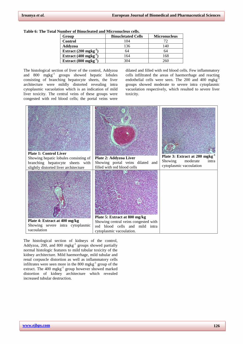
Table 6: The Total Number of Binucleated and Micronucleus cells.
Group Binucletated Cells Micronucleus
Control 104 72
Addyzoa 136 140
Extract (200 mgkg-1
) 64 64
Extract (400 mgkg-1
) 164 168
Extract (800 mgkg-1
) 304 260
The histological section of liver of the control, Addyzoa
and 800 mgkg-1
groups showed hepatic lobules
consisting of branching hepatocyte sheets, the liver
architecture were mildly distorted revealing intra
cytoplasmic vacuolation which is an indication of mild
liver toxicity. The central veins of these groups were
congested with red blood cells; the portal veins were
dilated and filled with red blood cells. Few inflammatory
cells infiltrated the areas of haemorrhage and reacting
endothelial cells were seen. The 200 and 400 mgkg-1
groups showed moderate to severe intra cytoplasmic
vacuolation respectively, which resulted to severe liver
toxicity.
Plate 1: Control Liver
Showing hepatic lobules consisting of
branching hepatocyte sheets with
slightly distorted liver architecture
Plate 2: Addyzoa Liver
Showing portal veins dilated and
filled with red blood cells
Plate 3: Extract at 200 mgkg
-1
Showing moderate intra
cytoplasmic vacoulation
Plate 4: Extract at 400 mg/kg
Showing severe intra cytoplasmic
vacoulation
Plate 5: Extract at 800 mg/kg
Showing central veins congested with
red blood cells and mild intra
cytoplasmic vacoulation.
The histological section of kidneys of the control,
Addyzoa, 200, and 800 mgkg-1
groups showed partially
normal histologic features to mild tubular toxicity of the
kidney architecture. Mild haemorrhage, mild tubular and
renal corpuscle distortion as well as inflammatory cells
infiltrates were seen more in the 800 mgkg-1
group of the
extract. The 400 mgkg-1
group however showed marked
distortion of kidney architecture which revealed
increased tubular destruction.

Iroanya et al. European Journal of Biomedical and Pharmaceutical Sciences
www.ejbps.com
127
Plate 6: Control Kidney Showing
partially normal histologic features
and mild tubular toxicity
Plate 7: Addyzoa Kidney: Showing partially normal
histologic features and mild tubular
toxicity
Plate 8: Extract at 200 mg/kg Showing mild tubular toxicity, mild
haemorrhage, mild tubular and
renal corpuscle distortion
Plate 9: Extract at 400 mg/kg Showing mild haemorrhage, mild
tubular and renal corpuscle
distortion as well as inflammatory
cells infiltrates
Plate 10: Extract at 800 mg/kg Showing marked distortion of
kidney architecture which revealed
increased tubular destruction
Plate 11: Control
Plate 12: Extract (200 mgkg
-1)
Plate 13: Extract (400 mgkg
-1)
Plate 14: Addyzoa group
Plate 15: Extract (800 mgkg
-1)
Plates 11 – 15 Shows the histological section of the testis of the experimental groups. Histological sections of
testicular specimens of all the groups showed normal histologic images.
The antioxidant effect on the testes of the rats exposed to
the ethanolic extract of the Jatropha tajorensis for sixty-
four (64) days are shown in table 7. There was
significant (p≤0.005) increase in the level of
Malondialdehyde (MDA) in the control group compared
to all groups while the extract at 800 mgkg-1
significantly
(p≤0.005) attenuated it compared to all groups. The level
of total protein was significantly (p≤0.005) reduced in
the groups that received 200 and 400 mgkg-1
of the
extract compared to the Addyzoa and control groups.
Addyzoa showed a significant (p≤0.005) increase in
catalase concentration compared to all the other groups
while the extract at 200 mgkg-1
demontrated a significant
(p≤0.005) decrease in catalase level compared to

Iroanya et al. European Journal of Biomedical and Pharmaceutical Sciences
www.ejbps.com
128
Addzoa, 400 and 800 mgkg-1
of the extract. The level of
GST was significantly (p≤0.005) increased in the extract
group administered 200 mgkg-1
compared to 400 and 800
mgkg-1
. The control group showed a significant
(p≤0.005) decreases in GSH compared to Addyzoa, 200
and 800 mgkg-1
. The SOD concentration at 400 mgkg-1
was significantly (p≤0.005) high compared to all the
groups.
Table 7: The antioxidant effect on the serum of the rats exposed to the ethanolic extract of the Jatropha
tajorensis for a duration of sixty four (64) days.
GROUPS MDA
(nmol/ml)
Total protein
(g/L)
CAT
(µmol/min/mg protein)
GST
(µmol/ml)
GSH
(µmol/ml)
SOD
(µmol/ml)
CONTROL 16.96± 0.95(b,c,d,e)
9.36± 0.97(c,d)
3.26 ±0.05(b,d,e)
4.42±0.09(b,c,d,e)
2.24 ±0.1(b,c,e)
2.74±0.05(b,c,d,e)
ADDYZOA 6.26 ±0. 1(a,c,e)
9.24 ±0.17(c,d)
11.46± 0.15(a,c,d,e)
2.82 ±0.06(a,d,e)
12 ±0.84(a,c,d,e)
6.36 ±0.1(a,c,d,e)
EXTRACT 200 10.12± 0.06(a,b,d,e)
6.56 ± 0.25(a,b)
3.32 ±0.18(b,d,e)
3.04 ±0.08(a,d,e)
6.7± 0.2(a,b)
7.16±0.11(a,b,d,e)
EXTRACT 400 7.4 ±0.51(a,c,e)
6.98 ±0.12(a,b)
4.66 ±0.11(a,b,c,e)
1.78 ±0.1(a,b,c)
4.6±0.2(b)
10.2 ±0.07(a,b,c,e)
EXTRACT 800 2.8± 0.23(a,b,c,d)
8.46 ±0.46 7.28 ±0.12(a,b,c,d)
2.04 ±0.12(a,b,c)
8.12 ±1.76(a,b)
3.7 ±0.14(a,b,c,d)
Values are expressed as Mean ± SEM for six rats. The
Mean difference is significant at the 0.05 level. (a) = p ≤
0.05 as compared with the normal control group. (b) = p
≤ 0.05 as compared to Addyzoa group. (c) = p ≤ 0.05 as
compared with the 200 mg kg
-1 group. (d) = p ≤ 0.05 as
compared with the 400 mg kg
-1 group. (e) = p ≤ 0.05 as
compared with the 800 mg kg
-1 group. The
significance
of differences among all groups was determined by the
Tukey HSD test.
DISCUSSION
Red blood cells (RBC) carry most of the body’s iron that
is vital for haemoglobin synthesis. There was a dose
dependent increase in hemoglobin level on
administration of the extract. Decreased haemoglobin
level is indicative of anaemia. The significant (p≤0.05)
dose-dependent increase in the red blood cell count
shows that the extract has hematopoietic properties and
might have increased the rate of production of
corpuscles. Erythropoietin affects the oxygen-carrying
capacity of the blood and the amount of oxygen
delivered to the tissues since red blood cells and
hemoglobin are very important in transferring respiratory
gases (Oyedeji and Bolarinwa, 2013). The Addyzoa
group however showed significant decrease in RBC
levels. The platelet count was increased suggesting that
the extract had a good effect on the oxygen-carrying
capacity of the blood and also on glycoprotein which is
the hormone responsible for the production of platelets
by the bone marrow (McLellan et al., 2003).
MCV and RDW are indices used to measure average size
of RBC’s in the blood and the variation in size of RBC’s
respectively, while MPV is used to measure the average
size of the platelets found in the blood. Low MPV levels
can be indicative of blood disorders or diseases. HCT is
the ratio of the volume of RBC’s to the volume of the
whole blood. The HCT levels were significantly (p≤0.05)
high in the extract groups at 800 mgkg-1
. The grup
administered the extract dose dependently caused an
increase in HGB, HCT and RBC levels compared to
control and Addyzoa groups. Normally, local tissue
anoxia apparently leads to the formation of
erythropoietin, thereby stimulating increased production
of erythrocytes (Bowman and Rand, 1980).
Erythropoietin is a glycoprotein hormone that stimulates
stem cells in the bone marrow producing red blood cells
(Ohlsson and Aher, 2009). It is most likely the extract
contains erythropoietin-like agent(s), compounds and or
phytochemicals that stimulate the formation or secretion
of erythropoietin in the stem cells of the animals that are
responsible for increased production of erythrocytes.
Previous studies by Okpuzor et al., (2009) and Kuppast
et al., (2009) indicated that an increase in the count of
erythrocytes and PCV is suggestive of polycythermia and
positive erythropoiesis. This is an indication of an
improved bone marrow function. This also implies
that the extract probably possesses anti-anemic potential
and can cause polychethermia.
The ALT, AST and ALP and serum bilirubin are
common biochemical parameter for detecting liver injury
(Girish et al., 2009). They are cytoplasmic in location
and released into circulation after cellular damages
indicative of development of hepatotoxicity. In this
study, treatment with ethanolic extract of Jatropha
tajorensis significantly (p ≤ 0.05) attenuated the increase
in ALP, AST and ALT levels in the test groups
compared to the control. This is an indication that the
extract improved plasma membrane stabilization and
protected against liver injury. The increase in albumin
activity shows high activity in the proliferation of cells
and high productivity of protein in the body. Cholesterol
is a critical fat that is a structural component of cell
membrane and plasma lipoproteins, and is important in
the synthesis of steroid hormones, glucocorticoids, and
bile acids. Decrease in the level of cholesterol is an
indicative of malnutrition, liver insufficiency,

Iroanya et al. European Journal of Biomedical and Pharmaceutical Sciences
www.ejbps.com
129
malignancies, anaemia and infection. There was also
increase in the level of HDL. The triglyceride level was
also reduced while the level of creatinine was increased
except from the Addyzoa group. The decreased
triglyceride level may be present in chronic obstructive
pulmonary disease, brain infarction, hyperthyroidism and
malnutrition. This indicates that the kidneys have
sufficient capacity to retain water content.
Based on the sperm count, the control and Addyzoa
groups, showed a significant increase, compared to the
groups administered the extract, which signifies that the
extract was not effective enough in boosting sperm
count. The group, administered the highest dose of 800
mgkg-1
showed a high reduction in the number of sperm
count compared to all groups. This might be an
indication that the highest dose of the extract decreased
the number of spermatogonium, spermatocytes,
spermatozoids and leydig cells. Sperm count is often
used as a measure of sperm production, testicular
function and male fertility. Low sperm count has been
associated with reduced fertility (Raji et al., 2003). For
the sperm abnormality, both the Addyzoa and the extract
groups showed abnormality. The highest extract of 800
mgkg-1
showed more abnormality compared to all
groups. More of banana shaped head and hook head was
found in the group administered 800 mgkg-1
of the
extract compared to other groups. Both sperm
abnormality and low sperm count is the major reason for
infertility in male reproduction process (Raji et al.,
2003).
In high quantities, endogenous reactive oxygen species
(ROS) overwhelm the innate antioxidant defence system
causing oxidative stress. Reduction in the level of free
radicals or antioxidation activity is important in the
protection against chemical substances that cause
testicular damage. The improved fertility observed in
male rats might be attributed to the antioxidant effect of
the extract. The level of Malondialdehyde (MDA) was
significantly high in the control group compared to all
groups. MDA a toxic and reactive metabolite produced
when increased reactive oxygen (ROS) reacts with
polyunsaturated fatty acids. It is a marker of lipid
peroxidation and oxidative stress. Superoxide dismutase
catalyses the dismutation of superoxide radicals to
hydrogen peroxide. The level of superoxide dismutase
was high in the group that received 400 mgkg-1
of the
extract compared to all groups. Elevated level in the
amount of superoxide dismutase is also a feature of
cancer properties. Addyzoa showed significant increase
(p≤0.005) in the level of catalase compared to all groups.
Catalase is an enzyme produced when a living organism
is exposed to oxygen. It catalyzes the decomposition of
water and oxygen. It is a very important enzyme in
protecting the cells from oxidative damage by reactive
oxygen species (ROS). One catalase molecule can
convert millions of hydrogen peroxide molecules to
water and oxygen each second (Boom et al., 1978). This
is an indicative that Addyzoa is more effective in
boosting the catalase level compared to all groups. The
control group also showed increase in the level of
glutathione compared to all groups. A high attenuation
was noticed in the extract 400 mgkg-1
. Glutathione is a
naturally occurring tripeptide in the body. It is important
to intermediary metabolism, immune response and
overall health. It repairs cellular damage from harmful
free radical. Low glutathione level indicates cellular
damage while increased levels are effective in combating
free radical induced cellular damage.
Micronucleaus assay, is a test used in identifying the
toxicological potential of a genotoxic compounds. In this
study, more of binucleated and fragmented nucleus was
found. The extract at 800 mgkg-1
had more of this
aberration compared to all groups. This showed that high
dose of this plant extract results in more of the aberrated
nucleus. Based on all these parameters, the ethanolic
extract of J. tajorensis showed increase in sperm count,
with sperm head abnormality basically at high dosage
concentration. The toxicity rate of the extract is not high,
only except at high concentration. The extract also
showed improvement in the protection of cells from
oxidative damage, especially in reducing the level of
MDA which is a toxic radical.
CONCLUSION
The results of this study show that Jatropha tanjorensis
probably possess hepatoprotective and hematopoietic
properties. The ethanolic extract of J. tajorensis also
showed diminution in sperm count, with sperm head
abnormality basically at high dosage concentration. Also,
it doesn’t support its use in enhancing male fertility at
high doses for a long duration. The use of this plant in
South Eastern Nigeria as a blood nourishing tonic and a
hepatoprotective drug has been corroborated by this
study using Wistar albino rats. More studies should be
carried out so as to ascertain the appropriate dose that is
safe and will produce the desired results.
REFERENCES
1. Boon, E.M., Downs, A., Marcey, D. (1978).
Mechanism of catalase in hydrogen peroxide,
Physiol. Rev., 13: 87-80.
2. Bowman WC and MT Rand, 1980. Textbook of
Pharmacology. Oxford: Blackwell Publishers. Drugs
affecting coagulation, fibrinolysis, haematopoiesis
and the functioning of blood cells, pp. 21.1–21.53.
3. Cacciapuoti, F., Scognamiglio, A. Palumbo, R.,
Forte, R. and Cacciapuoti, F (2013). Silymarin in
non- alcoholic fatty liver disease. World Journal of
Hepatology, 5: 109–113.
4. Chen X, B Li, Y Gao, J Ji, Z Wu, and S Chen, 2017.
Saponins from Sanguisorba officinalis Improve
Hematopoiesis by Promoting Survival through FAK
and Erk1/2 Activation and Modulating Cytokine
Production in Bone Marrow. Front Pharmacol, 8:
130. doi: 10.3389/fphar.2017.00130
5. D'Souza, U.J., A. Zain and S. Raju, 2002. Genotoxic
and cytotoxic effects in the bone marrow of rats
Iroanya et al. European Journal of Biomedical and Pharmaceutical Sciences
www.ejbps.com
130
exposed to a low dose of paraquat via the dermal
route. Mutat. Res./Genet. Toxicol. Environ.
Mutagen., 581: 187-190.
6. Ehimwenma SO and AU Osagie, 2007.
Phytochemd giical screening and anti-anaemic
effects of Jatropha tanjorensis leaf in protein
malnourished rats. Plant Archives, 7: 509-516.
7. Ellman GL, 1959. Tissue sulphydryl groups.
Archives of Biochemistry and Biophysics, 82:
70-77.
8. Girish C, BC Koner, S Jayanthi, KR Rao, B Rajesh
and SC Pradhan, 2009. Hepatoprotective activity of
six polyherbal formulations in paracetamol induced
liver toxicity in mice. The Indian Journal of Medical
Research, 129: 569-578.
9. Hagib WH, M.J. Pabst and WB Jakoby, 1974.
Glutathione-S-Transferase, the first enzymatic step
in mercapturic acid formation. Journal of Biological
Chemistry, 249: 7130-7139.
10. Hong B, Ji YH, Hong JH, Nam KY, Ahn TY, 2002.
A double-blind crossover study evaluating the
efficacy of korean red ginseng in patients with
erectile dysfunction: a preliminary report. J Urol.;
168(5): 2070-3.
11. Kakkar P, B. Das and PN Viswanathan, 1984. A
modified spectrophotometric assay of superoxide
dismutase (SOD). Indian Journal of Biochemistry
and Biophysics, 21: 130-132.
12. Keith, L. M,. Arthur, F. D. and Anne, M.R. A.
(2013). Clinically Oriented Anatomy. 7th
Ed,
Lipppincott Williams and Wilkins, USA. Pp:1168.
13. Kuppast IJ, NP Vasudeva, MC Ravi and SS Biradar,
2009. Studies on the hematological effect of the
extracts of Cordiadichotoma Forst F Fruits.
Research Journal of Agriculture Science and
Forestry, 1: 195-204.
14. Lee MS, Lee HW, You S, Ha KT. (2016). The use
of maca (Lepidium meyenii) to improve semen
quality: A systematic review. Maturitas, 92: 64-69.
doi: 10.1016/j.maturitas.2016.07.013.
15. Madubuike K.G., Yusuf N.O. and Ibekwe A.M.
(2015). Hepatoprotective activity of methanolic
extract of Jatropha tanjorensis in carbontetrachloride
– induced hepatotoxicity. Archives of Applied
Science Research, 7(5): 45-48.
16. Mallory FB, 1961. Pathological Technique, Hafner
Publishing., New York, c., pp. 152.
17. Marzouk, M.A., A.T.H. Mossa and F.S. Sabra,
2012. Cytogenetic effects of technical and
formulated tribenuron-methyl on rat bone-marrow
cells. J. Pharmacol. Toxicol., 7: 330-337.
18. McLellan, S. A., McClelland, D. B. L. and Walsh,
T. S. (2003). Anaemia and red blood cell transfusion
in the critically ill patient. Blood Rev., 17: 195-208.
19. Niehaus WG and B Samuelsson, 1968. Formation of
malondialdehyde from phospholipid arachidonate
during microsomal lipid peroxidation. European
Journal of Biochemistry, 6: 126-130.
20. Ohlsson A and SM Aher, 2009. Early erythropoietin
for preventing red blood cell transfusion in preterm
and/or low birth weight infants. Journal of Dietary
Supplements, 6: 227-251.
21. Okpuzor J, HA Ogbunugafor and GK Kareem, 2009.
Hepatoprotective and hematologic effects of
fractions of Globimetulabraunii (Engle) in normal
albino rats. EXCIL Journal, 8: 182-189.
22. Olayiwola G, EO Iwalewa, OR Omobuwajo, AC
Adebajo, AA Adeniyi and EJ Verspohl, 2004. The
Antidiabetic Potential of Jatropha
tanjorensis Leaves. Nigerian Journal of Natural
Products and Medicine, 8: 55-58.
23. Orhue ES, M Idu, JE Ataman and LE Ebite,
2008. Haematological and Histopathological
Studies of Jatropha tanjorensis (J.L. Ellis and
Soroja) Leaves in Rabbits. Asian Journal of
Biological Sciences, 1: 84-89.
24. Osuchukwu IW, CL Sakpa, J Ekezie, CU Okeke,
CC Eke and DN Ezejindu, 2016. Effect of leaf
extract of Jatropha tanjoensis on the testis of
wistar rats. IOSR Journal of Dental and Medical
Sciences, 15(4): 66-71.
25. Oyedeji KO and AF Bolarinwa, 2013. Effect of
Corchorusolitorius Extract on Haematological and
Plasma Biochemical Parameters in Male Albino
Rats. IOSR Journal of Dental and Medical Sciences
(IOSR-JDMS), 5: 68-71.
26. Raji Y, Udoh US, Mewoyaka OO, Ononye FC,
Bolarinwa AF. (2003) Implication of reproductive
endocrine malfunction in male antifertility efficacy
of Azadirachta indica extract in rats. Afri. J. Med.
Med. Sci., 32: 159-165.
27. Sinha KA, 1972. Colorimetric assay of catalase.
Analytical Biochemistry, 47: 389-394.