Hemostasis and Coagulation - Sinoe Medical …sinoemedicalassociation.org/AP2/Hemostasiscoag.pdf ·...
13
Hemostasis and Coagulation Danil Hammoudi.MD HEMATOLOGY PART 2 Hemostasis Process by which blood is maintained in a fluid state and confined to the circulatory system Goal is to stop bleeding and to do so only at the site of injury Components – Platelets • Involved in Primary Hemostasis – Coagulation system • Involved in Secondary Hemostasis – Fibrinolytic system – Inflammatory processes – Wound healing processes Platelets - small, anuclear cytoplasmic disks. In an unstimulated state, the shap is discoid. Hemostasis - the process in circulation where the blood is maintained fluid in vessels and without major loss in case of injury. Coagulation factors - Components that exist in the circulation and supply the necessary constituents for clot formation. Virchow’s Triad
Transcript of Hemostasis and Coagulation - Sinoe Medical …sinoemedicalassociation.org/AP2/Hemostasiscoag.pdf ·...

Hemostasis and Coagulation
Danil Hammoudi.MD
HEMATOLOGY PART 2
Hemostasis Process by which blood is maintained in a fluid state and confined to the circulatory system � Goal is to stop bleeding and to do so only at the site of injury � Components
– Platelets
• Involved in Primary Hemostasis
– Coagulation system
• Involved in Secondary Hemostasis
– Fibrinolytic system
– Inflammatory processes
– Wound healing processes
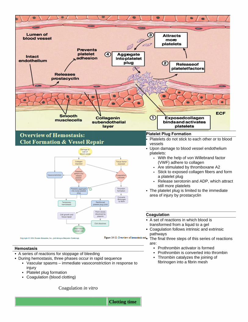
Platelets - small, anuclear cytoplasmic disks. In an unstimulated state, the shap is discoid. Hemostasis - the process in circulation where the blood is maintained fluid in vessels and without major loss in case of injury. Coagulation factors - Components that exist in the circulation and supply the necessary constituents for clot formation. Virchow’s Triad
Hem A s Du
ostasis series of reacuring hemosta
Vascular spinjury
Platelet plu Coagulation
ctions for stopasis, three phapasms – imm
g formation n (blood clotti
Coagu
page of bleedases occur inediate vasoco
ing)
ulation in vit
ding rapid sequenonstriction in
tro
Clo
nce response to
otting time
Platelet P Plateletvessels
Upon dplatelet
Wi(VW
Are Sti
a p Re
stil The plaarea of
Coagulat A set oftransfor
Coagulpathwa
The finaare:
Pro Pro Th
fib
Plug Formatits do not sticks damage to blots: th the help ofWF) adhere te stimulated bck to exposed
platelet plug elease serotonll more plateleatelet plug is lf injury by pro
tion f reactions in rmed from a lation follows
ays al three steps
othrombin actothrombin is crombin catalyrinogen into a
ion k to each othe
ood vessel en
f von Willebrao collagen by thromboxad collagen fib
nin and ADP,ets imited to the stacyclin
which blood liquid to a gelintrinsic and
s of this series
tivator is formconverted intoyzes the joinina fibrin mesh
er or to blood
dothelium
and factor
ane A2 bers and form
which attract
immediate
is extrinsic
s of reactions
med o thrombin ng of
t
s

Wh
Wh
Citr
Citr
Citr+ C
Citrthro
CoagDeta Coag Ma
Onfac
Coag Proact
Coag Th Ins Fib Fib
hole blood
hole blood +
rated platele
rated platele
rated plateleCa++
rated plateleomboplastin
gulation ailed Events o
gulation Phaay be initiated Triggered b Involves a s Each pathw
nce factor X hctor V to form
gulation Phaothrombin acttive enzyme t
gulation Pharombin cataly
soluble fibrin sbrin causes plbrin in the pre Cross-links Strengthen
EDTA or ci
et-poor plasm
et-poor plasm
et-poor plasm
et-poor plasm + Ca++
of Coagulatio
ase 1: Two Pad by either theby tissue-damseries of procway cascadesas been activprothrombin
ase 2: Pathwativator catalyzthrombin
ase 3: Commyzes the polymstrands form tlasma to becosence of calc
s fibrin s and stabiliz
itrate
ma + Ca++
ma + PL + C
ma + kaolin +
ma +
on
athways to Pe intrinsic or emaging eventscoagulants s toward factovated, it compactivator
ay to Thrombzes the transf
on Pathwaysmerization of the structural ome a gel-likecium ions acti
zes the clot
4-8
infin
2-4
a++ 60-8
+ PL 21-3(aPT
11-
Prothrombin extrinsic pathws
or X plexes with ca
bin formation of p
s to the Fibrifibrinogen intbasis of a clo
e trap vates factor X
min
nite
min
85 sec
32 sec TT)
12 sec (PT)
Activator way
alcium ions, P
prothrombin to
in Mesh to fibrin ot
XIII that:
PF3, and
o the
Cl
FaFo T
In
ot RetractionClot retractionby squeezingstrands Repair
Platelet-d(PDGF) blood ve
Fibroblastissue pa
Stimulategrowth facells muendothel
actors Limitinormation Two homeostclots from be
Swift rem Inhibition
factors
hibition of CFibrin acts asbinding throm
Positive
n and Repairn – stabilizati
g serum from
derived growtstimulates re
essel wall sts form a conatch ed by vasculaactor (VEGF)ltiply and restlial lining
ng Clot Grow
tatic mechancoming large
moval of clottin of activated
Clotting Factos an anticoagumbin and prev
feedback effe
r on of the clot the fibrin
th factor building of
nnective
ar endothelial, endothelial tore the
wth or
isms prevent
ng factors clotting
ors ulant by
venting its: ects of
coagulation Ability to speed up the production
of prothrombin activator via factor V
Acceleration of the intrinsic pathway by activating platelets
Thrombin not absorbed to fibrin is inactivated by antithrombin III
Heparin, another anticoagulant, also inhibits thrombin activity
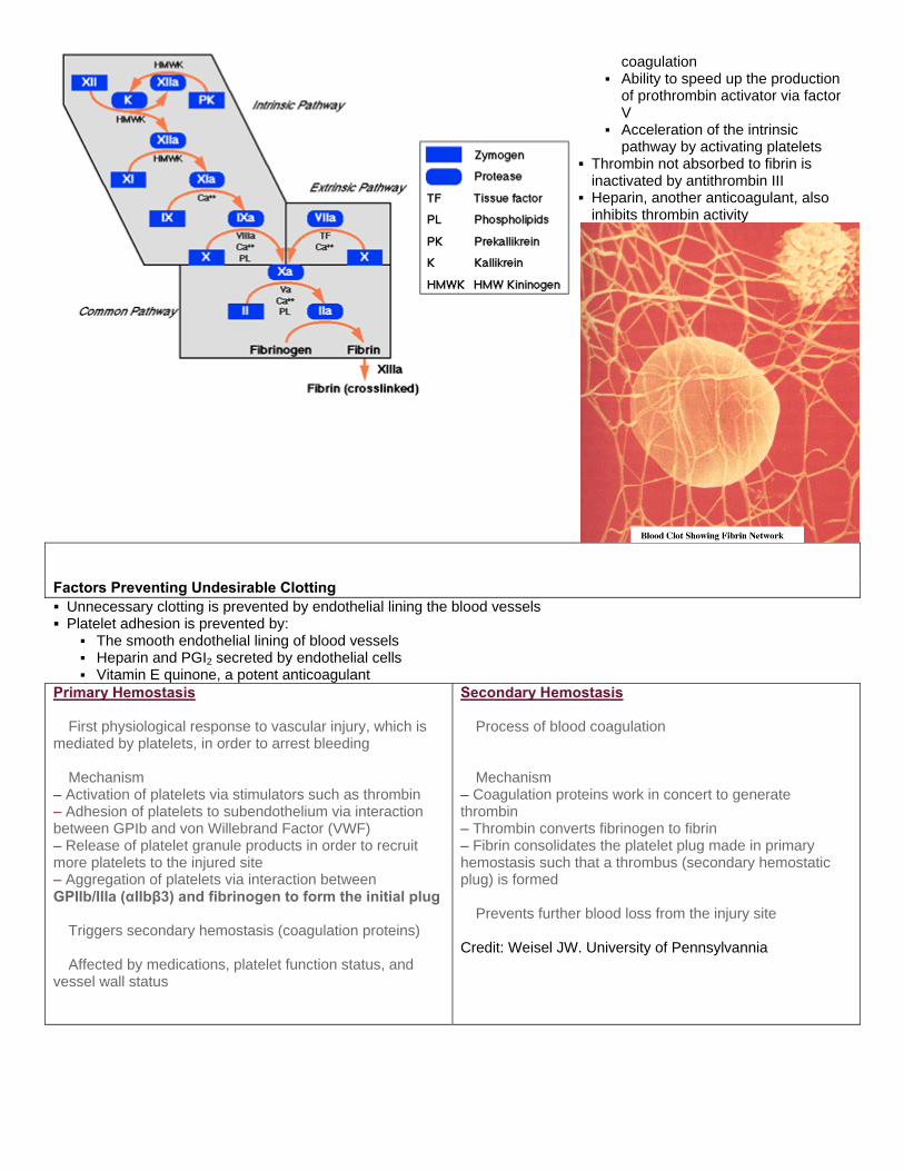
Factors Preventing Undesirable Clotting Unnecessary clotting is prevented by endothelial lining the blood vessels Platelet adhesion is prevented by:
The smooth endothelial lining of blood vessels Heparin and PGI2 secreted by endothelial cells Vitamin E quinone, a potent anticoagulant
Primary Hemostasis � First physiological response to vascular injury, which is mediated by platelets, in order to arrest bleeding � Mechanism – Activation of platelets via stimulators such as thrombin – Adhesion of platelets to subendothelium via interaction between GPIb and von Willebrand Factor (VWF) – Release of platelet granule products in order to recruit more platelets to the injured site – Aggregation of platelets via interaction between GPIIb/IIIa (αIIbβ3) and fibrinogen to form the initial plug � Triggers secondary hemostasis (coagulation proteins) � Affected by medications, platelet function status, and vessel wall status
Secondary Hemostasis � Process of blood coagulation � Mechanism – Coagulation proteins work in concert to generate thrombin – Thrombin converts fibrinogen to fibrin – Fibrin consolidates the platelet plug made in primary hemostasis such that a thrombus (secondary hemostatic plug) is formed � Prevents further blood loss from the injury site Credit: Weisel JW. University of Pennsylvannia

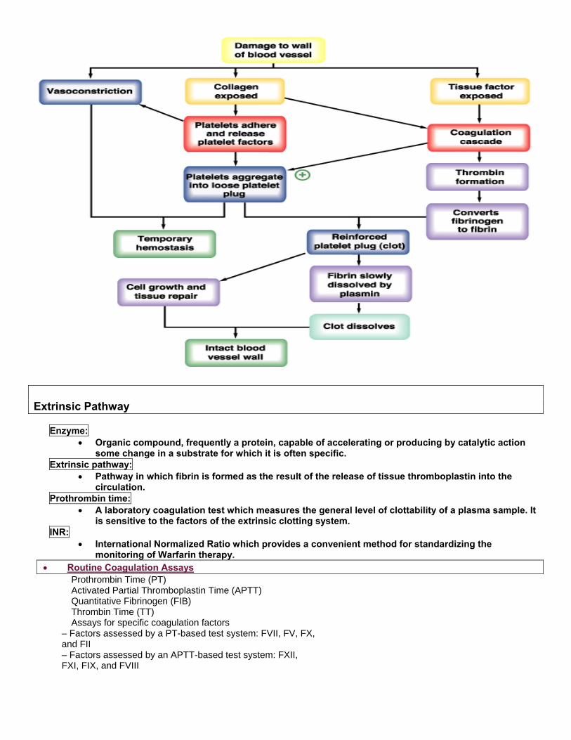
Extrinsic Pathway Enzyme:
• Organic compound, frequently a protein, capable of accelerating or producing by catalytic action some change in a substrate for which it is often specific.
Extrinsic pathway: • Pathway in which fibrin is formed as the result of the release of tissue thromboplastin into the
circulation. Prothrombin time:
• A laboratory coagulation test which measures the general level of clottability of a plasma sample. It is sensitive to the factors of the extrinsic clotting system.
INR: • International Normalized Ratio which provides a convenient method for standardizing the
monitoring of Warfarin therapy. • Routine Coagulation Assays
� Prothrombin Time (PT) � Activated Partial Thromboplastin Time (APTT) � Quantitative Fibrinogen (FIB) � Thrombin Time (TT) � Assays for specific coagulation factors – Factors assessed by a PT-based test system: FVII, FV, FX, and FII – Factors assessed by an APTT-based test system: FXII, FXI, FIX, and FVIII


Platelets are small fragments of bone marrow cells and are therefore not really classified as cells themselves.
Platelets have the following functions:
1. Secrete vasoconstrictors which constrict blood vessels, causing vascular spasms in broken blood vessels 2. Form temporary platelet plugs to stop bleeding 3. Secrete procoagulants (clotting factors) to promote blood clotting 4. Dissolve blood clots when they are no longer needed 5. Digest and destroy bacteria 6. Secrete chemicals that attract neutrophils and monocytes to sites of inflammation 7. Secrete growth factors to maintain the linings of blood vessels
The first three functions listed above refer to important haemostatic mechanisms in which platelets play a role in during bleeding - Vascular spasms, Platelet plug formation and Blood clotting (coagulation).
Vascular Spasm
This is a prompt constriction of the broken blood vessel and is the most immediate protection against blood loss. Injury stimulates pain receptors. Some of these receptors directly innervate nearby blood vessels and cause them to constrict. After a few minutes, other mechanisms take over. Injury to the smooth muscle of the blood vessel itself causes a longer-lasting vasoconstriction where platelets release a chemical vasoconstrictor called serotonin. This maintains vascular spasm long enough for the other haemostatic mechanisms to come into play.
Platelet plug formation
Under normal conditions, platelets do not usually adhere to the wall of undamaged blood vessels, since the vessel lining tends to be smooth and coated with a platelet repellent. When a vessel is broken, platelets put out long spiny extensions to adhere to the vessel wall as well as to other platelets. These extensions then contract and draw the walls of the vessel together. The mass of platelets formed is known as a platelet plug, and can reduce or stop minor bleeding.
Coagulation
This is the last and most effective defence against bleeding. During bleeding, it is important for the blood to clot quickly to minimise blood loss, but it is equally important for blood not to clot in undamaged vessels. Coagulation is a very complex process aimed at clotting the blood at appropriate amounts. The objective of coagulation is to convert plasma protein fibrinogen into fibrin, which is a sticky protein that adheres to the walls of a vessel. Blood cells and platelets become stuck to fibrin, and the resulting mass helps to seal the break in the blood vessel. The forming of fibrin is what makes coagulation so complicated, as it involved numerous chemicals reactions and many coagulation factors.
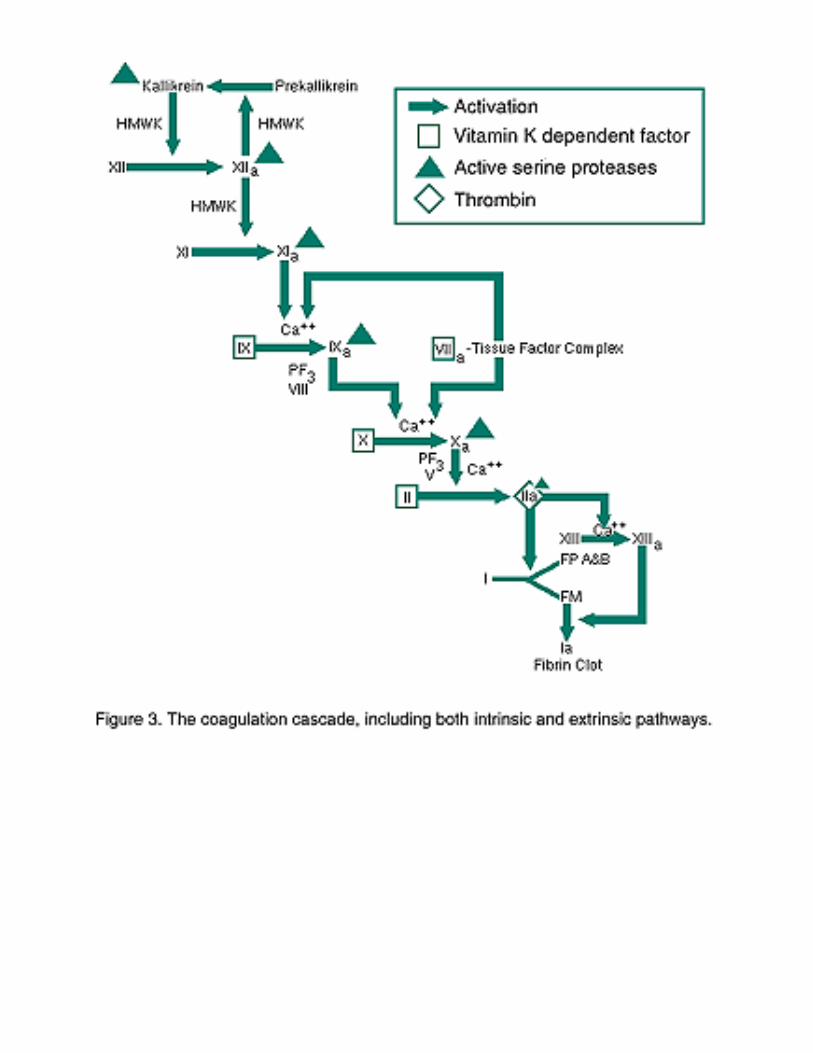

Intrinsic Pathway
Activated partial thromboplastin time (APTT)
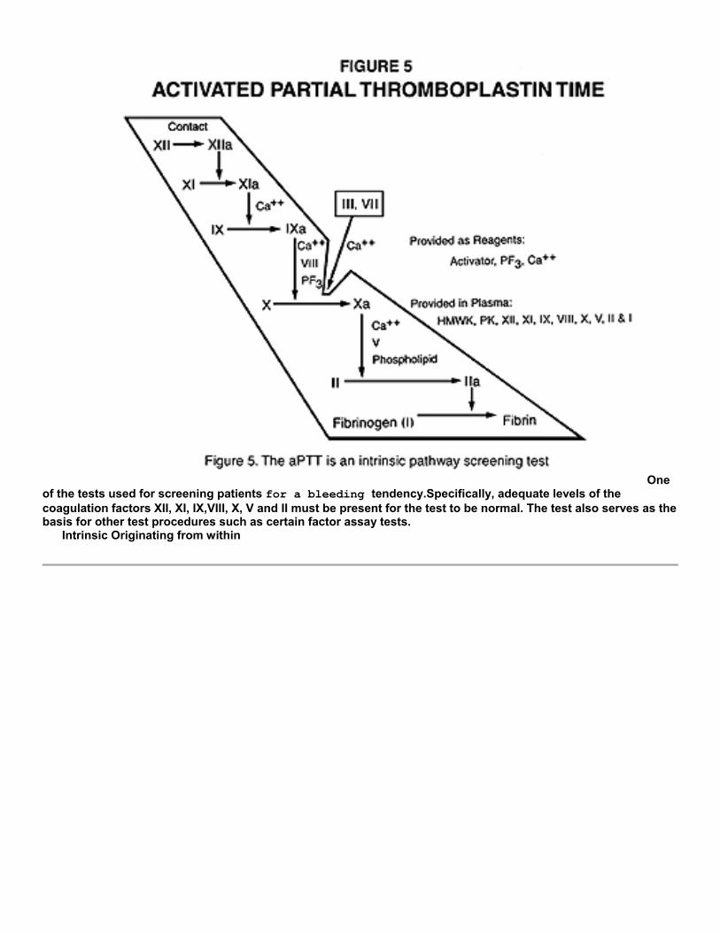
One of the tests used for screening patients for a bleeding tendency.Specifically, adequate levels of the coagulation factors XII, XI, IX,VIII, X, V and II must be present for the test to be normal. The test also serves as the basis for other test procedures such as certain factor assay tests. Intrinsic Originating from within
References :
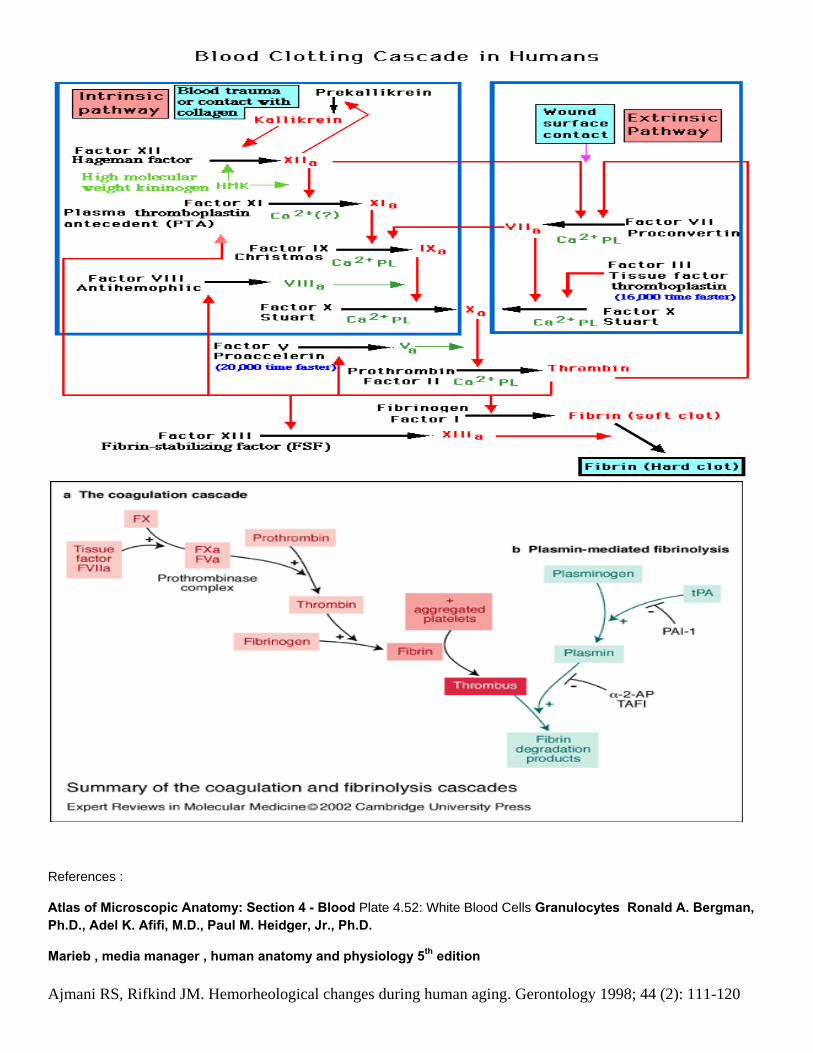
Atlas of Microscopic Anatomy: Section 4 - Blood Plate 4.52: White Blood Cells Granulocytes Ronald A. Bergman, Ph.D., Adel K. Afifi, M.D., Paul M. Heidger, Jr., Ph.D.
Marieb , media manager , human anatomy and physiology 5th edition
Ajmani RS, Rifkind JM. Hemorheological changes during human aging. Gerontology 1998; 44 (2): 111-120

Coagulation cascade [online]. 2003 [cited 2007 Sep 9]. Available from: URL: http://labtestsonline.org/ understanding/ analytes/ coag_cascade/ coagulation_cascade.html
Marieb EN. Human anatomy & physiology. 4th ed. Menlo Park, Calif.: Benjamin/Cummings; 1998.
Saladin KS. Anatomy and physiology - the unity of form and function. 3rd ed. New York: McGraw-Hill; 2004.
Sherwood L. Human physiology - from cells to systems. 5th ed. Belmont, Calif: Brooks/Cole; 2004.
Alex Munoz notes


![Hemostasis & Coagulation Disorders(Ringkas II) - Dicky [Compatibility Mode]](https://static.fdocuments.us/doc/165x107/577cc4cd1a28aba7119a7e52/hemostasis-coagulation-disordersringkas-ii-dicky-compatibility-mode.jpg)