Hemodynamic Monitoring in Sepsis Shao-Hsuan Hsia, MD Pediatric Critical Care Medicine Chang Gung...
55
Hemodynamic Monitori ng in Sepsis Shao-Hsuan Hsia, MD Pediatric Critical Care Medic ine Chang Gung Children’s Hospita l
-
Upload
alberta-knight -
Category
Documents
-
view
220 -
download
1
Transcript of Hemodynamic Monitoring in Sepsis Shao-Hsuan Hsia, MD Pediatric Critical Care Medicine Chang Gung...
Hemodynamic Monitoring in Sepsis
Shao-Hsuan Hsia, MD
Pediatric Critical Care Medicine
Chang Gung Children’s Hospital
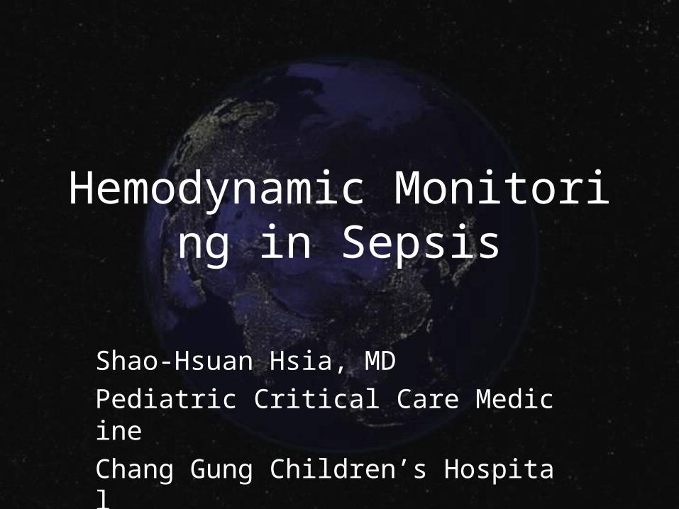
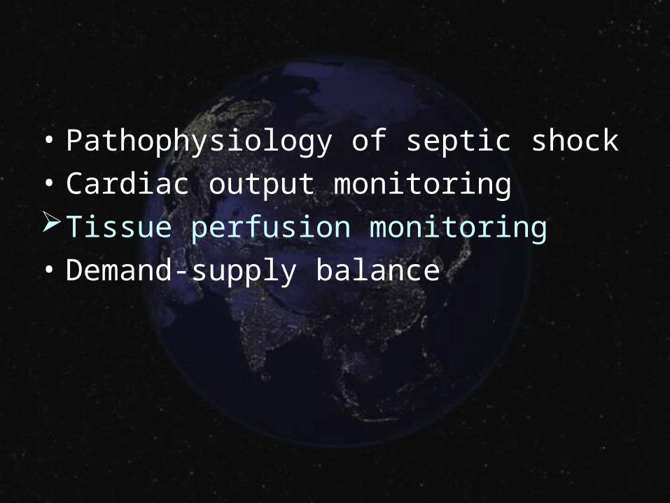
Pathophysiology of septic shock
• Cardiac output monitoring
• Tissue perfusion monitoring
• Demand-supply balance

Definition of sepsis
• Infection• Bacteremia• Systemic Inflammatory Response Syndrome (SI
RS)• Sepsis: systemic response to infection• Severe sepsis: organ dysfunction, hypoperfusion
or hypotension• Septic shock: hypotension• Multiple Organ Dysfunction Syndrome (MODS)
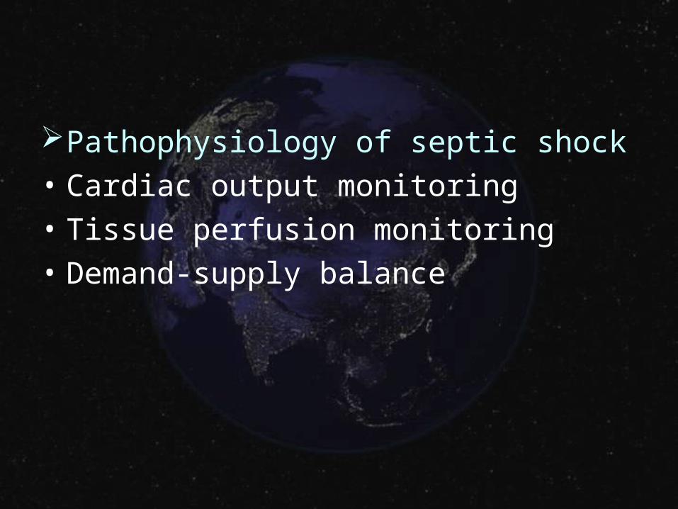
Activation of Macrophages to Bacterial Invasion
LBPLPS
Mitogen Activating Protein Kinase Pathway
Nucleus NFB
LBP - Lipopolysaccharide Binding Pro.LPS - EndotoxinNFB- Nuclear Transcription Factor
CD14
Gene Activation
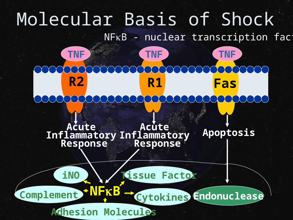
Molecular Basis of Shock
TNF TNF TNF
R2 R1 Fas
AcuteInflammatory
Response
NFB
AcuteInflammatory
ResponseApoptosis
Adhesion Molecules
CytokinesComplement
iNO Tissue Factor
Endonuclease
NFB - nuclear transcription factor
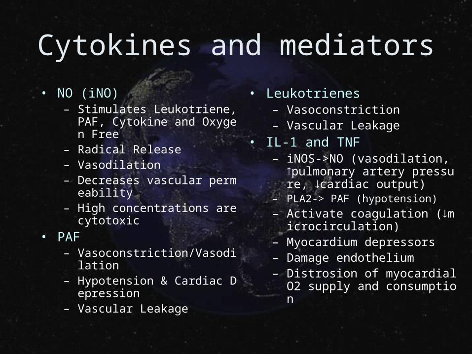
Cytokines and mediators
• NO (iNO) – Stimulates Leukotriene, PAF,
Cytokine and Oxygen Free– Radical Release– Vasodilation– Decreases vascular permeabil
ity– High concentrations are cytoto
xic
• PAF– Vasoconstriction/Vasodilation– Hypotension & Cardiac Depre
ssion– Vascular Leakage
• Leukotrienes– Vasoconstriction– Vascular Leakage
• IL-1 and TNF– iNOS->NO (vasodilation, pulmo
nary artery pressure, cardiac output)
– PLA2-> PAF (hypotension)
– Activate coagulation (microcirculation)
– Myocardium depressors– Damage endothelium– Distrosion of myocardial O2 sup
ply and consumption
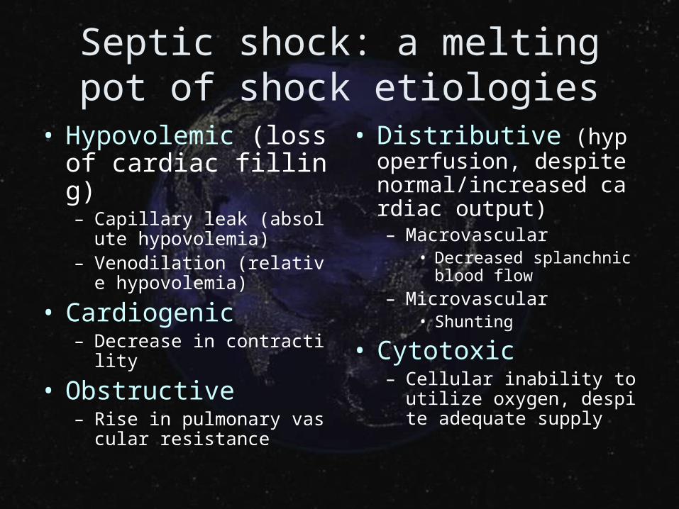
Septic shock: a melting pot of shock etiologies
• Hypovolemic (loss of cardiac filling)– Capillary leak (absolute hy
povolemia)– Venodilation (relative hypo
volemia)
• Cardiogenic– Decrease in contractility
• Obstructive– Rise in pulmonary vascular
resistance
• Distributive (hypoperfusion, despite normal/increased cardiac output)– Macrovascular
• Decreased splanchnic blood flow
– Microvascular• Shunting
• Cytotoxic– Cellular inability to utilize o
xygen, despite adequate supply
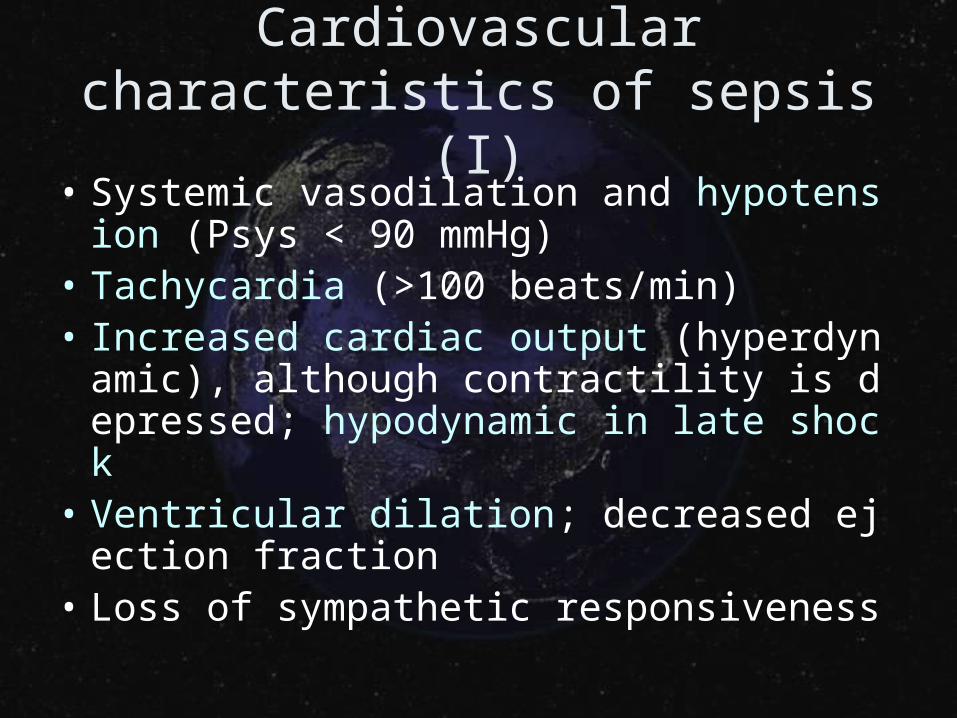
Cardiovascular characteristics of sepsis (I)
• Systemic vasodilation and hypotension (Psys < 90 mmHg)
• Tachycardia (>100 beats/min)• Increased cardiac output (hyperdynamic),
although contractility is depressed; hypodynamic in late shock
• Ventricular dilation; decreased ejection fraction
• Loss of sympathetic responsiveness
Cardiovascular characteristics of sepsis (II)
• Hypovolemia due to vascular leakage; central venous pressure may be decreased or increased depending upon fluid resuscitation
• Compromised nutrient blood flow (microcirculation) to organs; decreased organ oxygen extraction
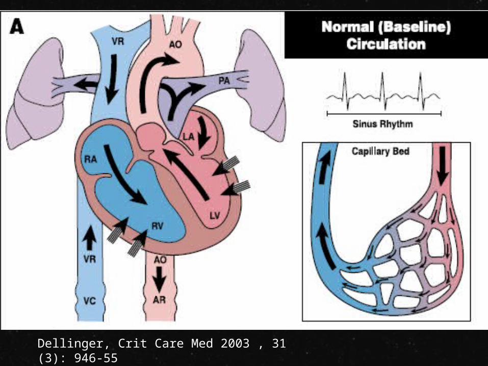
Dellinger, Crit Care Med 2003 , 31(3): 946-55
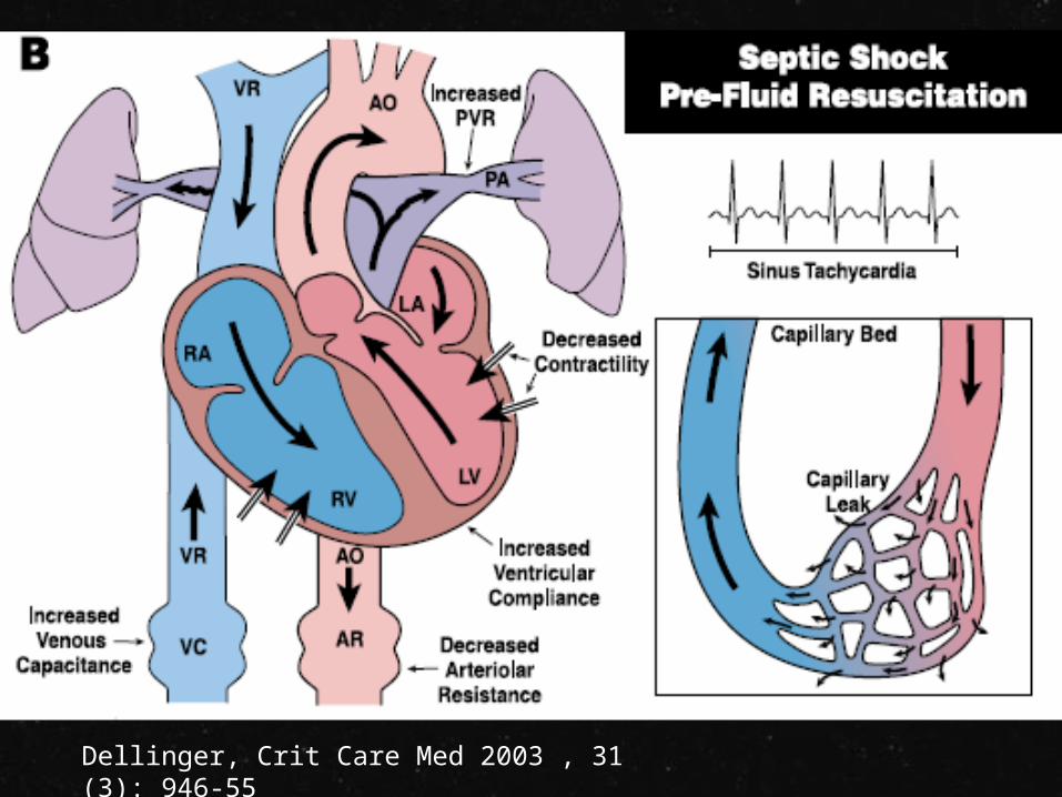
Dellinger, Crit Care Med 2003 , 31(3): 946-55
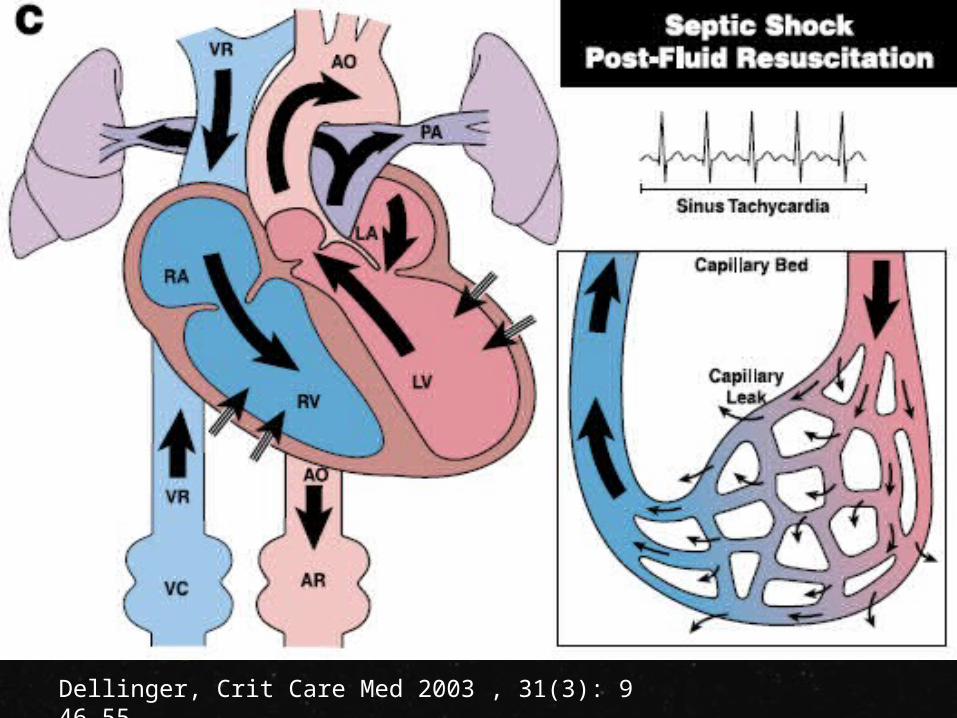
Dellinger, Crit Care Med 2003 , 31(3): 946-55
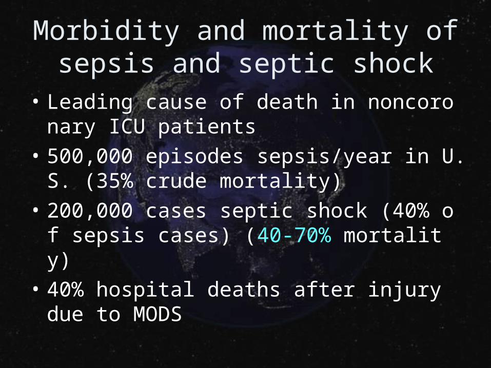
Morbidity and mortality of sepsis and septic shock
• Leading cause of death in noncoronary ICU patients
• 500,000 episodes sepsis/year in U.S. (35% crude mortality)
• 200,000 cases septic shock (40% of sepsis cases) (40-70% mortality)
• 40% hospital deaths after injury due to MODS
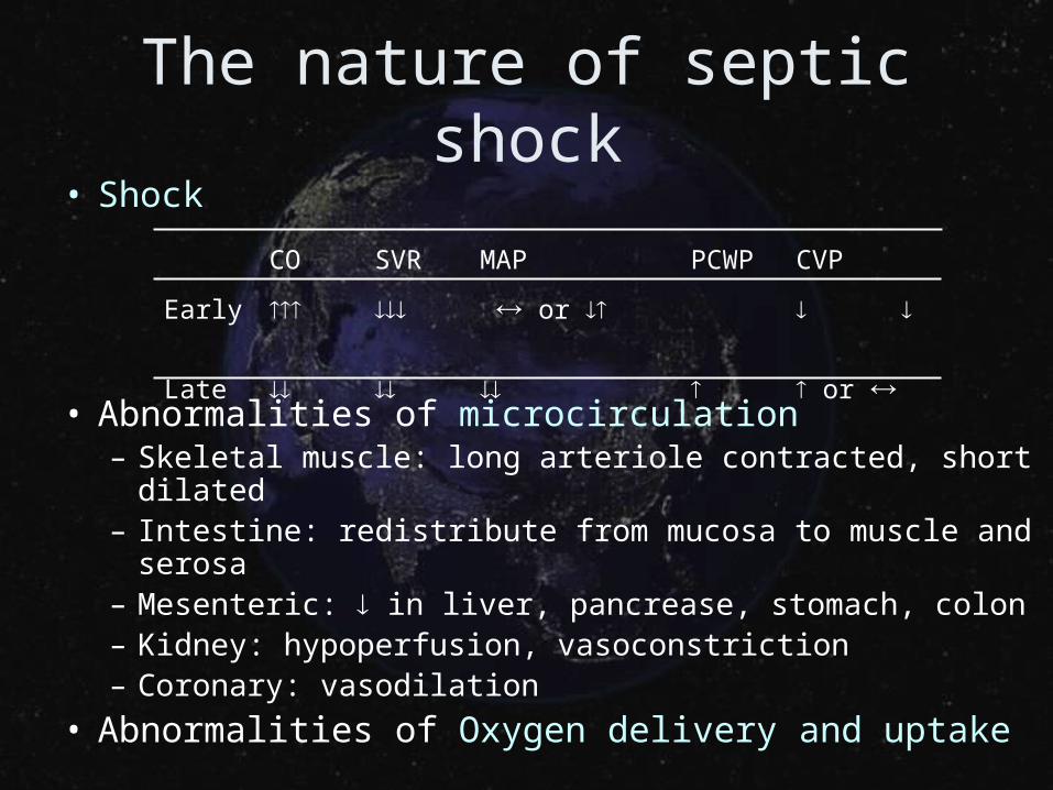
The nature of septic shock• Shock
• Abnormalities of microcirculation– Skeletal muscle: long arteriole contracted, short dilated– Intestine: redistribute from mucosa to muscle and serosa– Mesenteric: in liver, pancrease, stomach, colon– Kidney: hypoperfusion, vasoconstriction – Coronary: vasodilation
• Abnormalities of Oxygen delivery and uptake
CO SVR MAP PCWP CVP
Early or
Late or
• Pathophysiology of septic shockCardiac output monitoring
• Tissue perfusion monitoring
• Demand-supply balance
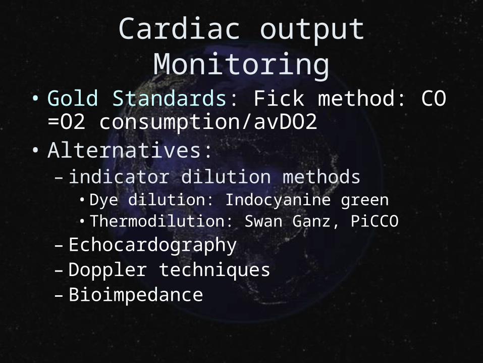
Cardiac output Monitoring
• Gold Standards: Fick method: CO=O2 consumption/avDO2
• Alternatives:– indicator dilution methods
• Dye dilution: Indocyanine green• Thermodilution: Swan Ganz, PiCCO
– Echocardography– Doppler techniques– Bioimpedance
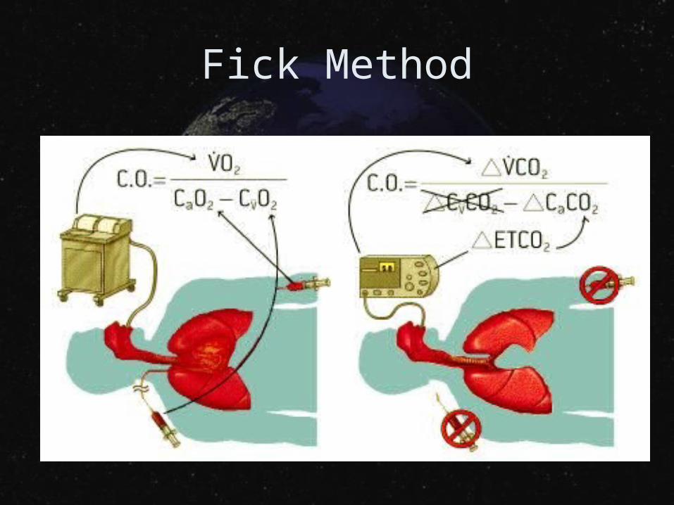
Fick Method
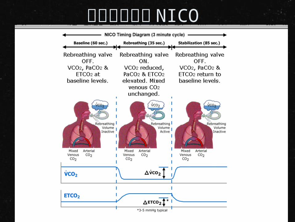
非侵入性監測 NICO
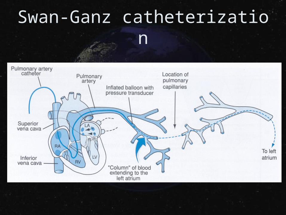
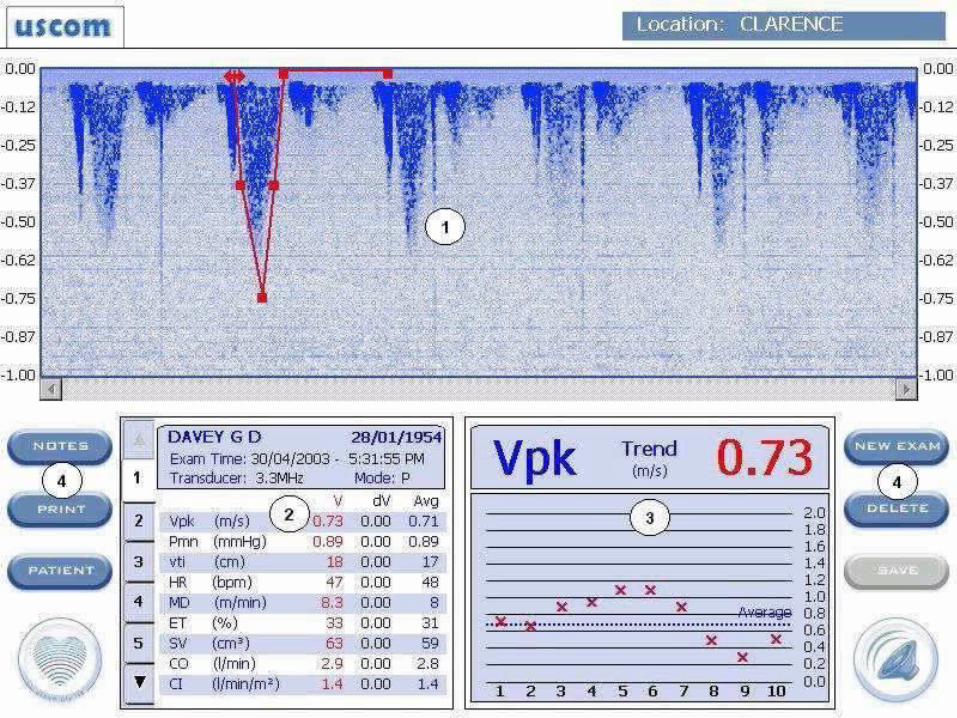
Swan-Ganz catheterization
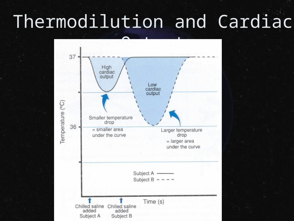
Thermodilution and Cardiac Output
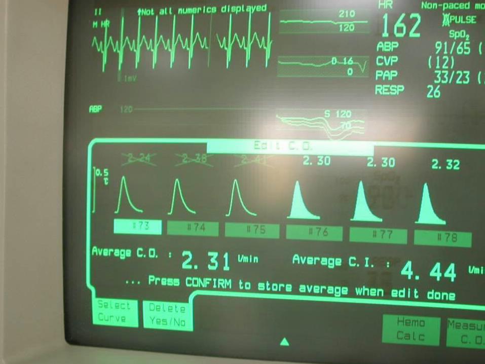
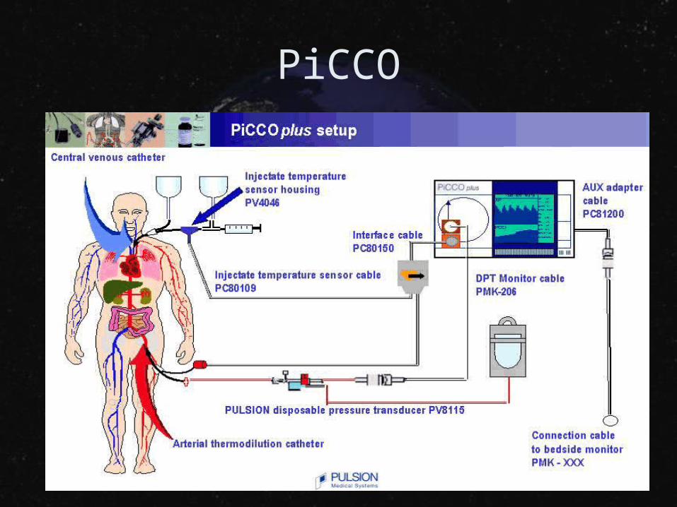
PiCCO
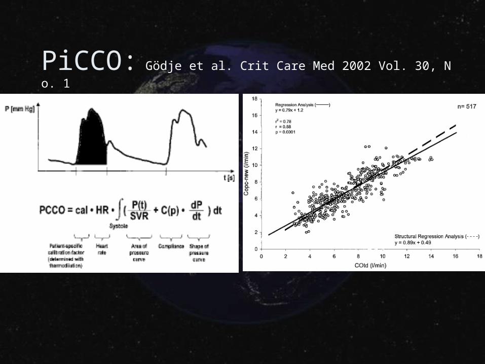
PiCCO: Gödje et al. Crit Care Med 2002 Vol. 30, No. 1
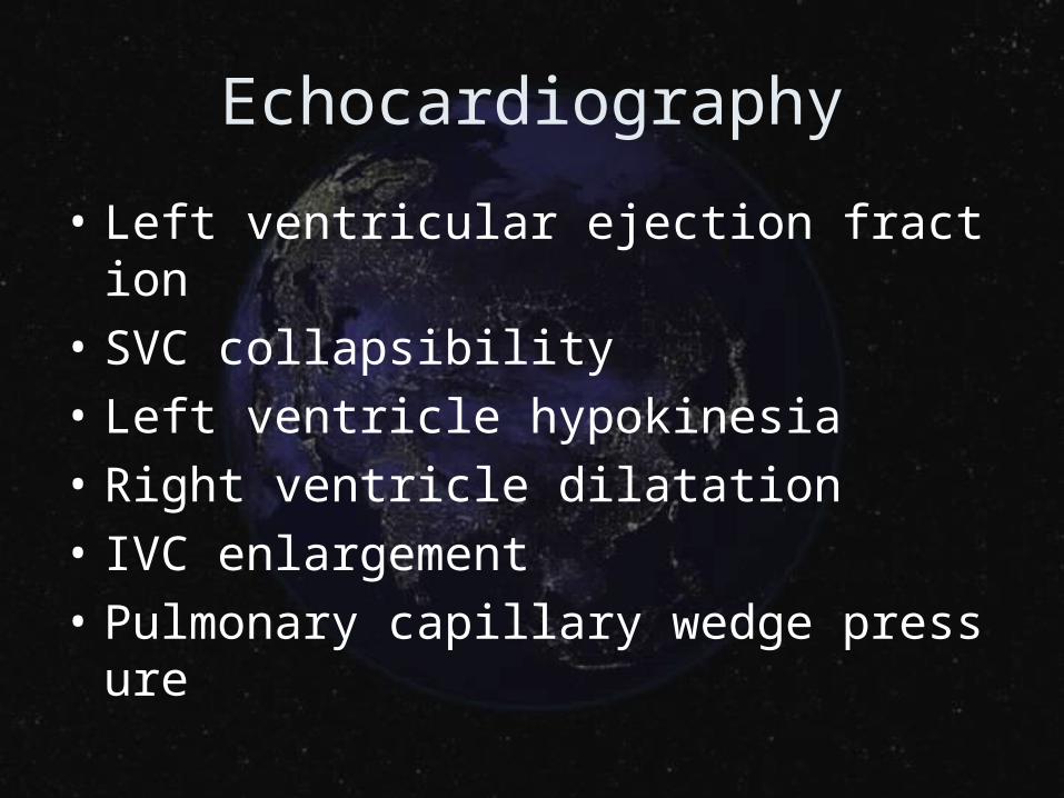
Echocardiography
• Left ventricular ejection fraction
• SVC collapsibility
• Left ventricle hypokinesia
• Right ventricle dilatation
• IVC enlargement
• Pulmonary capillary wedge pressure
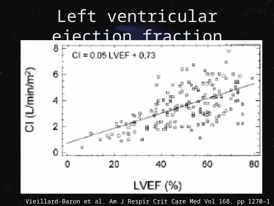
Left ventricular ejection fraction
Vieillard-Baron et al. Am J Respir Crit Care Med Vol 168. pp 1270–1276, 2003
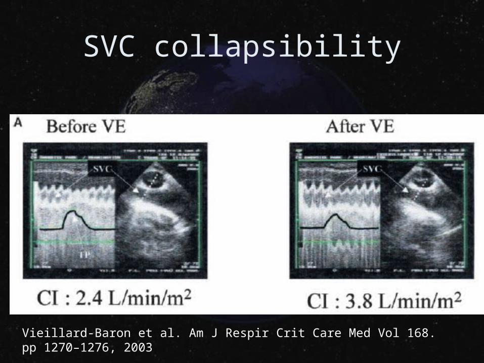
SVC collapsibility
Vieillard-Baron et al. Am J Respir Crit Care Med Vol 168. pp 1270–1276, 2003
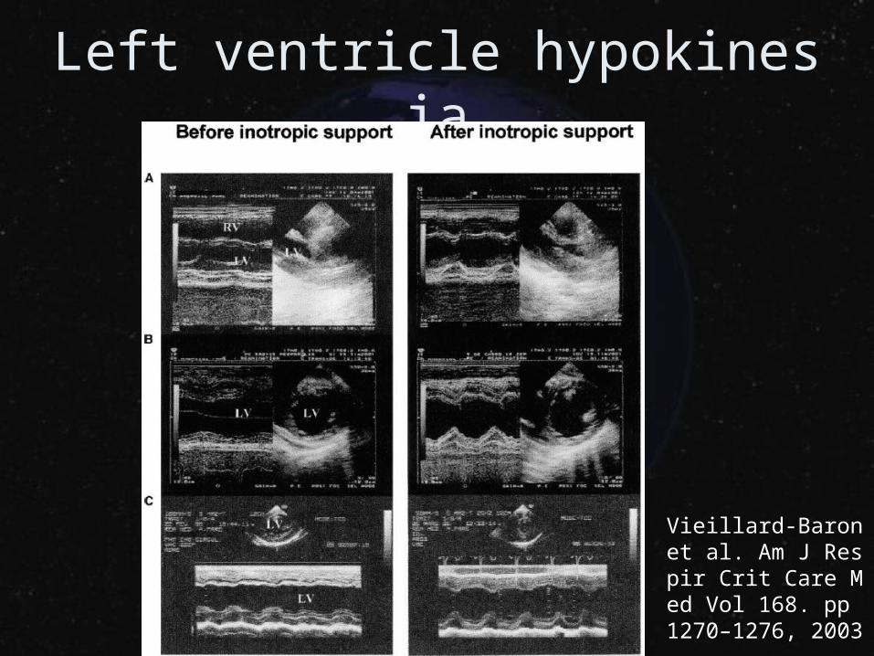
Left ventricle hypokinesia
Vieillard-Baron et al. Am J Respir Crit Care Med Vol 168. pp 1270–1276, 2003
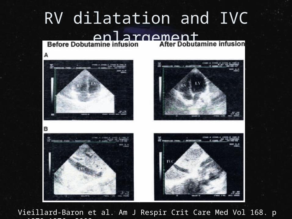
RV dilatation and IVC enlargement
Vieillard-Baron et al. Am J Respir Crit Care Med Vol 168. pp 1270–1276, 2003
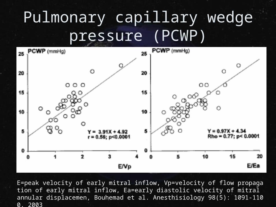
Pulmonary capillary wedge pressure (PCWP)
E=peak velocity of early mitral inflow, Vp=velocity of flow propagation of early mitral inflow, Ea=early diastolic velocity of mitral annular displacemen, Bouhemad et al. Anesthisiology 98(5): 1091-1100, 2003
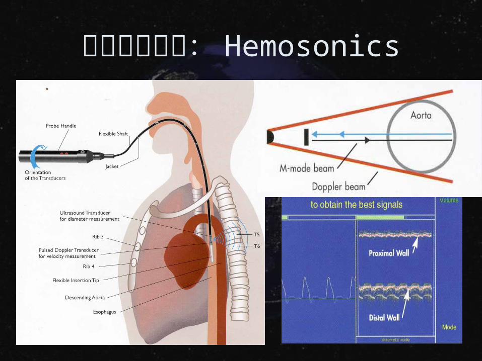
非侵入性監測: Hemosonics
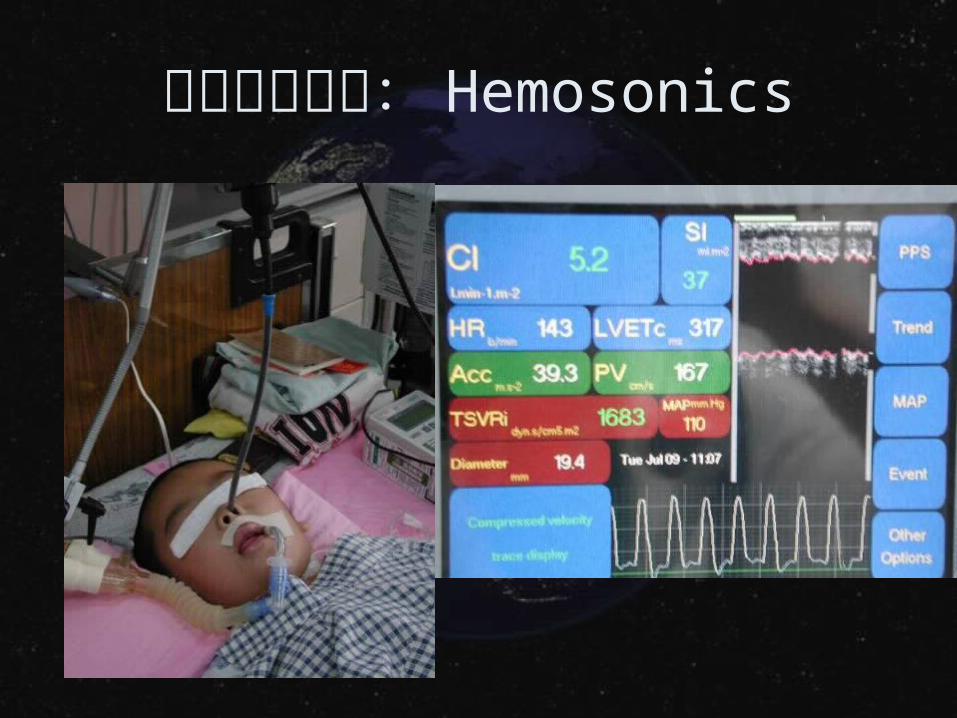
非侵入性監測: Hemosonics
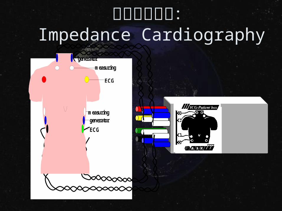
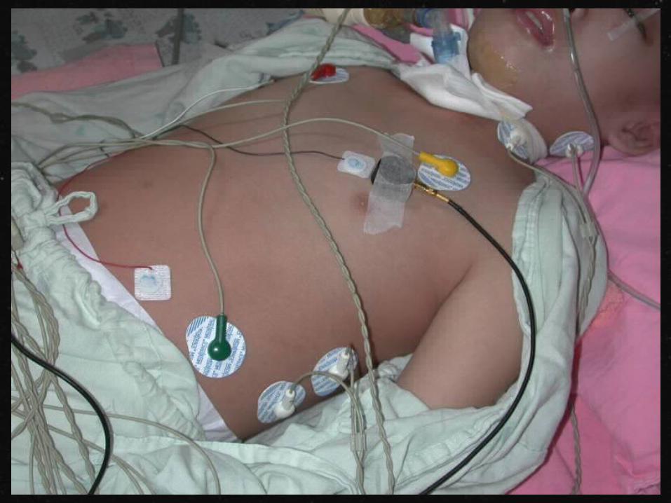
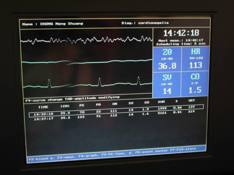
非侵入性監測:Impedance Cardiography
ECG
generator
measuring
measuringgenerator
ECG
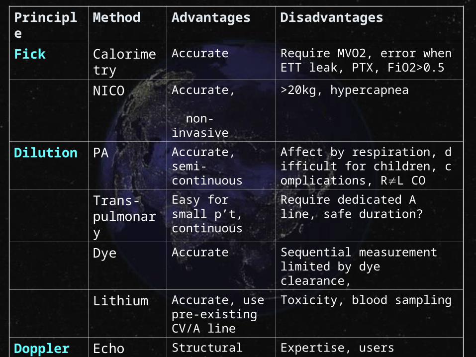
Principle Method Advantages Disadvantages
Fick Calorimetry Accurate Require MVO2, error when ETT leak, PTX, FiO2>0.5
NICO Accurate, non-invasive
>20kg, hypercapnea
Dilution PA Accurate, semi-continuous
Affect by respiration, difficult for children, complications, RL CO
Trans-pulmonary
Easy for small p’t, continuous
Require dedicated A line, safe duration?
Dye Accurate Sequential measurement limited by dye clearance,
Lithium Accurate, use pre-existing CV/A line
Toxicity, blood sampling
Doppler Echo Structural and function
Expertise, users variations
Trans-esophageal
Continuous, rapid insertion, less invasive
Probe fixation, individual errors, tracked accurately
Bio-impedance
Non-invasive Doubtful accuracy in critical illness
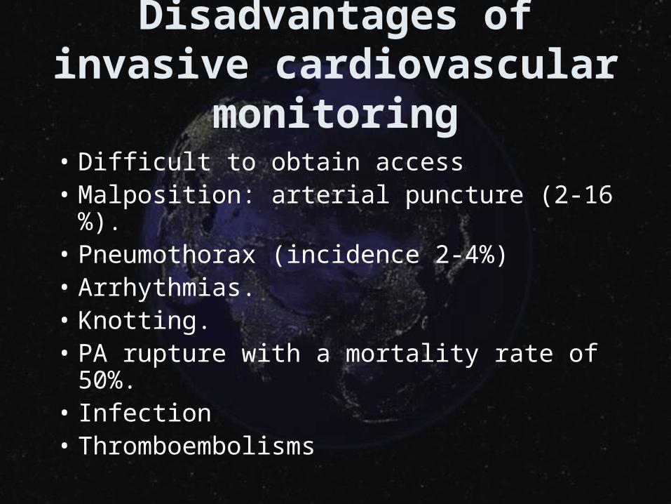
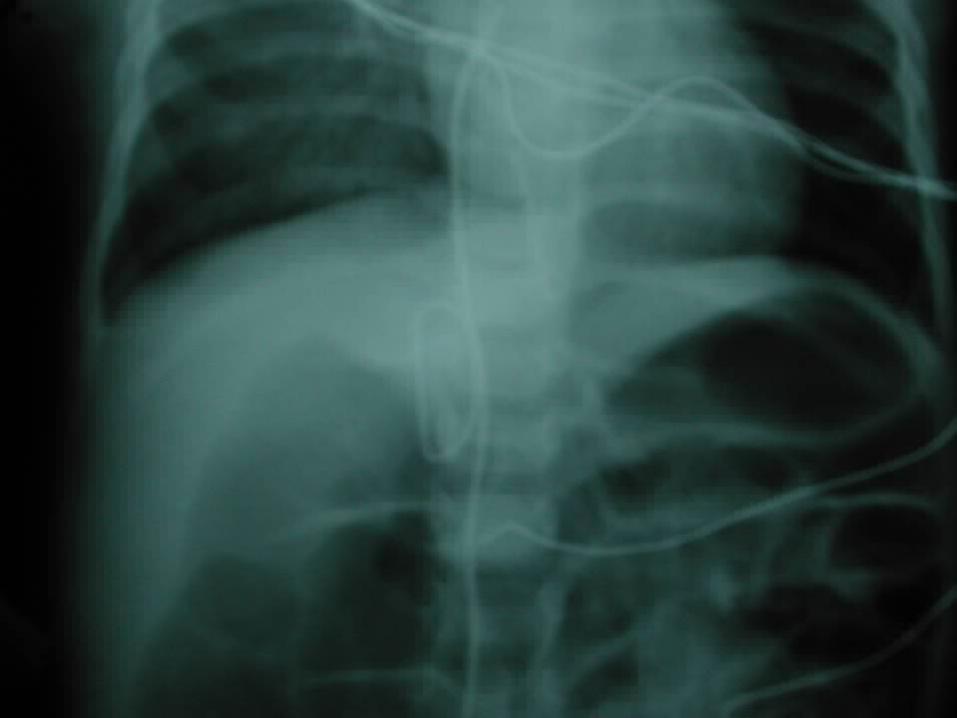
Disadvantages of invasive cardiovascular monitoring
• Difficult to obtain access• Malposition: arterial puncture (2-16%). • Pneumothorax (incidence 2-4%)• Arrhythmias. • Knotting. • PA rupture with a mortality rate of 50%. • Infection• Thromboembolisms
• Pathophysiology of septic shock
• Cardiac output monitoringTissue perfusion monitoring
• Demand-supply balance
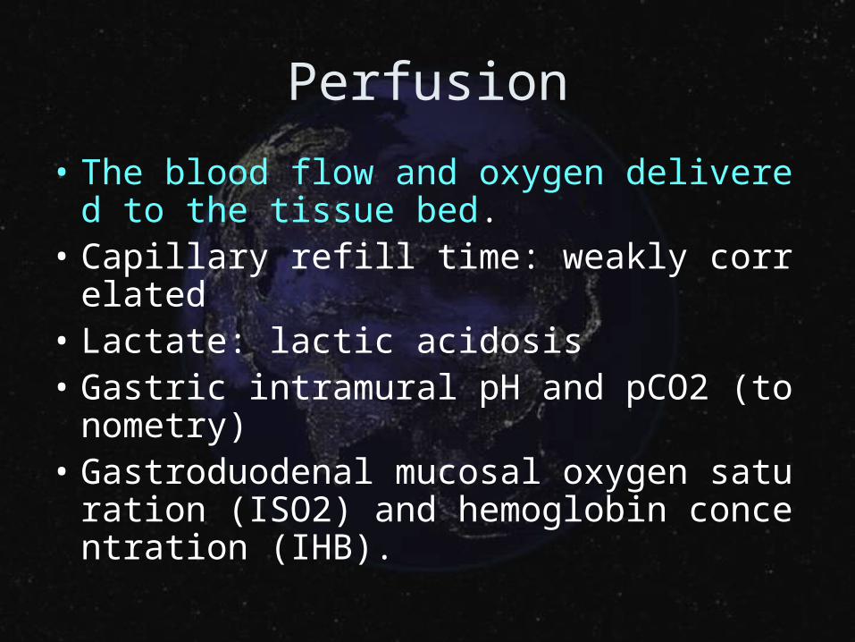
Perfusion
• The blood flow and oxygen delivered to the tissue bed.
• Capillary refill time: weakly correlated• Lactate: lactic acidosis• Gastric intramural pH and pCO2 (tonometr
y)• Gastroduodenal mucosal oxygen saturatio
n (ISO2) and hemoglobin concentration (IHB).
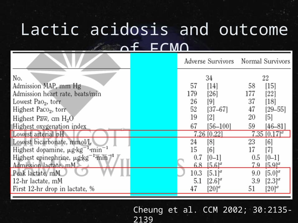
Lactic acidosis and outcome of ECMO
Cheung et al. CCM 2002; 30:2135-2139
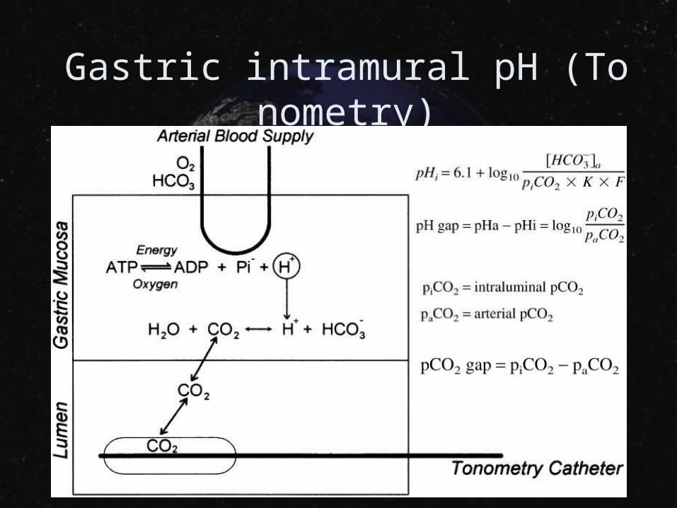
Gastric intramural pH (Tonometry)
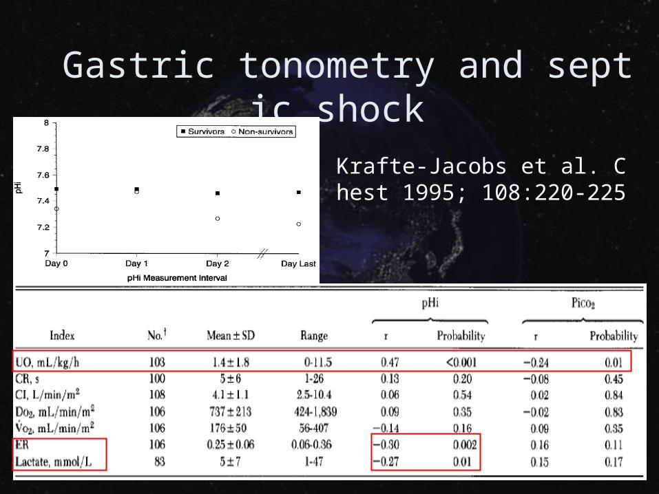
Gastric tonometry and septic shock
Krafte-Jacobs et al. Chest 1995; 108:220-225
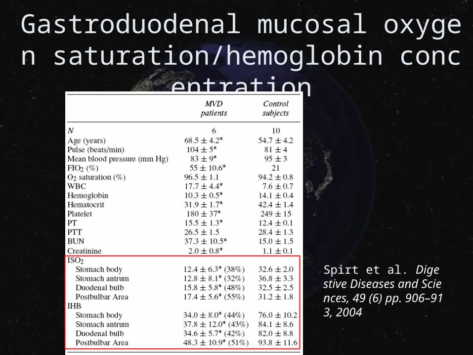
Gastroduodenal mucosal oxygen saturation/hemoglobin concentration
Spirt et al. Digestive Diseases and Sciences, 49 (6) pp. 906–913, 2004
• Pathophysiology of septic shock
• Cardiac output monitoring
• Tissue perfusion monitoringDemand-supply balance
Demand and supply balance
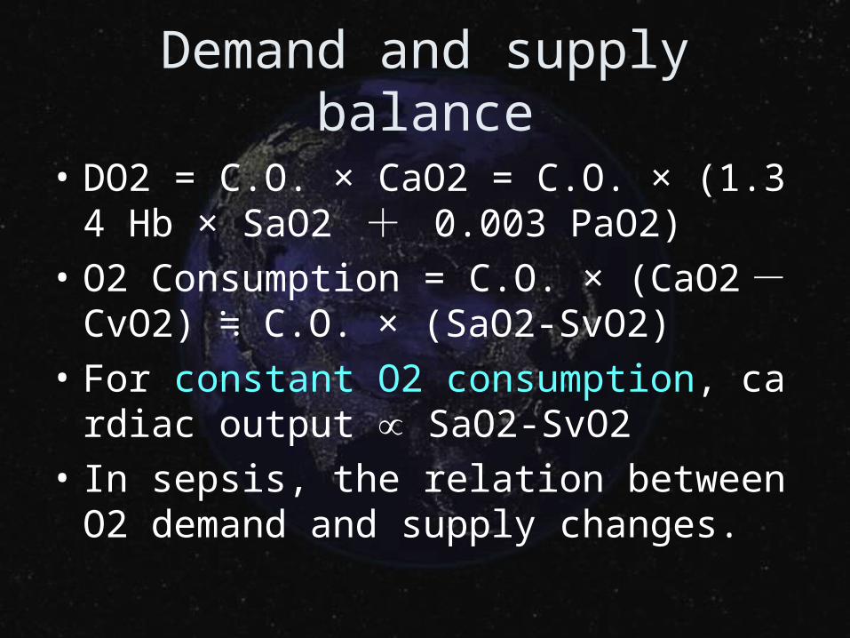
• DO2 = C.O. × CaO2 = C.O. × (1.34 Hb × SaO2 + 0.003 PaO2)
• O2 Consumption = C.O. × (CaO2 - CvO2) C.O. × (SaO2-SvO2)≒
• For constant O2 consumption, cardiac output SaO2-SvO2
• In sepsis, the relation between O2 demand and supply changes.
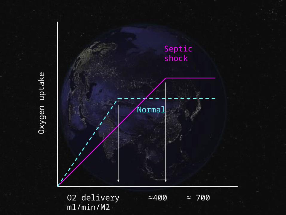
O2 delivery ≈400 ≈ 700 ml/min/M2
Oxy
gen
up
take
Normal
Septic shock
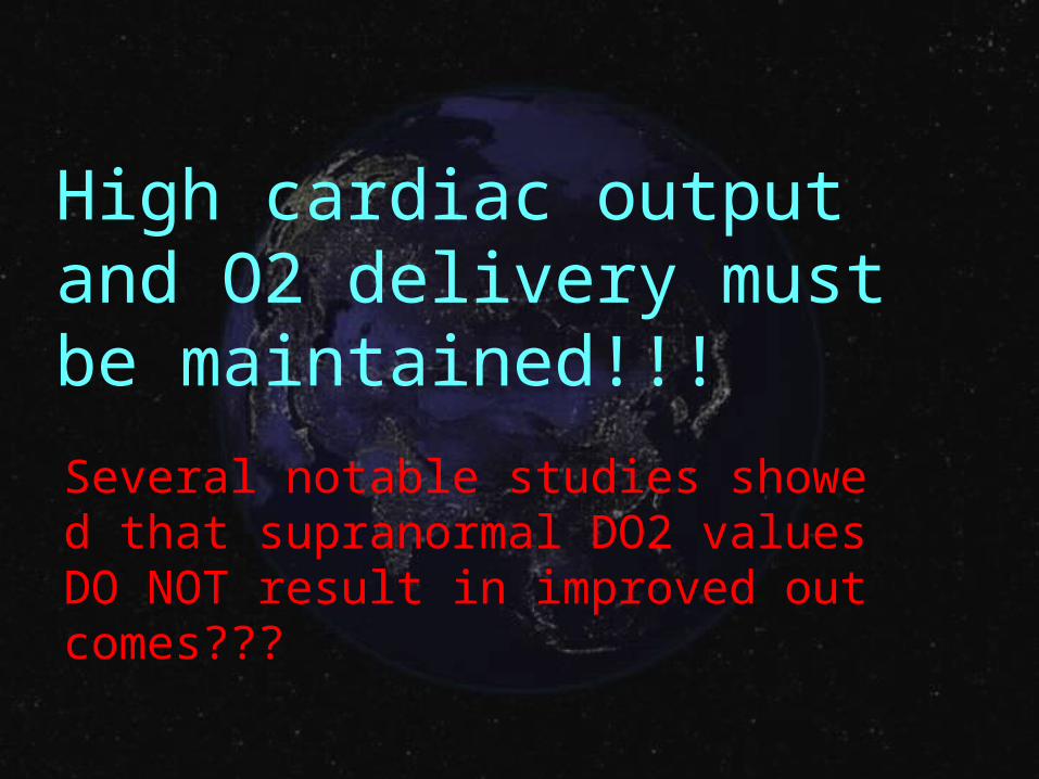
High cardiac output and O2 delivery must be maintained!!!
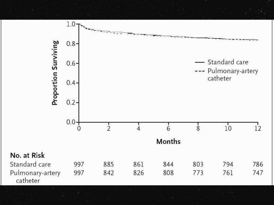
Several notable studies showed that supranormal DO2 values DO NOT result in improved outcomes???
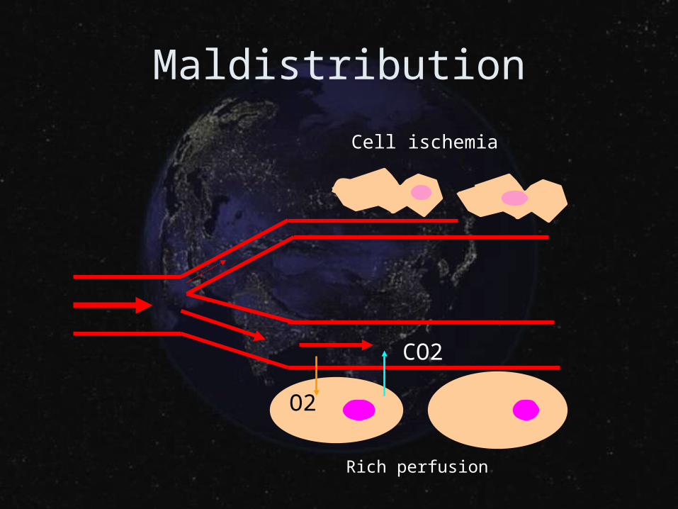
Maldistribution
O2
CO2
Cell ischemia
Rich perfusion
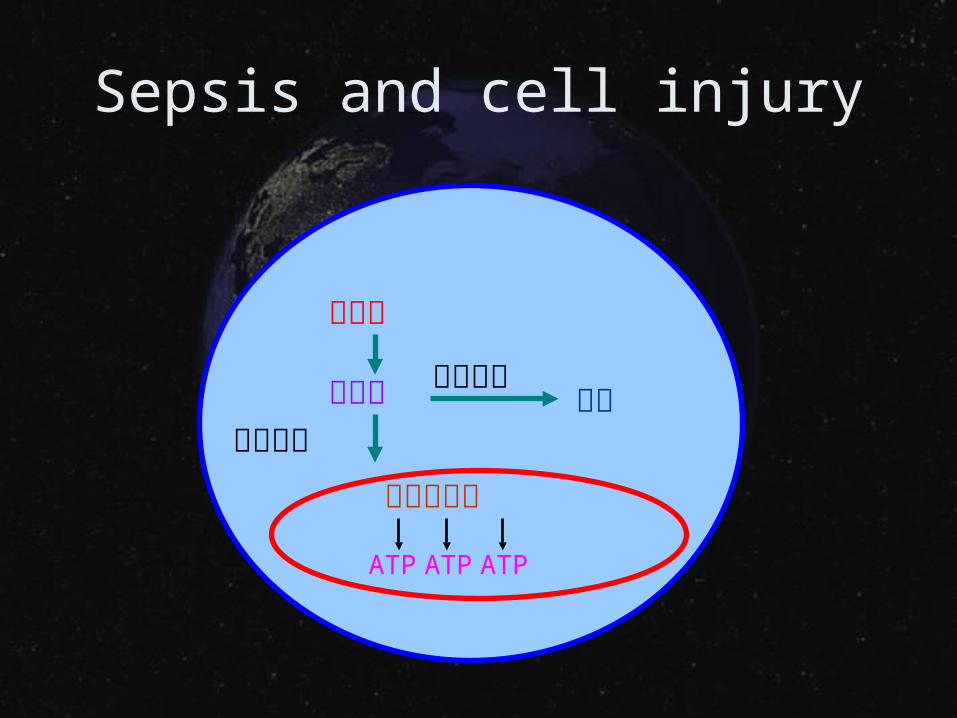
Sepsis and cell injury
糖分解
丙酮酸有氧呼吸
無氧呼吸乳酸
電子傳遞鏈
ATP ATP ATP
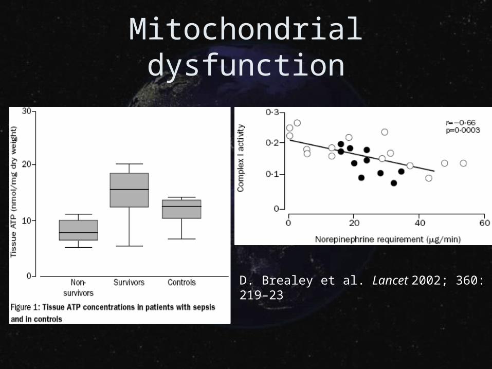
Mitochondrial dysfunction
D. Brealey et al. Lancet 2002; 360: 219–23
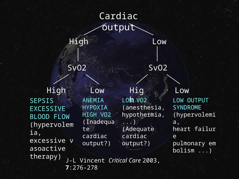
Cardiac output
High Low
SvO2 SvO2
High Low High LowSEPSISEXCESSIVEBLOOD FLOW(hypervolemia,excessive vasoactivetherapy)
ANEMIAHYPOXIAHIGH VO2(Inadequate cardiac output?)
LOW VO2(anesthesia,hypothermia, ...)(Adequate cardiac output?)
LOW OUTPUTSYNDROME(hypervolemia,heart failurepulmonary embolism ...)
J-L Vincent Critical Care 2003, 7:276-278
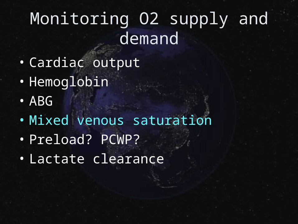
Monitoring O2 supply and demand
• Cardiac output
• Hemoglobin
• ABG
• Mixed venous saturation
• Preload? PCWP?
• Lactate clearance
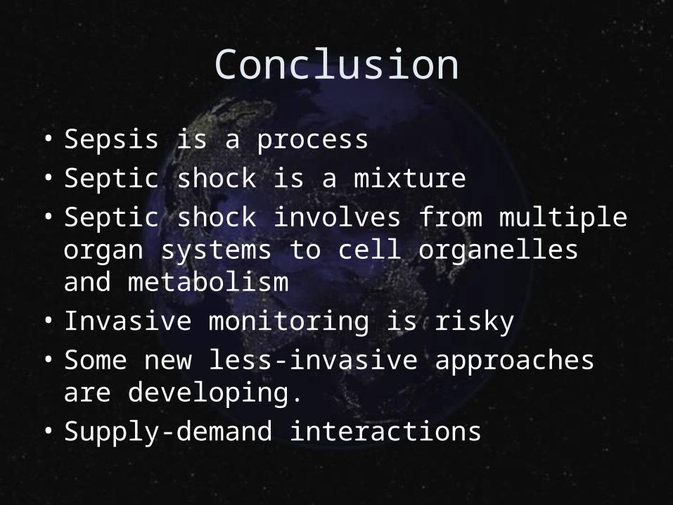
Conclusion
• Sepsis is a process
• Septic shock is a mixture
• Septic shock involves from multiple organ systems to cell organelles and metabolism
• Invasive monitoring is risky
• Some new less-invasive approaches are developing.
• Supply-demand interactions