Hemodynamic monitoring
23
BEDSIDE HEMODYNAMIC MONITORING
-
Upload
nursekim -
Category
Health & Medicine
-
view
2.083 -
download
2
description
Transcript of Hemodynamic monitoring
BEDSIDE HEMODYNAMIC MONITORING
• Indications and evidence for effectiveness– Sepsis – CHF– Peri-op
• Insertion• Complications• Measurement
Bedside Hemodynamic MonitoringOutline
Bedside Hemodynamic MonitoringDefinition
• The use of an indwelling catheter to measure– pulmonary artery pressure– pulmonary capillary wedge pressure– right atrial pressure– pulmonary artery oxygen saturation– thermodilution cardiac output
in the intensive care unit.
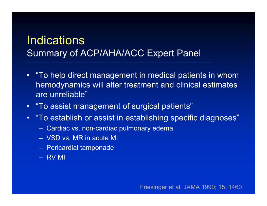
• “To help direct management in medical patients in whom hemodynamics will alter treatment and clinical estimates are unreliable”
• “To assist management of surgical patients”• “To establish or assist in establishing specific diagnoses”
– Cardiac vs. non-cardiac pulmonary edema– VSD vs. MR in acute MI– Pericardial tamponade– RV MI
IndicationsSummary of ACP/AHA/ACC Expert Panel
Friesinger et al. JAMA 1990; 15: 1460
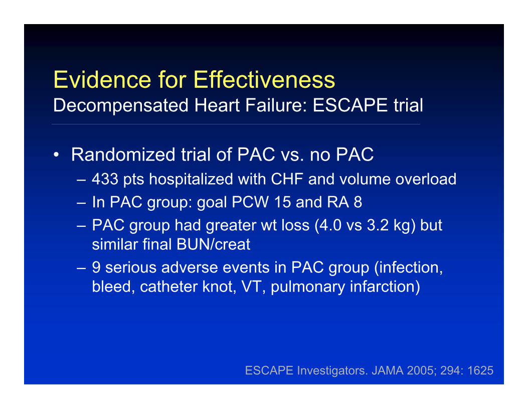
• Randomized trial of PAC vs. no PAC– 433 pts hospitalized with CHF and volume overload– In PAC group: goal PCW 15 and RA 8– PAC group had greater wt loss (4.0 vs 3.2 kg) but
similar final BUN/creat– 9 serious adverse events in PAC group (infection,
bleed, catheter knot, VT, pulmonary infarction)
Evidence for EffectivenessDecompensated Heart Failure: ESCAPE trial
ESCAPE Investigators. JAMA 2005; 294: 1625
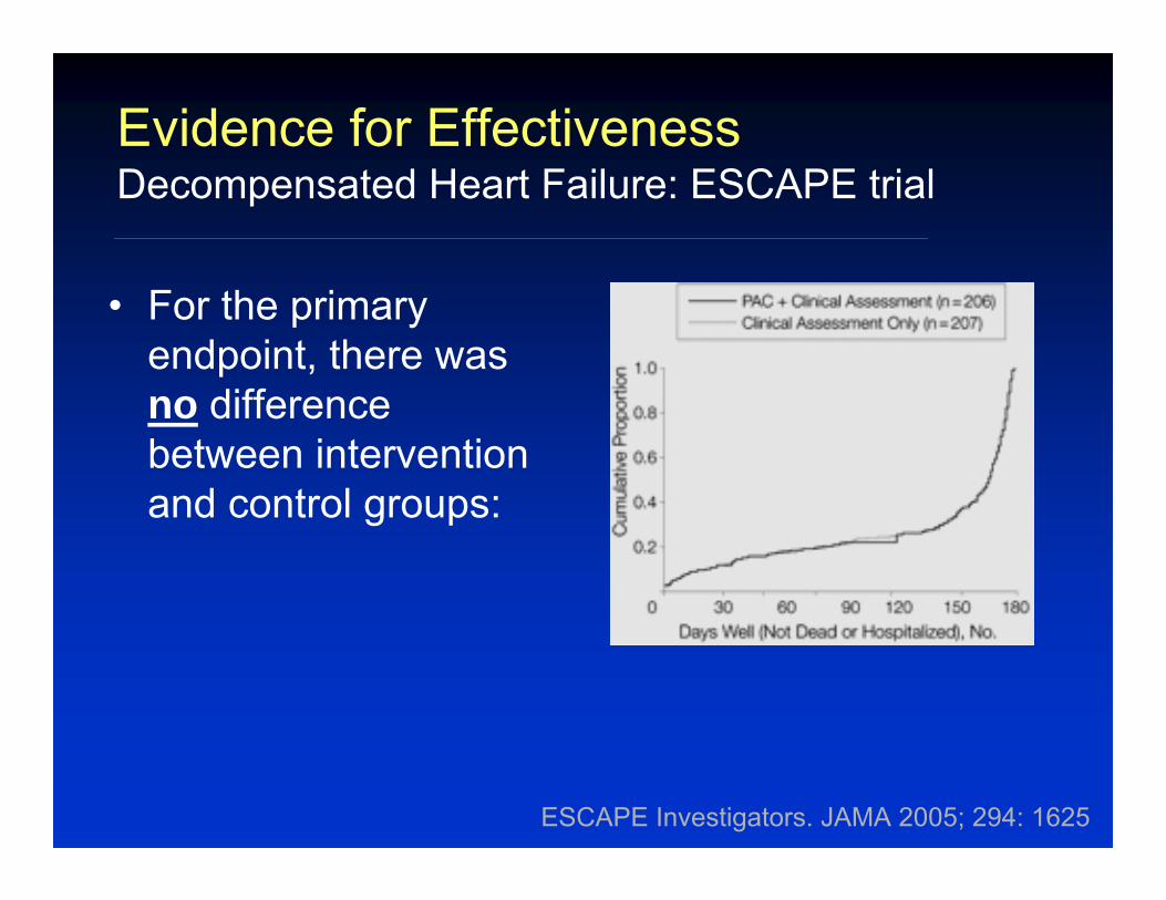
Evidence for EffectivenessDecompensated Heart Failure: ESCAPE trial
• For the primary endpoint, there was no difference between intervention and control groups:
ESCAPE Investigators. JAMA 2005; 294: 1625
ESCAPE Investigators. JAMA 2005; 294: 1625
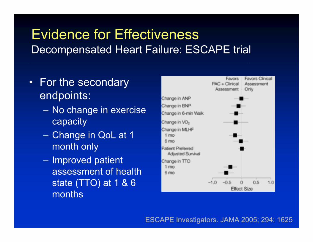
Evidence for EffectivenessDecompensated Heart Failure: ESCAPE trial
• For the secondary endpoints:– No change in exercise
capacity– Change in QoL at 1
month only– Improved patient
assessment of health state (TTO) at 1 & 6 months
ESCAPE Investigators. JAMA 2005; 294: 1625
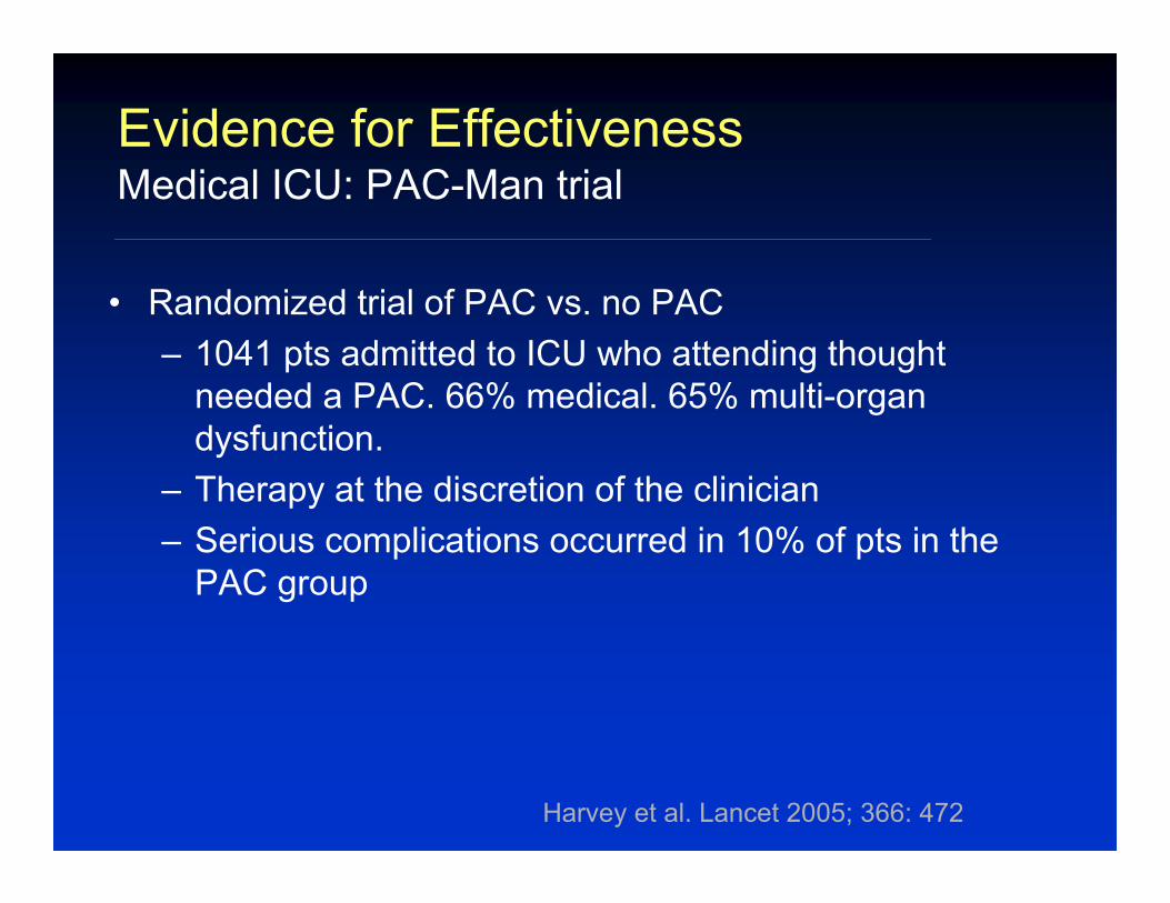
Evidence for EffectivenessMedical ICU: PAC-Man trial
• Randomized trial of PAC vs. no PAC– 1041 pts admitted to ICU who attending thought
needed a PAC. 66% medical. 65% multi-organ dysfunction.
– Therapy at the discretion of the clinician– Serious complications occurred in 10% of pts in the
PAC group
Harvey et al. Lancet 2005; 366: 472
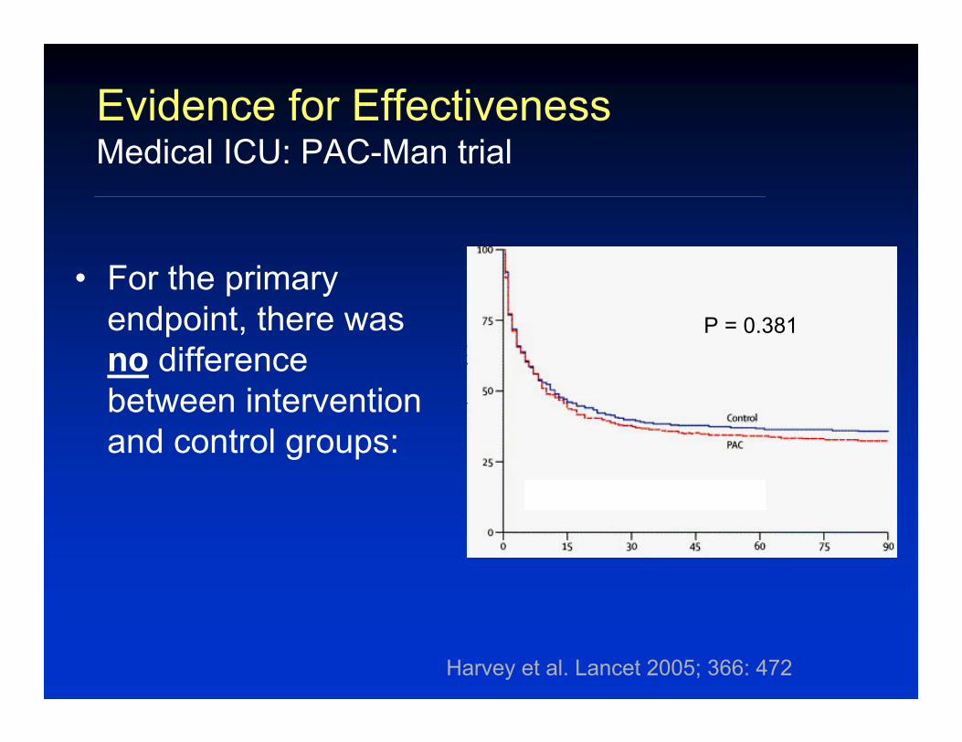
• For the primary endpoint, there was no difference between intervention and control groups:
Evidence for EffectivenessMedical ICU: PAC-Man trial
Harvey et al. Lancet 2005; 366: 472
P = 0.381
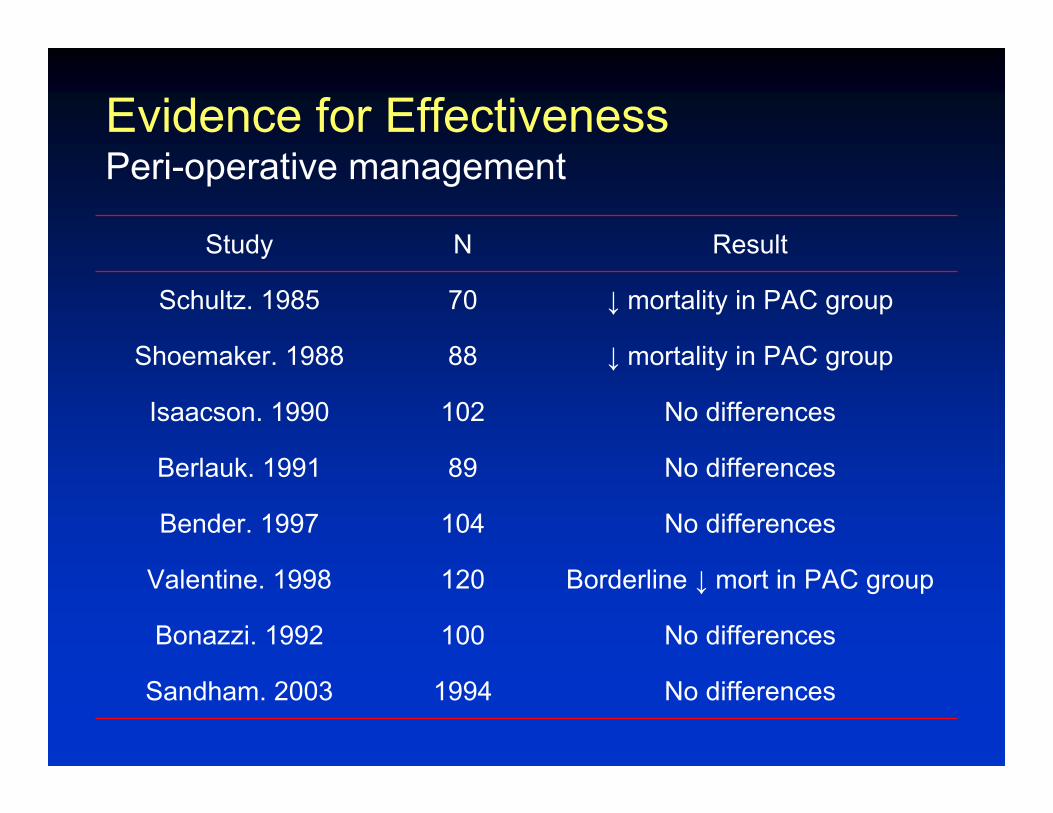
Evidence for EffectivenessPeri-operative management
ResultNStudy
No differences1994Sandham. 2003
No differences100Bonazzi. 1992
Borderline ↓ mort in PAC group120Valentine. 1998
No differences104Bender. 1997
No differences89Berlauk. 1991
No differences102Isaacson. 1990
↓ mortality in PAC group88Shoemaker. 1988
↓ mortality in PAC group70Schultz. 1985
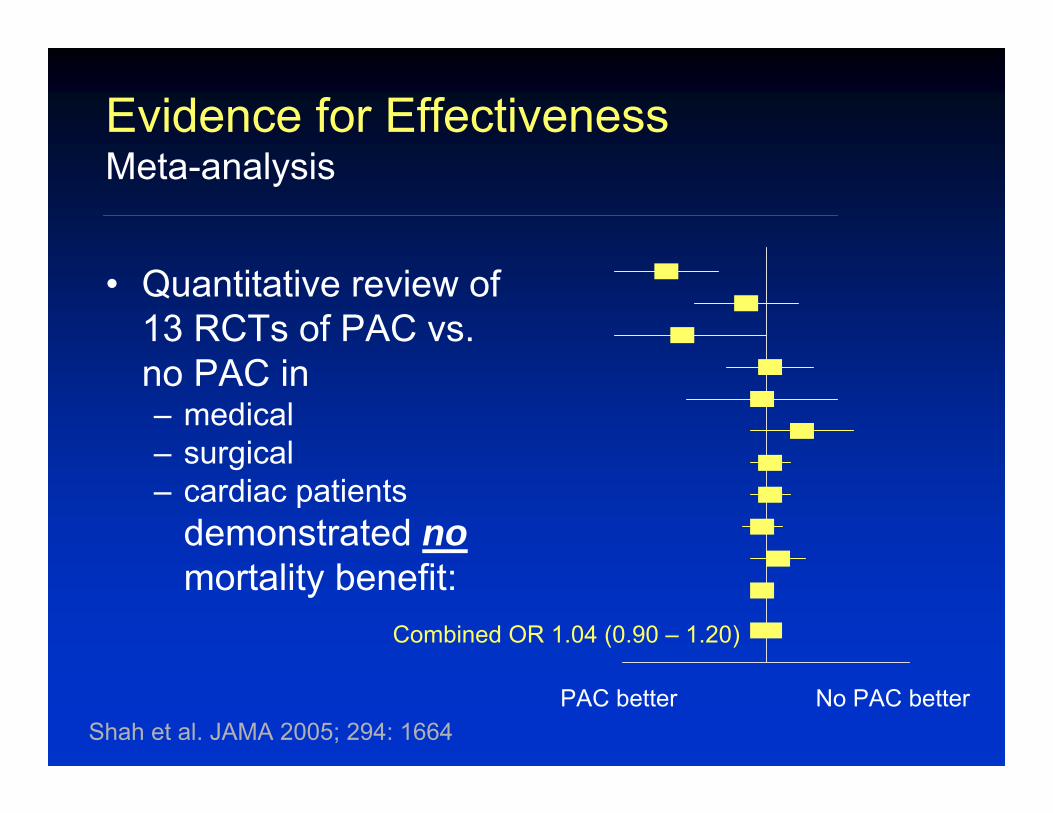
Evidence for EffectivenessMeta-analysis
Shah et al. JAMA 2005; 294: 1664
Combined OR 1.04 (0.90 – 1.20)
• Quantitative review of 13 RCTs of PAC vs. no PAC in – medical– surgical– cardiac patients
demonstrated nomortality benefit:
PAC better No PAC better
Bedside Hemodynamic MonitoringIndications and Evidence for Effectiveness
• Despite assertions by experts, there is no demonstrated benefit on mortality or length of stay associated with bedside hemodynamicmonitoring
• Its use should be limited to “rescue” situations where all other options have failed, and diagnostic situations where non-invasive options have been inconclusive

… So if you really feel like you absolutely have to use it …
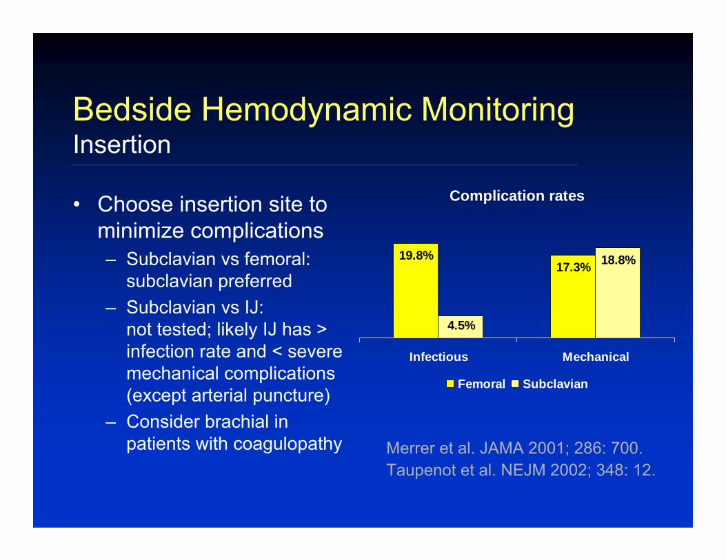
• Choose insertion site to minimize complications– Subclavian vs femoral:
subclavian preferred– Subclavian vs IJ:
not tested; likely IJ has > infection rate and < severe mechanical complications (except arterial puncture)
– Consider brachial in patients with coagulopathy
Bedside Hemodynamic MonitoringInsertion
Complication rates
19.8%17.3%
4.5%
18.8%
Infectious Mechanical
Femoral Subclavian
Taupenot et al. NEJM 2002; 348: 12.Merrer et al. JAMA 2001; 286: 700.
• Other potential complications:– Infection– Thrombosis and thromboembolism– Hemorrhage– Pneumothorax and hemothorax– Nerve injury– Pneumomediastinum– Air embolus– Foreign body embolus– RBBB– Ventricular arrhythmias– Pulmonary infarction– Pulmonary artery rupture– Heparin-induced thrombocytopenia (heparin-bonded catheter)
Bedside Hemodynamic MonitoringInsertion- complications
• Risk factors for complications:– Insertion site– Time taken for insertion– Duration of insertion– Operator experience (>50 vs <50)– Site preparation
• Scheduled insertion changes don’t decrease rates of complications
Bedside Hemodynamic MonitoringInsertion- complications
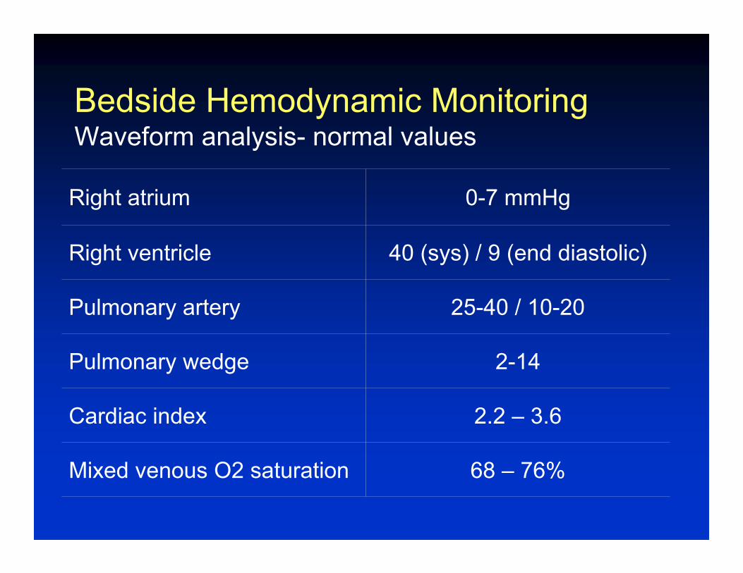
Bedside Hemodynamic MonitoringWaveform analysis- normal values
68 – 76%Mixed venous O2 saturation
2.2 – 3.6Cardiac index
2-14Pulmonary wedge
25-40 / 10-20Pulmonary artery
40 (sys) / 9 (end diastolic)Right ventricle
0-7 mmHgRight atrium
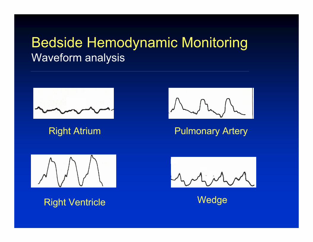
Bedside Hemodynamic MonitoringWaveform analysis
Right Atrium Pulmonary Artery
Right Ventricle Wedge
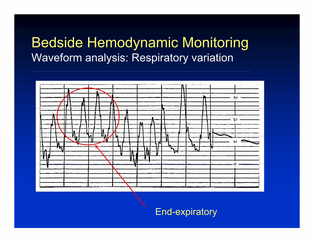
Bedside Hemodynamic MonitoringWaveform analysis: Respiratory variation
End-expiratory
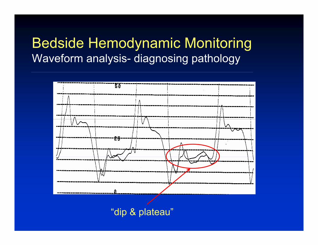
Bedside Hemodynamic MonitoringWaveform analysis- diagnosing pathology
“dip & plateau”
Bedside Hemodynamic MonitoringCardiac Output
• Measured by thermodilution:– Inject cool saline or warm the blood in the RA– Measure change in temperature with time in the PA– Integral of T vs. t curve is used to compute CO
• There are many pitfalls in bedside CO measurement; be skeptical when the “number”doesn’t match the clinical data
• Cardiac output is an imperfect indicator of circulatory function
• Useful check on accuracy of cardiac output• Can be used to confirm “wedged” position• Can be used to diagnose (relatively large)
intracardiac shunts
Bedside Hemodynamic MonitoringOxygen saturation