Hematologic System, Oncologic Disorders & Anemias
68
Hematologic System, Oncologic Disorders & Anemias 1
Transcript of Hematologic System, Oncologic Disorders & Anemias

Hematologic System,
Oncologic Disorders &
Anemias
1
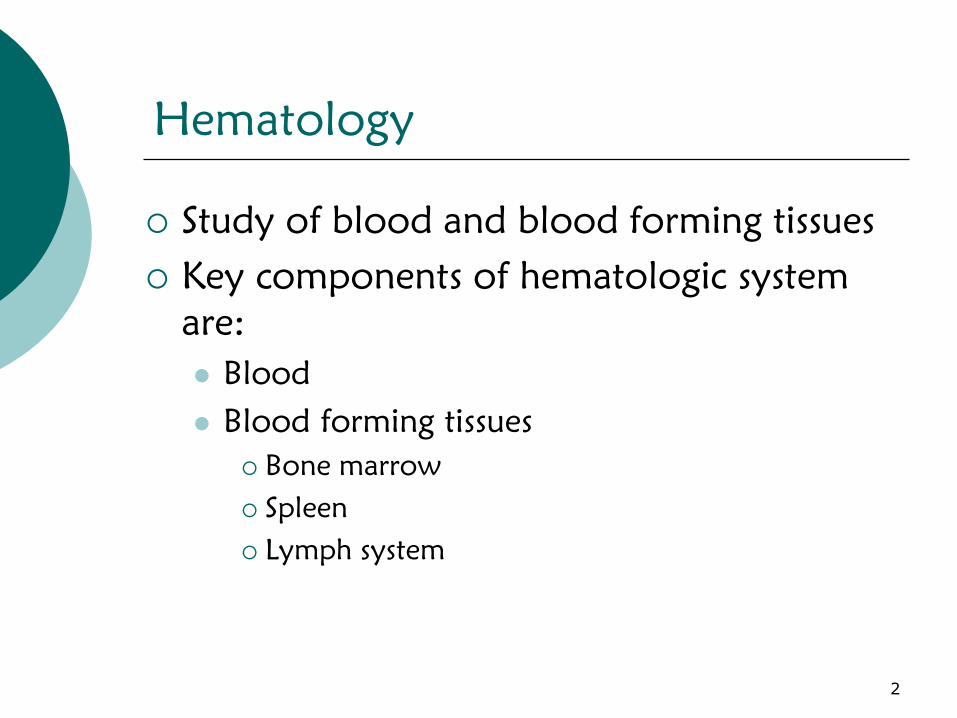
Hematology
Study of blood and blood forming tissues
Key components of hematologic system
are:
Blood
Blood forming tissues
Bone marrow
Spleen
Lymph system
2

What Does Blood Do?
Transportation
Oxygen
Nutrients
Hormones
Waste Products
Regulation
Fluid, electrolyte
Acid-Base balance
Protection
Coagulation
Fight Infections
3

Components of Blood
Plasma
55%
Blood Cells
45%
Three types
Erythrocytes/RBCs
Leukocytes/WBCs
Thrombocytes/Platelets
4

Erythrocytes/Red Blood Cells
Composed of hemoglobin
Erythropoiesis
= RBC production
Stimulated by hypoxia
Controlled by erythropoietin
Hormone synthesized in kidney
Hemolysis
= destruction of RBCs
Releases bilirubin into blood stream
Normal lifespan of RBC = 120 days
5

Leukocytes/White Blood Cells
5 types
Basophils
Eosinophils
Neutrophils
Monocytes
Lymphocytes
6
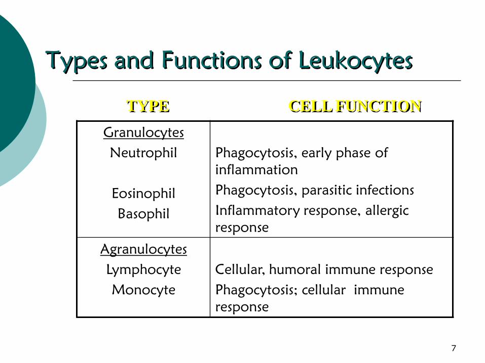
Types and Functions of Leukocytes
Granulocytes
Neutrophil
Eosinophil
Basophil
Phagocytosis, early phase of
inflammation
Phagocytosis, parasitic infections
Inflammatory response, allergic
response
Agranulocytes
Lymphocyte
Monocyte
Cellular, humoral immune response
Phagocytosis; cellular immune
response
TYPE CELL FUNCTION
7
Thrombocytes/Platelets
Must be present for clotting to occur
Involved in hemostasis
8

Normal Clotting Mechanisms
Hemostasis
Goal: Minimizing blood loss when injured
1. Vascular Response
vasoconstriction
2. Platelet response
Activated during injury
Form clumps (agglutination)
3. Plasma Clotting Factors
Factors I – XIII
Intrinsic pathway
Extrinsic pathway
9

Anticoagulation
Elements that interfere with blood
clotting
Countermechanism to blood clotting—
keeps blood liquid and able to flow
10

Structures of the Hematologic System
Bone Marrow
Liver
Lymph System
11

Bone Marrow
Bone Marrow
Soft substance in core of bones
Blood cell production (Hematopoiesis):The
production of all types of blood cells
generated by a remarkable self-regulated
system that is responsive to the demands
put upon it.
RBCs
WBCs
Platelets
12

Liver
Receives 24% of the cardiac output
(1500 ml of blood each minute)
Liver has many functions
Hematologic functions:
Liver synthesis plasma proteins
including clotting factors and
albumin
Liver clears damaged and non-
functioning RBCs/erythrocytes from
circulation
13

Spleen
Located in upper L quadrant of
abdomen
Functions
Hematopoietic function
Produces fetal RBCs
Filter function
Filter and reuse certain cells
Immune function
Lymphocytes, monocytes
Storage function
30% platelets stored in spleen
14

Effects of Aging on the Hematologic
System
CBC Studies
Hemoglobin (Hb or Hgb)
response to infection (WBC)
Platelets=no change
Clotting Studies
PTT
15

Assessment of the Hematologic System
Subjective Data
Important Health Information
Past health history
Medications
Surgery or other treatments
16

Assessment of
the Hematologic System (cont.)
Functional Health Patterns
Health perception – health management
Nutritional – metabolic
Elimination
Activity – exercise
Sleep – rest
Cognitive – perceptual
Self-perception – self-concept
Role – relationship
Sexuality – reproductive
Coping – stress tolerance
Value – belief
17
Assessment of
the Hematologic System (cont.)
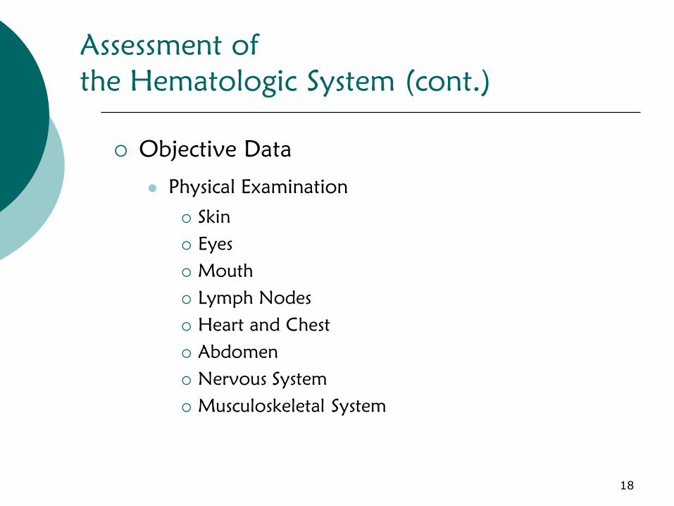
Objective Data
Physical Examination
Skin
Eyes
Mouth
Lymph Nodes
Heart and Chest
Abdomen
Nervous System
Musculoskeletal System
18

Diagnostic Studies of the Hematologic
System: Complete Blood Count (CBC)
WBCs
Normal 4,000 -11,000 µ/ℓ
Associated with infection, inflammation, tissue injury or
death
Leukopenia-- WBC
Neutropenia -- neutrophil count
RBC
♂ 4.5 – 5.5 x 10⁶/ℓ
♀ 4.0 – 5.0 x 10⁶/ℓ
Hematocrit (Hct)
The hematocrit is the percent of whole blood that is
composed of red blood cells. The hematocrit is a
measure of both the number of red blood cells and the
size of red blood cells.
19

Diagnostic Studies of the Hematologic System:
Complete Blood Count (CBC) Cont’d
Platelet count
Normal 150,000- 400,000
Thrombocytopenia- platelet count
Spontaneous hemorrhage likely when count is
below 20,000
Pancytopenia
Decrease in number of RBCs, WBCs, and
platelets
20

Diagnostic Studies
of the Hematologic System
Radiologic Studies
CT/MRI of lymph tissues
Biopsies
Bone Marrow examination
Lymph node biopsies
21

22
23

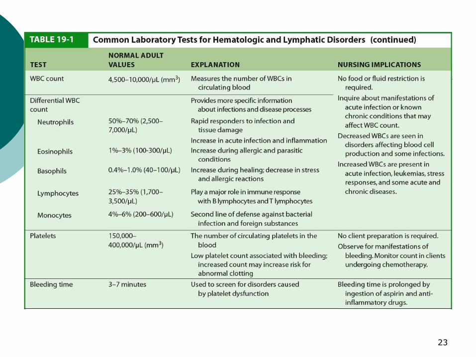
Common Laboratory Tests for Hematologic and Lymphatic Disorders
24

25

Common Laboratory Tests for Hematologic and Lymphatic Disorders
26

Anemia
Anemia is a reduction in the number of
RBCs, the quantity of hemoglobin, or
the volume of RBCs
Because the main function of RBCs is
oxygenation, anemia results in varying
degrees of hypoxia
27
Anemia
Prevalent conditions
Blood loss
Decreased production of erythrocytes
Increased destruction of erythrocytes
28
Anemia (cont’d)
Clinical Manifestations:
1. Pallor.
2. Fatigue, weakness.
3. Dyspnea.
4. Palpitations, tachycardia.
5. Headache, dizziness, and restlessness.
6. Slowing of thought.
7. Paresthesia.
29

Anemia (cont’d)
Nursing Management:
1. Direct general management toward addressing the
cause of anemia and replacing blood loss as needed
to sustain adequate oxygenation.
2. Promote optimal activity and protect from injury.
3. Reduce activities and stimuli that cause tachycardia
and increase cardiac output.
4. Provide nutritional needs.
5. Administer any prescribed nutritional supplements.
6. Patient and family education
30
Nursing Actions for a Patient who is
Anemic or Suffered Blood Loss
Administer oxygen as prescribed
Administer blood products as prescribed
Administer erythropoietin as prescribed
Allow for rest between periods of activity
Elevate the pt’s head on pillows during
episodes of shortness of breath
Provide extra blankets if the pt feels cool
Teach the pt/family about underlying
pathophysiology and how to manage the
symptoms of anemia
31

Anemia Caused by Decreased Erythrocyte
Production
Iron Deficiency Anemia
Thalassemia
Megablastic Anemia
32

Iron-Deficiency Anemia
Etiology
1. Inadequate dietary intake
Found in 30% of the
world’s population
2. Malabsorption
Absorbed in duodenum
GI surgery
3. Blood loss
2 mls blood contain 1mg iron
GI, GU losses
4. Hemolysis
33

Iron-Deficiency Anemia
Clinical Manifestations
Most common: pallor
Second most common: inflammation of the tongue
(glossistis)
Cheilitis=inflammation/fissures of lips
Sensitivity to cold
Weakness and fatigue
Diagnostic Studies
CBC
Iron studies Diagnostics:
Iron levels: Total iron-binding capacity (TIBC), Serum
Ferritin.
Endoscopy/Colonscopy
34

Iron-Deficiency Anemia
Collaborative Care
Treatment of underlying disease/problem
Replacing iron
Diet
Drug Therapy
Iron replacement
Oral iron
Feosol, DexFerrum, etc
Absorbed best in acidic environemtn
GI effects
Parenteral iron
IM or IV
Less desirable than PO
35
Iron-Deficiency Anemia
Nursing Management
Assess cardiovascular & respiratory status
Monitor vital signs
Recognizing s/s bleeding
Monitor stool, urine and emesis for occult blood
Diet teaching—foods rich in iron
Provide periods of rest
Supplemental iron
Discuss diagnostic studies
Emphasize compliance
Iron therapy for 2-3 months after the
hemoglobin levels return to normal
36

Thalassemia
Etiology
Autosomal recessive genetic disorder of
inadequate production of normal hemoglobin
Found in Mediterranean ethnic groups
Clinical Manifestations
Asymptomatic major retardation life
threatening
Splenomegaly, hepatomegaly
37
Thalassemia
Collaborative Care
No specific drug or diet are effective in
treating thalassemia
Thalassemia minor
Body adapts to ↓ Hgb
Thalassemia major
Blood transfusions with IV deferoxamine
(used to remove excess iron from the body)
38

Megaloblastic Anemias
Characterized by large
RBCs which are fragile
and easily destroyed
Common forms of
megaloblastic anemia
1. Cobalamin deficiency
2. Folic acid deficiencyThis picture shows large, dense,
oversized, red blood cells (RBCs)
that are seen in megaloblastic
anemia.
39

Cobalamin (Vitamin B12
) Deficiency
Cobalamin Deficiency--formerly known as
pernicious anemia
Vitamin B12
(cobalamin) is an important water-
soluble vitamin.
Intrinsic factor (IF) is required for cobalamin
absorption
Causes of cobalamin deficiency
Gastric mucosa not secreting IF
GI surgery loss of IF-secreting gastric mucosal cells
Long-term use of H2-histamine receptor blockers cause
atrophy or loss of gastric mucosa.
Nutritional deficiency
Hereditary defects of cobalamine utilization40

Cobalamin (Vitamin B12
) Deficiency
Clinical manifestations
General symptoms of anemia
Sore tongue
Anorexia
Weakness
Parathesias of the feet and hands
Altered thought processes
Confusion dementia
41

Cobalamin Deficiency
Diagnostic Studies
RBCs appear large
Abnormal shapes
Structure contributes to erythrocyte
destruction
Schilling Test: a medical investigation used
for patients with vitamin B12 deficiency. The
purpose of the test is to determine if the
patient has pernicious anemia.
42

Cobalamin Deficiency
Collaborative Care
Parenteral administration of cobalamin
↑ Dietary cobalamin does not correct the anemia
Still important to emphasize adequate dietary intake
Intranasal form of cyanocobalamin (Nascobal) is
available
High dose oral cobalamin and SL cobalamin can
use be used
43
Cobalamin Deficiency
Nursing Management
Familial disposition
Early detection and treatment can lead to reversal of
symptoms
Potential for Injury r/t patient’s diminished
sensations to heat and pain
Compliance with medication regime
Ongoing evaluation of GI and neuro status
Evaluate patient for gastric carcinoma frequently
44

Folic Acid Deficiency
Folic Acid Deficiency also causes megablastic
anemia (RBCs that are large and fewer in
number)
Folic Acid required for RBC formation and
maturation
Causes
Poor dietary intake
Malabsorption syndromes
Drugs that inhibit absorption
Alcohol abuse
Hemodialysis
45

Folic Acid Deficiency
Clinical manifestations are similar to those of
cobalamin deficiency
Insidious onset: progress slowly
Absence of neurologic problems
Treated by folate replacement therapy
Encourage patient to eat foods with large amounts
of folic acid
Leafy green vegetables
Liver
Mushrooms
Oatmeal (الشوفان المجروش)
Peanut butter
Red beans
46
Anemia of Chronic Disease
Underproduction of RBCs, shortening of RBC
survival
2nd
most common cause of anemia (after iron
deficiency anemia
Generally develops after 1-2 months of sustained
disease
Causes
Impaired renal function
Chronic, inflammatory, infectious or malignant disease
Chronic liver disease
Folic acid deficiencies
Splenomegaly
Hepatitis47
Aplastic Anemia
Characterized by Pancytopenia
↓ of all blood cell types
RBCs
White blood cells (WBCs)
Platelets
Hypocellular bone marrow
Etiology
Congenital
Chromosomal alterations
Acquired
Results from exposure to ionizing radiation, chemical
agents, viral and bacterial infections48
Aplastic Anemia
Etiology
Low incidence
Affecting 4 of every 1 million persons
Manageable with erythropoietin or blood transfusion
Can be a critical condition
Hemorrhage
Sepsis
49

Aplastic Anemia
Clinical Manifestations
Gradual development
Symptoms caused by suppression of any or all bone
marrow elements
General manifestations of anemia
Fatigue
Dyspnea
Pale skin
Frequent or prolonged infections
Unexplained or easy bruising
Nosebleed and bleeding gums
Prolonged bleeding from cuts
Dizziness
headache50

Aplastic Anemia
Diagnosis
Blood tests
CBC
Bone marrow biopsy
51
Aplastic Anemia
Treatment
Identifying cause
Blood transfusions
Antibiotics
Immunosuppressants (neoral, sandimmune)
Corticosteroids (Medrol, solu-medrol)
Bone marrow stimulants
Filgrastim (Neupogen)
Epoetin alfa (Epogen, Procrit)
Bone marrow transplantation
52
Aplastic Anemia
Nursing Management
Preventing complications from infection and
hemorrhage
Prognosis is poor if untreated
75% fatal
53

Anemia Caused By Blood Loss
Acute Blood Loss
Chronic Blood Loss
54

Acute Blood Loss
Result of sudden hemorrhage
Trauma, surgery, vascular disruption
Collaborative Care
1. Replacing blood volume
2. Identifying source of hemorrhage
3. Stopping blood loss
55
Chronic Blood Loss
Sources/Symptoms
Similar to iron deficiency anemia
GI bleeding, hemorrhoids, menstrual blood loss
Diagnostic Studies
Identifying source
Stopping bleeding
Collaborative Care
Supplemental iron administration
56

Anemia caused by Increased Erythrocyte
Destruction
Hemolytic Anemia
Sickle Cell disease (peds)
Acquired Hemolytic Anemia
Hemochromatosis
Polycythemia
57
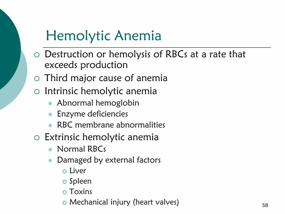
Hemolytic Anemia
Destruction or hemolysis of RBCs at a rate that
exceeds production
Third major cause of anemia
Intrinsic hemolytic anemia
Abnormal hemoglobin
Enzyme deficiencies
RBC membrane abnormalities
Extrinsic hemolytic anemia
Normal RBCs
Damaged by external factors
Liver
Spleen
Toxins
Mechanical injury (heart valves)58
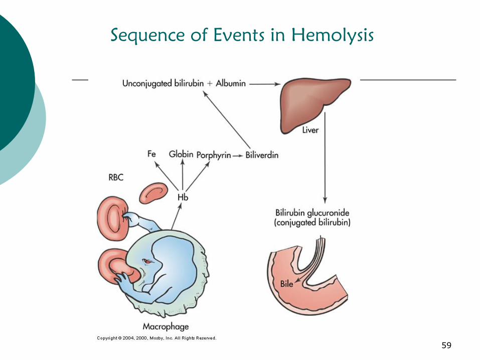
Sequence of Events in Hemolysis
59
Acquired Hemolytic Anemia
Causes
Medications
Infections
Manifestations
S/S of anemia
Complications
Accumulation of hemoglobin molecules can
obstruct renal tubules Tubular necrosis
Treatment
Eliminating the causative agent
60

Potential Nursing Dx for Patients with
Anemia
Activity Intolerance r/t weakness, malaise m/b
difficulty tolerating ↑’d activity
Imbalance nutrition: less than body requirements
r/t poor intake, anorexia, etc. m/b wt loss, serum albumin, iron levels, vitamin
deficiencies, below ideal body wt.
Ineffective therapeutic regimen management r/t
lack of knowledge about nutrition/medications
etc. m/b ineffective lifestyle/diet/medication
adjustments
Collaborative Problem: Hypoxemia r/t
hemoglobin
61

Hemochromatosis
Iron overload disease
Over absorption and
storage of iron causing
damage especially to
liver, heart and
pancreas
62
Polycythemia
Polycythemia is a condition in which
there is a net increase in the total number
of red blood cells
Overproduction of red blood cells may
be due to
a primary process in the bone marrow (a so-
called myeloproliferative syndrome)
or it may be a reaction to chronically low
oxygen levels or
malignancy
63

Polycythemia
Complications
↑d viscosity of blood
hemorrhage and thrombosis
Treatment
Phlebotomy
Myelosupressive agents: A number of new
therapeutic agents such as, interferon alfa-2b (Intron A)
therapy, agents that target platelet number (e.g.,
anagrelide [Agrylin]), and platelet function (e.g., aspirin).
64
Thrombocytopenia
Disorder of decreased platelets
platelet count below 150,000
Causes
Low production of platelets
Increased breakdown of platelets
Symptoms
Bruising
Nosebleeds
Petechiae (pinpoint microhemorrhages)
65
Thrombocytopenia
Types of Thrombocytopenia
Immune Thrombocytopenic Purpura
Abnormal destruction of circulating platelets
Autoimmune disorder
Destroyed in hosts’ spleen by macrophages
Thrombotic Thrombocytopenic Purpura
d agglutination of platelets that from microthrombi
66

Heparin-Induced Thrombocytopenia
(HIT)
HIT
Associated with administration of heparin
Develops when the body develops an antibody, or allergy
to heparin
Heparin (paradoxically) causes thrombosis
Immune mediated response that casues intense platelet
activation and relaese of procoaggulation particles.
Clinical features
Thrombocytopenia
Possible thrombosis after heparin therapy
Can be triggered by any type, route or amount of heparin
67

Thrombocytopenia
Diagnostic Studies
Platelet count
Prothrombin Time (PT)
Activated Partial Thromboplastin Time (aPTT)
Hgb/Hct
Treatment
Based on cause
Corticosteroids
Plasmaphoresis
Splenectomy
Platelet transfusion
68