
Helical Peptide Arrays on Self-Assembled Monolayer Surfaces through Soft and Reactive Landing of...
3
Peptide Arrays DOI: 10.1002/anie.200801366 Helical Peptide Arrays on Self-Assembled Monolayer Surfaces through Soft and Reactive Landing of Mass-Selected Ions** Peng Wang and Julia Laskin* The a helix, a common building block of the protein secondary structure, plays an important role in determining protein structure and function. The biological function of the a helix is mainly attributed to its large macrodipole [1] originating from the alignment of individual dipole moments of peptide bonds. Preparation of directionally aligned a- helical peptide layers on substrates has attracted significant attention because the resulting strong net dipole is useful for a variety of applications in photonics, [2, 3] molecular electron- ics, [4] and catalysis. [5–7] In addition, conformationally-selected a-helical peptide arrays can be used for detailed character- ization of molecular recognition steps critical for protein folding, enzyme function, and DNA binding by proteins. Existing technologies for the production of a-helical peptide surfaces are based on a variety of solution-phase synthetic strategies [2, 5, 8] that usually require relatively large quantities of purified materials. Preparative mass spectrometry based on soft landing (SL) [9–18] of mass-selected ions is a viable alternative to the existing surface modification approaches. It has been dem- onstrated that SL enables highly specific preparation of uniform thin films of biological molecules on substrates. [19–21] In addition, reactive landing (RL), in which SL is followed by covalent linking of molecules to chemically reactive surfaces, can be used for controlled immobilization of peptides and proteins on solid supports. [22, 23] Because SL is a relatively gentle ion deposition technique, it is easy to preserve the primary structure of deposited species. However, it is very difficult to control the secondary structure of soft-landed biomolecules, because electrospray ionization (ESI) utilized in these experiments generates ions in a variety of different conformations. Previous studies reported retention of the secondary and possibly tertiary structure by soft-landed proteins. [19a, 21, 22] Herein, we demonstrate that SL can be used to prepare peptides on substrates in stable conforma- tions that do not exist in solution. This study focuses on the preparation of conformation- ally-selected peptide arrays using SL of mass selected peptide ions on self-assembled monolayer (SAM) surfaces. The singly protonated Ac-A 15 K peptide was selected as a model system for this study because ion mobility measurements and molecular dynamics (MD) simulations demonstrated that this peptide forms a very stable a-helical conformation in the gas phase, which is stabilized by the interaction between the protonated C-terminal lysine residue and the dipole of the helix. [24] Formation of the a-helical peptide array is demon- strated on an inert SAM of alkylthiol on gold (HSAM) and covalent immobilization of the Ac-A 15 K peptide on a reactive SAM of N-hydroxysuccinimidyl ester terminated alkylthiol on gold (NHS-SAM) with retention of the secondary structure. Because the NHS-SAM surface readily reacts with primary amino groups in proteins or peptides by forming amide bonds [25, 26] this substrate has been previously used for efficient covalent immobilization of soft-landed peptides onto SAMs by the formation of an amide bond between the SAM and the amino group of the lysine side chain. [23] Experiments were performed using an ion deposition apparatus described in detail elsewhere. [20] Experimental details are given in the Supporting Information. Infrared reflection absorption spectroscopy (IRRAS) was used for characterization of the secondary structure of peptides on SAM surfaces based on the presence and position of amide I and amide A bands originating from peptide bonds. [11, 27] The amide I band is dominated by the C =O stretching vibrations of amide groups, and gives rise to infrared absorption in the region between 1600 and 1700 cm À1 , whereas the amide A band responsible for absorption in the 3200–3300 cm À1 region corresponds to the N À H stretching mode. In Figure 1, standard circular dichroism (CD) spectra of the a helix, b sheet, and random coil are compared with the spectrum obtained for the ESI solution of Ac-A 15 K. The CD spectrum of the Ac-A 15 K solution shows the presence of a mixture of conformations dominated by the b sheet and a small fraction of the a helix and random coil. Similarly, the IRRAS spectrum of the Ac-A 15 K layer prepared on the HSAM surface using ESI deposition is also dominated by the characteristic features of the b-sheet structure. Specifically, the IRRAS spectrum (blue trace, Figure 2) shows a broad amide I band with dominant features at 1632 and 1697 cm À1 which is characteristic of the b-sheet conformation. [28] In addition, the position of the amide A band centered at 3280 cm À1 indicates that NH groups are involved in C = O···H À N hydrogen bonds typical for the b-sheet structure. The features in the center of the amide I band (ca. 1670 cm À1 ) and the high-frequency tailing of the amide A band can be assigned to contributions of the a-helix and the random-coil [*] P. Wang, J. Laskin Fundamental Sciences Division Pacific Northwest National Laboratory P.O.Box 999K8-88, Richland, WA 99352 (USA) Fax: (+ 1) 509-371-6139 E-mail: [email protected] Homepage: http://emslbios.pnl.gov/id/laskin_j [**] This work was supported by the Laboratory Directed Research and Development Program at Pacific Northwest National Laboratory and a grant from the Chemical Sciences Division, Office of Basic Energy Sciences of the U.S. Department of Energy. The authors thank Dr. John Cort for technical assistance and helpful discussions. Supporting information for this article is available on the WWW under http://dx.doi.org/10.1002/anie.200801366. Communications 6678 # 2008 Wiley-VCH Verlag GmbH & Co. KGaA, Weinheim Angew. Chem. Int. Ed. 2008, 47, 6678 –6680
Transcript of Helical Peptide Arrays on Self-Assembled Monolayer Surfaces through Soft and Reactive Landing of...
Peptide ArraysDOI: 10.1002/anie.200801366
Helical Peptide Arrays on Self-AssembledMonolayer Surfaces throughSoft and Reactive Landing of Mass-Selected Ions**Peng Wang and Julia Laskin*
The a helix, a common building block of the proteinsecondary structure, plays an important role in determiningprotein structure and function. The biological function of thea helix is mainly attributed to its large macrodipole[1]
originating from the alignment of individual dipole momentsof peptide bonds. Preparation of directionally aligned a-helical peptide layers on substrates has attracted significantattention because the resulting strong net dipole is useful for avariety of applications in photonics,[2,3] molecular electron-ics,[4] and catalysis.[5–7] In addition, conformationally-selecteda-helical peptide arrays can be used for detailed character-ization of molecular recognition steps critical for proteinfolding, enzyme function, and DNA binding by proteins.Existing technologies for the production of a-helical peptidesurfaces are based on a variety of solution-phase syntheticstrategies[2,5, 8] that usually require relatively large quantitiesof purified materials.
Preparative mass spectrometry based on soft landing(SL)[9–18] of mass-selected ions is a viable alternative to theexisting surface modification approaches. It has been dem-onstrated that SL enables highly specific preparation ofuniform thin films of biological molecules on substrates.[19–21]
In addition, reactive landing (RL), in which SL is followed bycovalent linking of molecules to chemically reactive surfaces,can be used for controlled immobilization of peptides andproteins on solid supports.[22,23] Because SL is a relativelygentle ion deposition technique, it is easy to preserve theprimary structure of deposited species. However, it is verydifficult to control the secondary structure of soft-landedbiomolecules, because electrospray ionization (ESI) utilizedin these experiments generates ions in a variety of differentconformations. Previous studies reported retention of thesecondary and possibly tertiary structure by soft-landedproteins.[19a,21,22] Herein, we demonstrate that SL can beused to prepare peptides on substrates in stable conforma-tions that do not exist in solution.
This study focuses on the preparation of conformation-ally-selected peptide arrays using SL of mass selected peptideions on self-assembled monolayer (SAM) surfaces. The singlyprotonated Ac-A15K peptide was selected as a model systemfor this study because ion mobility measurements andmolecular dynamics (MD) simulations demonstrated thatthis peptide forms a very stable a-helical conformation in thegas phase, which is stabilized by the interaction between theprotonated C-terminal lysine residue and the dipole of thehelix.[24] Formation of the a-helical peptide array is demon-strated on an inert SAM of alkylthiol on gold (HSAM) andcovalent immobilization of the Ac-A15K peptide on a reactiveSAM of N-hydroxysuccinimidyl ester terminated alkylthiolon gold (NHS-SAM) with retention of the secondarystructure. Because the NHS-SAM surface readily reactswith primary amino groups in proteins or peptides by formingamide bonds[25,26] this substrate has been previously used forefficient covalent immobilization of soft-landed peptides ontoSAMs by the formation of an amide bond between the SAMand the amino group of the lysine side chain.[23]
Experiments were performed using an ion depositionapparatus described in detail elsewhere.[20] Experimentaldetails are given in the Supporting Information. Infraredreflection absorption spectroscopy (IRRAS) was used forcharacterization of the secondary structure of peptides onSAM surfaces based on the presence and position of amide Iand amide A bands originating from peptide bonds.[11,27] Theamide I band is dominated by the C=O stretching vibrationsof amide groups, and gives rise to infrared absorption in theregion between 1600 and 1700 cm�1, whereas the amide Aband responsible for absorption in the 3200–3300 cm�1 regioncorresponds to the N�H stretching mode.
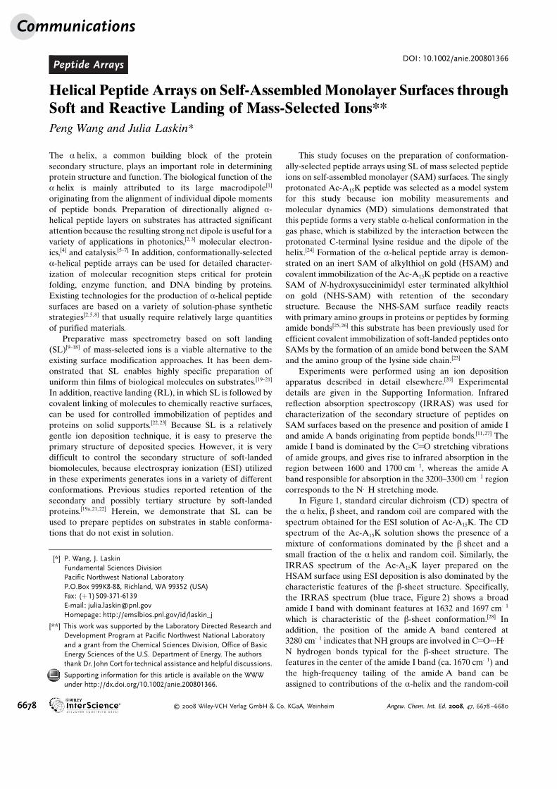
In Figure 1, standard circular dichroism (CD) spectra ofthe a helix, b sheet, and random coil are compared with thespectrum obtained for the ESI solution of Ac-A15K. The CDspectrum of the Ac-A15K solution shows the presence of amixture of conformations dominated by the b sheet and asmall fraction of the a helix and random coil. Similarly, theIRRAS spectrum of the Ac-A15K layer prepared on theHSAM surface using ESI deposition is also dominated by thecharacteristic features of the b-sheet structure. Specifically,the IRRAS spectrum (blue trace, Figure 2) shows a broadamide I band with dominant features at 1632 and 1697 cm�1
which is characteristic of the b-sheet conformation.[28] Inaddition, the position of the amide A band centered at3280 cm�1 indicates that NH groups are involved in C=O···H�N hydrogen bonds typical for the b-sheet structure. Thefeatures in the center of the amide I band (ca. 1670 cm�1) andthe high-frequency tailing of the amide A band can beassigned to contributions of the a-helix and the random-coil
[*] P. Wang, J. LaskinFundamental Sciences DivisionPacific Northwest National LaboratoryP.O.Box 999K8-88, Richland, WA 99352 (USA)Fax: (+1)509-371-6139E-mail: [email protected]: http://emslbios.pnl.gov/id/laskin_j
[**] This work was supported by the Laboratory Directed Research andDevelopment Program at Pacific Northwest National Laboratoryand a grant from the Chemical Sciences Division, Office of BasicEnergy Sciences of the U.S. Department of Energy. The authorsthank Dr. John Cort for technical assistance and helpful discussions.
Supporting information for this article is available on the WWWunder http://dx.doi.org/10.1002/anie.200801366.
Communications
6678 � 2008 Wiley-VCH Verlag GmbH & Co. KGaA, Weinheim Angew. Chem. Int. Ed. 2008, 47, 6678 –6680
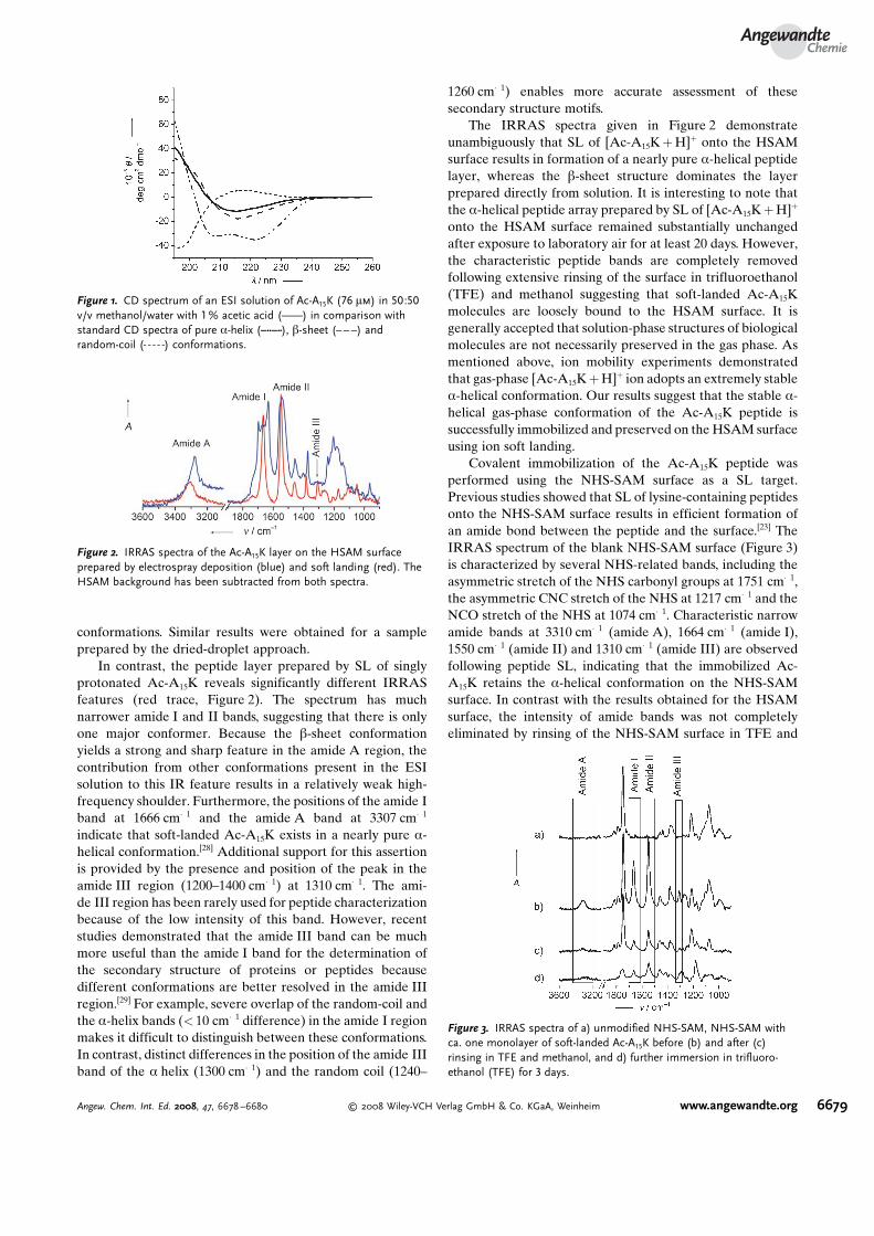
conformations. Similar results were obtained for a sampleprepared by the dried-droplet approach.
In contrast, the peptide layer prepared by SL of singlyprotonated Ac-A15K reveals significantly different IRRASfeatures (red trace, Figure 2). The spectrum has muchnarrower amide I and II bands, suggesting that there is onlyone major conformer. Because the b-sheet conformationyields a strong and sharp feature in the amide A region, thecontribution from other conformations present in the ESIsolution to this IR feature results in a relatively weak high-frequency shoulder. Furthermore, the positions of the amide Iband at 1666 cm�1 and the amide A band at 3307 cm�1
indicate that soft-landed Ac-A15K exists in a nearly pure a-helical conformation.[28] Additional support for this assertionis provided by the presence and position of the peak in theamide III region (1200–1400 cm�1) at 1310 cm�1. The ami-de III region has been rarely used for peptide characterizationbecause of the low intensity of this band. However, recentstudies demonstrated that the amide III band can be muchmore useful than the amide I band for the determination ofthe secondary structure of proteins or peptides becausedifferent conformations are better resolved in the amide IIIregion.[29] For example, severe overlap of the random-coil andthe a-helix bands (< 10 cm�1 difference) in the amide I regionmakes it difficult to distinguish between these conformations.In contrast, distinct differences in the position of the amide IIIband of the a helix (1300 cm�1) and the random coil (1240–
1260 cm�1) enables more accurate assessment of thesesecondary structure motifs.
The IRRAS spectra given in Figure 2 demonstrateunambiguously that SL of [Ac-A15K+H]+ onto the HSAMsurface results in formation of a nearly pure a-helical peptidelayer, whereas the b-sheet structure dominates the layerprepared directly from solution. It is interesting to note thatthe a-helical peptide array prepared by SL of [Ac-A15K+H]+
onto the HSAM surface remained substantially unchangedafter exposure to laboratory air for at least 20 days. However,the characteristic peptide bands are completely removedfollowing extensive rinsing of the surface in trifluoroethanol(TFE) and methanol suggesting that soft-landed Ac-A15Kmolecules are loosely bound to the HSAM surface. It isgenerally accepted that solution-phase structures of biologicalmolecules are not necessarily preserved in the gas phase. Asmentioned above, ion mobility experiments demonstratedthat gas-phase [Ac-A15K+H]+ ion adopts an extremely stablea-helical conformation. Our results suggest that the stable a-helical gas-phase conformation of the Ac-A15K peptide issuccessfully immobilized and preserved on the HSAM surfaceusing ion soft landing.
Covalent immobilization of the Ac-A15K peptide wasperformed using the NHS-SAM surface as a SL target.Previous studies showed that SL of lysine-containing peptidesonto the NHS-SAM surface results in efficient formation ofan amide bond between the peptide and the surface.[23] TheIRRAS spectrum of the blank NHS-SAM surface (Figure 3)is characterized by several NHS-related bands, including theasymmetric stretch of the NHS carbonyl groups at 1751 cm�1,the asymmetric CNC stretch of the NHS at 1217 cm�1 and theNCO stretch of the NHS at 1074 cm�1. Characteristic narrowamide bands at 3310 cm�1 (amide A), 1664 cm�1 (amide I),1550 cm�1 (amide II) and 1310 cm�1 (amide III) are observedfollowing peptide SL, indicating that the immobilized Ac-A15K retains the a-helical conformation on the NHS-SAMsurface. In contrast with the results obtained for the HSAMsurface, the intensity of amide bands was not completelyeliminated by rinsing of the NHS-SAM surface in TFE and
Figure 2. IRRAS spectra of the Ac-A15K layer on the HSAM surfaceprepared by electrospray deposition (blue) and soft landing (red). TheHSAM background has been subtracted from both spectra.
Figure 3. IRRAS spectra of a) unmodified NHS-SAM, NHS-SAM withca. one monolayer of soft-landed Ac-A15K before (b) and after (c)rinsing in TFE and methanol, and d) further immersion in trifluoro-ethanol (TFE) for 3 days.
Figure 1. CD spectrum of an ESI solution of Ac-A15K (76 mm) in 50:50v/v methanol/water with 1% acetic acid (c) in comparison withstandard CD spectra of pure a-helix (l), b-sheet (b) andrandom-coil (a) conformations.
AngewandteChemie
6679Angew. Chem. Int. Ed. 2008, 47, 6678 –6680 � 2008 Wiley-VCH Verlag GmbH & Co. KGaA, Weinheim www.angewandte.org
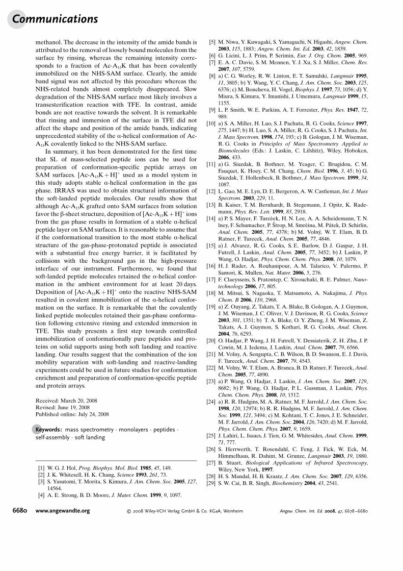
methanol. The decrease in the intensity of the amide bands isattributed to the removal of loosely boundmolecules from thesurface by rinsing, whereas the remaining intensity corre-sponds to a fraction of Ac-A15K that has been covalentlyimmobilized on the NHS-SAM surface. Clearly, the amideband signal was not affected by this procedure whereas theNHS-related bands almost completely disappeared. Slowdegradation of the NHS-SAM surface most likely involves atransesterification reaction with TFE. In contrast, amidebonds are not reactive towards the solvent. It is remarkablethat rinsing and immersion of the surface in TFE did notaffect the shape and position of the amide bands, indicatingunprecedented stability of the a-helical conformation of Ac-A15K covalently linked to the NHS-SAM surface.
In summary, it has been demonstrated for the first timethat SL of mass-selected peptide ions can be used forpreparation of conformation-specific peptide arrays onSAM surfaces. [Ac-A15K+H]+ used as a model system inthis study adopts stable a-helical conformation in the gasphase. IRRAS was used to obtain structural information ofthe soft-landed peptide molecules. Our results show thatalthough Ac-A15K grafted onto SAM surfaces from solutionfavor the b-sheet structure, deposition of [Ac-A15K+H]+ ionsfrom the gas phase results in formation of a stable a-helicalpeptide layer on SAM surfaces. It is reasonable to assume thatif the conformational transition to the most stable a-helicalstructure of the gas-phase-protonated peptide is associatedwith a substantial free energy barrier, it is facilitated bycollisions with the background gas in the high-pressureinterface of our instrument. Furthermore, we found thatsoft-landed peptide molecules retained the a-helical confor-mation in the ambient environment for at least 20 days.Deposition of [Ac-A15K+H]+ onto the reactive NHS-SAMresulted in covalent immobilization of the a-helical confor-mation on the surface. It is remarkable that the covalentlylinked peptide molecules retained their gas-phase conforma-tion following extensive rinsing and extended immersion inTFE. This study presents a first step towards controlledimmobilization of conformationally pure peptides and pro-teins on solid supports using both soft landing and reactivelanding. Our results suggest that the combination of the ionmobility separation with soft-landing and reactive-landingexperiments could be used in future studies for conformationenrichment and preparation of conformation-specific peptideand protein arrays.
Received: March 20, 2008Revised: June 19, 2008Published online: July 24, 2008
.Keywords: mass spectrometry · monolayers · peptides ·self-assembly · soft landing
[1] W. G. J. Hol, Prog. Biophys. Mol. Biol. 1985, 45, 149.[2] J. K. Whitesell, H. K. Chang, Science 1993, 261, 73.[3] S. Yasutomi, T. Morita, S. Kimura, J. Am. Chem. Soc. 2005, 127,
14564.[4] A. E. Strong, B. D. Moore, J. Mater. Chem. 1999, 9, 1097.
[5] M. Niwa, Y. Kuwagaki, S. Yamaguchi, N. Higashi,Angew. Chem.2003, 115, 1883; Angew. Chem. Int. Ed. 2003, 42, 1839.
[6] G. Licini, L. J. Prins, P. Scrimin, Eur. J. Org. Chem. 2005, 969.[7] E. A. C. Davie, S. M. Mennen, Y. J. Xu, S. J. Miller, Chem. Rev.
2007, 107, 5759.[8] a) C. G. Worley, R. W. Linton, E. T. Samulski, Langmuir 1995,
11, 3805; b) Y. Wang, Y. C. Chang, J. Am. Chem. Soc. 2003, 125,6376; c) M. Boncheva, H. Vogel, Biophys. J. 1997, 73, 1056; d) Y.Miura, S. Kimura, Y. Imanishi, J. Umemura, Langmuir 1999, 15,1155.
[9] L. P. Smith, W. E. Parkins, A. T. Forrester, Phys. Rev. 1947, 72,989.
[10] a) S. A. Miller, H. Luo, S. J. Pachuta, R. G. Cooks, Science 1997,275, 1447; b) H. Luo, S. A. Miller, R. G. Cooks, S. J. Pachuta, Int.J. Mass Spectrom. 1998, 174, 193; c) B. Gologan, J. M. Wiseman,R. G. Cooks in Principles of Mass Spectrometry Applied toBiomolecules (Eds.: J. Laskin, C. Lifshitz), Wiley, Hoboken,2006, 433.
[11] a) G. Siuzdak, B. Bothner, M. Yeager, C. Brugidou, C. M.Fauquet, K. Hoey, C. M. Chang, Chem. Biol. 1996, 3, 45; b) G.Siuzdak, T. Hollenbeck, B. Bothner, J. Mass Spectrom. 1999, 34,1087.
[12] L. Gao, M. E. Lyn, D. E. Bergeron, A. W. Castleman, Int. J. MassSpectrom. 2003, 229, 11.
[13] B. Kaiser, T. M. Bernhardt, B. Stegemann, J. Opitz, K. Rade-mann, Phys. Rev. Lett. 1999, 83, 2918.
[14] a) P. S. Mayer, F. TureHek, H. N. Lee, A. A. Scheidemann, T. N.lney, F. Schumacher, P. Strop, M. SmrHina, M. PJtek, D. Schirlin,Anal. Chem. 2005, 77, 4378; b) M. Volny, W. T. Elam, B. D.Ratner, F. Turecek, Anal. Chem. 2005, 77, 4846.
[15] a) J. Alvarez, R. G. Cooks, S. E. Barlow, D. J. Gaspar, J. H.Futrell, J. Laskin, Anal. Chem. 2005, 77, 3452; b) J. Laskin, P.Wang, O. Hadjar, Phys. Chem. Chem. Phys. 2008, 10, 1079.
[16] H. J. Rader, A. Rouhanipour, A. M. Talarico, V. Palermo, P.Samori, K. Mullen, Nat. Mater. 2006, 5, 276.
[17] F. Claeyssens, S. Pratontep, C. Xirouchaki, R. E. Palmer, Nano-technology 2006, 17, 805.
[18] M. Mitsui, S. Nagaoka, T. Matsumoto, A. Nakajima, J. Phys.Chem. B 2006, 110, 2968.
[19] a) Z. Ouyang, Z. Takats, T. A. Blake, B. Gologan, A. J. Guymon,J. M. Wiseman, J. C. Oliver, V. J. Davisson, R. G. Cooks, Science2003, 301, 1351; b) T. A. Blake, O. Y. Zheng, J. M. Wiseman, Z.Takats, A. J. Guymon, S. Kothari, R. G. Cooks, Anal. Chem.2004, 76, 6293.
[20] O. Hadjar, P. Wang, J. H. Futrell, Y. Dessiaterik, Z. H. Zhu, J. P.Cowin, M. J. Iedema, J. Laskin, Anal. Chem. 2007, 79, 6566.
[21] M. Volny, A. Sengupta, C. B. Wilson, B. D. Swanson, E. J. Davis,F. Turecek, Anal. Chem. 2007, 79, 4543.
[22] M. Volny, W. T. Elam, A. Branca, B. D. Ratner, F. Turecek,Anal.Chem. 2005, 77, 4890.
[23] a) P. Wang, O. Hadjar, J. Laskin, J. Am. Chem. Soc. 2007, 129,8682; b) P. Wang, O. Hadjar, P. L. Gassman, J. Laskin, Phys.Chem. Chem. Phys. 2008, 10, 1512.
[24] a) R. R. Hudgins, M. A. Ratner, M. F. Jarrold, J. Am. Chem. Soc.1998, 120, 12974; b) R. R. Hudgins, M. F. Jarrold, J. Am. Chem.Soc. 1999, 121, 3494; c) M. Kohtani, T. C. Jones, J. E. Schneider,M. F. Jarrold, J. Am. Chem. Soc. 2004, 126, 7420; d) M. F. Jarrold,Phys. Chem. Chem. Phys. 2007, 9, 1659.
[25] J. Lahiri, L. Isaacs, J. Tien, G. M. Whitesides, Anal. Chem. 1999,71, 777.
[26] S. Herrwerth, T. Rosendahl, C. Feng, J. Fick, W. Eck, M.Himmelhaus, R. Dahint, M. Grunze, Langmuir 2003, 19, 1880.
[27] B. Stuart, Biological Applications of Infrared Spectroscopy,Wiley, New York, 1997.
[28] H. S. Mandal, H. B. Kraatz, J. Am. Chem. Soc. 2007, 129, 6356.[29] S. W. Cai, B. R. Singh, Biochemistry 2004, 43, 2541.
Communications
6680 www.angewandte.org � 2008 Wiley-VCH Verlag GmbH & Co. KGaA, Weinheim Angew. Chem. Int. Ed. 2008, 47, 6678 –6680