Heel spur treatment plantar fasciitis home remedies, chronic plantar fasciitis
Upload
kevin-ambadanCategory
view
352download
2
HEEL PAIN AND PLANTAR FASCIITIS
BY
JITHIN ABRAHAM

INTRODUCTION
The foot is really unique to human being. The structure of the foot allows the foot to sustain large weight bearing stresses under a variety of surfaces and activities that maximize stability and mobility.
Arches of the foot help in fast walking, running, jumping, weight bearing
and in providing upright posture.
Arches are supported by intrinsic and extrinsic muscles of the sole in addition to ligaments, aponeurosis and shape of the bones.

Commonest of all causes
•Plantar fascitis•Achilles tendinitis•Achilles bursitis•Calcanel spur•Heel pad fat atrophy

PLANTAR FASCIA
• The plantar fascia is the thick connective tissue which supports the arch on the bottom of the foot
• It runs from the tuberosity of the calcaneus forward to the heads of the metatarsal bones
•The plantar fascia contributes to support of arch of the foot•The plantar fascia also has an important role in dynamic function during gait

What is PLANTAR FASCIITIS?• Plantar fasciitis is a painful foot condition caused by
inflammation of insertion of the plantar fascia on the medial process of the calcaneal tuberosity.
• This associated with• Pain• Swelling• Warmth of the affected area• Redness of the adjacent skin

AETIOLOGY
•Excessive pronation of the foot.•Poor arch support in the shoe•Flat foot•Prolonged standing•Fat pad atrophy•Tight triceps surae•Repetitive strength imbalances•Over use may cause microtears and inflammation•Congenital problems such as Pescavus and Pesplanus•Obesity •Reiters disease,Ankylosing spondylitis,Diffuse idiopathic skeletal hyperostosis

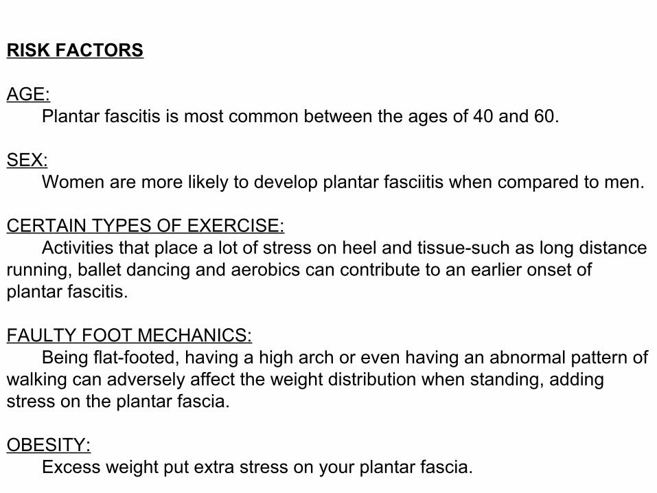
RISK FACTORS
AGE: Plantar fascitis is most common between the ages of 40 and 60.
SEX: Women are more likely to develop plantar fasciitis when compared to men.
CERTAIN TYPES OF EXERCISE: Activities that place a lot of stress on heel and tissue-such as long distance running, ballet dancing and aerobics can contribute to an earlier onset of plantar fascitis.
FAULTY FOOT MECHANICS: Being flat-footed, having a high arch or even having an abnormal pattern of walking can adversely affect the weight distribution when standing, adding stress on the plantar fascia.
OBESITY: Excess weight put extra stress on your plantar fascia.

RELEVANT ANATOMY
The os calcis is elevated anteriorly so that during heel strike, the posterior tubercle contacts the ground 1st and transmits full body weight.
This make the calcaneum vulnerable to trauma or micro trauma
The heel fat pad has many fat globules enclosed by multiple fibroelastic septa
And after 40 years this fat pad begin to atrophy and degenerate

The plantar aponeurosis is an inelastic facia that arises from the os calcis and is composed of three segments
Anteriomedial tuberosity
Covers undersurface of abductor haluucis
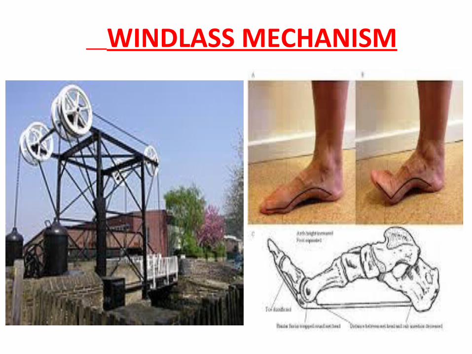
WINDLASS MECHANISM

Windlass mechanism of the plantar fascia as the toes are dorsiflexed
Plantar fascia inserts through several slips into the plantar plates of the metatarsophalangeal joints, the flexor tendon sheaths, and the bases of the proximal phalanges of the digits, is under constant traction as it is pulled distally around the drum of the windlass (metatarsal heads). This tightening elevates the longitudinal arch, inverts the hind foot and externally rotates the leg. This mechanism is passive and depends entirely on bony and ligamentous instabilty.
Pathomechanics of Plantar fascia

•Excessive foot pronation:
• Excessive pronation or inward rolling of the foot also inhibits efficient use of the windlass mechanism. This decreases shock absorption through the plantar fascia which in turn increases the tension on the plantar fascia.

•Tight calf muscles: Having tight calf muscles can cause excessive foot pronation contributing to excessive foot mobility which increases the level of stresses on the plantar fascia.

•High arched foot:
•A high arched foot lacks the normal joint mobility which reduces the foot’s ability to absorb shock from the ground, thereby increasing the stresses on the plantar fascia.

•Ill-fitting or worn out shoes:
•Wearing ill-fitting or worn out shoes may change the foot biomechanics, causing undue strain on the plantar fascia.

•Excessive walking and running on hard surfaces: This increases the shock transmitted to the plantar fascia, increasing the strain on the plantar fascia.
•Overweight: Being overweight increases the level of stresses applied to the fascia due to the added body weight on the foot, increasing the strain on the plantar fascia.

INVESTIGATIONS
X-RAYS
•An X-ray may be taken to rule out a stress fracture of the heel bone
•X-rays of patient with heel pain sometimes reveal a calcification of the plantar aponeurosis at the origin on the calcaneus, commonly referred to as a heel spur
MRI: Show thickening of plantar fascia
BONE SCAN: It show increase uptake at the calcaneus
RHEUMATOLOGIC SCREENING: It can be important to rule out inflammatory arthrides.

plantar aponeurosis as uniform bandlike structure of low signal intensity.
thickening of central component of plantar aponeurosis . Extensive edema infiltrates perifascial soft tissue .

SPECIAL TEST
•Plantar fascitis have more tenderness in the plantar fascia when it is stretched and less tenderness when the fascia is relaxed. The plantar fascitis test uses this property to diagnose patients with plantar fascitis. •To perform this test, first stretch plantar fascia. Then use your thumb or finger to feel the plantar fascia. If plantar fascia is tender, then try the same maneuver with plantar fascia relaxed.
•If pushing the stretched plantar fascia causes more tenderness than pushing on the relaxed plantar fascia, then the plantar fascia is likely the source of the pain and the patient have plantar fascitis.

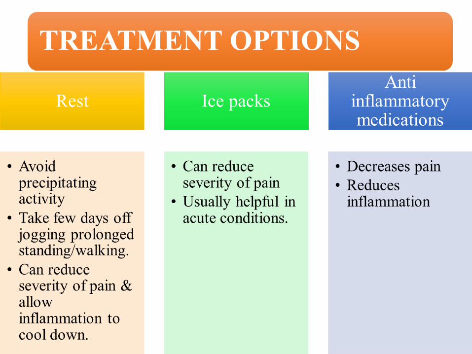
CONSERVATIVE TREATMENT

Not all of these treatments are appropriate for every condition, but they may be helpful in certain situation.

Modified Foot wearCan help to soothe pain
Shoe inserts

Stretching

TAPING
• Calcaneal taping or low-dye taping used for short-term pain relief. Taping does cause improvement in function.

Injection Therapy : Local corticosteroid injections can help
to reduce inflammation and relieve pain.
Weight reduction

Modalities
•Therapeutic ultrasound •Extracorporeal shockwave therapy (ESWT).

ELECTROTHERAPY

NIGHT SPLINTS
• It is used for patient with symptoms greater than 6 months in duration.• The desire length of time for wearing the splint is 1
to 3months.• This splint maintain ankle in neutral position and
toes in slight extension.

SUMMARY
• Plantar Fasciitis is a painful condition caused by overuse of the plantar fascia or arch tendon of the foot. • The most common cause of plantar fasciitis is very
tight calf muscles• Treatment can last from several months to 2 years• Some people may need surgery.