Heat shock causes a decrease in polysomes and the appearance of
13
3002 Research Article Introduction When cells are subjected to stress, such as heat shock, genes associated with growth are downregulated and expression of proteins believed to increase cellular survival, such as HSP70, are selectively upregulated. In eukaryotes, this regulation operates at multiple levels: transcription initiation may be enhanced via factors specific for heat shock-genes, whereas for other genes there is selective inhibition of splicing and nuclear export of mRNAs (Bond, 2006). Stress also influences the fate of cytoplasmic mRNA in eukaryotes. Treatment of mammalian cells with a variety of stresses, such as arsenite or heat shock, results in the relocation of most mRNAs from polysomes to cytoplasmic foci called stress granules (Kedersha et al., 1999). These contain the small ribosomal subunit (Kedersha et al., 2002), the translation-initiation factors eIF2, eIF3, eIF4E and eIF4G (Kedersha et al., 2002; Kimball et al., 2003), the poly(A)-binding protein (Kedersha et al., 1999), DHH1 (also known as DDX6, Rck, ATP-dependent RNA helicase p54) (Wilczynska et al., 2005), the 5-3 exonuclease XRN1 (Kedersha et al., 2005) and RNA-binding proteins, including SCD6 (also known as LSM14 and RAP55) (Yang et al., 2006; Kedersha and Anderson, 2007). Formation of stress granules in response to heat shock and metabolic stress is dependent on, and might be initiated by, the phosphorylation of eukaryotic translation-initiation factor eIF2α on a conserved serine residue (S51 in mammals and yeast) by a family of stress-activated kinases (Kedersha et al., 1999; McEwen et al., 2005). The modification inhibits initiation of translation by increasing the affinity of eIF2α for the guanine nucleotide exchange factor eIF2B, resulting in its sequestration. Stress-granule formation can also be induced independently of eIF2α phosphorylation by pateamine or hippuristanol, which interfere with the action of eIF4A, an RNA helicase necessary for translation initiation (Dang et al., 2006; Mazroui et al., 2006). The fate of mRNAs in stress granules remains unclear; mRNAs may be degraded or stored until the stress is over. Inhibitors of translation elongation that stabilise polysomes cause the dissociation of stress granules, whereas inhibitors that destabilise polysomes promote the assembly of stress granules, suggesting that the mRNA in stress granules is in equilibrium with polysomes (Kedersha et al., 2000). Stress granules have been shown to interact with processing bodies (P-bodies) (Kedersha et al., 2005; Wilczynska et al., 2005). P-bodies are cytoplasmic granules thought to be sites of mRNA degradation (Parker and Sheth, 2007). P-bodies share some, but not all, components with stress granules, for instance XRN1, DHH1 and SCD6. The decapping enzyme (a complex of DCP1 and DCP2) is specific to P-bodies while the small ribosomal subunit, eIF2, eIF3 and poly(A)-binding protein are absent (Bashkirov et al., 1997; Cougot et al., 2004; Ingelfinger et al., 2002; Kedersha and Anderson, 2007; Sheth and Parker, 2003). It is believed that XRN1 is the major degradative enzyme in P-bodies but the precise function of other components has yet to be fully characterised. Trypanosoma brucei is a unicellular flagellate that causes African trypanosomiasis (sleeping sickness) in humans and a range of diseases in animals. Synthesis of mRNA in trypanosomes is atypical; tandem arrays of genes (Berriman et al., 2005; El Sayed et al., 2003; Hall et al., 2003; McDonagh et al., 2000) are transcribed In trypanosomes there is an almost total reliance on post- transcriptional mechanisms to alter gene expression; here, heat shock was used to investigate the response to an environmental signal. Heat shock rapidly and reversibly induced a decrease in polysome abundance, and the consequent changes in mRNA metabolism were studied. Both heat shock and polysome dissociation were necessary for (1) a reduction in mRNA levels that was more rapid than normal turnover, (2) an increased number of P-body-like granules that contained DHH1, SCD6 and XRNA, (3) the formation of stress granules that remained largely separate from the P-body-like granules and localise to the periphery of the cell and, (4) an increase in the size of a novel focus located at the posterior pole of the cell that contain XRNA, but neither DHH1 nor SCD6. The response differed from mammalian cells in that neither the decrease in polysomes nor stress-granule formation required phosphorylation of eIF2α at the position homologous to that of serine 51 in mammalian eIF2α and in the occurrence of a novel XRNA-focus. Supplementary material available online at http://jcs.biologists.org/cgi/content/full/121/18/3002/DC1 Key words: Heat shock, Trypanosoma brucei, Stress granules, eIF2 alpha, P-bodies Summary Heat shock causes a decrease in polysomes and the appearance of stress granules in trypanosomes independently of eIF2α phosphorylation at Thr169 Susanne Kramer 1 , Rafael Queiroz 2,3 , Louise Ellis 1 , Helena Webb 1 , Jörg D. Hoheisel 3 , Christine Clayton 2 and Mark Carrington 1, * 1 Department of Biochemistry, University of Cambridge, 80 Tennis Court Road, Cambridge CB2 1GA, UK 2 ZMBH, Im Neuenheimer Feld 282, 69120 Heidelberg, Germany 3 Deutsches Krebsforschungszentrum, Im Neuenheimer Feld 580, 69120 Heidelberg, Germany *Author for correspondence (e-mail: [email protected]) Accepted 30 June 2008 Journal of Cell Science 121, 3002-3014 Published by The Company of Biologists 2008 doi:10.1242/jcs.031823 Journal of Cell Science
Transcript of Heat shock causes a decrease in polysomes and the appearance of

3002 Research Article
IntroductionWhen cells are subjected to stress, such as heat shock, genes
associated with growth are downregulated and expression of
proteins believed to increase cellular survival, such as HSP70, are
selectively upregulated. In eukaryotes, this regulation operates at
multiple levels: transcription initiation may be enhanced via factors
specific for heat shock-genes, whereas for other genes there is
selective inhibition of splicing and nuclear export of mRNAs (Bond,
2006).
Stress also influences the fate of cytoplasmic mRNA in
eukaryotes. Treatment of mammalian cells with a variety of stresses,
such as arsenite or heat shock, results in the relocation of most
mRNAs from polysomes to cytoplasmic foci called stress granules
(Kedersha et al., 1999). These contain the small ribosomal subunit
(Kedersha et al., 2002), the translation-initiation factors eIF2, eIF3,
eIF4E and eIF4G (Kedersha et al., 2002; Kimball et al., 2003), the
poly(A)-binding protein (Kedersha et al., 1999), DHH1 (also
known as DDX6, Rck, ATP-dependent RNA helicase p54)
(Wilczynska et al., 2005), the 5�-3� exonuclease XRN1 (Kedersha
et al., 2005) and RNA-binding proteins, including SCD6 (also
known as LSM14 and RAP55) (Yang et al., 2006; Kedersha and
Anderson, 2007).
Formation of stress granules in response to heat shock and
metabolic stress is dependent on, and might be initiated by, the
phosphorylation of eukaryotic translation-initiation factor eIF2α on
a conserved serine residue (S51 in mammals and yeast) by a family
of stress-activated kinases (Kedersha et al., 1999; McEwen et al.,
2005). The modification inhibits initiation of translation by
increasing the affinity of eIF2α for the guanine nucleotide exchange
factor eIF2B, resulting in its sequestration. Stress-granule formation
can also be induced independently of eIF2α phosphorylation by
pateamine or hippuristanol, which interfere with the action of eIF4A,
an RNA helicase necessary for translation initiation (Dang et al.,
2006; Mazroui et al., 2006).
The fate of mRNAs in stress granules remains unclear; mRNAs
may be degraded or stored until the stress is over. Inhibitors of
translation elongation that stabilise polysomes cause the dissociation
of stress granules, whereas inhibitors that destabilise polysomes
promote the assembly of stress granules, suggesting that the mRNA
in stress granules is in equilibrium with polysomes (Kedersha et
al., 2000).
Stress granules have been shown to interact with processing
bodies (P-bodies) (Kedersha et al., 2005; Wilczynska et al., 2005).
P-bodies are cytoplasmic granules thought to be sites of mRNA
degradation (Parker and Sheth, 2007). P-bodies share some, but not
all, components with stress granules, for instance XRN1, DHH1
and SCD6. The decapping enzyme (a complex of DCP1 and DCP2)
is specific to P-bodies while the small ribosomal subunit, eIF2, eIF3
and poly(A)-binding protein are absent (Bashkirov et al., 1997;
Cougot et al., 2004; Ingelfinger et al., 2002; Kedersha and Anderson,
2007; Sheth and Parker, 2003). It is believed that XRN1 is the major
degradative enzyme in P-bodies but the precise function of other
components has yet to be fully characterised.
Trypanosoma brucei is a unicellular flagellate that causes African
trypanosomiasis (sleeping sickness) in humans and a range of
diseases in animals. Synthesis of mRNA in trypanosomes is
atypical; tandem arrays of genes (Berriman et al., 2005; El Sayed
et al., 2003; Hall et al., 2003; McDonagh et al., 2000) are transcribed
In trypanosomes there is an almost total reliance on post-
transcriptional mechanisms to alter gene expression; here, heat
shock was used to investigate the response to an environmental
signal. Heat shock rapidly and reversibly induced a decrease
in polysome abundance, and the consequent changes in mRNA
metabolism were studied. Both heat shock and polysome
dissociation were necessary for (1) a reduction in mRNA levels
that was more rapid than normal turnover, (2) an increased
number of P-body-like granules that contained DHH1, SCD6
and XRNA, (3) the formation of stress granules that remained
largely separate from the P-body-like granules and localise to
the periphery of the cell and, (4) an increase in the size of a
novel focus located at the posterior pole of the cell that contain
XRNA, but neither DHH1 nor SCD6. The response differed
from mammalian cells in that neither the decrease in polysomes
nor stress-granule formation required phosphorylation of eIF2αat the position homologous to that of serine 51 in mammalian
eIF2α and in the occurrence of a novel XRNA-focus.
Supplementary material available online at
http://jcs.biologists.org/cgi/content/full/121/18/3002/DC1
Key words: Heat shock, Trypanosoma brucei, Stress granules, eIF2
alpha, P-bodies
Summary
Heat shock causes a decrease in polysomes and theappearance of stress granules in trypanosomesindependently of eIF2α phosphorylation at Thr169Susanne Kramer1, Rafael Queiroz2,3, Louise Ellis1, Helena Webb1, Jörg D. Hoheisel3, Christine Clayton2 andMark Carrington1,*1Department of Biochemistry, University of Cambridge, 80 Tennis Court Road, Cambridge CB2 1GA, UK2ZMBH, Im Neuenheimer Feld 282, 69120 Heidelberg, Germany3Deutsches Krebsforschungszentrum, Im Neuenheimer Feld 580, 69120 Heidelberg, Germany*Author for correspondence (e-mail: [email protected])
Accepted 30 June 2008Journal of Cell Science 121, 3002-3014 Published by The Company of Biologists 2008doi:10.1242/jcs.031823
Jour
nal o
f Cel
l Sci
ence

3003Heat shock stress granules and P-bodies in trypanosomes
from a single promoter (Johnson et al., 1987; Kooter et al., 1987;
Martinez-Calvillo et al., 2004; Martinez-Calvillo et al., 2003), and
monocistronic mRNAs result from trans-splicing of a short, capped
leader to the 5� end and linked 3� cleavage and polyadenylation of
the upstream mRNA (Campbell et al., 1984; LeBowitz et al., 1993;
Liang et al., 2003; Matthews et al., 1994; Schürch et al., 1994; Ullu
et al., 1993). Consequently, the regulation of gene expression in
trypanosomes is predominantly post-transcriptional (Clayton and
Shapira, 2007). There is evidence for the presence of P-bodies:
DHH1, XRNA and one Pumilio-family protein are located in
cytoplasmic granules in normally growing cells (Caro et al., 2006;
Cassola et al., 2007; Dallagiovanna et al., 2007; Dallagiovanna et
al., 2005; Holetz et al., 2007), although any role in the regulation
of gene expression has yet to be determined. In contrast to mRNA
synthesis, the mechanisms of translation initiation and elongation
appear to be typical for a eukaryote; all the factors identified in
metazoa and yeast are present in the trypanosome genome (Ivens
et al., 2005), although a functional analysis has only been performed
on a small number (Dhalia et al., 2006; Dhalia et al., 2005) and
very little is known about regulation of translation (Clayton and
Shapira, 2007).
Little is known about how the overall rate of gene expression is
regulated, for example when trypanosomes stop growth and enter
stationary phase. The only such phenomenon that has been
investigated in any detail is the response to heat shock (Lee, 1998;
Lee and Van der Ploeg, 1990; Muhich and Boothroyd, 1988; Muhich
and Boothroyd, 1989; Muhich et al., 1989). There is a reduction of
up to 50% in the rate of transcription and a selective block in trans-splicing of tubulin mRNAs, whereas HSP70 and HSP85 mRNA
maturation continues efficiently. There is a reduction in steady-state
levels of tubulin and several other mRNAs, but the steady-state
levels of HSP70 and HSP85 mRNAs are either stable or perhaps
slightly increased.
Recent work has described the appearance of stress granules in
trypanosomes in response to carbon-source starvation. The granules
contained polyadenylated mRNA, poly(A)-binding protein, one
isoform of eIF4E, the XRN1 homologue XRNA, DHH1 and
several proteins with RNA recognition motifs (Cassola et al., 2007).
However, unlike in mammalian cells, the stress granules did not
contain the ribosomal small subunit, arguing against the presence
of arrested initiation complexes. The RNA in the granules was stable
and it has been suggested that the granules are involved in mRNA
storage and stabilisation during stress (Cassola et al., 2007).
Here, the behaviour of cytoplasmic mRNA during heat shock
and recovery in T. brucei has been characterised. The onset of heat
shock caused a reduction in polysomes and a decrease in mRNA
levels; the majority of mRNAs decreased at a rate greater than could
be explained by a block in synthesis. These changes in mRNA levels
were associated with alterations in cytoplasmic granules. Heat-shock
stress granules of similar composition to mammalian stress granules
appeared, the number of P-body-like granules increased and XRNA
accumulated in a novel granule at the posterior pole of the cell.
However, unlike in mammalian cells these changes were not
dependent on the phosphorylation of eIF2α at the position
homologous to S51.
ResultsHeat shock reversibly reduces translation of most proteinsProcyclic form Trypanosoma brucei are normally cultured at 27°C
but some procyclic cell lines can grow indefinitely at 37°C
(Guttinger et al., 2007). The T. brucei Lister 427 procyclic cell line
used in our experiments was cultured at 27°C and was able to
proliferate for at least 1 day at 37°C. At 41°C, the trypanosomes
underwent a heat-shock response that resulted in a cessation of
proliferation. The response was reversible for at least 2 hours at
41°C and, after a lag, normal growth resumed on return to 27°C;
a typical growth curve is shown in Fig. 1. The heat-shock treatments
above did not cause significant cell death.
The effect of heat shock on gene expression was investigated by
metabolic labelling with [35S]methionine. Cells were labelled for
20-minute windows over a time course of heat shock (41°C for 60
minutes) and recovery (27°C for 5 hours; Fig. 2A). There was a
decrease in the synthesis of most polypeptides within the first 30
minutes. Some polypeptides were unaffected, most noticeably two
with the molecular mass of 55 kDa and 70 kDa (solid arrows in
Fig. 2A). On return to 27°C, the incorporation of [35S]methionine
into most polypeptides returned to the level before the heat shock.
However, even after 5 hours recovery some polypeptides were still
synthesised at a lower rate (dashed arrows in Fig. 2A) and the 55
kDa protein was still synthesised at an increased rate. Reduced
protein synthesis during heat shock could result from an inhibition
of translation initiation or elongation, or from increased proteolysis
of newly synthesised proteins. Polysome analysis of cells after 15
or 30 minutes at 41°C showed a large decrease in the polysome
fraction, consistent with a reduced rate of initiation of translation
(Fig. 2B).
Heat shock reversibly reduces steady-state levels of mostmRNAsThe rapid reduction in number of polysomes during heat shock must
increase the pool of non-polysome-associated mRNAs. To
investigate the fate of mRNA during heat shock, RNA was prepared
and northern blots were probed for the 5�-spliced leader that is
present on all cytoplasmic mRNAs (De Lange et al., 1984). At 41°C,
steady-state mRNA was reduced to 51% after 60 minutes and to
28% after 120 minutes. At 37°C, mRNA levels remained unaffected
for the first 60 minutes but began to decrease slightly between 60
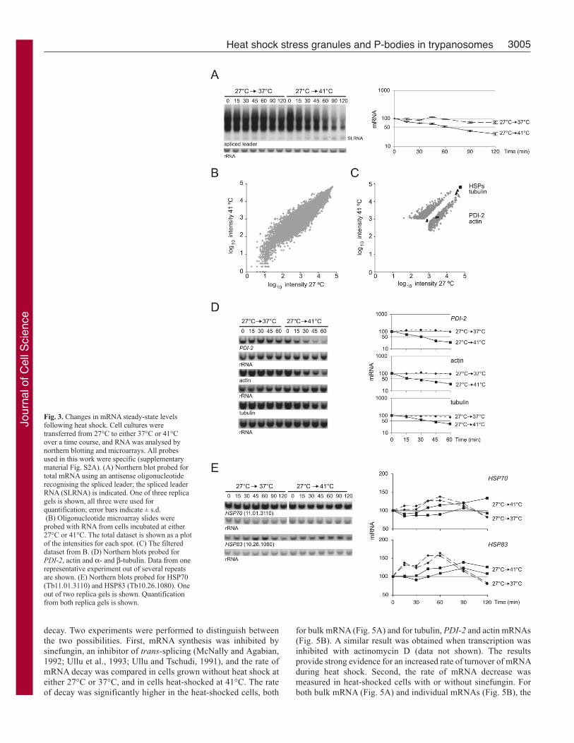
and 120 minutes (Fig. 3A).
To determine global effects of heat shock on steady-state levels
of mRNA in more detail, a microarray was performed to compare
the mRNAs in cells incubated at 41°C for 60 minutes with those
in cells at 27°C (ArrayExpress submission accession number: E-
MEXP-1476). The data were normalised and filtered for quality
and reproducibility. The data are shown as a plot of the intensities
for each spot in each condition for the total (Fig. 3B) and filtered
Fig. 1. Growth recovery following heat shock. Trypanosome cultures wereincubated a 41°C for 60, 90 or 120 minutes. Growth was monitored aftercultures were returned to 27°C. One representative growth curve from threeexperiments is shown.
Jour
nal o
f Cel
l Sci
ence

3004
dataset (Fig. 3C). Data normalisation was based on the standard
assumption that the total mRNA levels remain unchanged under
the two conditions. Since, in reality, incubation at 41°C for 60
minutes resulted in a 50% loss of total mRNA (Fig. 3A), an mRNA
with a ratio outcome of 1.0 (41°C/27°C) in the plot of the array
data was actually reduced twofold; an mRNA with a ratio outcome
of 2.0 was unaffected by heat shock and an mRNA with a ratio
outcome of 0.5 was reduced fourfold. In an initial analysis we
selected genes that behaved significantly differently from the
average mRNA using cut-off values of �2.5 and �0.4
(supplementary material Table S1). The heat-shock-regulated genes
were randomly distributed throughout the genome.
The list of genes with a ratio outcome of �2.5 represents the
mRNAs that escaped the general reduction in mRNA levels upon
heat shock. There were 371 genes in this category (supplementary
material Table S2). 230 genes had no assigned function (half of all
open reading frames in the genome are unassigned). The annotated
genes included several stress-response genes including one HSP100,
four E2 ubiquitin ligases, two proteosome subunits, and two DnaJ-
domain-containing chaperones. Genes involved in mRNA metabolism
included a polyU polymerase (Stagno et al., 2007), an RNA helicase
and three mRNA-binding proteins (RBP6, RBP10 and hnRNP/F).
The 317 genes with a ratio outcome of �0.4 included both mRNAs
that were downregulated after heat shock and those which normally
have a high turnover rate. More than half of these were functionally
annotated (supplementary material Tables S1 and S2). Core metabolic
functions were strongly represented. There were eleven amino acyl
tRNA synthases (three more have ratio outcomes between 0.41 and
0.45) and five translation initiation factors. In addition, there were
three subunits of the t-complex chaperone (three further subunits have
ratio outcomes between 0.41 and 0.45).
Response of individual mRNAs to heat shockThe kinetics of the response of individual mRNAs was investigated
in further detail. Four genes associated with cell growth were used:
actin, α-tubulin and β-tubulin (probed together), and protein
Journal of Cell Science 121 (18)
disulphide isomerase 2 (PDI-2) (Rubotham et al., 2005). After 60
minutes of heat shock, steady-state levels of these mRNAs were
reduced to between 50% and 25% (Fig. 3D).
Next, the expression of putative heat shock protein genes was
investigated. The T. brucei genome encodes a range of different
HSP70 genes (Folgueira and Requena, 2007), and a tandem array
of identical HSP83 genes (Mottram et al., 1989). Specific probes
were used to screen northern blots of RNA prepared over a time
course of heat shock. The steady-state levels of HSP70(Tb11.01.3110) and HSP83 (Tb10.26.1080) mRNAs were
maintained during heat shock (Fig. 3E). HSP70 and HSP83 mRNAs
increased slightly at 37°C, whereas the housekeeping genes tested
did not. The results with HSP70 and HSP83 mRNAs confirmed
earlier observations (Lee, 1998; Muhich et al., 1989). Other HSP70
isoforms were investigated: mitochondrial HSP70s (Tb927.6.3740,
Tb927.6.3750 and Tb927.6.3800) and cytoplasmic HSP70.4(Tb927.7.710) (Searle and Smith, 1993), which all decreased in
response to heat shock, similar to the majority of mRNAs
(supplementary material Fig. S1). The changes in mRNA levels
determined by northern blot for actin and PDI-2 were very similar
to those determined by in the microarray experiment. The tubulin,
HSP70 (Tb11.01.3110) and HSP83 could not be accurately
measured in the microarray experiments because the mRNAs are
very abundant and saturated the spots (Fig. 3C and ArrayExpress
submission accession number: E-MEXP-1476).
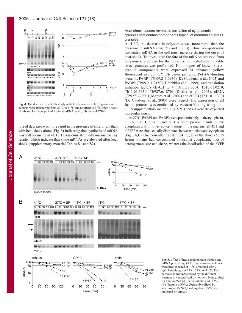
The decrease in mRNA steady-state levels during heat shock was
fully reversible. Total mRNA returned to the pre-heat shock levels
after ~3 hours of recovery (Fig. 4). Individual mRNAs had variable
kinetics of return with tubulin being noticeably slower than the mean.
Together, the data from metabolic labelling, microarray experiments
and northern blots show that trypanosomes undergo a defined and
reversible change in gene expression in response to heat shock.
Bulk mRNA decay rate is increased during heat shockThe decrease in steady-state levels of mRNAs during heat shock
could have resulted from decreased synthesis and/or increased
Fig. 2. Effect of heat shock on translation and polysomes. (A) Changes in translation during heat-shock treatment. Cellswere labelled for 20-minute windows at 27°C and during aheat-shock time course followed by recovery at 27°C. Anautoradiograph of the SDS-PAGE analysis is shown. Twopolypeptides with unchanged synthesis are marked with solidarrows. Examples of proteins that have reduced synthesisafter 5 hours recovery are marked with dashed arrows. Thestained gel was used to control for equal loading (bottom).One lane of the gel has been removed. (B) Changes inpolysomes during heat shock treatment. Absorbance profilesat 254 nm of sucrose density gradients of cells incubated at41°C for 15 and 30 minutes. 50 mM EDTA was added to thecell lysate to dissociate polysomes. Representative resultsfrom several experiments are shown.
Jour
nal o
f Cel
l Sci
ence
3005Heat shock stress granules and P-bodies in trypanosomes
decay. Two experiments were performed to distinguish between
the two possibilities. First, mRNA synthesis was inhibited by
sinefungin, an inhibitor of trans-splicing (McNally and Agabian,
1992; Ullu et al., 1993; Ullu and Tschudi, 1991), and the rate of
mRNA decay was compared in cells grown without heat shock at
either 27°C or 37°C, and in cells heat-shocked at 41°C. The rate
of decay was significantly higher in the heat-shocked cells, both
for bulk mRNA (Fig. 5A) and for tubulin, PDI-2 and actin mRNAs
(Fig. 5B). A similar result was obtained when transcription was
inhibited with actinomycin D (data not shown). The results
provide strong evidence for an increased rate of turnover of mRNA
during heat shock. Second, the rate of mRNA decrease was
measured in heat-shocked cells with or without sinefungin. For
both bulk mRNA (Fig. 5A) and individual mRNAs (Fig. 5B), the
Fig. 3. Changes in mRNA steady-state levelsfollowing heat shock. Cell cultures weretransferred from 27°C to either 37°C or 41°Cover a time course, and RNA was analysed bynorthern blotting and microarrays. All probesused in this work were specific (supplementarymaterial Fig. S2A). (A) Northern blot probed fortotal mRNA using an antisense oligonucleotiderecognising the spliced leader; the spliced leaderRNA (SLRNA) is indicated. One of three replicagels is shown, all three were used forquantification; error bars indicate ± s.d.(B) Oligonucleotide microarray slides wereprobed with RNA from cells incubated at either27°C or 41°C. The total dataset is shown as a plotof the intensities for each spot. (C) The filtereddataset from B. (D) Northern blots probed forPDI-2, actin and α- and β-tubulin. Data from onerepresentative experiment out of several repeatsare shown. (E) Northern blots probed for HSP70(Tb11.01.3110) and HSP83 (Tb10.26.1080). Oneout of two replica gels is shown. Quantificationfrom both replica gels is shown.
Jour
nal o
f Cel
l Sci
ence
3006
rate of decrease was more rapid in the presence of sinefungin than
with heat shock alone (Fig. 5) indicating that synthesis of mRNA
was still occurring at 41°C. This is consistent with our microarray
results, which indicate that some mRNAs are elevated after heat
shock (supplementary material Tables S1 and S2).
Journal of Cell Science 121 (18)
Heat shock causes reversible formation of cytoplasmicgranules that contain components typical of mammalian stressgranulesAt 41°C, the decrease in polysomes was more rapid than the
decrease in mRNA (Fig. 2B and Fig. 3). Thus, non-polysome
associated mRNA in the cell must increase during the onset of
heat shock. To investigate the fate of the mRNAs released from
polysomes, a screen for the presence of heat-shock-inducible
stress granules was performed. Homologues of known stress-
granule components were expressed as enhanced yellow
fluorescent protein (eYFP)-fusion proteins. Poly(A)-binding
proteins, PABP1 (Tb09.211.0930) (De Gaudenzi et al., 2005) and
PABP2 (Tb09.211.2150) (Hotchkiss et al., 1999), and translation
initiation factors eIF4E1 to 4 (Tb11.18.0004, Tb10.61.0210,
Tb11.01.3630, Tb927.6.1870) (Dhalia et al., 2005), eIF2A
(Tb927.3.2900) (Moraes et al., 2007) and eIF3B (Tb11.01.1370)
(De Gaudenzi et al., 2005) were tagged. The expression of all
fusion proteins was confirmed by western blotting using anti-
GFP (supplementary material Fig. S2B) and all were the expected
molecular mass.
At 27°C, PABP1 and PABP2 were predominantly in the cytoplasm,
eIF2A, eIF3B, eIF4E3 and eIF4E4 were present mainly in the
cytoplasm and in lower concentrations in the nucleus, eIF4E1 and
eIF4E2 were about equally distributed between nucleus and cytoplasm
(Fig. 6A,B). One hour after transfer to 41°C, all of the above eYFP-
fusion proteins had concentrated in distinct cytoplasmic foci of
heterogenous size and shape, whereas the localisation of the eYFP
Fig. 4. The decrease in mRNA steady-state levels is reversible. Trypanosomecultures were transferred from 27°C to 41°C and returned to 27°C after 1 hour.Northern blots were probed for total mRNA, actin, tubulin and PDI-2.
Fig. 5. Effect of heat shock on transcription andmRNA processing. (A,B) Trypanosome cultureswere heat-shocked at 41°C or treated with 2μg/ml sinefungin at 27°C, 37°C or 41°C. Thedecrease in mRNAs caused by the differenttreatments was analysed by northern blots probedfor total mRNA (A), actin, tubulin and PDI-2(B). Tubulin mRNAs aberrantly spliced bysinefungin (McNally and Agabian, 1992) areindicated by arrows.
Jour
nal o
f Cel
l Sci
ence

3007Heat shock stress granules and P-bodies in trypanosomes
control was unaffected (Fig. 6A,B). Granule formation occurred
within 30 minutes, increased up to 120 minutes (Fig. 6B) and required
a minimum temperature of 40°C (data not shown). Inhibition of new
mRNA synthesis using actinomycin D for up to 2 hours did not
prevent the formation of heat-shock stress granules (Fig. 6B),
indicating that granule formation does not require newly synthesised
mRNAs.
The formation of granules was fully reversible and they
disappeared within 5-6 hours after return to 27°C (Fig. 6C). Optical
sections through a heat-shocked cell that expressed either eIF4E3-
eYFP or PABP1-eYFP revealed that the granules were concentrated
towards the cell periphery (Fig. 6D). eIF4E3 and eIF3B were tagged
in the same cell line and colocalised in granules (Fig. 6E).
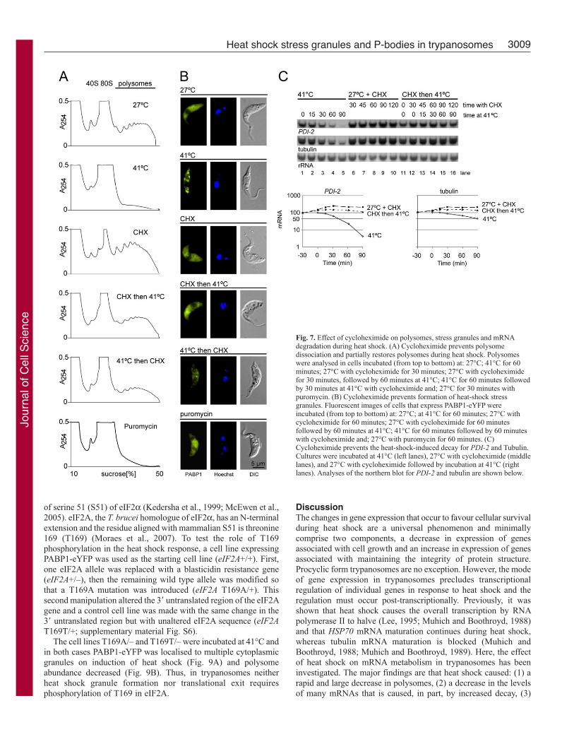
Cycloheximide prevents the formation of heat-shock granulesand can reverse the decline in the polysome pool during heatshockCycloheximide was added before and during heat shock to
investigate the relationship between polysome dissociation and
stress-granule formation. Polysome analyses were performed on
cells incubated at: (1) 27°C, (2) 41°C, (3) 27°C with
cycloheximide, (4) 27°C with cycloheximide followed by an
increase in temperature to 41°C, (5) 41°C with cycloheximide (6)
27°C with puromycin (Fig. 7A). Stress-granule formation was
monitored in similar conditions using cells expressing PABP1-
eYFP (Fig. 7B). Cycloheximide stabilised and puromycin
dissociated polysomes as expected, and neither caused stress-
granule formation. Thus, polysome dissociation by puromycin is
not sufficient for granule formation. Addition of cycloheximide
prior to heat shock largely prevented polysome dissociation and
granule formation was blocked providing evidence that polysome
dissociation is necessary for stress granule formation.
Cycloheximide addition during heat shock caused an increase
in polysomes. The increase could result from newly synthesised
mRNAs becoming trapped and/or ‘old’ mRNAs returning to
polysomes. However, the rapid increase in polysomes after
cycloheximide addition favours a return of old mRNA.
Cycloheximide also inhibited heat-shock-induced decrease in PDI-2 and tubulin mRNAs (Fig. 7C) providing evidence that polysome
dissociation is required for mRNA decay during heat shock.
Heat shock increases the number of P-bodies and causesrelocalisation of XRNA, but not of DHH1 and SCD6, to a focusat the posterior pole of the cellHomologues of P-body components were screened by expression
of eYFP fusions from extragenic loci. At 27°C, three proteins
localised to discrete foci within the cytoplasm: DHH1
(Tb10.70.3290), SCD6 (Tb11.03.0530) and XRNA (Li et al., 2006).
The putative P-bodies differed from the stress granules induced by
heat shock in that they were constitutively present and more defined
with a clearly spherical shape.
The localisation of DHH1 and SCD6 to cytoplasmic foci was
confirmed using antisera raised against recombinant proteins (for
details, see supplementary material Fig. S3). The foci visualised
using antibodies were less apparent against the background signal
from the cytoplasm than the foci visualised using fluoresecence
from the cognate fusion protein. The same was true when anti-
eYFP antibody was used. The most possible explanation for this
discrepancy is that the antibodies only labelled the surface of the
foci, whereas the fluorescent proteins labelled the foci uniformly,
giving a stronger signal relative to the cytoplasm.
For further analysis cell lines were made with one endogenous
allele of either DHH1 or SCD6 that had been modified to express
a fluorescent protein fusion. The expression levels from the
modified allele were either similar (SCD6) or less (DHH1) than
from the remaining wild-type allele (supplementary material Fig.
S3A). DHH1 was expressed with eYFP at the N-terminus
(eYFP-DHH1) and this fusion protein was shown to be functional
as cells grew at the normal rate after deletion of the second allele
whereas knockdown of DHH1 by RNA interference was lethal
(Schwede et al., 2008). SCD6 was expressed as either an N- or
C-terminal eYFP-fusion protein (eYFP-SCD6 or SCD6-eYFP)
and both localised into discrete foci in the cytoplasm. There was
an average of 2.9 foci per cell (Fig. 8A; supplementary material
Fig. S4A). DHH1 and SCD6 were expressed as eYFP and
mCherry fluorescent protein fusions (eYFP-DHH1 and SCD6-
mChFP) in the same cell line (Fig. 8B, left panel). Both proteins
colocalised to the same cytoplasmic foci and no foci were found
that did not contain both fluorescent proteins. In response to
protein synthesis inhibitors, the foci behaved in a similar manner
to P-bodies in yeast and cultured mammalian cells. Puromycin
caused an increase in size of the foci but not in the number, and
cycloheximide caused the foci to fully disappear. Heat shock at
41°C caused an increase in the number and the size of the foci
(Fig. 8B).
XRNA was expressed as either an N- or C-terminal eYFP-fusion
protein by modification of one endogenous allele (eYFP-XRNA,
XRNA-eYFP; supplementary material Fig. S5); both localised to
foci distributed throughout the cytoplasm but, in contrast to DHH1
and SCD6, one focus was always located at the posterior pole of
the cell. A cell line expressing XRNA-eYFP and mChFP-DHH1
(Fig. 8B, right panel; supplementary material Fig. S5) confirmed
that the two proteins localised to the same foci in the cytoplasm
(dashed arrows in Fig. 8B) but only XRNA-eYFP localised to the
focus present at the posterior pole of cells (solid arrows in Fig. 8B).
Cycloheximide caused all XRNA-containing foci to disappear (Fig.
8B right panel), whereas puromycin caused an increase in size of
all (Fig. 8B, right panel; supplementary material Fig. S4B). By
contrast, heat shock caused a preferential increase in the size of the
XRNA-containing focus at the posterior pole, which always became
the brightest focus in the cell (Fig. 8B, right panel; supplementary
material Fig. S4B).
Heat-shock stress granules do not colocalise with P-bodiesAny interaction between P-bodies and heat-shock stress granules
was investigated using cell lines expressing PABP1-eYFP and either
mChFP-DHH1 or SCD6-mChFP (supplementary material Fig. S5).
At 27°C, PABP1-eYFP was uniformly distributed throughout the
cytoplasm, whereas mChFP-DHH1 and SCD6-mChFP were
concentrated in cytoplasmic foci (Fig. 8C). At 41°C, PABP1
localised to heat-shock stress granules, and the DHH1 and SCD6
foci increased in size and number (Fig. 8C). The majority of the
PABP1 foci did not colocalise with the DHH1 and SCD6 foci. Only
a small number of heat-shock stress granules (PABP1-foci) and P-
bodies (DHH1-SCD6 foci) were found to either colocalise or lie
adjacent to each other (dashed arrows in Fig. 8C).
Formation of heat-shock granules is independent of eIF2Aphosphorylation at T169, the equivalent of S51 in mammalianeIF2αOne of the first steps in the heat shock response in mammalian cells
is the inhibition of the initiation of translation after phosphorylation
Jour
nal o
f Cel
l Sci
ence

3008 Journal of Cell Science 121 (18)
Fig. 6. Characterisation of T. brucei heat-shock stress granules. (A) Homologues of stress granule markers were expressed as eYFP fusion proteins. Fluorescentimages of unstressed (27°C) and heat-shocked (1 hour 41°C) cells are shown. (B) The effect of heat shock on cells that express PABP1-eYFP from a modifiedendogenous locus; treatments are indicated. (C) Disappearance of heat-shock stress granules after a return to 27°C in cells that express PABP1-eYFP from amodified endogenous locus. The percentage of cells with visible granules was determined during recovery and the average values (n=100) from three slides isshown, error bars indicate ± s.d. (D) Optical sections through T. brucei cells that express PAPB1-eYFP or eIF4E3-eYFP after 60 minutes at 41°C are shown asprojection (left) and some selected single plane images (right). (E) Colocalisation of eIF3B-eYFP and eIF4E3-mChFP to heat-shock stress granules.
Jour
nal o
f Cel
l Sci
ence
3009Heat shock stress granules and P-bodies in trypanosomes
of serine 51 (S51) of eIF2α (Kedersha et al., 1999; McEwen et al.,
2005). eIF2A, the T. brucei homologue of eIF2α, has an N-terminal
extension and the residue aligned with mammalian S51 is threonine
169 (T169) (Moraes et al., 2007). To test the role of T169
phosphorylation in the heat shock response, a cell line expressing
PABP1-eYFP was used as the starting cell line (eIF2A+/+). First,
one eIF2A allele was replaced with a blasticidin resistance gene
(eIF2A+/–), then the remaining wild type allele was modified so
that a T169A mutation was introduced (eIF2A T169A/+). This
second manipulation altered the 3� untranslated region of the eIF2A
gene and a control cell line was made with the same change in the
3� untranslated region but with unaltered eIF2A sequence (eIF2AT169T/+; supplementary material Fig. S6).
The cell lines T169A/– and T169T/– were incubated at 41°C and
in both cases PABP1-eYFP was localised to multiple cytoplasmic
granules on induction of heat shock (Fig. 9A) and polysome
abundance decreased (Fig. 9B). Thus, in trypanosomes neither
heat shock granule formation nor translational exit requires
phosphorylation of T169 in eIF2A.
DiscussionThe changes in gene expression that occur to favour cellular survival
during heat shock are a universal phenomenon and minimally
comprise two components, a decrease in expression of genes
associated with cell growth and an increase in expression of genes
associated with maintaining the integrity of protein structure.
Procyclic form trypanosomes are no exception. However, the mode
of gene expression in trypanosomes precludes transcriptional
regulation of individual genes in response to heat shock and the
regulation must occur post-transcriptionally. Previously, it was
shown that heat shock causes the overall transcription by RNA
polymerase II to halve (Lee, 1995; Muhich and Boothroyd, 1988)
and that HSP70 mRNA maturation continues during heat shock,
whereas tubulin mRNA maturation is blocked (Muhich and
Boothroyd, 1988; Muhich and Boothroyd, 1989). Here, the effect
of heat shock on mRNA metabolism in trypanosomes has been
investigated. The major findings are that heat shock caused: (1) a
rapid and large decrease in polysomes, (2) a decrease in the levels
of many mRNAs that is caused, in part, by increased decay, (3)
Fig. 7. Effect of cycloheximide on polysomes, stress granules and mRNAdegradation during heat shock. (A) Cycloheximide prevents polysomedissociation and partially restores polysomes during heat shock. Polysomeswere analysed in cells incubated (from top to bottom) at: 27°C; 41°C for 60minutes; 27°C with cycloheximide for 30 minutes; 27°C with cycloheximidefor 30 minutes, followed by 60 minutes at 41°C; 41°C for 60 minutes followedby 30 minutes at 41°C with cycloheximide and; 27°C for 30 minutes withpuromycin. (B) Cycloheximide prevents formation of heat-shock stressgranules. Fluorescent images of cells that express PABP1-eYFP wereincubated (from top to bottom) at: 27°C; at 41°C for 60 minutes; 27°C withcycloheximide for 60 minutes; 27°C with cycloheximide for 60 minutesfollowed by 60 minutes at 41°C; 41°C for 60 minutes followed by 60 minuteswith cycloheximide and; 27°C with puromycin for 60 minutes. (C)Cycloheximide prevents the heat-shock-induced decay for PDI-2 and Tubulin.Cultures were incubated at 41°C (left lanes), 27°C with cycloheximide (middlelanes), and 27°C with cycloheximide followed by incubation at 41°C (rightlanes). Analyses of the northern blot for PDI-2 and tubulin are shown below.
Jour
nal o
f Cel
l Sci
ence

3010
increased levels of some mRNAs, (4) the formation
of heat-shock stress granules at the cell periphery,
(5) an increase in the number and size of P-bodies
and, (6) an increase in size of a novel XRNA focus
at the posterior pole of the cell. The decrease in
polysomes and appearence of stress granules were
not dependent on phosphorylation of eIF2A at T169
(equivalent to S51 in mammalian eIF2α). The
inhibition of stress-granule formation by
cycloheximide indicates that polysome-free mRNA
is required for their formation. The P-bodies
contained XRNA, DHH1 and SCD6; by contrast, a
novel focus at the posterior pole of the cell contained
XRNA but neither DHH1 nor SCD6. Importantly,
the changes on heat shock were fully reversible and
the kinetics of return to proliferative growth,
restoration of starting mRNA levels, and
disappearance of heat shock stress granules were
similar.
Changes in mRNA metabolism following heatshockWithin 15 minutes of heat shock the polysome pool
and the translation of most proteins had decreased.
Heat shock also reduced steady-state levels of most
mRNAs but with slower kinetics than the decrease
in polysomes. Thus, the dissociation of polysomes
on heat shock must result in increased levels of non-
polysome-associated mRNA in the cell. The
appearance of heat-shock stress granules and the
increase in the number of P-bodies following heat
shock occurred with similar kinetics to polysome
dissociation. One interpretation of the kinetics is that
the mRNAs released from polysomes were directed
to either or both of these cytoplasmic mRNP
particles, but this remains to be proven.
Journal of Cell Science 121 (18)
Fig. 8. T. brucei P-bodies. (A) T. brucei cells have inaverage 2.9 P-bodies. Projection of optical sections throughpuromycin-treated cells that express SCD6-eYFP. Similar Z-stacks (supplementary material Fig. S5A) were used todetermine the number of P-bodies per cell: the average was2.9 per cell (n=125) with little variation in different cell-cycle stages: 2.7 in 1K1N (n=95), 3.3 in 2K1N (n=17) and3.3 in 2K2N (n=12) cells. (B) P-body components DHH1,SCD6 and XRNA. eYFP-DHH1 and SCD6-mChFP (leftpanel) or mChFP-DHH1 and XRNA-eYFP (right panel)were expressed in the same cell line from endogenous loci.Representative fluorescent images of untreated, and cellsafter 1 hour with puromycin, cycloheximide or heat shock(41°C) are shown. Solid arrows indicate the posterior poleof the cell. An enlargement of a heat-shocked cellsexpressing mChFP-DHH1 and XRNA-eYFP is shown toindicate the colocalisation of DHH1 and XRNA in all spots(dashed arrows), except at the posterior pole. (C) P-bodiesand heat-shock stress granules. mChFP-DHH1 and PABP1-eYFP (left) or mChFP-SCD6 and PABP1-eYFP (right) wereexpressed in the same cell line from their endogenous loci.Representative fluorescent images of one untreated cell andseveral images of cells that have been heat shocked (1 hour41°C) are shown. Dashed arrows point at granules thatcontain both a P-body marker (DHH1 or SCD6) and thestress granule marker PABP1, either colocalised or close toeach other.
Jour
nal o
f Cel
l Sci
ence

3011Heat shock stress granules and P-bodies in trypanosomes
What causes the decrease in mRNA steady-state levels following
heat shock? The microarray experiment showed a wide range of
responses for individual mRNA steady-state levels; some were
unaffected or even increased, most decreased and some decreased
very rapidly. Experiments measuring the rate of decay strongly
suggested that the decrease in mRNAs at 41°C was due to both
increased degradation, and reduced transcription and maturation.
Inhibition of polysome dissociation by cycloheximide blocked
both the decrease in PDI-2 and tubulin mRNAs and heat shock
stress granule formation. Dissociation of polysomes by puromycin
was not sufficient to induce either heat shock stress granule
formation or a decrease in mRNAs. These results provide evidence
that dissociation of polysomes is necessary but not sufficient for
the heat-shock response. The addition of cycloheximide during heat
shock resulted in an increase in polysomes (Fig. 7A); one
explanation for this is that mRNAs are in equilibrium between
polysomes and stress granules as found in mammals (Kedersha et
al., 2000). If this is correct, exit from the polysome pool was not
irreversible and did not inevitably lead to degradation.
P-bodiesIn yeast and mammalian cells, P-bodies contain the activities
involved in 5�-3� exonucleolytic digestion of mRNA: the decapping
enzyme components DCP1 and DCP2, XRN1 exonuclease, and
accessory proteins including DHH1 and SCD6 (reviewed in Parker
and Sheth, 2007). Decapping activity has been detected in
trypanosomatid cell lysates (Milone et al., 2002) but the genes
encoding this activity have not been identified as there are no readily
identifiable orthologues of DCP1 or DCP2 in the trypanosome
genome (Berriman et al., 2005). P-body components identified in
trypanosomes include: DHH1 (Cassola et al., 2007; Holetz et al.,
2007), SCD6 (this work), XRNA (Cassola et al., 2007), one
member of the PUF protein family (Caro et al., 2006;
Dallagiovanna et al., 2007; Dallagiovanna et al.,
2005) and the RNA binding protein RBP3 (A.
Robles, D. Gudjonsdottir-Planck, C. Hartmann and
C.E.C., unpublished). eIF4E is found in P-bodies in
yeast and mammalian cells but has not been reported
to be present in P-bodies in trypanosomes (Cassola
et al., 2007). There is no LSM1 gene in the genome,
and the LSM1 to LSM7 complex is absent whereas
the LSM2 to LSM8 splicing factor is present in the
nucleus (Liu et al., 2004).
The most unexpected result from this work was
the localisation of XRNA, but not DHH1 or SCD6,
to a spot at the posterior cell pole in addition to the
cytoplasm and P-bodies. This focus shared
properties with P-bodies: cycloheximide caused the
focus to dissolve and puromycin caused it to increase
in size. In response to heat shock, this focus
increased in size to become the largest focus of
XRNA in the cell. It is not clear why XRNA is found
there, nor why heat shock induces XRNA to further
accumulate there. The posterior pole contains the
Fig. 9. Formation of heat-shockstress granules is independent oneIF2α phosphorylation at T169.Heat shock treatment (1 hour41°C) of cells expressing PABP1-eYFP from the endogenous locusand with either a single wild-type(T169T) or mutated (T169A)eIF2A gene (supplementarymaterial Fig. S6). (A) Fluorescentimages of two clonal cellpopulations expressing wild-typeeIF2A (T2 and T3) or mutanteIF2A (A1 and A2) are shown. (B) Polysome analysis of theeIF2A T169A/– cells incubated ateither 27°C or 41°C for 60minutes.
Fig. 10. Model of compartments that may determine mRNA fate in growing cells (left) and inheat-shocked cells (right).
Jour
nal o
f Cel
l Sci
ence
3012
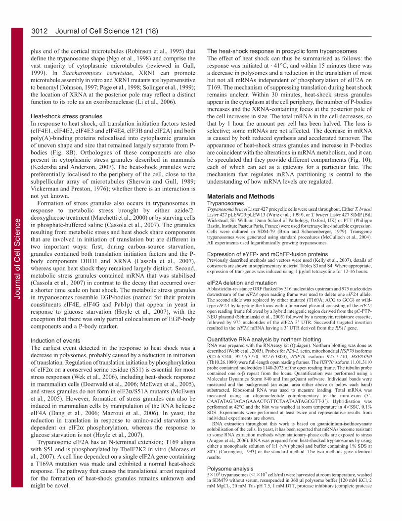
plus end of the cortical microtubules (Robinson et al., 1995) that
define the trypanosome shape (Ngo et al., 1998) and comprise the
vast majority of cytoplasmic microtubules (reviewed in Gull,
1999). In Saccharomyces cerevisiae, XRN1 can promote
microtubule assembly in vitro and XRN1 mutants are hypersensitive
to benomyl (Johnson, 1997; Page et al., 1998; Solinger et al., 1999);
the location of XRNA at the posterior pole may reflect a distinct
function to its role as an exoribonuclease (Li et al., 2006).
Heat-shock stress granulesIn response to heat shock, all translation initiation factors tested
(eIF4E1, eIF4E2, eIF4E3 and eIF4E4, eIF3B and eIF2A) and both
poly(A)-binding proteins relocalised into cytoplasmic granules
of uneven shape and size that remained largely separate from P-
bodies (Fig. 8B). Orthologues of these components are also
present in cytoplasmic stress granules described in mammals
(Kedersha and Anderson, 2007). The heat-shock granules were
preferentially localised to the periphery of the cell, close to the
subpellicular array of microtubules (Sherwin and Gull, 1989;
Vickerman and Preston, 1976); whether there is an interaction is
not yet known.
Formation of stress granules also occurs in trypanosomes in
response to metabolic stress brought by either azide/2-
deoxyglucose treatment (Marchetti et al., 2000) or by starving cells
in phosphate-buffered saline (Cassola et al., 2007). The granules
resulting from metabolic stress and heat shock share components
that are involved in initiation of translation but are different in
two important ways: first, during carbon-source starvation,
granules contained both translation initiation factors and the P-
body components DHH1 and XRNA (Cassola et al., 2007),
whereas upon heat shock they remained largely distinct. Second,
metabolic stress granules contained mRNA that was stabilised
(Cassola et al., 2007) in contrast to the decay that occurred over
a shorter time scale on heat shock. The metabolic stress granules
in trypanosomes resemble EGP-bodies (named for their protein
constituents eIF4E, eIF4G and Pab1p) that appear in yeast in
response to glucose starvation (Hoyle et al., 2007), with the
exception that there was only partial colocalisation of EGP-body
components and a P-body marker.
Induction of eventsThe earliest event detected in the response to heat shock was a
decrease in polysomes, probably caused by a reduction in initiation
of translation. Regulation of translation initiation by phosphorylation
of eIF2α on a conserved serine residue (S51) is essential for most
stress responses (Wek et al., 2006), including heat-shock response
in mammalian cells (Doerwald et al., 2006; McEwen et al., 2005),
and stress granules do not form in eIF2α/S51A mutants (McEwen
et al., 2005). However, formation of stress granules can also be
induced in mammalian cells by manipulation of the RNA helicase
eIF4A (Dang et al., 2006; Mazroui et al., 2006). In yeast, the
reduction in translation in response to amino-acid starvation is
dependent on eIF2α phosphorylation, whereas the response to
glucose starvation is not (Hoyle et al., 2007).
Trypanosome eIF2A has an N-terminal extension; T169 aligns
with S51 and is phosphorylated by TbeIF2K2 in vitro (Moraes et
al., 2007). A cell line dependent on a single eIF2A gene containing
a T169A mutation was made and exhibited a normal heat-shock
response. The pathway that causes the translational arrest required
for the formation of heat-shock granules remains unknown and
might be novel.
Journal of Cell Science 121 (18)
The heat-shock response in procyclic form trypanosomesThe effect of heat shock can thus be summarised as follows: the
response was initiated at ~41°C, and within 15 minutes there was
a decrease in polysomes and a reduction in the translation of most
but not all mRNAs independent of phosphorylation of eIF2A on
T169. The mechanism of suppressing translation during heat shock
remains unclear. Within 30 minutes, heat-shock stress granules
appear in the cytoplasm at the cell periphery, the number of P-bodies
increases and the XRNA-containing focus at the posterior pole of
the cell increases in size. The total mRNA in the cell decreases, so
that by 1 hour the amount per cell has been halved. The loss is
selective; some mRNAs are not affected. The decrease in mRNA
is caused by both reduced synthesis and accelerated turnover. The
appearance of heat-shock stress granules and increase in P-bodies
are coincident with the alterations in mRNA metabolism, and it can
be speculated that they provide different compartments (Fig. 10),
each of which can act as a gateway for a particular fate. The
mechanism that regulates mRNA partitioning is central to the
understanding of how mRNA levels are regulated.
Materials and MethodsTrypanosomesTrypanosoma brucei Lister 427 procyclic cells were used throughout. Either T. bruceiLister 427 pLEW29:pLEW13 (Wirtz et al., 1999), or T. brucei Lister 427 SIMP (BillWickstead, Sir William Dunn School of Pathology, Oxford, UK) or PTT (PhilippeBastin, Institute Pasteur Paris, France) were used for tetracycline-inducible expression.Cells were cultured in SDM-79 (Brun and Schonenberger, 1979). Transgenictrypanosomes were generated using standard procedures (McCulloch et al., 2004).All experiments used logarithmically growing trypanosomes.
Expression of eYFP- and mChFP-fusion proteinsPreviously described methods and vectors were used (Kelly et al., 2007), details ofconstructs are shown in supplementary material Tables S3 and S4. Where appropriate,expression of transgenes was induced using 1 μg/ml tetracycline for 12-16 hours.
eIF2A deletion and mutationA blasticidin-resistance ORF flanked by 316 nucleotides upstream and 975 nucleotidesdownstream of the eIF2A open reading frame was used to delete one eIF2A allele.The second allele was replaced by either mutated (T169A; ACG to GCG) or wild-type eIF2A by targeting the locus with a linearised plasmid consisting of the eIF2Aopen reading frame followed by a hybrid intergenic region derived from the pC-PTP-NEO plasmid (Schimanski et al., 2005) followed by a neomycin resistance cassette,followed by 975 nucleotides of the eIF2A 3� UTR. Successful targeted insertionresulted in the eIF2A mRNA having a 3� UTR derived from the RPA1 gene.
Quantitative RNA analysis by northern blottingRNA was prepared with the RNAeasy kit (Quiagen). Northern blotting was done asdescribed (Webb et al., 2005). Probes for PDI-2, actin, mitochondrial HSP70 isoforms(927.6.3740, 927.6.3750, 927.6.3800), HSP70 isoform 927.7.710, HSP83/90(Tb10.26.1080) were full-length open reading frames. The HSP70 isoform 11.01.3110probe contained nucleotides 1140-2073 of the open reading frame. The tubulin probecontained one α-β repeat from the locus. Quantification was performed using aMolecular Dynamics Storm 840 and ImageQuant software. Individual bands weremeasured and the background (an equal area either above or below each band)substracted. Ribosomal RNA was used to measure loading. Total mRNA wasmeasured using an oligonucleotide complementary to the mini-exon (5�-CAATATAGTACAGAAACTGTTCTAATAATAGCGTT-3�). Hybridisation wasperformed at 42°C and the blot was washed at room temperature in 4�SSC, 0.1%SDS. Experiments were performed at least twice and representative results fromindividual experiments are shown.
RNA extraction throughout this work is based on guanidinium-isothiocyanatesolubilisation of the cells. In yeast, it has been reported that mRNAs become resistantto some RNA extraction methods when stationary-phase cells are exposed to stress(Aragon et al., 2006). RNA was prepared from heat-shocked trypanosomes by usingeither a monophasic solution of 1:1 (v/v) phenol and buffer containing 1% SDS at80°C (Carrington, 1993) or the standard method. The two methods gave identicalresults.
Polysome analysis5�108 trypanosomes (<1�107 cells/ml) were harvested at room temperature, washedin SDM79 without serum, resuspended in 360 μl polysome buffer [120 mM KCl, 2mM MgCl2, 20 mM Tris pH 7.5, 1 mM DTT, protease inhibitors (complete protease
Jour
nal o
f Cel
l Sci
ence
3013Heat shock stress granules and P-bodies in trypanosomes
inhibitor cocktail, Roche)] and lysed by adding 40 μl 10% n-octyl glycoside. The
lysate was cleared by centrifugation at 16,000 g (4°C, 10 min), layered onto a 10-
50% sucrose gradient (12 ml) prepared in polysome buffer, and centrifuged at 4°C
for 2 hours at 36,000 rpm in a Beckman SW 40 rotor. Absorption at 254 nm was
monitored using the UVICORD SII system. Both cycloheximide and puromycin were
used at 50 μg/ml.
Metabolic labelling5�106 cells were transferred into 500 μl methionine-free RPMI-1640 (R7513, Sigma)
with 3.7 kBq/ml [35S]methionine (>37 TBq/mmol) for 20 minutes. Cells were pelleted,
washed and lysed in SDS-PAGE sample buffer. Samples were analysed by SDS-
PAGE and autoradiography.
Microscopic ImagingCells were washed with SDM79 without serum and fixed at a density of 1�107 cells/ml
with 2.4% paraformaldehyde overnight, washed once in PBS and stained with Hoechst
H33258. Fluorescence microscopy was carried out using a Zeiss Axioskop microscope
equipped with a Plan-Apochromat 100�/1.4 Oil DIC objective. Images were taken
with the monochrome CCD camera AxioCam MR using AxioVision software (Zeiss).
Confocal images were made with a BioRad Radiance 2100 on a Nikon Eclipse E800
upright microscope using a 100�/1.4 Oil DIC objective.
MicroarraysProcyclic cultures (~5�106 cells/ml) at 27°C were added to one volume SDM79 at
53°C and incubated at 41°C in a waterbath in a closed tube for 60 minutes. The
control cells were added to one volume medium at 27°C and also incubated for 60
minutes in a closed tube at 27°C. Total RNA was prepared using the Qiagen RNAeasy
Midi kit, including the DNAse digestion on the column. Two biological replicates
were performed on different days. 10 μg of total RNA of each condition (41°C and
27°C) were labelled as previously described (Brems et al., 2005; Diehl et al., 2002;
Luu et al., 2006). After checking dye incorporation using a fluorimeter (Nanodrop
ND-1000 3.3), samples were mixed and hybridised onto the array (JCVI PFGRC T.brucei 37K v2) according to the M008 TIGR protocol. Five technical replicates were
performed. Image acquisition and data analysis were performed as previously
described (Diehl et al., 2002). The MCHiPS software package (Fellenberg et al.,
2002; Fellenberg et al., 2001) was used for data normalisation and analysis. After
normalising the original signal intensities by logarithmic regression, each individual
signal was filtered for quality and reproducibility. Only spots with a fitted intensity
of at least twice the standard deviation above background, a P-value �0.05 and a
minimum-maximum separation value of 0.1 survived the filtering.
This work was funded by the Wellcome Trust. The authors thankAlex Sossick and Nicola Lawrence (Gurdon Institute, Cambridge, UK)for help with confocal microscopy, Julie Aspen and Richard Jacksonfor help with polysomes, Rachana Ramarao for helpful discussions,Jenny Reed for ever-patient and excellent assistance, Nancy Standartfor critical reading of the manuscript (all Department of Biochemistry,Cambridge, UK), Elisabetta Ullu (Yale University School of Medicine,New Haven, CT) for helpful discussions on polysomes, Bill Wickstead(Sir William Dunn School of Pathology, University of Oxford, UK)for the SIMP cell line, Phillipe Bastin (Pasteur Institute, Paris, France)for the PTT cell line, Jay Bangs (University of Wisconsin-Madison,Madison, WI) for kindly providing the BiP antibody, Derek Nolan(Trinity College, Dublin, Ireland) for providing us with informationabout PDI-2. We thank the Pathogen Functional Genomics ResourceCentre (PFGRC), NIAID and the J. Craig Venter Institute (JCVI) forproviding T. brucei microarrays. R.Q. holds a stipend from the DeutscheAkademische Austauschdienst.
ReferencesAragon, A. D., Quinones, G. A., Thomas, E. V., Roy, S. and Werner-Washburne, M.
(2006). Release of extraction-resistant mRNA in stationary phase Saccharomyces
cerevisiae produces a massive increase in transcript abundance in response to stress.
Genome Biol. 7, R9.
Bashkirov, V. I., Scherthan, H., Solinger, J. A., Buerstedde, J. M. and Heyer, W. D.
(1997). A mouse cytoplasmic exoribonuclease (mXRN1p) with preference for G4
tetraplex substrates. J. Cell Biol. 136, 761-773.
Berriman, M., Ghedin, E., Hertz-Fowler, C., Blandin, G., Renauld, H., Bartholomeu,
D. C., Lennard, N. J., Caler, E., Hamlin, N. E., Haas, B. et al. (2005). The genome
of the African trypanosome Trypanosoma brucei. Science 309, 416-422.
Bond, U. (2006). Stressed out! Effects of environmental stress on mRNA metabolism. FEMSYeast Res. 6, 160-170.
Brems, S., Guilbride, D. L., Gundlesdodjir-Planck, D., Busold, C., Luu, V. D.,
Schanne, M., Hoheisel, J. and Clayton, C. (2005). The transcriptomes of Trypanosoma
brucei Lister 427 and TREU927 bloodstream and procyclic trypomastigotes. Mol.Biochem. Parasitol. 139, 163-172.
Brun, R. and Schonenberger. (1979). Cultivation and in vitro cloning or procyclic culture
forms of Trypanosoma brucei in a semi-defined medium. Short communication. ActaTrop. 36, 289-292.
Campbell, D. A., Thornton, D. A. and Boothroyd, J. C. (1984). Apparent discontinuous
transcription of Trypanosoma brucei variant surface antigen genes. Nature 311, 350-
355.
Caro, F., Bercovich, N., Atorrasagasti, C., Levin, M. J. and Vazquez, M. P. (2006).
Trypanosoma cruzi: analysis of the complete PUF RNA-binding protein family. Exp.Parasitol. 113, 112-124.
Carrington, M. (1993). Preparation of DNA and RNA from Trypanosoma brucei. MethodsMol. Biol. 21, 101-111.
Cassola, A., De Gaudenzi, J. G. and Frasch, A. C. (2007). Recruitment of mRNAs to
cytoplasmic ribonucleoprotein granules in trypanosomes. Mol. Microbiol. 65, 655-670.
Clayton, C. and Shapira, M. (2007). Post-transcriptional regulation of gene expression
in trypanosomes and leishmanias. Mol. Biochem. Parasitol. 156, 93-101.
Cougot, N., Babajko, S. and Seraphin, B. (2004). Cytoplasmic foci are sites of mRNA
decay in human cells. J. Cell Biol. 165, 31-40.
Dallagiovanna, B., Perez, L., Sotelo-Silveira, J., Smircich, P., Duhagon, M. A. and
Garat, B. (2005). Trypanosoma cruzi: molecular characterization of TcPUF6, a Pumilio
protein. Exp. Parasitol. 109, 260-264.
Dallagiovanna, B., Correa, A., Probst, C. M., Holetz, F., Smircich, P., de Aguiar, A.
M., Mansur, F., da Silva, C. V., Mortara, R. A., Garat, B. et al. (2007). Functional
genomic characterization of mRNAs associated with TcPUF6, a pumilio-like protein
from Trypanosoma cruzi. J. Biol. Chem. 283, 8266-8273.
Dang, Y., Kedersha, N., Low, W. K., Romo, D., Gorospe, M., Kaufman, R., Anderson,
P. and Liu, J. O. (2006). Eukaryotic initiation factor 2alpha-independent pathway of
stress granule induction by the natural product pateamine A. J. Biol. Chem. 281, 32870-
32878.
De Gaudenzi, J., Frasch, A. C. and Clayton, C. (2005). RNA-binding domain proteins
in Kinetoplastids: a comparative analysis. Eukaryotic Cell 4, 2106-2114.
De Lange, T., Michels, P. A. M., Veerman, H. J. G., Cornelissen, A. C. W. A. and
Borst, P. (1984). Many trypanosome mRNAs share a common 5� terminal sequence.
Nucleic Acids Res. 12, 3777-3790.
Dhalia, R., Reis, C. R., Freire, E. R., Rocha, P. O., Katz, R., Muniz, J. R., Standart,
N. and de Melo Neto, O. P. (2005). Translation initiation in Leishmania major:
characterisation of multiple eIF4F subunit homologues. Mol. Biochem. Parasitol. 140,
23-41.
Dhalia, R., Marinsek, N., Reis, C. R., Katz, R., Muniz, J. R., Standart, N., Carrington,
M. and de Melo Neto, O. P. (2006). The two eIF4A helicases in Trypanosoma brucei
are functionally distinct. Nucleic Acids Res. 34, 2495-2507.
Diehl, S., Diehl, F., El-Sayed, N. M., Clayton, C. and Hoheisel, J. D. (2002). Analysis
of stage-specific gene expression in the bloodstream and the procyclic form of
Trypanosoma brucei using a genomic DNA-microarray. Mol. Biochem. Parasitol. 123,
115-123.
Doerwald, L., van Genesen, S. T., Onnekink, C., Marin-Vinader, L., de Lange, F., de
Jong, W. W. and Lubsen, N. H. (2006). The effect of alphaB-crystallin and Hsp27 on
the availability of translation initiation factors in heat-shocked cells. Cell Mol. Life Sci.63, 735-743.
El Sayed, N. M., Ghedin, E., Song, J., MacLeod, A., Bringaud, F., Larkin, C., Wanless,
D., Peterson, J., Hou, L., Taylor, S. et al. (2003). The sequence and analysis of
Trypanosoma brucei chromosome II. Nucleic Acids Res. 31, 4856-4863.
Fellenberg, K., Hauser, N. C., Brors, B., Neutzner, A., Hoheisel, J. D. and Vingron,
M. (2001). Correspondence analysis applied to microarray data. Proc. Natl. Acad. Sci.USA 98, 10781-10786.
Fellenberg, K., Hauser, N. C., Brors, B., Hoheisel, J. D. and Vingron, M. (2002).
Microarray data warehouse allowing for inclusion of experiment annotations in statistical
analysis. Bioinformatics 18, 423-433.
Folgueira, C. and Requena, J. M. (2007). A postgenomic view of the heat shock proteins
in kinetoplastids. FEMS Microbiol. Rev. 31, 359-377.
Gull, K. (1999). The cytoskeleton of trypanosomatid parasites. Annu. Rev. Microbiol. 53,
629-655.
Guttinger, A., Schwab, C., Morand, S., Roditi, I. and Vassella, E. (2007). A mitogen-
activated protein kinase of Trypanosoma brucei confers resistance to temperature stress.
Mol. Biochem. Parasitol. 153, 203-206.
Hall, N., Berriman, M., Lennard, N. J., Harris, B. R., Hertz-Fowler, C., Bart-Delabesse,
E. N., Gerrard, C. S., Atkin, R. J., Barron, A. J., Bowman, S. et al. (2003). The
DNA sequence of chromosome I of an African trypanosome: gene content, chromosome
organisation, recombination and polymorphism. Nucleic Acids Res. 31, 4864-4873.
Holetz, F. B., Correa, A., Avila, A. R., Nakamura, C. V., Krieger, M. A. and
Goldenberg, S. (2007). Evidence of P-body-like structures in Trypanosoma cruzi.
Biochem. Biophys. Res. Commun. 356, 1062-1067.
Hotchkiss, T. L., Nerantzakis, G. E., Dills, S. C., Shang, L. and Read, L. K. (1999).
Trypanosoma brucei poly(A) binding protein I cDNA cloning, expression, and binding
to 5 untranslated region sequence elements. Mol. Biochem. Parasitol. 98, 117-129.
Hoyle, N. P., Castelli, L. M., Campbell, S. G., Holmes, L. E. and Ashe, M. P. (2007).
Stress-dependent relocalization of translationally primed mRNPs to cytoplasmic granules
that are kinetically and spatially distinct from P-bodies. J. Cell Biol. 179, 65-74.
Ingelfinger, D., Arndt-Jovin, D. J., Luhrmann, R. and Achsel, T. (2002). The human
LSm1-7 proteins colocalize with the mRNA-degrading enzymes Dcp1/2 and Xrnl in
distinct cytoplasmic foci. RNA 8, 1489-1501.
Jour
nal o
f Cel
l Sci
ence

Ivens, A. C., Peacock, C. S., Worthey, E. A., Murphy, L., Aggarwal, G., Berriman,
M., Sisk, E., Rajandream, M. A., Adlem, E., Aert, R. et al. (2005). The genome of
the kinetoplastid parasite, Leishmania major. Science 309, 436-442.
Johnson, A. W. (1997). Rat1p and Xrn1p are functionally interchangeable exoribonucleases
that are restricted to and required in the nucleus and cytoplasm, respectively. Mol. Cell.Biol. 17, 6122-6130.
Johnson, P. J., Kooter, J. M. and Borst, P. (1987). Inactivation of transcription by UV
irradiation of T. brucei provides evidence for a multicistronic transcription unit including
a VSG gene. Cell 51, 273-281.
Kedersha, N. and Anderson, P. (2007). Mammalian stress granules and processing bodies.
Meth. Enzymol. 431, 61-81.
Kedersha, N. L., Gupta, M., Li, W., Miller, I. and Anderson, P. (1999). RNA-binding
proteins TIA-1 and TIAR link the phosphorylation of eIF-2 alpha to the assembly of
mammalian stress granules. J. Cell Biol. 147, 1431-1442.
Kedersha, N., Cho, M. R., Li, W., Yacono, P. W., Chen, S., Gilks, N., Golan, D. E. and
Anderson, P. (2000). Dynamic shuttling of TIA-1 accompanies the recruitment of mRNA
to mammalian stress granules. J. Cell Biol. 151, 1257-1268.
Kedersha, N., Chen, S., Gilks, N., Li, W., Miller, I. J., Stahl, J. and Anderson, P. (2002).
Evidence that ternary complex (eIF2-GTP-tRNA(i)(Met))-deficient preinitiation
complexes are core constituents of mammalian stress granules. Mol. Biol. Cell 13, 195-
210.
Kedersha, N., Stoecklin, G., Ayodele, M., Yacono, P., Lykke-Andersen, J., Fritzler, M.
J., Scheuner, D., Kaufman, R. J., Golan, D. E. and Anderson, P. (2005). Stress granules
and processing bodies are dynamically linked sites of mRNP remodeling. J. Cell Biol.169, 871-884.
Kelly, S., Reed, J., Kramer, S., Ellis, L., Webb, H., Sunter, J., Salje, J., Marinsek, N.,
Gull, K., Wickstead, B. et al. (2007). Functional genomics in Trypanosoma brucei: a
collection of vectors for the expression of tagged proteins from endogenous and ectopic
gene loci. Mol. Biochem. Parasitol. 154, 103-109.
Kimball, S. R., Horetsky, R. L., Ron, D., Jefferson, L. S. and Harding, H. P. (2003).
Mammalian stress granules represent sites of accumulation of stalled translation initiation
complexes. Am. J. Physiol. Cell Physiol. 284, C273-C284.
Kooter, J. M., Van der Spek, H. J., Wagter, R., d’Oliviera, C. E., Van der Hoeven, F.,
Johnson, P. J. and Borst, P. (1987). The anatomy and transcription of a telomeric
expression site for variant specific surface antigens in Trypanosoma brucei. Cell 51,
261-272.
LeBowitz, J. H., Smith, H. Q., Rusche, L. and Beverley, S. M. (1993). Coupling of
poly(A) site selection and trans-splicing in Leishmania. Genes Dev. 7, 996-1007.
Lee, M. G. (1995). Heat shock does not increase the transcriptional efficiency of the Hsp
70 genes of Trypanosoma brucei. Exp. Parasitol. 81, 608-613.
Lee, M. G. (1998). The 3� untranslated region of the hsp 70 genes maintains the level of
steady state mRNA in Trypanosoma brucei upon heat shock. Nucleic Acids Res. 26,
4025-4033.
Lee, M. G. and Van der Ploeg, L. H. (1990). Transcription of the heat shock 70 locus in
Trypanosoma brucei. Mol. Biochem. Parasitol. 41, 221-231.
Li, C. H., Irmer, H., Gudjonsdottir-Planck, D., Freese, S., Salm, H., Haile, S., Estevez,
A. M. and Clayton, C. (2006). Roles of a Trypanosoma brucei 5�->3� exoribonuclease
homolog in mRNA degradation. RNA 12, 2171-2186.
Liang, X. H., Haritan, A., Uliel, S. and Michaeli, S. (2003). trans and cis splicing in
trypanosomatids: mechanism, factors, and regulation. Eukaryotic Cell 2, 830-840.
Liu, Q., Liang, X. H., Uliel, S., Belahcen, M., Unger, R. and Michaeli, S. (2004).
Identification and functional characterization of lsm proteins in Trypanosoma brucei. J.Biol. Chem. 279, 18210-18219.
Luu, V. D., Brems, S., Hoheisel, J. D., Burchmore, R., Guilbride, D. L. and Clayton,
C. (2006). Functional analysis of Trypanosoma brucei PUF1. Mol. Biochem. Parasitol.150, 340-349.
Marchetti, M. A., Tschudi, C., Kwon, H., Wolin, S. L. and Ullu, E. (2000). Import of
proteins into the trypanosome nucleus and their distribution at karyokinesis. J. Cell Sci.113, 899-906.
Martinez-Calvillo, S., Yan, S., Nguyen, D., Fox, M., Stuart, K. and Myler, P. J. (2003).
Transcription of Leishmania major Friedlin chromosome 1 initiates in both directions
within a single region. Mol. Cell 11, 1291-1299.
Martinez-Calvillo, S., Nguyen, D., Stuart, K. and Myler, P. J. (2004). Transcription
initiation and termination on Leishmania major chromosome 3. Eukaryotic Cell. 3, 506-
517.
Matthews, K. R., Tschudi, C. and Ullu, E. (1994). A common pyrimidine-rich motif
governs trans-splicing and polyadenylation of tubulin polycistronic pre-mRNA in
trypanosomes. Genes Dev. 8, 491-501.
Mazroui, R., Sukarieh, R., Bordeleau, M. E., Kaufman, R. J., Northcote, P., Tanaka,
J., Gallouzi, I. and Pelletier, J. (2006). Inhibition of ribosome recruitment induces
stress granule formation independently of eukaryotic initiation factor 2alpha
phosphorylation. Mol. Biol. Cell 17, 4212-4219.
McCulloch, R., Vassella, E., Burton, P., Boshart, M. and Barry, J. D. (2004).
Transformation of monomorphic and pleomorphic Trypanosoma brucei. Methods Mol.Biol. 262, 53-86.
McDonagh, P. D., Myler, P. J. and Stuart, K. (2000). The unusual gene organisation of
Leishmania major chromosome 1 may reflect novel transcription processes. Nucleic AcidsRes. 28, 2800-2803.
McEwen, E., Kedersha, N., Song, B., Scheuner, D., Gilks, N., Han, A., Chen, J. J.,
Anderson, P. and Kaufman, R. J. (2005). Heme-regulated inhibitor kinase-mediated
phosphorylation of eukaryotic translation initiation factor 2 inhibits translation, induces
stress granule formation, and mediates survival upon arsenite exposure. J. Biol. Chem.280, 16925-16933.
McNally, K. P. and Agabian, N. (1992). Trypanosoma brucei spliced-leader RNA
methylations are required for trans splicing in vivo. Mol. Cell. Biol. 12, 4844-4851.
Milone, J., Wilusz, J. and Bellofatto, V. (2002). Identification of mRNA decapping
activities and an ARE-regulated 3� to 5� exonuclease activity in trypanosome extracts.
Nucleic Acids Res. 30, 4040-4050.
Moraes, M. C., Jesus, T. C., Hashimoto, N. N., Dey, M., Schwartz, K. J., Alves, V. S.,
Avila, C. C., Bangs, J. D., Dever, T. E., Schenkman, S. et al. (2007). Novel membrane-
bound eIF2alpha kinase in the flagellar pocket of Trypanosoma brucei. Eukaryotic Cell6, 1979-1991.
Mottram, J. C., Murphy, W. J. and Agabian, N. (1989). A transcriptional analysis of
the Trypanosoma brucei hsp83 gene cluster. Mol. Biochem. Parasitol. 37, 115-127.
Muhich, M. L. and Boothroyd, J. C. (1988). Polycistronic transcripts in trypanosomes
and their accumulation during heat shock: evidence for a precursor role in mRNA
synthesis. Mol. Cell. Biol. 8, 3837-3846.
Muhich, M. L. and Boothroyd, J. C. (1989). Synthesis of trypanosome hsp70 mRNA
is resistant to disruption of trans-splicing by heat shock. J. Biol. Chem. 264, 7107-
7110.
Muhich, M. L., Hsu, M. P. and Boothroyd, J. C. (1989). Heat-shock disruption of trans-
splicing in trypanosomes: effect on Hsp70, Hsp85 and tubulin mRNA synthesis. Gene82, 169-175.
Ngo, H., Tschudi, C., Gull, K. and Ullu, E. (1998). Double-stranded RNA induces mRNA
degradation in Trypanosoma brucei. Proc. Natl. Acad. Sci. USA 95, 14687-14692.
Page, A. M., Davis, K., Molineux, C., Kolodner, R. D. and Johnson, A. W. (1998).
Mutational analysis of exoribonuclease I from Saccharomyces cerevisiae. Nucleic AcidsRes. 26, 3707-3716.
Parker, R. and Sheth, U. (2007). P bodies and the control of mRNA translation and
degradation. Mol. Cell 25, 635-646.
Robinson, D. R., Sherwin, T., Ploubidou, A., Byard, E. H. and Gull, K. (1995).
Microtubule polarity and dynamics in the control of organelle positioning, segregation,
and cytokinesis in the trypanosome cell cycle. J. Cell Biol. 128, 1163-1172.
Rubotham, J., Woods, K., Garcia-Salcedo, J. A., Pays, E. and Nolan, D. P.
(2005). Characterization of two protein disulfide isomerases from the endocytic
pathway of bloodstream forms of Trypanosoma brucei. J. Biol. Chem. 280, 10410-
10418.
Schimanski, B., Nguyen, T. N. and Gunzl, A. (2005). Highly efficient tandem affinity
purification of trypanosome protein complexes based on a novel epitope combination.
Eukaryotic Cell 4, 1942-1950.
Schürch, N., Hehl, A., Vassella, E., Braun, R. and Roditi, I. (1994). Accurate
polyadenylation of procyclin mRNAs in Trypanosoma brucei is determined by
pyrimidine-rich elements in the intergenic regions. Mol. Cell. Biol. 14, 3668-3675.
Schwede, A., Ellis, L., Luther, L., Carrington, M., Stoecklin, G. and Clayton, C. E.
(2008). A role for CAF1 in eukaryotic mRNA deadenylation and decay. Nulceic AcidsRes. 36, 3374-3388.
Searle, S. and Smith, D. F. (1993). Leishmania major: characterisation and expression of
a cytoplasmic stress-related protein. Exp. Parasitol. 77, 43-52.
Sherwin, T. and Gull, K. (1989). Visualization of detyrosination along single microtubules
reveals novel mechanisms of assembly during cytoskeletal duplication in trypanosomes.
Cell 57, 211-221.
Sheth, U. and Parker, R. (2003). Decapping and decay of messenger RNA occur in
cytoplasmic processing bodies. Science 300, 805-808.
Solinger, J. A., Pascolini, D. and Heyer, W. D. (1999). Active-site mutations in the Xrn1p
exoribonuclease of Saccharomyces cerevisiae reveal a specific role in meiosis. Mol.Cell. Biol. 19, 5930-5942.
Stagno, J., Aphasizheva, I., Rosengarth, A., Luecke, H. and Aphasizhev, R. (2007).
UTP-bound and Apo structures of a minimal RNA uridylyltransferase. J. Mol. Biol. 366,
882-899.
Ullu, E. and Tschudi, C. (1991). Trans splicing in trypanosomes requires methylation of
the 5� end of the spliced leader RNA. Proc. Natl. Acad. Sci. USA 88, 10074-10078.
Ullu, E., Matthews, K. R. and Tschudi, C. (1993). Temporal order of RNA-processing
reactions in trypanosomes: rapid trans splicing precedes polyadenylation of newly
synthesised tubulin transcripts. Mol. Cell. Biol. 13, 720-725.
Vickerman, K. and Preston, T. (1976). Comparative cell biology of the kinetoplastid
flagellates. Biol. Kinetoplastida 1, 35-130.
Webb, H., Burns, R., Ellis, L., Kimblin, N. and Carrington, M. (2005). Developmentally
regulated instability of the GPI-PLC mRNA is dependent on a short lived protein factor.
Nucleic Acids Res. 33, 1503-1512.
Wek, R. C., Jiang, H. Y. and Anthony, T. G. (2006). Coping with stress: eIF2 kinases
and translational control. Biochem. Soc. Trans. 34, 7-11.
Wilczynska, A., Aigueperse, C., Kress, M., Dautry, F. and Weil, D. (2005). The
translational regulator CPEB1 provides a link between dcp1 bodies and stress granules.
J. Cell Sci. 118, 981-992.
Wirtz, E., Leal, S., Ochatt, C. and Cross, G. A. (1999). A tightly regulated inducible
expression system for conditional gene knock-outs and dominant-negative genetics in
Trypanosoma brucei. Mol. Biochem. Parasitol. 99, 89-101.
Yang, W. H., Yu, J. H., Gulick, T., Bloch, K. D. and Bloch, D. B. (2006). RNA-associated
protein 55 (RAP55) localizes to mRNA processing bodies and stress granules. RNA 12,
547-554.
Journal of Cell Science 121 (18)3014
Jour
nal o
f Cel
l Sci
ence