Healing - GMCH
19
Healing • Body response to injury in an attempt to restore normal structure and function. • Involves 2 processes: – Regeneration: healing takes place by proliferation of parenchymal cells, results in complete restoration of the original tissues. requires intact C.T. scaffold e.g- hematopoietic system, epithelia of skin, GIT – Repair: healing takes place by proliferation of connective tissue elements, results in fibrosis and scarring. e.g- incisional skin wounds, MI, constrictive pericarditis, cirrhosis
Transcript of Healing - GMCH

Healing
• Body response to injury in an attempt to restore normal structure and function.
• Involves 2 processes: – Regeneration: healing takes place by proliferation of
parenchymal cells, results in complete restoration of the original tissues.
requires intact C.T. scaffold e.g- hematopoietic system, epithelia of skin, GIT
– Repair: healing takes place by proliferation of connective tissue elements, results in fibrosis and scarring. e.g- incisional skin wounds, MI, constrictive pericarditis, cirrhosis

right lobe to
be resected
left lobe now
enlarged

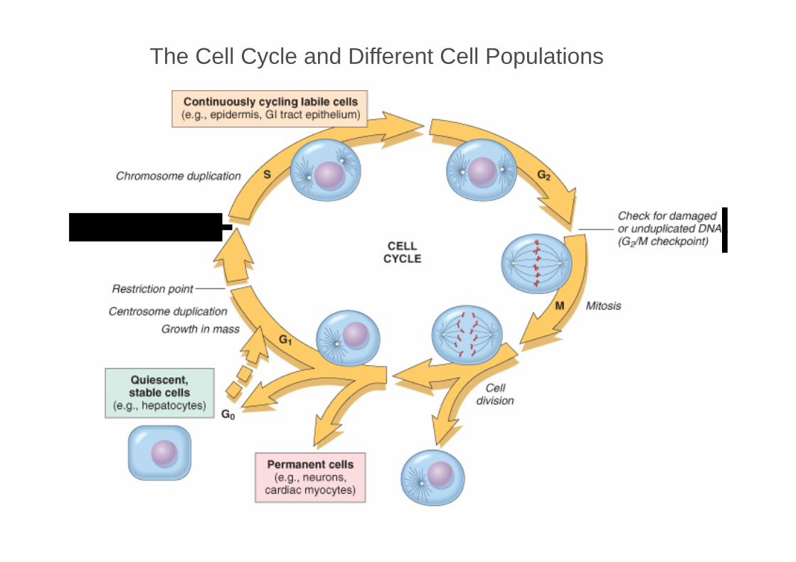
Tissue proliferative activity
Cell cycle
- G1 (pre-synthetic) - S (DNA synthesis) - G2 (pre- mitotic) - M (mitotic) - G0

Chromosome dupUcalion
Check for DNA idamage, ,(G,IS checkpoint) --
Restriction point-
Centrosome duplication,
Cell division
Check for damaged --or unduplicaled DNA
(Gy M ,checkpoint)
Mitosis
The Cell Cycle

Types of cells
• Labile (always dividing) cells: – Replace dying cells – Epithelia: skin, oral cavity, exocrine ducts, GIT, FGT,
hematopoietic cells • Stable (quiescent) cells:
– Usually G0 and low rate of division – Driven into G1 and rapid proliferation – Liver, kidney, pancreas, endothelium, fibroblasts

Types of cells
• Permanent (non-dividing) cells: – Permanently removed from cell cycle – Irreversible injury leads only to scar – Nerve cells, myocardium, skeletal muscle
tinl!ll ously eye ling lab ·1e oe Is. (e.g. , epidermis GI tract e-pi~heli um)
. ~---i------------· r::~
au ies,eenl, srablo. cons
(e.g., hepatooytes)
\ ! :-.:
G1
o ·~ G . ,m
Permanent cells (e.g., 11 eurons,
card I ac yooytes)
CELl CYC E
The Cell Cycle and Different Cell Populations

Growth Factors
• Very important in tissue repair.
• Actions: • stimulate cell division and proliferation • promote cell survival
• Epidermal growth factor (EGF)- Keratinocytes,fibroblasts • Vascular endothelial growth factor (VEGF)-Angiogenesis • Transforming growth factor (TGF)-Fibrogenesis • Platelet-derived growth factor (PDGF)
– Migration and proliferation of fibroblasts, smooth muscle, and monocytes

Repair
• Repair is the replacement of injured tissue by fibrous tissue. Two processes are involved in repair: – Granulation tissue formation – Contraction of wounds
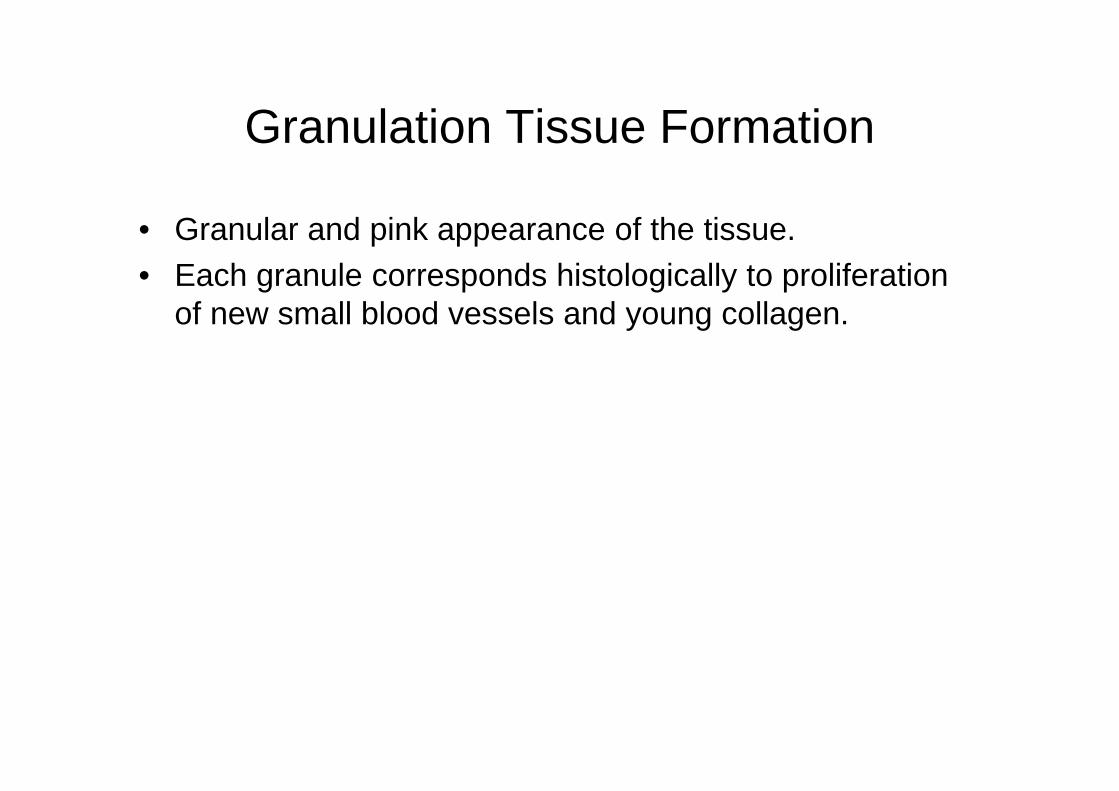
Granulation Tissue Formation
• Granular and pink appearance of the tissue. • Each granule corresponds histologically to proliferation
of new small blood vessels and young collagen.

Phases in the formation of granulation tissue
• Phase of inflammation- Following trauma, blood clots at site of injury. There is acute inflammatory response with exudation of plasma, neutrophils and some monocyteswithin 24 hours.
• Phase of clearance- proteolytic enzymes liberated from neutrophils, and phagocytic activity of macrophages clear off the necrotic tissue, debris and red blood cells.
• Phase of in growth of granulation tissue- consists of 2 main processes: - angiogenesis or neovascularisation - fibrogenesis
Angiogenesis (neovascularisation)
• Formation of new blood vessels takes place by proliferation of endothelial cells
• Initially, proliferated endothelial cells are solid buds butwithin few hours develop lumen.
• Newly formed blood vessels are more leaky, accounting for oedematous appearance of granulation tissue.
• Soon, blood vessels differentiate into arterioles, venules and capillaries.
• Factors: VEGF, PDGF, TGF-b, FGF and surface integrins

Fibrogenesis
• Proliferation of fibroblasts. • Myofibroblasts - some of the fibroblasts have
morphologic and functional characteristics of smooth muscle cells.
• Collagen fibrils begin to appear by about 6th day. • As maturation proceeds, more and more of collagen is
formed while the number of active fibroblasts and new blood vessels decreases. – Growth factors (TGF-, PDGF, EGF, FGF) – Cytokines (IL-1, TNF-)

Granulation tissue

Contraction of Wounds
• The wound starts contracting after 2-3 days and process is completed by the 14th day.
• The wound is reduced by approximately 80% of its original size.
• Mechanism of wound contraction – Dehydration – Contraction of collagen – Myofibroblasts- have features intermediate between
those of fibroblasts and smooth muscle cells. Their migration into the wound area and their active contraction decreases the size of the defect.

1
I
Conneclive issue reticular b:ers
Portal 1rl.ad: hepatlc a ery; pone.I
vei1n, bite, duct
11 1
·njury to cells lnj,ury to celJs and maltix .. t
I Prolrff3f'alion of residual Cf:llls
-thin intacl mal!rii:ic
'
REGENERATION
I DeposiUon of ,oonl"l,eclive !issue;
proliferation of residual ceu:s, . "lhin disrup,ted mailmc
+·
R:EPAIR BY SCARR NG

Extracellular Matrix (ECM)
• ECM is the network that surrounds cells
• Two forms: interstitial matrix and basement membrane • ECM provides support, adhesion substrate, reservoir for
factors; protein components – Collagens: most common, 14 types
• I-III: interstitial/fibrillar, most abundant • IV-VI: non-fibrillar, basement membranes
– Adhesive glycoproteins: e.g., Laminin, fibronectin, integrins which bind ECM components to each other, and to other cells
– Proteoglycans: sugars linked to proteins; influence ECM permeability and structure
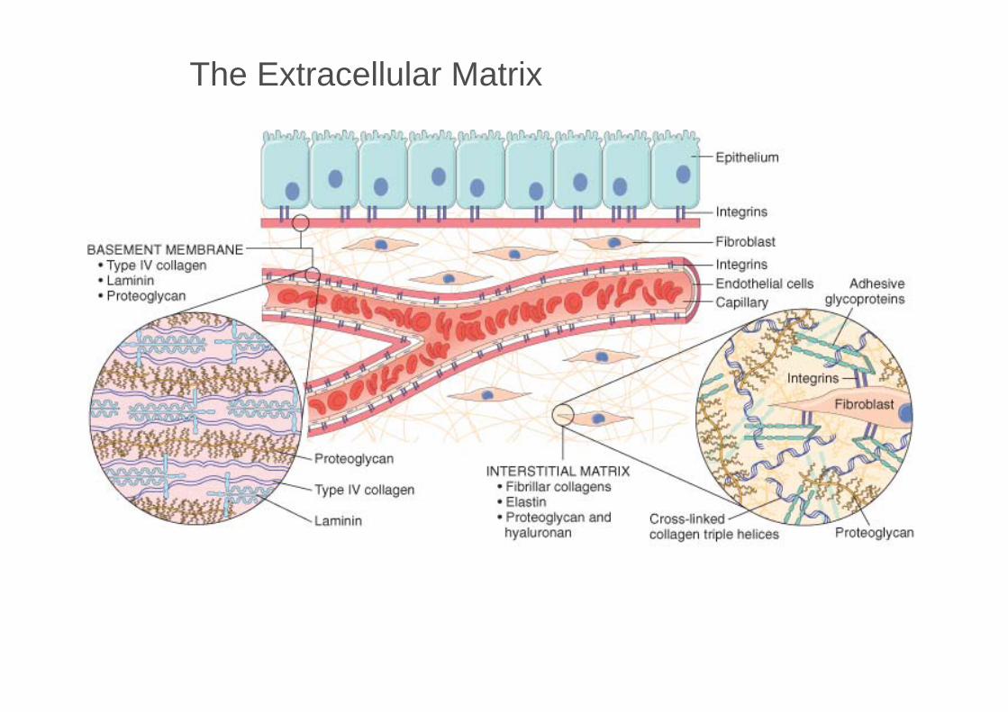
BASEME .IT MEMBRANE-----'-~. • Type IV collagen ., Lamtnin • P r-0teoglycan
--Epitil eUum
lntegriins
, ::][:::~---Flbmblas!
IINTEFISTITI Al MATFI IX • Fibrmar coll:egen$ • Elastin • Prote.ogtycan and
hya:lu1rnnain
lntegrjns 11:ndothella! cells Adhesave Ca pill ry __...-----n-......... •g lycopro eins
Cross--linkedl coJla.gen ·Hip'Je heJices
The Extracellular Matrix

... .... r
..
.--
...
■
"'!!--
-..
...... --
"!
,.
r
'I -_, J
.. • --,._
'II,
-..
'I
--. •
.. 'I
_ .. .,
..
;
... . - ..
..
·-.
■
d
.._
!'"
-. "'=.
"!
-·
..
r
.. -
•
-·
-.
fil =·
"!
,F
■ •
...
-.
!b
. -I
..
.. .. --. -
.,,.
.r
■
"!
-·
,.
... . ...
"! .. .-
--
---·--.
r:
- r" I
'I
---
.-
.-,i ...
"i -
i..
.,. .... • -. ...
-- .. -- ---· .. ..
-· -- __.. 'I
-.._
•
.....
"1
Scar