Head and Neck 1-2011 (Final)Head and Neck Table of Contents Discussion NCCN Guidelines™ Version...
145
Version 2.2011, 03/30/11 © National Comprehensive Cancer Network, Inc. 2011,All rights reserved. The NCCN Guidelines™ and this illustration may not be reproduced in any form without the express written permission of NCCN®. NCCN.org Continue NCCN Clinical Practice Guidelines in Oncology (NCCN Guidelines™) Head and Neck Cancers Version 2.2011
Transcript of Head and Neck 1-2011 (Final)Head and Neck Table of Contents Discussion NCCN Guidelines™ Version...

Version 2.2011, 03/30/11 © National Comprehensive Cancer Network, Inc. 2011, All rights reserved. The NCCN Guidelines™ and this illustration may not be reproduced in any form without the express written permission of NCCN®.
NCCN Guidelines IndexHead and Neck Table of Contents
Discussion
NCCN.org
Continue
NCCN Clinical Practice Guidelines in Oncology (NCCN Guidelines™)
Head and NeckCancers
Version 2.2011
Version 2.2011, 03/30/11 © National Comprehensive Cancer Network, Inc. 2011, All rights reserved. The NCCN Guidelines™ and this illustration may not be reproduced in any form without the express written permission of NCCN®.
NCCN Guidelines IndexHead and Neck Table of Contents
Discussion
NCCN Guidelines™ Version 2.2011 Panel MembersHead and Neck Cancers
David G. Pfister, MD /ChairMemorial Sloan-Kettering Cancer Center
Kie-Kian Ang, MD, PhDThe University of TexasMD Anderson Cancer Center
David Brizel, MDDuke Cancer Institute
Barbara A. Burtness, MDFox Chase Cancer Center
Anthony J. Cmelak, MDVanderbilt-Ingram Cancer Center
Alexander D. Colevas, MDStanford Comprehensive Cancer Center
Frank Dunphy, MDDuke Cancer Institute
David W. Eisele, MDUCSF Helen Diller FamilyComprehensive Cancer Center
Jill Gilbert, MD
Maura L. Gillison, MD, PhD
Robert I. Haddad, MDDana-Farber/Brigham and Women’s CancerCenter | Massachusetts General HospitalCancer Center
† Þ
§
§
†
§
†
†
¶
¶
†Vanderbilt-Ingram Cancer Center
The Ohio State University ComprehensiveCancer Center - James Cancer Hospital andSolove Research Institute
†
Harlan A. Pinto, MDStanford Comprehensive Cancer Center
John A. Ridge, MD, PhDFox Chase Cancer Center
Sandeep Samant, MDSt. Jude Children's Research Hospital/University of Tennessee Cancer Institute
Giuseppe Sanguineti, MDThe Sidney Kimmel ComprehensiveCancer Center at Johns Hopkins
David E. Schuller, MDThe Ohio State University ComprehensiveCancer Center - James Cancer Hospital andSolove Research Institute
† Þ
¶
¶
§
¶
¶
§
§
¶
¶
¶ †
Jatin P. Shah, MDMemorial Sloan-Kettering Cancer Center
Sharon Spencer, MDUniversity of Alabama at BirminghamComprehensive Cancer Center
Andy Trotti, III, MDH. Lee Moffitt Cancer Center& Research Institute
Randal S. Weber, MDThe University of TexasMD Anderson Cancer Center
Gregory T. Wolf, MDUniversity of MichiganComprehensive Cancer Center
Frank Worden, MDUniversity of MichiganComprehensive Cancer Center
�
Bruce H. Haughey, MBChB, MSSiteman Cancer Center at Barnes-Jewish Hospitaland Washington University School of medicine
Wesley L. Hicks, Jr., MDRoswell Park Cancer Institute
¶
¶
§Ying J. Hitchcock, MDHuntsman Cancer Instituteat the University of Utah
Merrill S. Kies, MDThe University of TexasMD Anderson Cancer Center
William M. Lydiatt, MDUNMC Eppley Cancer Centerat The Nebraska Medical Center
Ellie Maghami, MDCity of Hope Comprehensive Cancer Center
Renato Martins, MD, MPHFred Hutchinson Cancer Research Center/Seattle Cancer Care Alliance
Thomas McCaffrey, MD, PhDH. Lee Moffitt Cancer Center &Research Institute
Bharat B. Mittal, MDRobert H. Lurie Comprehensive Cancer Centerof Northwestern University
†
¶
¶
†
§
�
�
�
*
† Medical Oncology
¶ Surgery/Surgical oncology
§ Radiation oncology
Otolaryngology
Þ Internal medicine
�
* Writing Committee MemberNCCN Guidelines Panel Disclosures
*
*
*
*
*
ContinueNCCNMiranda Hughes, PhDNicole McMillian, MS
*
Printed by Luke Reid on 6/27/2011 12:07:17 AM. For personal use only. Not approved for distribution. Copyright © 2011 National Comprehensive Cancer Network, Inc., All Rights Reserved.

Version 2.2011, 03/30/11 © National Comprehensive Cancer Network, Inc. 2011, All rights reserved. The NCCN Guidelines™ and this illustration may not be reproduced in any form without the express written permission of NCCN®.
NCCN Guidelines IndexHead and Neck Table of Contents
Discussion
NCCN Guidelines™ Version 2.2011 Sub-CommitteesHead and Neck Cancers
Continue
Mucosal Melanoma
William M. Lydiatt, MD /Lead
UNMC Eppley Cancer Centerat The Nebraska Medical Center
Jatin P. Shah, MD
Memorial Sloan-Kettering Cancer Center
Andy Trotti, III, MDH. Lee Moffitt Cancer Center &Research Institute
¶ �
¶
§
† Medical Oncology
¶ Surgery/Surgical oncology
§ Radiation oncology
Otolaryngology
Þ Internal medicine
�
NCCN Guidelines Panel Disclosures
Principles of Radiation Therapy
Andy Trotti, III, MD
H. Lee Moffitt Cancer Center &
Research Institute
Kie-Kian Ang, MD, PhD
The University of Texas
MD Anderson Cancer Center
David Brizel, MD
Duke Cancer Institute
Anthony J. Cmelak, MD
Vanderbilt-Ingram Cancer Center
Bharat B. Mittal, MD
Robert H. Lurie Comprehensive Cancer
Center of Northwestern University
Giuseppe Sanguineti, MDThe Sidney Kimmel ComprehensiveCancer Center at Johns Hopkins
Sharon Spencer, MDUniversity of Alabama at BirminghamComprehensive Cancer Center
§
§
§
§
§
§
/Lead
§
Principles of Surgery
Gregory T. Wolf, MDUniversity of MichiganComprehensive Cancer Center
David Brizel, MDDuke Cancer Institute
David W. Eisele, MDUCSF Helen Diller FamilyComprehensive Cancer Center
William M. Lydiatt, MDUNMC Eppley Cancer Centerat The Nebraska Medical Center
David E. Schuller, MDThe Ohio State UniversityComprehensive Cancer Center - JamesCancer Hospital and Solove ResearchInstitute
Randal S. Weber, MDThe University of TexasMD Anderson Cancer Center
¶
§
¶
¶
¶
�/Lead
¶ �
Principles of Systemic Therapy
Alexander D. Colevas, MD
Stanford Comprehensive Cancer Center
Frank Dunphy, MDDuke Cancer Institute
Renato Martins, MD, MPHFred Hutchinson Cancer Research Center/Seattle Cancer Care Alliance
†
†
†
Printed by Luke Reid on 6/27/2011 12:07:17 AM. For personal use only. Not approved for distribution. Copyright © 2011 National Comprehensive Cancer Network, Inc., All Rights Reserved.

Version 2.2011, 03/30/11 © National Comprehensive Cancer Network, Inc. 2011, All rights reserved. The NCCN Guidelines™ and this illustration may not be reproduced in any form without the express written permission of NCCN®.
NCCN Guidelines IndexHead and Neck Table of Contents
Discussion
NCCN Head Neck Cancers Panel Members
NCCN Head and Cancers Sub-Committee Members
Summary of the Guidelines UpdatesMultidisciplinary Team Approach and Support Modalities (TEAM-1)
Cancer of the Lip (LIP-1)
Cancer of the Oral Cavity (OR-1)
Cancer of the Oropharynx (ORPH-1)
Cancer of the Hypopharynx (HYPO-1)
Cancer of the Nasopharynx (NASO-1)
Cancer of the Glottic Larynx (GLOT-1)
Cancer of the Supraglottic Larynx (SUPRA-1)
Ethmoid Sinus Tumors (ETHM-1)
Maxillary Sinus Tumors (MAXI-1)
Very Advanced Head and Neck Cancer (ADV-1)
Recurrent/Persistent Head and Neck Cancer (ADV-2)
Occult Primary (OCC-1)
Salivary Gland Tumors (SALI-1)
Mucosal Melanoma (MM-1)
Follow-up Recommendations (FOLL-A)
Principles of Surgery (SURG-A)
Radiation Techniques (RAD-A)
Principles of Systemic Therapy (CHEM-A)
Staging (ST-1)
�
�
�
�
�
�
�
�
�
�
�
�
�
�
�
�
�
�
�
Clinical Trials:
Categories of Evidence andConsensus:NCCN
Thebelieves that the best managementfor any cancer patient is in a clinicaltrial. Participation in clinical trials isespecially encouraged.
All recommendationsare Category 2A unless otherwisespecified.
NCCN
To find clinical trials online at NCCNmember institutions, click here:nccn.org/clinical_trials/physician.html
See NCCN Categories of Evidenceand Consensus
The NCCN Guidelines™ are a statement of evidence and consensus of the authors regarding their views of currently accepted approaches to
treatment. Any clinician seeking to apply or consult the NCCN Guidelines is expected to use independent medical judgment in the context of individual
clinical circumstances to determine any patient’s care or treatment. The National Comprehensive Cancer Network® (NCCN®) makes no
representations or warranties of any kind regarding their content, use or application and disclaims any responsibility for their application or use in any
way. The NCCN Guidelines are copyrighted by National Comprehensive Cancer Network®. All rights reserved. The NCCN Guidelines and the
illustrations herein may not be reproduced in any form without the express written permission of NCCN. ©2011.
NCCN Guidelines™ Version 2.2011 Table of ContentsHead and Neck Cancers
Printed by Luke Reid on 6/27/2011 12:07:17 AM. For personal use only. Not approved for distribution. Copyright © 2011 National Comprehensive Cancer Network, Inc., All Rights Reserved.

Version 2.2011, 03/30/11 © National Comprehensive Cancer Network, Inc. 2011, All rights reserved. The NCCN Guidelines™ and this illustration may not be reproduced in any form without the express written permission of NCCN®.
NCCN Guidelines IndexHead and Neck Table of Contents
Discussion
NCCN Guidelines™ Version 2.2011 UpdatesHead and Neck Cancers
The 2.2011 Version of the NCCN Head and Neck Cancers Guidelines represents the addition of the Discussion text correspondent to the
changes in the algorithm. (MS-1)
UPDATES1 of 3
Global Changes
� The Principles of Radiation Therapy pages for all sites were revised.
Continued on next page
Cancer of the Lip
Cancer of the Oral Cavity
LIP-1
LIP-2
OR-1
OR-2
OR-3
�
�
�
�
�
�
�
�
Workup: CT/MRI changed to “CT/MRI of primary and neck as
indicated”.
Footnote e “Consider re-excision to achieve negative margins, if
feasible” is new to the algorithm. This footnote was also added to
other sites in the guidelines.
Workup: Sixth bullet changed to “Examination under anesthesia
, if indicated”. Eighth bullet changed to
“Dental/ evaluation, including as
indicated”.
T1-2, N0: Primary treatment changed to “
N0, N1, N2a-b, N3 pathway: Primary treatment changed to
with endoscopy
prosthodontic jaw imaging
Excision of primary
(preferred) ± ipsilateral or bilateral neck dissection
”.
After Adverse features, the pathway for Positive margin (T1, N0 only)
was removed. “Re-excision” changed from category 1 to
category 2A.
The T3, N0 pathway was deleted from this page and incorporated
into page OR-3.
“Excision
of primary, ipsilateral neck dissection
”.
Footnote d: “selected pT2, N0, N1 disease” was removed as an
adverse feature”. (Also for other sites)
(guided by tumor
thickness)
or bilateral (guided by tumor
thickness, extent of disease)
Cancer of the Oropharynx
and audiogram HYPO-1
NASO-1, GLOT-1, SUPRA-1
� Workup: Eighth bullet changed to “Nutrition, speech & swallowing
evaluation/therapy as indicated” (Also for ,
). Examination under anesthesia with
endoscopy is now “as indicated”.
“Selected T2, N0 (requiring laryngectomy)” was added to the clinical
staging column.
Treatment: Induction chemotherapy changed from category 1 to
category 2A.
Cancer of the Hypopharynx
�
�
ORPH-1
ORPH-2
ORPH-3
HYPO-1
HYPO-3
�
�
�
�
Adverse features pathway: Extracapsular spread and/or positive
margin changed to “Extracapsular spread
Footnote h referring to the Discussion for more information on
induction chemotherapy is new to the algorithm. (Also for other
sites.)
Surgery: primary and neck:N1, N2a-b, N3: Recommendation changed to “Excision of primary,
ipsilateral neck dissection”N2c: After “Excision of primary and bilateral neck dissection, the
following text was removed: “(bilateral is category 3 if neck nodes
contralateral only)”.
Workup: “Consider videostrobe for select patients” was added.
(Also for , )
± positive margin”.
“Positive margin pathway: “T1, N0 only” was removed from “Positive
margin”. (Also for )HYPO-2
�
�
or bilateral
GLOT-1 SUPRA-1
ORPH-4
Updates in Version 1.2011 of the NCCN Head and Neck Cancer Guidelines from Version 2.2010 include:
Printed by Luke Reid on 6/27/2011 12:07:17 AM. For personal use only. Not approved for distribution. Copyright © 2011 National Comprehensive Cancer Network, Inc., All Rights Reserved.

Version 2.2011, 03/30/11 © National Comprehensive Cancer Network, Inc. 2011, All rights reserved. The NCCN Guidelines™ and this illustration may not be reproduced in any form without the express written permission of NCCN®.
NCCN Guidelines IndexHead and Neck Table of Contents
Discussion
NCCN Guidelines™ Version 2.2011 UpdatesHead and Neck Cancers
UPDATES2 of 3
Continued on next page
Cancer of the Nasopharynx
Cancer of the Glottic Larynx
or patient declines surgery
�
�
�
T1, N1-3; T2-T4, any N: Recommendations simplified as “Concurrent
chemo/RT (category 1)” and “Adjuvant chemotherapy”.
Any T, Any N, M1:Platinum-based combination therapy pathway: “If complete
clinical response” was removed. Chemo/RT is now “as clinically
indicated”.“Concurrent chemo/RT” was added as an option for primary
treatment. Corresponding footnote d is new to the algorithm.
Clinical Presentation; Third row: Changed to “Newly diagnosed, T4b
”.
�
�
Cancer of the Supraglottic Larynx
Ethmoid Sinus Tumors
�
NASO-2
GLOT-1
GLOT-3
ETHM-1
ETHM-2
�
�
�
�
The Clinical staging listings were revised for clarity.
After Surgery; N0: the option of “
“in selected T3, N2-N3 patients” was removed after “Induction
chemotherapy followed by chemo/RT (category 2B)”.
Footnote a: “Esthesioneuroblastomas” was removed from the list of
histologies (Also for ). “Consider referral to a major medical
center that specializes in these diseases” was added.
± unilateral or bilateral neck
dissection” was removed after “Laryngectomy with ipsilateral
thyroidectomy”.
Surgery; N1: “Laryngectomy with ipsilateral thyroidectomy,
ipsilateral neck dissection ± contralateral neck dissection” changed
to “Laryngectomy with ipsilateral thyroidectomy, ipsilateral neck
dissection neck dissection”.or bilateral
MAXI-1
SUPRA-6
Ethmoid Sinus Tumors
Occult Primary
---continued
�
�
�
�
Primary Treatment for newly diagnosed T3, T4a pathway:
Surgical resection is now “preferred” and Chemo/RT was added
as an option.
Footnote d was revised to include minor salivary gland tumors.
“Principles of Radiation Therapy” is a new page that provides
specific RT recommendations and dosing for ethmoid sinus
tumors.
Footnote f regarding adenoid cystic tumors and minor salivary
gland tumors is new to the algorithm
(Very Advanced Head and Neck Cancer)
After neck dissection in patients who have N1 disease without
extracapsular spread (for all levels):
Maxillary Sinus Tumors
Advanced Head and Neck Cancer
�
�
�
“Observation” was added as an option.“RT to neck only (category 3)” was removed as an option.
ETHM-2
MAXI-2
OCC-1
ETHM-A
ADV-1
OCC-4
�
�
�
�
For Performance status(PS) 0-1 patients: The recommendation
changed to “Induction chemotherapy followed by
chemoradiation category 3)”.
For PS 3 patients, “single-agent chemotherapy” was added as a
treatment option.
Workup: Fifth bullet changed to “Thyroglobulin
staining...”
Footnote c “Human papilloma virus or Epstein-Barr virus positive
status may help to define the radiation fields” is new to the
algorithm.
RT or
and calcitonin
Printed by Luke Reid on 6/27/2011 12:07:17 AM. For personal use only. Not approved for distribution. Copyright © 2011 National Comprehensive Cancer Network, Inc., All Rights Reserved.
Version 2.2011, 03/30/11 © National Comprehensive Cancer Network, Inc. 2011, All rights reserved. The NCCN Guidelines™ and this illustration may not be reproduced in any form without the express written permission of NCCN®.
NCCN Guidelines IndexHead and Neck Table of Contents
Discussion
NCCN Guidelines™ Version 2.2011 UpdatesHead and Neck Cancers
UPDATES3 of 3
Salivary Gland Tumors
Mucosal Melanoma
�
�
�
�
Clinically benign or carcinoma T1, T2 pathway: After complete surgical excision, for low grade tumors, the option to “If tumor spillage,
consider RT” was added.
Parotid gland; Clinical N0 pathway: After parotidectomy, “± neck dissection for high-grade and high-stage tumors” was added.
Locoregional recurrence without prior RT; Resectable; Completely excised: “RT” was added as a treatment option.
Workup: “Consider PET-CT scan to rule out metastatic disease” was added. Chest imaging is now listed “as indicated”.
Principles of Systemic Therapy
Follow-up Recommendations
Principles of Surgery
Radiation Techniques
� The following sentence was added to the first paragraph “Close cooperation and interdisciplinary management are critical to treatment planning and
radiation targeting, especially in the postoperative setting or after induction chemotherapy.”
SALI-2
SALI-3
SALI-4
MM-1
FOLL-A
SURG-A
RAD-A
CHEM-A
�
�
�
Under Dental evaluation: “Not recommended for other sites” changed to “As indicated for other sites, if significant intraoral radiation”.
These pages were extensively revised.
Recurrent, Unresectable, or Metastatic (incurable)Combination therapy: Regimens with cetuximab were clarified as “non-nasopharyngeal”.Single agents: Cetuximab was clarified as “non-nasopharyngeal”.�
�
Printed by Luke Reid on 6/27/2011 12:07:17 AM. For personal use only. Not approved for distribution. Copyright © 2011 National Comprehensive Cancer Network, Inc., All Rights Reserved.

Version 2.2011, 03/30/11 © National Comprehensive Cancer Network, Inc. 2011, All rights reserved. The NCCN Guidelines™ and this illustration may not be reproduced in any form without the express written permission of NCCN®.
NCCN Guidelines IndexHead and Neck Table of Contents
Discussion
Note: All recommendations are category 2A unless otherwise indicated.
Clinical Trials: NCCN believes that the best management of any cancer patient is in a clinical trial. Participation in clinical trials is especially encouraged.
NCCN Guidelines™ Version 2.2011Team Approach
Follow-up should be performed by a physician and other health care professionals with expertise in
the management and prevention of treatment sequelae. It should include a comprehensive head and
neck exam. The management of head and neck cancer patients may involve the following:
SUPPORT AND SERVICES
TEAM-1
����������
���
Head and neck surgeryRadiation oncologyMedical oncologyPlastic and reconstructive surgerySpecialized nursing careDentistry/prosthodonticsPhysical medicine and rehabilitationSpeech and swallowing therapyClinical social workNutrition support
Pathology (including cytopathology)Diagnostic radiologyAdjunctive services�
�
�
�
�
�
NeurosurgeryOphthalmologyPsychiatryAddiction servicesAudiologyPalliative care
MULTIDISCIPLINARY TEAM
The management of patients with head and neck cancers is complex. All patients need
access to the full range of specialists and support services
for optimal treatment and follow-up.
with expertise in the
management of patients with head and neck cancer
���
��
General medical carePain and symptom managementNutritional support
Dental care for RT effectsXerostomia management
Smoking and alcohol cessation
Speech and swallowing therapy
Audiology
Tracheotomy care
Wound management
Depression assessment and management
Social work and case management
Supportive care
�
�
Enteral feedingOral supplements
�
�
�
�
�
�
�
�
(See NCCN Palliative Care Guideline)
Printed by Luke Reid on 6/27/2011 12:07:17 AM. For personal use only. Not approved for distribution. Copyright © 2011 National Comprehensive Cancer Network, Inc., All Rights Reserved.
Version 2.2011, 03/30/11 © National Comprehensive Cancer Network, Inc. 2011, All rights reserved. The NCCN Guidelines™ and this illustration may not be reproduced in any form without the express written permission of NCCN®.
NCCN Guidelines IndexHead and Neck Table of Contents
Discussion
Note: All recommendations are category 2A unless otherwise indicated.
Clinical Trials: NCCN believes that the best management of any cancer patient is in a clinical trial. Participation in clinical trials is especially encouraged.
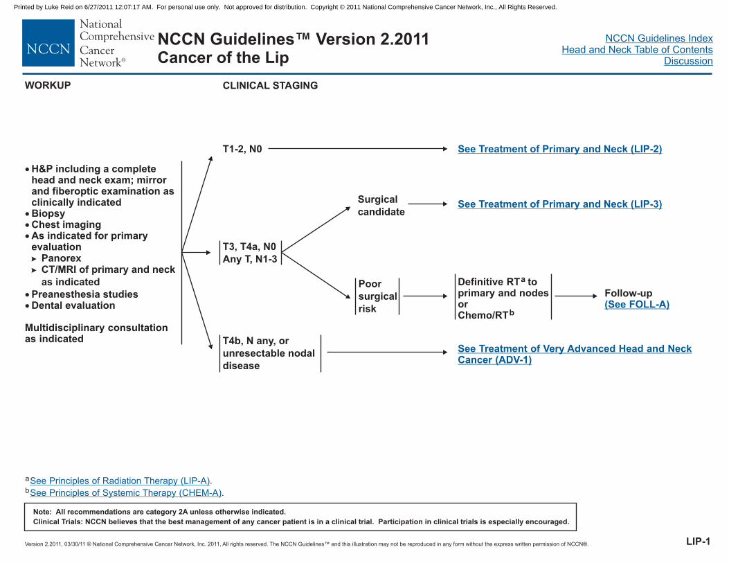
NCCN Guidelines™ Version 2.2011Cancer of the Lip
�
���
��
H&P
BiopsyChest imagingAs indicated for primaryevaluation
Preanesthesia studiesDental evaluation
including a completehead and neck exam; mirrorand fiberoptic examination asclinically indicated
PanorexCT/MRI of primary and neck
as indicated
Multidisciplinary consultationas indicated
�
�
WORKUP CLINICAL STAGING
T1-2, N0
T3, T4a, N0
Any T, N1-3
See Treatment of Primary and Neck (LIP-2)
See Treatment of Primary and Neck (LIP-3)
T4b, N any, or
unresectable nodal
disease
See Treatment of Very Advanced Head and NeckCancer (ADV-1)
LIP-1
Surgical
candidate
Poor
surgical
risk
Definitive RT toprimary and nodesorChemo/RT
a
b
Follow-up(See FOLL-A)
a
b
.
.
See Principles of Radiation Therapy (LIP-A)
See Principles of (CHEM-A)Systemic Therapy
Printed by Luke Reid on 6/27/2011 12:07:17 AM. For personal use only. Not approved for distribution. Copyright © 2011 National Comprehensive Cancer Network, Inc., All Rights Reserved.

Version 2.2011, 03/30/11 © National Comprehensive Cancer Network, Inc. 2011, All rights reserved. The NCCN Guidelines™ and this illustration may not be reproduced in any form without the express written permission of NCCN®.
NCCN Guidelines IndexHead and Neck Table of Contents
Discussion
Note: All recommendations are category 2A unless otherwise indicated.
Clinical Trials: NCCN believes that the best management of any cancer patient is in a clinical trial. Participation in clinical trials is especially encouraged.
NCCN Guidelines™ Version 2.2011Cancer of the Lip
LIP-2
TREATMENT OF PRIMARY AND NECKCLINICAL STAGING
T1-2, N0
Surgical excision
(preferred)
(elective neck
dissection not
recommended)
or
External-beam RT
to primary site
brachytherapy
d
a,c
±
FOLLOW-UP
Follow-up(See FOLL-A)
Residual or
recurrent tumor
post-RT
Positive margins,
perineural/vascular/
lymphatic invasion
No adverse
pathologic findings
Re-excision
or
RT
e
a
Surgery /
reconstruction
d
ADJUVANT TREATMENT
a
c
d
e
.
No elective treatment to neck preferred for T1-2, N0.
Consider re-excision to achieve negative margins, if feasible.
See Principles of Radiation Therapy (LIP-A)
See Principles of Surgery (SURG-A).
RecurrentorPersistentDisease(See ADV-2)
Printed by Luke Reid on 6/27/2011 12:07:17 AM. For personal use only. Not approved for distribution. Copyright © 2011 National Comprehensive Cancer Network, Inc., All Rights Reserved.

Version 2.2011, 03/30/11 © National Comprehensive Cancer Network, Inc. 2011, All rights reserved. The NCCN Guidelines™ and this illustration may not be reproduced in any form without the express written permission of NCCN®.
NCCN Guidelines IndexHead and Neck Table of Contents
Discussion
Note: All recommendations are category 2A unless otherwise indicated.
Clinical Trials: NCCN believes that the best management of any cancer patient is in a clinical trial. Participation in clinical trials is especially encouraged.
NCCN Guidelines™ Version 2.2011Cancer of the Lip
Treatment of Primary and Neck (LIP-4)
CLINICAL STAGING:
T3,T4a, N0; Any T, N1-3
Excision of primary ± ipsilateral or
bilateral neck dissectiondN0
External RT
± brachytherapy
or
a
Chemo/RTb
Excision of primary and bilateral
neck dissectiond
Excision of primary, ipsilateral neck
dissection ± contralateral neck
dissectiond
N2c
(bilateral)
N2a-b,
N3
RT (optional)aOne positive node without
adverse featuresf
Follow-up(See FOLL-A)
FOLLOW-UP
a
b
d
f
.
.
eConsider re-excision to achieve negative margins, if feasible.
Adverse features: extracapsular nodal spread, positive margins, multiple positive nodes or perineural/lymphatic/vascular invasion.
See Principles of Radiation Therapy (LIP-A)
See Principles of (CHEM-A)
See Principles of Surgery (SURG-A)
Systemic Therapy
.
Surgery
(preferred)
d
ADJUVANT
TREATMENT
Adverse
featuresf
Other risk
features
RTa
or
Consider
chemo/RTb
N1
or
Excision of primary, ipsilateral neck
dissection ± contralateral neck
dissectiond
N0
Chemo/RTb
preferred
(category 1)
RT
or
Re-excision
or
e
a
Extracapsular
spread and/or
positive
margin
TREATMENT OF PRIMARY AND NECK
RecurrentorPersistentDisease(See ADV-2)
LIP-3
Printed by Luke Reid on 6/27/2011 12:07:17 AM. For personal use only. Not approved for distribution. Copyright © 2011 National Comprehensive Cancer Network, Inc., All Rights Reserved.

Version 2.2011, 03/30/11 © National Comprehensive Cancer Network, Inc. 2011, All rights reserved. The NCCN Guidelines™ and this illustration may not be reproduced in any form without the express written permission of NCCN®.
NCCN Guidelines IndexHead and Neck Table of Contents
Discussion
Note: All recommendations are category 2A unless otherwise indicated.
Clinical Trials: NCCN believes that the best management of any cancer patient is in a clinical trial. Participation in clinical trials is especially encouraged.
NCCN Guidelines™ Version 2.2011Cancer of the Lip
TREATMENT OF PRIMARY AND NECKCLINICAL STAGING:
T3, T4a, N0; Any T, N1-3
External RT
± brachytherapy
or
a
Chemo/RTb
Follow-up(See FOLL-A)
FOLLOW-UP
a
b
d
g
.
.
See Principles of Radiation Therapy (LIP-A)
See Principles of (CHEM-A)
See Principles of Surgery (SURG-A)
See Post Chemoradiation or RT Neck Evaluation (SURG-A 6 of 6)
Systemic Therapy
.
.
Residual tumor
in neck
Complete clinicalresponse of neck
Primary site:Completeclinicalresponse (N+ atinitial staging)
Primary site:< completeclinicalresponse
Salvage surgery + neckdissection as indicatedd
Neckdissectiond
ADJUVANT
TREATMENT
Post-treatment
evaluationg
Negative
Positive
Observe
Neck
dissectiond
Primary site:Complete clinicalresponse(N0 at initial staging)
RecurrentorPersistentDisease(See ADV-2)
LIP-4
Printed by Luke Reid on 6/27/2011 12:07:17 AM. For personal use only. Not approved for distribution. Copyright © 2011 National Comprehensive Cancer Network, Inc., All Rights Reserved.

Version 2.2011, 03/30/11 © National Comprehensive Cancer Network, Inc. 2011, All rights reserved. The NCCN Guidelines™ and this illustration may not be reproduced in any form without the express written permission of NCCN®.
NCCN Guidelines IndexHead and Neck Table of Contents
Discussion
Note: All recommendations are category 2A unless otherwise indicated.
Clinical Trials: NCCN believes that the best management of any cancer patient is in a clinical trial. Participation in clinical trials is especially encouraged.
NCCN Guidelines™ Version 2.2011Cancer of the Lip
PRINCIPLES OF RADIATION THERAPY1
Uninvolved nodal stations:
44-64 Gy (1.6-2.0 Gy/fraction)
Involved nodal stations:
60-66 Gy (2.0 Gy/fraction)
44-64 Gy (1.6-2.0 Gy/fraction)
Definitive RT
Postoperative RT
�
�
��
Primary and gross adenopathy:Conventional fractionation: 66-74 Gy (2.0 Gy/fraction;daily Monday-Friday) in 7 weeksExternal-beam RT ± brachytherapy50-60 Gy with brachytherapyNeck
Primary: 60-66 Gy (2.0 Gy/fraction)Neck
�
�
� Uninvolved nodal stations:
1 .See Radiation Techniques (RAD-A) and Discussion
LIP-A
Printed by Luke Reid on 6/27/2011 12:07:17 AM. For personal use only. Not approved for distribution. Copyright © 2011 National Comprehensive Cancer Network, Inc., All Rights Reserved.

Version 2.2011, 03/30/11 © National Comprehensive Cancer Network, Inc. 2011, All rights reserved. The NCCN Guidelines™ and this illustration may not be reproduced in any form without the express written permission of NCCN®.
NCCN Guidelines IndexHead and Neck Table of Contents
Discussion
Note: All recommendations are category 2A unless otherwise indicated.
Clinical Trials: NCCN believes that the best management of any cancer patient is in a clinical trial. Participation in clinical trials is especially encouraged.
NCCN Guidelines™ Version 2.2011Cancer of the Oral Cavity
�
��
�
�
��
�
H&P
BiopsyChest imaging
Examination under anesthesia withendoscopy, if indicatedPreanesthesia studiesDental/prosthodontic evaluation,including jaw imaging as indicated
including a complete head and neckexam; mirror and fiberoptic examinationas clinically indicated
CT with contrast and/or MRI with contrast
of primary and neck as indicated
Consider PET-CT for stage III-IV disease
Nutrition, speech & swallowingevaluation/therapy as indicated
Multidisciplinary consultation as indicated
�
a
WORKUP CLINICAL STAGING
T1–2, N0
T3, N0
T1–3, N1–3
T4a, any N
See Treatment of Primary and Neck (OR-2)
See Treatment of Primary and Neck (OR-3)
See Treatment of Primary and Neck (OR-3)
See Treatment of Primary and Neck (OR-3)
See Treatment of Very Advanced Head and NeckCancer (ADV-1)
Buccal mucosa, floor of mouth, anterior tongue, alveolar ridge, retromolar trigone, hard palate
T4b, N any, or
unresectable nodal
disease
aSee Discussion.
OR-1
Printed by Luke Reid on 6/27/2011 12:07:17 AM. For personal use only. Not approved for distribution. Copyright © 2011 National Comprehensive Cancer Network, Inc., All Rights Reserved.

Version 2.2011, 03/30/11 © National Comprehensive Cancer Network, Inc. 2011, All rights reserved. The NCCN Guidelines™ and this illustration may not be reproduced in any form without the express written permission of NCCN®.
NCCN Guidelines IndexHead and Neck Table of Contents
Discussion
Note: All recommendations are category 2A unless otherwise indicated.
Clinical Trials: NCCN believes that the best management of any cancer patient is in a clinical trial. Participation in clinical trials is especially encouraged.
NCCN Guidelines™ Version 2.2011Cancer of the Oral Cavity
Buccal mucosa, floor of mouth, anterior tongue, alveolar ridge, retromolar trigone, hard palate
CLINICAL
STAGING
TREATMENT OF PRIMARY AND NECK
T1–2, N0
Excision of primary
(preferred) ± ipsilateral or
bilateral neck dissection
(guided by tumor thickness)b
or
External-beam RT
± brachytherapy
c
One positive node without
adverse featuresd RT optional (category 2B)c
b
c
d
e
Adverse risk features: extracapsular nodal spread, positive margins, pT3 or pT4 primary, N2 or N3 nodal disease, nodal disease in levels IV or V, perineural invasion,vascular embolism .
Consider re-excision to achieve negative margins, if feasible.
.f
See Principles of Surgery (SURG-A)
See Principles of (CHEM-A)
.
( )See Discussion
Systemic Therapy
See Principles of Radiation Therapy (OR-A).
FOLLOW-UP
Follow-up(See FOLL-A)
RecurrentorPersistentDisease(See ADV-2)
No adverse featuresd
ADJUVANT TREATMENT
Adverse
featuresd
Residual disease Salvage surgery
No residual disease
Chemo/RT
Re-excision
c,e
f
(preferred) (category 1)
or
RT
or
c
Extracapsular
spread and/or
positive margin
Other risk
features
RTc
or
Consider chemo/RTc,e
OR-2
Printed by Luke Reid on 6/27/2011 12:07:17 AM. For personal use only. Not approved for distribution. Copyright © 2011 National Comprehensive Cancer Network, Inc., All Rights Reserved.

Version 2.2011, 03/30/11 © National Comprehensive Cancer Network, Inc. 2011, All rights reserved. The NCCN Guidelines™ and this illustration may not be reproduced in any form without the express written permission of NCCN®.
NCCN Guidelines IndexHead and Neck Table of Contents
Discussion
Note: All recommendations are category 2A unless otherwise indicated.
Clinical Trials: NCCN believes that the best management of any cancer patient is in a clinical trial. Participation in clinical trials is especially encouraged.
NCCN Guidelines™ Version 2.2011Cancer of the Oral Cavity
Buccal mucosa, floor of mouth, anterior tongue, alveolar ridge, retromolar trigone, hard palate
T3,N0;
T4a, Any N;
T1-3, N1-3
Excision of primary
and bilateral neck
dissectionb
N2c
(bilateral)
Excision of primary,
ipsilateral or
bilateral neck
dissection
(guided by tumor
thickness, extent of
disease)
bN0, N1,
N2a-b,
N3
CLINICAL
STAGING
TREATMENT OF PRIMARY AND NECK FOLLOW-UP
Surgeryb
ADJUVANT
TREATMENT
No adverse
featuresd RT (optional)c
Adverse
featuresd
Other risk
features
RTc
or
Consider
chemo/RTc,e
Chemo/RT
(preferred)
Re-excision
c,e
f
(category 1)
or
RT
or
c
b
c
d
e
Adverse risk features: extracapsular nodal spread, positive margins, pT3 or pT4 primary, N2 or N3 nodal disease, nodal disease in levels IV or V, perineural invasion,vascular embolism .
Consider re-excision to achieve negative margins, if feasible.f
See Principles of Surgery (SURG-A)
See Principles of Radiation Therapy (OR-A)
See Principles of (CHEM-A)
.
.
.( )See Discussion
Systemic Therapy
Extracapsular
spread and/or
positive
margin Follow-up(See FOLL-A)
RecurrentorPersistentDisease(See ADV-2)
OR-3
Printed by Luke Reid on 6/27/2011 12:07:17 AM. For personal use only. Not approved for distribution. Copyright © 2011 National Comprehensive Cancer Network, Inc., All Rights Reserved.
Version 2.2011, 03/30/11 © National Comprehensive Cancer Network, Inc. 2011, All rights reserved. The NCCN Guidelines™ and this illustration may not be reproduced in any form without the express written permission of NCCN®.
NCCN Guidelines IndexHead and Neck Table of Contents
Discussion
Note: All recommendations are category 2A unless otherwise indicated.
Clinical Trials: NCCN believes that the best management of any cancer patient is in a clinical trial. Participation in clinical trials is especially encouraged.
NCCN Guidelines™ Version 2.2011Cancer of the Oral Cavity
PRINCIPLES OF RADIATION THERAPY1
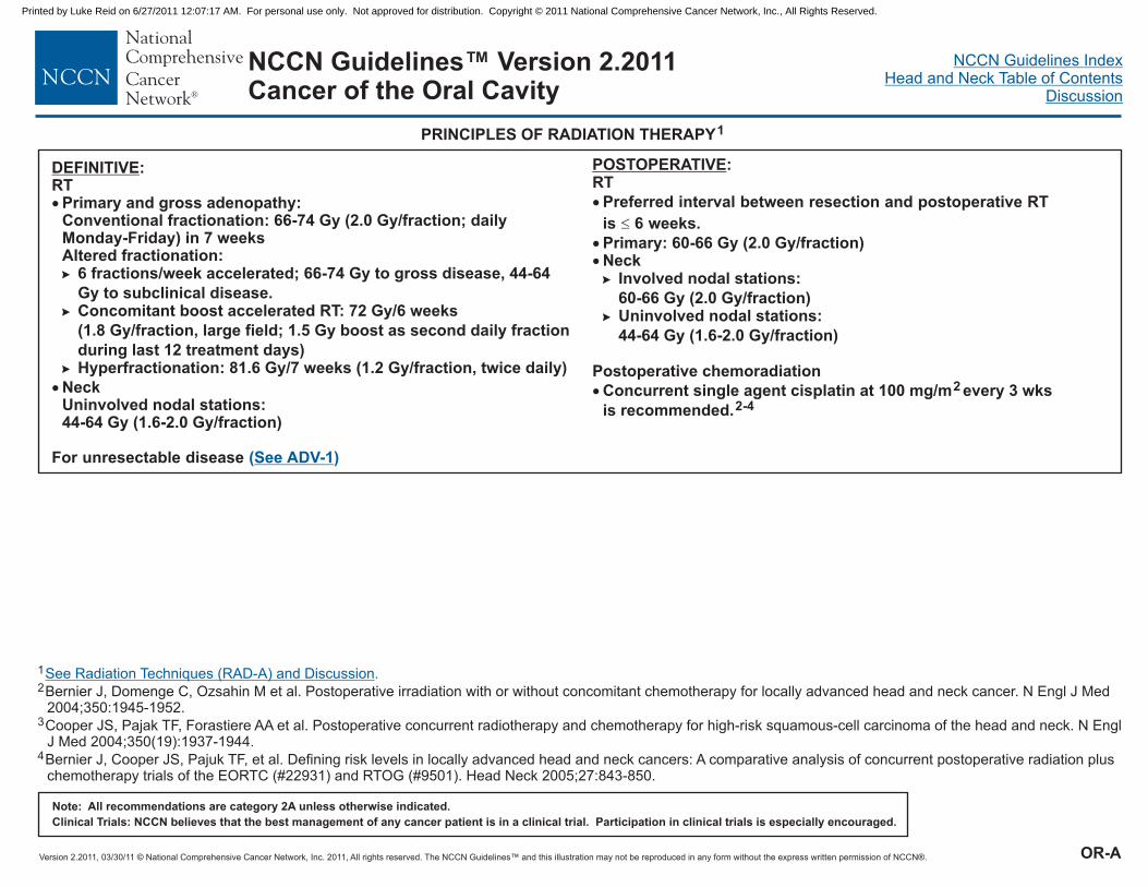
DEFINITIVE:RT
Conventional fractionation: 66-74 Gy (2.0 Gy/fraction; dailyMonday-Friday) in 7 weeks
6 fractions/week accelerated; 66-74 Gy to gross disease, 44-64
Gy to subclinical disease.Concomitant boost accelerated RT: 72 Gy/6 weeks
(1.8 Gy/fraction, large field; 1.5 Gy boost as second daily fraction
during last 12 treatment days)Hyperfractionation: 81.6 Gy/7 weeks (1.2 Gy/fraction, twice daily)
For unresectable disease
�
�
Primary and gross adenopathy:
Altered fractionation:
NeckUninvolved nodal stations:44-64 Gy (1.6-2.0 Gy/fraction)
�
�
�
( )See ADV-1
1
2
3
4
Bernier J, Domenge C, Ozsahin M et al. Postoperative irradiation with or without concomitant chemotherapy for locally advanced head and neck cancer. N Engl J Med2004;350:1945-1952.
Cooper JS, Pajak TF, Forastiere AA et al. Postoperative concurrent radiotherapy and chemotherapy for high-risk squamous-cell carcinoma of the head and neck. N EnglJ Med 2004;350(19):1937-1944.
Bernier J, Cooper JS, Pajuk TF, et al. Defining risk levels in locally advanced head and neck cancers: A comparative analysis of concurrent postoperative radiation pluschemotherapy trials of the EORTC (#22931) and RTOG (#9501). Head Neck 2005;27:843-850.
See Radiation Techniques (RAD-A) and Discussion.
POSTOPERATIVE:RT
Preferred interval between resection and postoperative RT
is 6 weeks.
Involved nodal stations:
60-66 Gy (2.0 Gy/fraction)Uninvolved nodal stations:
44-64 Gy (1.6-2.0 Gy/fraction)
Postoperative chemoradiation
Concurrent single agent cisplatin at 100 mg/m every 3 wks
is recommended.
�
�
��
�
Primary: 60-66 Gy (2.0 Gy/fraction)Neck�
�
2
2-4
OR-A
Printed by Luke Reid on 6/27/2011 12:07:17 AM. For personal use only. Not approved for distribution. Copyright © 2011 National Comprehensive Cancer Network, Inc., All Rights Reserved.

Version 2.2011, 03/30/11 © National Comprehensive Cancer Network, Inc. 2011, All rights reserved. The NCCN Guidelines™ and this illustration may not be reproduced in any form without the express written permission of NCCN®.
NCCN Guidelines IndexHead and Neck Table of Contents
Discussion
Note: All recommendations are category 2A unless otherwise indicated.
Clinical Trials: NCCN believes that the best management of any cancer patient is in a clinical trial. Participation in clinical trials is especially encouraged.
NCCN Guidelines™ Version 2.2011Cancer of the Oropharynx
Base of tongue/tonsil/posterior pharyngeal wall/soft palate
CLINICAL STAGING
T1-2, N0-1
Any T, N2-3
T3-4a, N0-1
WORKUP
�
�
�
�
�
�
�
�
H&P including a complete head and neck
exam; mirror and fiberoptic examination
as clinically indicated
Biopsy
Tumor HPV testing suggested
Chest imaging
CT with contrast and/or MRI with
contrast of primary and neck
Consider PET-CT for
stage III-IV disease
Dental evaluation, including panorex as
indicated
Nutrition, speech & swallowing
evaluation/therapy and audiogram as
indicated
Examination under anesthesia with
endoscopy as indicated
Preanesthesia studies
a
�
�
b
Multidisciplinary consultation as indicated
See Treatment of Primary and Neck (ORPH-2)
See Treatment of Primary and Neck (ORPH-3)
See Treatment of Primary and Neck (ORPH-4)
T4b, N any, or
unresectable nodal
disease
See Treatment of Very AdvancedHead and Neck Cancer (ADV-1)
aImmunohistochemical staining for p16 is recommended. Although not used to guide treatment, HPV testing is valuable prognostically. The results of
HPV testing should not change management decisions except in the context of a clinical trial.Anatomical imaging is also recommended.b
ORPH-1
Printed by Luke Reid on 6/27/2011 12:07:17 AM. For personal use only. Not approved for distribution. Copyright © 2011 National Comprehensive Cancer Network, Inc., All Rights Reserved.

Version 2.2011, 03/30/11 © National Comprehensive Cancer Network, Inc. 2011, All rights reserved. The NCCN Guidelines™ and this illustration may not be reproduced in any form without the express written permission of NCCN®.
NCCN Guidelines IndexHead and Neck Table of Contents
Discussion
Note: All recommendations are category 2A unless otherwise indicated.
Clinical Trials: NCCN believes that the best management of any cancer patient is in a clinical trial. Participation in clinical trials is especially encouraged.
NCCN Guidelines™ Version 2.2011Cancer of the Oropharynx
CLINICAL
STAGING
T1-2, N0-1
TREATMENT OF PRIMARY AND NECK
No adverse featuresf
One positive node without
adverse featuresf Consider RTc
Complete clinical response
Residual disease Salvagesurgery
Definitive RTc
c
d
e
f
g
.
Adverse features: extracapsular nodal spread, positive margins, pT3 or pT4 primary, N2 or N3 nodal disease, nodal disease in levels IV or V, perineural invasion,vascular embolism .
Consider re-excision to achieve negative margins, if feasible.
See Principles of Radiation Therapy (ORPH-A).
See Principles of Surgery (SURG-A).
See Principles of Systemic Therapy (CHEM-A)
See Discussion( )
Excision of primary
± ipsilateral or bilateral
neck dissectiond
or
RT + systemic
therapy (category 2B
for systemic therapy)
For T2, N1 only,c
e
Residual disease Salvagesurgery
ADJUVANT TREATMENT
Adverse
featuresf
Other risk
features
RTc
or
Consider chemo/RTc,e
Complete clinical
response
Follow-up(See FOLL-A)Chemo/RTc,e
(category 1)
Positive margin Re-excision or RTcg
Extracapsular
spread
positive margin±
or
RecurrentorPersistentDisease(See ADV-2)
Base of tongue/tonsil/posterior pharyngeal wall/soft palate
ORPH-2
Printed by Luke Reid on 6/27/2011 12:07:17 AM. For personal use only. Not approved for distribution. Copyright © 2011 National Comprehensive Cancer Network, Inc., All Rights Reserved.
Version 2.2011, 03/30/11 © National Comprehensive Cancer Network, Inc. 2011, All rights reserved. The NCCN Guidelines™ and this illustration may not be reproduced in any form without the express written permission of NCCN®.
NCCN Guidelines IndexHead and Neck Table of Contents
Discussion
Note: All recommendations are category 2A unless otherwise indicated.
Clinical Trials: NCCN believes that the best management of any cancer patient is in a clinical trial. Participation in clinical trials is especially encouraged.
NCCN Guidelines™ Version 2.2011Cancer of the Oropharynx
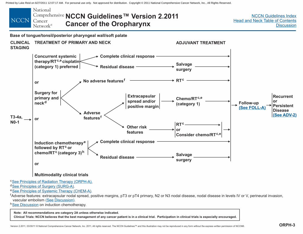
T3-4a,
N0-1
Salvage
surgeryResidual disease
Complete clinical response
Surgery for
primary and
neckd
CLINICAL
STAGING
TREATMENT OF PRIMARY AND NECK
No adverse featuresf
Concurrent systemic
therapy/RT cisplatin
(category 1) preferred
c,e
or
or
ADJUVANT TREATMENT
Induction chemotherapyfollowed by RT or
chemo/RT (category 3)
e
c
c h
Multimodality clinical trials
or
Salvage
surgeryResidual disease
Complete clinical response
RTc
Adverse
featuresf
Other risk
features
RTc
or
Consider chemo/RTc,e
Extracapsular
spread and/or
positive margin
Chemo/RTc,e
(category 1)
c
d
e
f
h
.
Adverse features: extracapsular nodal spread, positive margins, pT3 or pT4 primary, N2 or N3 nodal disease, nodal disease in levels IV or V, perineural invasion,vascular embolism .
on induction chemotherapy.
See Principles of Radiation Therapy (ORPH-A).
See Principles of Surgery (SURG-A).
See Principles of Systemic Therapy (CHEM-A)
See Discussion
See Discussion
( )
Follow-up(See FOLL-A)
RecurrentorPersistentDisease(See ADV-2)
Base of tongue/tonsil/posterior pharyngeal wall/soft palate
ORPH-3
Printed by Luke Reid on 6/27/2011 12:07:17 AM. For personal use only. Not approved for distribution. Copyright © 2011 National Comprehensive Cancer Network, Inc., All Rights Reserved.
Version 2.2011, 03/30/11 © National Comprehensive Cancer Network, Inc. 2011, All rights reserved. The NCCN Guidelines™ and this illustration may not be reproduced in any form without the express written permission of NCCN®.
NCCN Guidelines IndexHead and Neck Table of Contents
Discussion
Note: All recommendations are category 2A unless otherwise indicated.
Clinical Trials: NCCN believes that the best management of any cancer patient is in a clinical trial. Participation in clinical trials is especially encouraged.
NCCN Guidelines™ Version 2.2011Cancer of the Oropharynx
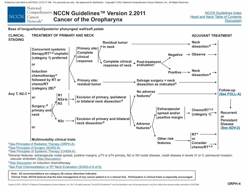
Any T, N2-3
Concurrent systemic
therapy/RT cisplatin
(category 1) preferred
c,e
CLINICAL
STAGING
TREATMENT OF PRIMARY AND NECK
or
N2c
Excision of primary, ipsilateral
or bilateral neck dissectiond
Excision of primary and bilateral
neck dissectiond
N1
N2a-b
N3Surgery:
primary and
neck
d
or
ADJUVANT TREATMENT
Induction
chemotherapy
followed by RT or
chemo/RT
(category 2B)
e
h
or
Multimodality clinical trials
Residual tumor
in neck
Complete clinicalresponse of neck
Primary site:
Complete
clinical
response
Primary site:residual tumor
Salvage surgery + neckdissection as indicatedd
Neck
dissectiond
Negative
Positive
Observe
Neck
dissectiond
No adverse
featuresf
Adverse
featuresf
Extracapsular
spread and/or
positive margin
Other risk
features
RTc
c,e
or
Consider
chemo/RT
c
d
e
f
h
i
.
Adverse features: extracapsular nodal spread, positive margins, pT3 or pT4 primary, N2 or N3 nodal disease, nodal disease in levels IV or V, perineural invasion,vascular embolism .
on induction chemotherapy.
See Principles of Radiation Therapy (ORPH-A).
See Principles of Surgery (SURG-A)
See Post Chemoradiation or RT Neck Evaluation (SURG-A 6 of 6)
.
.
See Principles of Systemic Therapy (CHEM-A)
See Discussion
See Discussion
( )
Chemo/RTc,e
(category 1)
Follow-up(See FOLL-A)
Post-treatmentevaluationi
RecurrentorPersistentDisease(See ADV-2)
Base of tongue/tonsil/posterior pharyngeal wall/soft palate
ORPH-4
Printed by Luke Reid on 6/27/2011 12:07:17 AM. For personal use only. Not approved for distribution. Copyright © 2011 National Comprehensive Cancer Network, Inc., All Rights Reserved.

Version 2.2011, 03/30/11 © National Comprehensive Cancer Network, Inc. 2011, All rights reserved. The NCCN Guidelines™ and this illustration may not be reproduced in any form without the express written permission of NCCN®.
NCCN Guidelines IndexHead and Neck Table of Contents
Discussion
Note: All recommendations are category 2A unless otherwise indicated.
Clinical Trials: NCCN believes that the best management of any cancer patient is in a clinical trial. Participation in clinical trials is especially encouraged.
NCCN Guidelines™ Version 2.2011Cancer of the Oropharynx
PRINCIPLES OF RADIATION THERAPY1
1
2
2
5
Based on published data, concurrent chemoradiation most commonly uses
conventional fractionation at 2.0 Gy per fraction to 70 Gy in 7 wks with singleagent cisplatin given every 3 wks at 100 mg/m x 3 doses. Other fraction sizes(eg, 1.8 Gy, conventional), multiagent chemotherapy, other dosing schedules ofcisplatin; altered fractionation with chemotherapy are efficacious, and there is noconsensus on the optimal approach. In general, the use of concurrentchemoradiation carries a high toxicity burden; altered fractionation or multiagentchemotherapy will likely further increase the toxicity burden. For anychemoradiation approach, close attention should be paid to published reports forthe specific chemotherapy agent, dose, and schedule of administration.Chemoradiation should be performed by an experienced team and shouldinclude substantial supportive care.
Bernier J, Cooper JS, Pajak TF, et al. Defining risk levels in locally advanced headand neck cancers: A comparative analysis of concurrent postoperative radiationplus chemotherapy trials of the EORTC (#22931) and RTOG (#9501). Head Neck2005;27:843-850.
�
3
4
Bernier J, Domenge C, Ozsahin M, et al. Postoperative irradiation with or withoutconcomitant chemotherapy for locally advanced head and neck cancer. N Engl JMed 2004;350:1945-1952.
Cooper JS, Pajak TF, Forastiere AA, et al. Postoperative concurrent radiotherapyand chemotherapy for high-risk squamous-cell carcinoma of the head and neck.N Engl J Med 2004;350:1937-1944.
See Radiation Techniques (RAD-A) and Discussion.
DEFINITIVERT
Concurrent chemoradiation
�
�
�
Conventional fractionation: 66-74 Gy
(2.0 Gy/fraction; daily Monday-Friday) in 7 weeks
Altered fractionation:6 fractions/week accelerated; 66-74 Gy to gross disease,
44-64 Gy to subclinical disease.Concomitant boost accelerated RT: 72 Gy/6 weeks
(1.8 Gy/fraction, large field; 1.5 Gy boost as second daily
fraction during last 12 treatment days)Hyperfractionation: 81.6 Gy/7 weeks
(1.2 Gy/fraction, twice daily)
Primary and gross adenopathy: 70 Gy (2.0 Gy/fraction)Neck
Uninvolved nodal stations: 44-64 Gy (1.6-2.0 Gy/fraction)
RT
Involved nodal stations: 60-66 Gy (2.0 Gy/fraction)Uninvolved nodal stations: 44-64 Gy (1.6-2.0 Gy/fraction)
Postoperative chemoradiation
�
�
�
�
�
�
POSTOPERATIVE
�
�
Conventional fractionation:
Preferred interval between resection and postoperative RT
is 6 weeks.
Primary: 60-66 Gy (2.0 Gy/fraction)
Neck
Concurrent single agent cisplatin at
100 mg/m every 3 wks x 3 doses is recommended.
2
2
�
�
�
�
�3-5
IMRT is a preferred technique for cancers of the oropharynx in order to minimize dose to critical structures.
ORPH-A
Printed by Luke Reid on 6/27/2011 12:07:17 AM. For personal use only. Not approved for distribution. Copyright © 2011 National Comprehensive Cancer Network, Inc., All Rights Reserved.

Version 2.2011, 03/30/11 © National Comprehensive Cancer Network, Inc. 2011, All rights reserved. The NCCN Guidelines™ and this illustration may not be reproduced in any form without the express written permission of NCCN®.
NCCN Guidelines IndexHead and Neck Table of Contents
Discussion
Note: All recommendations are category 2A unless otherwise indicated.
Clinical Trials: NCCN believes that the best management of any cancer patient is in a clinical trial. Participation in clinical trials is especially encouraged.
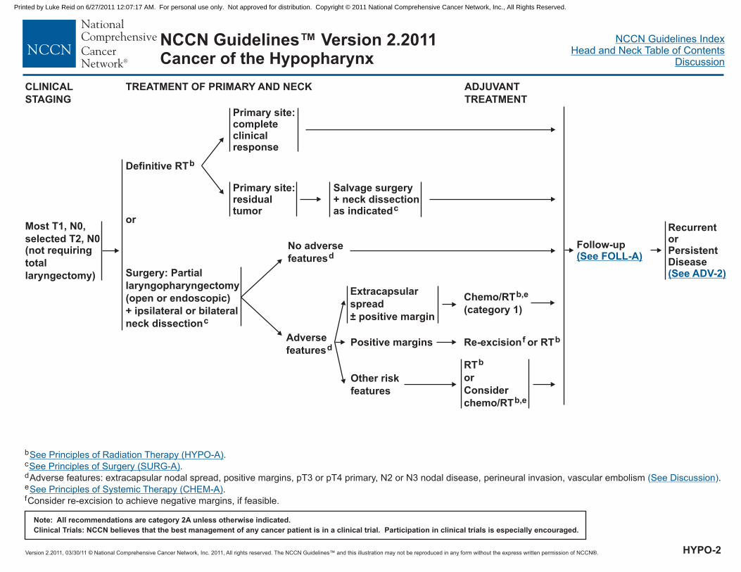
NCCN Guidelines™ Version 2.2011Cancer of the Hypopharynx
T1, N+;
T2-3, Any N
T4a, Any N
WORKUP CLINICAL STAGING
Advanced cancer requiring
total laryngectomy
�
�
�
�
�
�
�
�
�
�
H&P including a complete
head and neck exam; mirror
and fiberoptic examination as
clinically indicated
Biopsy
Chest imaging
CT with contrast and/or MRI
with contrast of primary and
neck
Consider PET-CT for stage
III-IV disease
Examination under
anesthesia with endoscopy
Preanesthesia studies
Nutrition, speech &
swallowing
evaluation/therapy and
audiogram as indicated
Dental evaluation
Consider videostrobe for
select patients
a
Multidisciplinary consultation
as indicated
See Treatment of Primary andNeck (HYPO-2)
See Treatment of Primary andNeck (HYPO-3)
See Treatment of Primary andNeck (HYPO-5)
See Treatment of VeryAdvanced Head and NeckCancer (ADV-1)
aAnatomical imaging is also recommended.
T4b, N any, or
unresectable nodal
disease
HYPO-1
Most T1, N0, selected T2, N0(not requiring total laryngectomy)
Printed by Luke Reid on 6/27/2011 12:07:17 AM. For personal use only. Not approved for distribution. Copyright © 2011 National Comprehensive Cancer Network, Inc., All Rights Reserved.
Version 2.2011, 03/30/11 © National Comprehensive Cancer Network, Inc. 2011, All rights reserved. The NCCN Guidelines™ and this illustration may not be reproduced in any form without the express written permission of NCCN®.
NCCN Guidelines IndexHead and Neck Table of Contents
Discussion
Note: All recommendations are category 2A unless otherwise indicated.
Clinical Trials: NCCN believes that the best management of any cancer patient is in a clinical trial. Participation in clinical trials is especially encouraged.
NCCN Guidelines™ Version 2.2011Cancer of the Hypopharynx
Primary site:completeclinicalresponse
Primary site:residualtumor
Salvage surgery+ neck dissectionas indicatedc
Most T1, N0,
selected T2, N0(not requiring
total
laryngectomy)
Definitive RTb
CLINICAL
STAGING
TREATMENT OF PRIMARY AND NECK
Surgery: Partial
laryngopharyngectomy
(open or endoscopic)
+ ipsilateral or bilateral
neck dissectionc
or
No adverse
featuresd
b .c
e
f
dAdverse features: extracapsular nodal spread, positive margins, pT3 or pT4 primary, N2 or N3 nodal disease, perineural invasion, vascular embolism .
.
Consider re-excision to achieve negative margins, if feasible.
See Principles of Radiation Therapy
See Discussion
See Principles of Systemic Therapy (CHEM-A)
(HYPO-A)
See Principles of Surgery (SURG-A).
( )
ADJUVANT
TREATMENT
Adverse
featuresd
Other risk
features
RTb
or
Consider
chemo/RTb,e
Follow-up(See FOLL-A)
Extracapsular
spread
positive margin±
Chemo/RTb,e
(category 1)
Positive margins Re-excision or RTbf
RecurrentorPersistentDisease(See ADV-2)
HYPO-2
Printed by Luke Reid on 6/27/2011 12:07:17 AM. For personal use only. Not approved for distribution. Copyright © 2011 National Comprehensive Cancer Network, Inc., All Rights Reserved.

Version 2.2011, 03/30/11 © National Comprehensive Cancer Network, Inc. 2011, All rights reserved. The NCCN Guidelines™ and this illustration may not be reproduced in any form without the express written permission of NCCN®.
NCCN Guidelines IndexHead and Neck Table of Contents
Discussion
Note: All recommendations are category 2A unless otherwise indicated.
Clinical Trials: NCCN believes that the best management of any cancer patient is in a clinical trial. Participation in clinical trials is especially encouraged.
NCCN Guidelines™ Version 2.2011Cancer of the Hypopharynx
Induction chemotherapye,g See Response After InductionChemotherapy (HYPO-4)
Selected T2, N0
(requiring
laryngectomy)T1, N+;
T2-3, any N
(if total
laryngectomy
required)
CLINICAL
STAGING
TREATMENT OF PRIMARY AND NECK ADJUVANT TREATMENT
Residual tumor
in neck
Completeclinicalresponseof neck
Primary site:
complete
clinical
response
Primary site:
residual
tumor
Salvage surgery + neckdissection as indicatedc
Neck dissectionc
Multimodality clinical trials
Laryngopharyngectomy
+ neck dissection,
including level VI
c
Concurrent systemic
therapy/RT (cisplatin
preferred)b,e
or
or
or
b .c
d
e
h
Adverse features: extracapsular nodal spread, positive margins, pT3 or pT4 primary, N2 or N3 nodal disease, perineural invasion, vascular embolism .
.gIn randomized clinical trials, assessment of response has been done after 2 or 3 cycles.
See Principles of Radiation Therapy
(See Discussion)
See Principles of Systemic Therapy (CHEM-A)
(HYPO-A)
See Principles of Surgery (SURG-A)
See Post Chemoradiation or RT Neck Evaluation (SURG-A 6 of 6)
.
.
No adverse
featuresd
Adverse
featuresd
Other risk
features
RTb
or
Consider chemo/RTb,e
Extracapsular
spread and/or
positive margin
Chemo/RTb,e (category 1)
Negative
Positive
Observe
Neck
dissectionc
Follow-up(See FOLL-A)
RecurrentorPersistentDisease(See ADV-2)
Post-treatmentevaluationh
HYPO-3
Printed by Luke Reid on 6/27/2011 12:07:17 AM. For personal use only. Not approved for distribution. Copyright © 2011 National Comprehensive Cancer Network, Inc., All Rights Reserved.

Version 2.2011, 03/30/11 © National Comprehensive Cancer Network, Inc. 2011, All rights reserved. The NCCN Guidelines™ and this illustration may not be reproduced in any form without the express written permission of NCCN®.
NCCN Guidelines IndexHead and Neck Table of Contents
Discussion
Note: All recommendations are category 2A unless otherwise indicated.
Clinical Trials: NCCN believes that the best management of any cancer patient is in a clinical trial. Participation in clinical trials is especially encouraged.
NCCN Guidelines™ Version 2.2011Cancer of the Hypopharynx
b
c
d
e
g
h
Adverse features: extracapsular nodal spread, positive margins, pT3 or pT4 primary, N2 or N3 nodal disease, perineural invasion, vascular embolism .
In randomized clinical trials, assessment of response has been done after 2 or 3 cycles.
See Principles of Radiation Therapy (HYPO-A).See Principles of Surgery (SURG-A)
See Post Chemoradiation or RT Neck Evaluation (SURG-A 6 of 6)
.
.
(See Discussion)
See Principles of Systemic Therapy (CHEM-A).
Response
after
induction
chemo-
therapye,g
Primary site:
Partial
response
(PR)
Primary site:
< Partial
response
Surgeryc
Definitive RTb
b,e
(category 1)
or
Consider
chemo/RT
(category 2B)
Residual
tumor in neck
Complete
clinical
response
of neck
Neck dissectionc
Primary site:
Complete
response
(CR)
Chemo/RTb,e
(category 2B)
CR Observe
Salvage
surgery
Residual
disease
Negative
Positive
Observe
Neck
dissectionc
No adverse
featuresd
Adverse
featuresdRTb
or
Consider chemo/RTb,e
Extracapsular
spread and/or
positive margin
Chemo/RTb,e (category 1)
RTb
Post-treatmentevaluationh
RESPONSE
ASSESSMENT
Other risk
features
Follow-up(See FOLL-A)
RecurrentorPersistentDisease(See ADV-2)
HYPO-4
Printed by Luke Reid on 6/27/2011 12:07:17 AM. For personal use only. Not approved for distribution. Copyright © 2011 National Comprehensive Cancer Network, Inc., All Rights Reserved.

Version 2.2011, 03/30/11 © National Comprehensive Cancer Network, Inc. 2011, All rights reserved. The NCCN Guidelines™ and this illustration may not be reproduced in any form without the express written permission of NCCN®.
NCCN Guidelines IndexHead and Neck Table of Contents
Discussion
Note: All recommendations are category 2A unless otherwise indicated.
Clinical Trials: NCCN believes that the best management of any cancer patient is in a clinical trial. Participation in clinical trials is especially encouraged.
NCCN Guidelines™ Version 2.2011Cancer of the Hypopharynx
Surgery + neck dissection
(preferred)
c
CLINICAL
STAGING
TREATMENT OF PRIMARY AND NECK
RTorChemo/RT
b
b,e
ADJUVANT TREATMENT
T4a,any N
Residual
tumor in neckPrimary site:
complete
clinical
response
Primary site:
residual tumorSalvage surgery + neckdissection as indicatedc
Neck dissectionc
Multimodality clinical trials
or
or
Concurrent systemic
therapy/RT
(category 3)b,e
Induction
chemo-
therapy
(category 3)
e,g
i
or
b .c
e .
See Principles of Radiation Therapy
See Principles of Systemic Therapy (CHEM-A)
(HYPO-A)
See Principles of Surgery (SURG-A).
Complete
clinical
response
of neck
Negative
Positive
Observe
Neck
dissectionc
Primary site:
CR or PR
and stable
disease in
neck
�
Primary site:
< PR or
progression
in neck
Salvage surgery + neckdissection as indicatedc
For CR:
For PR:
RT orconsiderchemo/RT;
Chemo/RT
b,e
b,e
Residual
tumor in
neck
Primary site:
clinical
complete
response
Primary site:
residual tumor
Salvage surgery + neckdissection as indicatedc
Neck dissectionc
Complete
clinical
response
of neck
Negative
Positive
Observe
Neck
dissectionc
RTorChemo/RT
b
b,e
RecurrentorPersistentDisease(See ADV-2)
Post-treatmentevaluationh
Follow-up(See FOLL-A)
Post-treatment
evaluationh
g
h
i
In randomized clinical trials, assessment of response has been done after2 or 3 cycles.
on induction chemotherapy.
See Post Chemoradiation or RT Neck Evaluation (SURG-A 6 of 6).
See Discussion
HYPO-5
Printed by Luke Reid on 6/27/2011 12:07:17 AM. For personal use only. Not approved for distribution. Copyright © 2011 National Comprehensive Cancer Network, Inc., All Rights Reserved.

Version 2.2011, 03/30/11 © National Comprehensive Cancer Network, Inc. 2011, All rights reserved. The NCCN Guidelines™ and this illustration may not be reproduced in any form without the express written permission of NCCN®.
NCCN Guidelines IndexHead and Neck Table of Contents
Discussion
Note: All recommendations are category 2A unless otherwise indicated.
Clinical Trials: NCCN believes that the best management of any cancer patient is in a clinical trial. Participation in clinical trials is especially encouraged.
NCCN Guidelines™ Version 2.2011Cancer of the Hypopharynx
RT
6 fractions/week accelerated; 66-74 Gy to gross disease, 44-64
Gy to subclinical disease.Concomitant boost accelerated RT:
72 Gy/6 weeks (1.8 Gy/fraction, large field; 1.5 Gy boost as
second daily fraction during last 12 treatment days)Hyperfractionation: 81.6 Gy/7 weeks (1.2 Gy/fraction, twice daily)
Uninvolved odal stations: 44-64 Gy (1.6-2.0 Gy/fraction)
Concurrent chemoradiation
Primary and gross adenopathy: 70 Gy (2.0 Gy/fraction)Neck
Uninvolved nodal stations: 44-64 Gy (1.6-2.0 Gy/fraction)
DEFINITIVE
�
�
�
Primary and gross adenopathy:Conventional fractionation: 66-74 Gy (2.0 Gy/fraction; dailyMonday-Friday) in 7 weeksAltered fractionation:�
�
�
�
Neckn
Conventional fractionation� 3
�
�
PRINCIPLES OF RADIATION THERAPY1,2
1
2.
Particular attention to speech and swallowing needs during therapy.
Based on published data, concurrent chemoradiation most commonly uses
conventional fractionation at 2.0 Gy per fraction to 70 Gy in 7 wks with singleagent cisplatin given every 3 wks at 100 mg/m x 3 doses. Other fraction sizes(eg, 1.8 Gy, conventional), multiagent chemotherapy, other dosing schedules ofcisplatin; altered fractionation with chemotherapy are efficacious, and there is noconsensus on the optimal approach. In general, the use of concurrentchemoradiation carries a high toxicity burden; altered fractionation or multiagentchemotherapy will likely further increase the toxicity burden. For anychemoradiation approach, close attention should be paid to published reports forthe specific chemotherapy agent, dose, and schedule of administration.Chemoradiation should be performed by an experienced team and should includesubstantial supportive care.
3
2�
See Radiation Techniques (RAD-A) and Discussion
POSTOPERATIVERT
Preferred interval between resection and postoperative RT
is 6 weeks.
Involved nodal stations: 60-66 Gy (2.0 Gy/fraction)Uninvolved nodal stations: 44-64 Gy (1.6-2.0 Gy/fraction)
Concurrent single agent cisplatin at 100 mg/m every 3 wks is
recommended.
�
�
��
�
Primary: 60-66 Gy (2.0 Gy/fraction)Neck�
�
Postoperative chemoradiation2
4-6
4
5
6
Bernier J, Domenge C, Ozsahin M, et al. Postoperative irradiation with or withoutconcomitant chemotherapy for locally advanced head and neck cancer. N Engl JMed 2004;350:1945-1952.
Cooper JS, Pajak TF, Forastiere AA, et al. Postoperative concurrent radiotherapyand chemotherapy for high-risk squamous-cell carcinoma of the head and neck.N Engl J Med 2004;350:1937-1944.
Bernier J, Cooper JS, Pajak TF, et al. Defining risk levels in locally advanced headand neck cancers: A comparative analysis of concurrent postoperative radiationplus chemotherapy trials of the EORTC (#22931) and RTOG (#9501). Head Neck2005;27:843-850.
HYPO-A
Printed by Luke Reid on 6/27/2011 12:07:17 AM. For personal use only. Not approved for distribution. Copyright © 2011 National Comprehensive Cancer Network, Inc., All Rights Reserved.

Version 2.2011, 03/30/11 © National Comprehensive Cancer Network, Inc. 2011, All rights reserved. The NCCN Guidelines™ and this illustration may not be reproduced in any form without the express written permission of NCCN®.
NCCN Guidelines IndexHead and Neck Table of Contents
Discussion
Note: All recommendations are category 2A unless otherwise indicated.
Clinical Trials: NCCN believes that the best management of any cancer patient is in a clinical trial. Participation in clinical trials is especially encouraged.
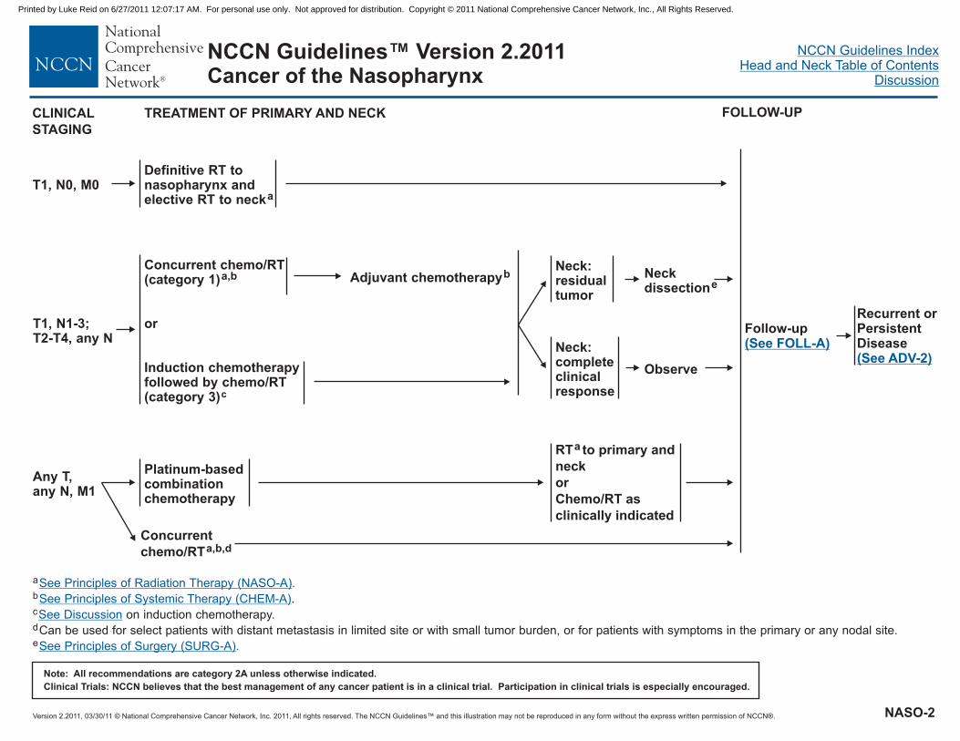
NCCN Guidelines™ Version 2.2011Cancer of the Nasopharynx
T1, N0, M0
T1, N1-3; T2-T4,Any N
Any T, Any N, M1
�
�
�
H&P
Nasopharyngeal exam and biopsy
including a complete head and neckexam; mirror and fiberoptic examinationas clinically indicated
Chest imaging
Consider PET-CT for stage III-IV disease
Dental evaluation as indicated
Nutrition, speech & swallowing
evaluation/therapy, and audiogram as
indicated
WHO class 2-3/N2-3
disease (may include PET scan and/or CT)
�
�
�
�
�
MRI with gadolinium of nasopharynx and
base of skull to clavicles and CT (as
indicated) with contrast
Imaging for distant metastases
(chest, liver, bone) for
Multidisciplinary consultation as indicated
WORKUP CLINICAL STAGING
See Treatment of Primaryand Neck (NASO-2)
See Treatment of Primaryand Neck (NASO-2)
See Treatment of Primaryand Neck (NASO-2)
NASO-1
Printed by Luke Reid on 6/27/2011 12:07:17 AM. For personal use only. Not approved for distribution. Copyright © 2011 National Comprehensive Cancer Network, Inc., All Rights Reserved.
Version 2.2011, 03/30/11 © National Comprehensive Cancer Network, Inc. 2011, All rights reserved. The NCCN Guidelines™ and this illustration may not be reproduced in any form without the express written permission of NCCN®.
NCCN Guidelines IndexHead and Neck Table of Contents
Discussion
Note: All recommendations are category 2A unless otherwise indicated.
Clinical Trials: NCCN believes that the best management of any cancer patient is in a clinical trial. Participation in clinical trials is especially encouraged.
NCCN Guidelines™ Version 2.2011Cancer of the Nasopharynx
T1, N0, M0Definitive RT tonasopharynx andelective RT to necka
CLINICAL
STAGING
TREATMENT OF PRIMARY AND NECK
Follow-up(See FOLL-A)
FOLLOW-UP
a
d
e
b
c
.
on induction chemotherapy.
Can be used for select patients with distant metastasis in limited site or with small tumor burden, or for patients with symptoms in the primary or any nodal site.
See Principles of Radiation Therapy
See Principles of Systemic Therapy (CHEM-A)
See Discussion
(NASO-A)
See Principles of Surgery (SURG-A)
.
.
Concurrent chemo/RT(category 1)
or
Induction chemotherapyfollowed by chemo/RT(category 3)
a,b
c
Neck:residualtumor
Neck:completeclinicalresponse
Neckdissectione
Adjuvant chemotherapyb
Platinum-basedcombinationchemotherapy
RT to primary and
neck
or
Chemo/RT as
clinically indicated
a
Any T,any N, M1
Observe
T1, N1-3;T2-T4, any N
Recurrent orPersistentDisease(See ADV-2)
Concurrent
chemo/RTa,b,d
NASO-2
Printed by Luke Reid on 6/27/2011 12:07:17 AM. For personal use only. Not approved for distribution. Copyright © 2011 National Comprehensive Cancer Network, Inc., All Rights Reserved.
Version 2.2011, 03/30/11 © National Comprehensive Cancer Network, Inc. 2011, All rights reserved. The NCCN Guidelines™ and this illustration may not be reproduced in any form without the express written permission of NCCN®.
NCCN Guidelines IndexHead and Neck Table of Contents
Discussion
Note: All recommendations are category 2A unless otherwise indicated.
Clinical Trials: NCCN believes that the best management of any cancer patient is in a clinical trial. Participation in clinical trials is especially encouraged.
NCCN Guidelines™ Version 2.2011Cancer of the Nasopharynx
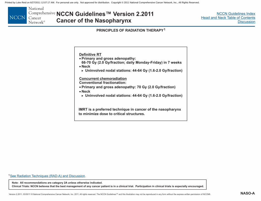
Definitive RT�
�
Primary and gross adenopathy:66-70 Gy (2.0 Gy/fraction; daily Monday-Friday) in 7 weeksNeck�
�
Uninvolved nodal stations: 44-64 Gy (1.6-2.0 Gy/fraction)
Conventional fractionation:Concurrent chemoradiation
�
�
Primary and gross adenopathy: 70 Gy (2.0 Gy/fraction)
NeckUninvolved nodal stations: 44-64 Gy (1.6-2.0 Gy/fraction)
PRINCIPLES OF RADIATION THERAPY1
1See Radiation Techniques (RAD-A) and Discussion.
IMRT is a preferred technique in cancer of the nasopharynx
to minimize dose to critical structures.
NASO-A
Printed by Luke Reid on 6/27/2011 12:07:17 AM. For personal use only. Not approved for distribution. Copyright © 2011 National Comprehensive Cancer Network, Inc., All Rights Reserved.

Version 2.2011, 03/30/11 © National Comprehensive Cancer Network, Inc. 2011, All rights reserved. The NCCN Guidelines™ and this illustration may not be reproduced in any form without the express written permission of NCCN®.
NCCN Guidelines IndexHead and Neck Table of Contents
Discussion
Note: All recommendations are category 2A unless otherwise indicated.
Clinical Trials: NCCN believes that the best management of any cancer patient is in a clinical trial. Participation in clinical trials is especially encouraged.
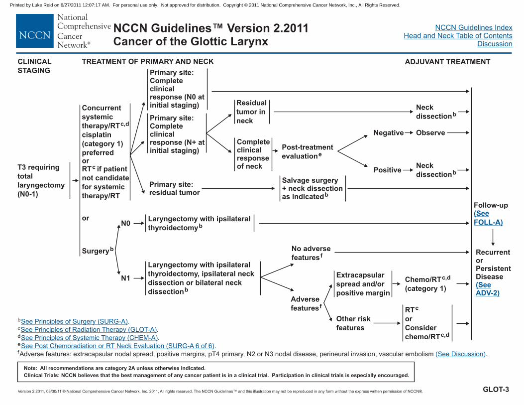
NCCN Guidelines™ Version 2.2011Cancer of the Glottic Larynx
WORKUPa
Total laryngectomy
not required
T3 requiring total
laryngectomy
(N0-1)
Carcinoma in situ
T4a disease
�
�
�
�
�
�
�
�
�
H&P
Biopsy
Chest imaging
CT with contrast and thin cuts through
larynx and/or MRI of primary and neck
Consider PET-CT for stage III-IV disease
Examination under anesthesia with
endoscopy
Preanesthesia studies
Dental/evaluation as indicated
Multidisciplinary consultation as indicated
including a complete head and neck
exam; mirror and fiberoptic examination as
clinically indicated
Nutrition, speech & swallowing
evaluation/therapy and audiogram as
indicated
Consider videostrobe for select patients�
CLINICAL STAGING TREATMENT OF PRIMARY AND NECK
See Treatment (GLOT-2)
See Treatment (GLOT-2)
See Treatment of Primary and Neck(GLOT-6)
aComplete workup not indicated for Tis, T1.
See Treatment of Primary and Neck(GLOT-3)
See Treatment of Very AdvancedHead and Neck Cancer (ADV-1)
T3 requiring total
laryngectomy
(N2-3)
See Treatment of Primary and Neck(GLOT-4)
T4b, N any, or
unresectable nodal
disease
GLOT-1
Printed by Luke Reid on 6/27/2011 12:07:17 AM. For personal use only. Not approved for distribution. Copyright © 2011 National Comprehensive Cancer Network, Inc., All Rights Reserved.

Version 2.2011, 03/30/11 © National Comprehensive Cancer Network, Inc. 2011, All rights reserved. The NCCN Guidelines™ and this illustration may not be reproduced in any form without the express written permission of NCCN®.
NCCN Guidelines IndexHead and Neck Table of Contents
Discussion
Note: All recommendations are category 2A unless otherwise indicated.
Clinical Trials: NCCN believes that the best management of any cancer patient is in a clinical trial. Participation in clinical trials is especially encouraged.
NCCN Guidelines™ Version 2.2011Cancer of the Glottic Larynx
CLINICAL STAGING TREATMENT OF PRIMARY AND NECK
N0 Observe
Total laryngectomy
not required
Carcinoma in situ
Clinical trialorEndoscopic resectionorRTc
RTorPartial laryngectomy/endoscopic or openresection as indicated
c
b
FOLLOW-UP
Follow-up(See FOLL-A)
b
c .
See Principles of Surgery (SURG-A)
See Principles of Radiation Therapy
.
(GLOT-A)
RecurrentorPersistentDisease(See ADV-2)
GLOT-2
Printed by Luke Reid on 6/27/2011 12:07:17 AM. For personal use only. Not approved for distribution. Copyright © 2011 National Comprehensive Cancer Network, Inc., All Rights Reserved.
Version 2.2011, 03/30/11 © National Comprehensive Cancer Network, Inc. 2011, All rights reserved. The NCCN Guidelines™ and this illustration may not be reproduced in any form without the express written permission of NCCN®.
NCCN Guidelines IndexHead and Neck Table of Contents
Discussion
Note: All recommendations are category 2A unless otherwise indicated.
Clinical Trials: NCCN believes that the best management of any cancer patient is in a clinical trial. Participation in clinical trials is especially encouraged.
NCCN Guidelines™ Version 2.2011Cancer of the Glottic Larynx
Residual
tumor in
neck
Completeclinicalresponseof neck
Primary site:residual tumor
Salvage surgery+ neck dissectionas indicatedb
Neck
dissectionb
T3 requiring
total
laryngectomy
(N0-1)
CLINICAL
STAGING
TREATMENT OF PRIMARY AND NECK
Surgeryb
Laryngectomy with ipsilateral
thyroidectomyb
N1
N0
Laryngectomy with ipsilateral
thyroidectomy, ipsilateral neck
dissection bilateral neck
dissection
orb
b
d
e
f
c .
.
Adverse features: extracapsular nodal spread, positive margins, pT4 primary, N2 or N3 nodal disease, perineural invasion, vascular embolism .
See Principles of Surgery (SURG-A)
See Principles of Radiation Therapy
See Principles of Systemic Therapy (CHEM-A)
See Discussion
.
( )
(GLOT-A)
See Post Chemoradiation or RT Neck Evaluation (SURG-A 6 of 6).
ADJUVANT TREATMENT
or
Concurrent
systemic
therapy/RT
cisplatin
(category 1)
preferred
c,d
orRT if patient
not candidate
for systemic
therapy/RT
c
Follow-up(See
FOLL-A)
Negative
Positive
Observe
Neck
dissectionb
No adverse
featuresf
Adverse
featuresf
Other risk
features
RTc
or
Consider
chemo/RTc,d
Extracapsular
spread and/or
positive margin
Chemo/RTc,d
(category 1)
Primary site:Completeclinicalresponse (N+ atinitial staging)
Primary site:Completeclinicalresponse (N0 atinitial staging)
RecurrentorPersistentDisease(SeeADV-2)
Post-treatment
evaluatione
GLOT-3
Printed by Luke Reid on 6/27/2011 12:07:17 AM. For personal use only. Not approved for distribution. Copyright © 2011 National Comprehensive Cancer Network, Inc., All Rights Reserved.
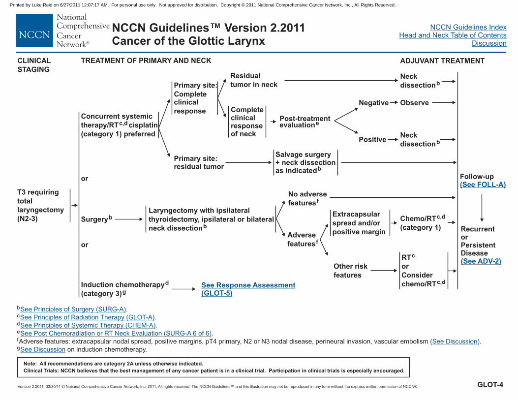
Version 2.2011, 03/30/11 © National Comprehensive Cancer Network, Inc. 2011, All rights reserved. The NCCN Guidelines™ and this illustration may not be reproduced in any form without the express written permission of NCCN®.
NCCN Guidelines IndexHead and Neck Table of Contents
Discussion
Note: All recommendations are category 2A unless otherwise indicated.
Clinical Trials: NCCN believes that the best management of any cancer patient is in a clinical trial. Participation in clinical trials is especially encouraged.
NCCN Guidelines™ Version 2.2011Cancer of the Glottic Larynx
Residual
tumor in neck
Completeclinicalresponseof neck
Primary site:
Completeclinical
response
Primary site:residual tumor
Salvage surgery+ neck dissectionas indicatedb
Neck
dissectionb
T3 requiring
total
laryngectomy
(N2-3) SurgerybLaryngectomy with ipsilateral
thyroidectomy, ipsilateral or bilateral
neck dissectionb
or
Concurrent systemic
therapy/RT cisplatin
(category 1) preferred
c,d
Negative
Positive
Observe
Neck
dissectionb
CLINICAL
STAGING
TREATMENT OF PRIMARY AND NECK ADJUVANT TREATMENT
or
No adverse
featuresf
Adverse
featuresf
Other risk
features
RTc
or
Consider
chemo/RTc,d
Extracapsular
spread and/or
positive margin
Chemo/RTc,d
(category 1)
Induction chemotherapy
(category 3)
d
gSee Response Assessment(GLOT-5)
b
d
e
f
g
c .
.
Adverse features: extracapsular nodal spread, positive margins, pT4 primary, N2 or N3 nodal disease, perineural invasion, vascular embolism .
on induction chemotherapy.
See Principles of Surgery (SURG-A)
See Principles of Radiation Therapy
See Principles of Systemic Therapy (CHEM-A)
See Discussion
See Discussion
.
( )
(GLOT-A)
See Post Chemoradiation or RT Neck Evaluation (SURG-A 6 of 6).
RecurrentorPersistentDisease(See ADV-2)
Follow-up(See FOLL-A)
Post-treatmentevaluatione
GLOT-4
Printed by Luke Reid on 6/27/2011 12:07:17 AM. For personal use only. Not approved for distribution. Copyright © 2011 National Comprehensive Cancer Network, Inc., All Rights Reserved.

Version 2.2011, 03/30/11 © National Comprehensive Cancer Network, Inc. 2011, All rights reserved. The NCCN Guidelines™ and this illustration may not be reproduced in any form without the express written permission of NCCN®.
NCCN Guidelines IndexHead and Neck Table of Contents
Discussion
Note: All recommendations are category 2A unless otherwise indicated.
Clinical Trials: NCCN believes that the best management of any cancer patient is in a clinical trial. Participation in clinical trials is especially encouraged.
NCCN Guidelines™ Version 2.2011Cancer of the Glottic Larynx
Response
after
induction
chemo-
therapyd,h
b
c
d
e
f
h
Adverse features: extracapsular nodal spread, positive margins, pT4 primary, N2 or N3 nodal disease, perineural invasion, vascular embolism .
In randomized clinical trials, assessment of response has been done after 2 or 3 cycles.
See Principles of Surgery (SURG-A)
See Principles of Systemic Therapy (CHEM-A)
See Discussion
.
.
( )
See Post Chemoradiation or RT Neck Evaluation (SURG-A 6 of 6).
See Principles of Radiation Therapy (GLOT-A).
Primary site:
Partial
response
(PR)
Primary site:
< Partial
response
Surgeryb
Definitive
RTc
c,d
(category 1)
or
Consider
chemo/RT
(category 2B)
Residual
tumor in neck
Complete
clinical
response
of neck
Neck dissectionb
Primary site:
Complete
response
(CR)
Chemo/RTc,d
(category 2B)
CR Observe
Salvage
surgery
Residual
disease
Negative
Positive
Observe
Neck
dissectionb
No adverse
featuresf
Adverse
featuresfRTc
or
Consider chemo/RTc,d
Extracapsular
spread and/or
positive margin
Chemo/RTc,d (category 1)
RTc
Follow-up(See FOLL-A)
RecurrentorPersistentDisease(See ADV-2)
Post-treatmentevaluatione
RESPONSE
ASSESSMENT
Other risk
features
GLOT-5
Printed by Luke Reid on 6/27/2011 12:07:17 AM. For personal use only. Not approved for distribution. Copyright © 2011 National Comprehensive Cancer Network, Inc., All Rights Reserved.

Version 2.2011, 03/30/11 © National Comprehensive Cancer Network, Inc. 2011, All rights reserved. The NCCN Guidelines™ and this illustration may not be reproduced in any form without the express written permission of NCCN®.
NCCN Guidelines IndexHead and Neck Table of Contents
Discussion
Note: All recommendations are category 2A unless otherwise indicated.
Clinical Trials: NCCN believes that the best management of any cancer patient is in a clinical trial. Participation in clinical trials is especially encouraged.
NCCN Guidelines™ Version 2.2011Cancer of the Glottic Larynx
CLINICAL
STAGING
TREATMENT OF PRIMARY AND NECK
Selected T4apatients whodeclinesurgery
Consider concurrentchemoradiationc,d
T4a, Any N
ADJUVANT TREATMENT
Chemo/RT(category 1)
c,d
Laryngectomy with ipsilateral thyroidectomy
unilateral or bilateral neck dissection± b
N2-3
N1
N0
Laryngectomy with ipsilateral thyroidectomy,
ipsilateral or bilateral neck dissectionb
Laryngectomy with ipsilateral thyroidectomy,
ipsilateral neck dissection
± contralateral neck dissectionb
or
Induction chemotherapy
followed by chemo/RT
(category 2B)
d
c,d
g
Residual
tumor in neck
Completeclinicalresponseof neck
Primary site:
Complete
clinical
response
Primary site:residual tumor
Salvage surgery + neckdissection as indicatedb
Neck
dissectionb
Post-treatmentevaluatione
Negative
Positive
Observe
Neck
dissectionb
Surgeryb
See Response Assessment(GLOT-5)
b
d
e
g
c .
.
on induction chemotherapy.
.
See Principles of Surgery (SURG-A)
See Principles of Radiation Therapy
See Principles of Systemic Therapy (CHEM-A)
See Discussion
.
(GLOT-A)
See Post Chemoradiation or RT Neck Evaluation (SURG-A 6 of 6)
Follow-up(See FOLL-A)
RecurrentorPersistentDisease(See ADV-2)
Clinical trial forfunction preservingsurgical or nonsurgicalmanagement
or
GLOT-6
Printed by Luke Reid on 6/27/2011 12:07:17 AM. For personal use only. Not approved for distribution. Copyright © 2011 National Comprehensive Cancer Network, Inc., All Rights Reserved.

Version 2.2011, 03/30/11 © National Comprehensive Cancer Network, Inc. 2011, All rights reserved. The NCCN Guidelines™ and this illustration may not be reproduced in any form without the express written permission of NCCN®.
NCCN Guidelines IndexHead and Neck Table of Contents
Discussion
Note: All recommendations are category 2A unless otherwise indicated.
Clinical Trials: NCCN believes that the best management of any cancer patient is in a clinical trial. Participation in clinical trials is especially encouraged.
NCCN Guidelines™ Version 2.2011Cancer of the Glottic Larynx
RT
dailyMonday-Friday
Concomitant boost accelerated RT: 72 Gy/6 weeks
(1.8 Gy/fraction, large field; 1.5 Gy boost as second daily fraction
during last 12 treatment days)Hyperfractionation: 79.2-81.6 Gy/7 weeks
(1.2 Gy/fraction, twice daily)
Uninvolved nodal stations:
Concurrent chemoradiation
Conventional fractionation:
Primary and gross adenopathy: 70 Gy (2.0 Gy/fraction)Neck
Uninvolved nodal stations: 44-64 Gy (1.6-2.0 Gy/fraction)
DEFINITIVE
���
�
�
T1, N0: 63-66 Gy in 2.25-2.0 Gy/fractionT1-2: > 66 Gy using conventional fractionation (2.0 Gy/fraction)
T2 and gross adenopathy:Conventional fractionation: 66-74 Gy (2.0 Gy/fraction;
) in 7 weeksAltered fractionation:
�
�
�
� 44-64 Gy (1.6-2.0 Gy/fraction)
2
�
�
�
PRINCIPLES OF RADIATION THERAPY1
1
2Based on published data, concurrent chemoradiation most commonly uses conventional fractionation at 2.0 Gy per fraction to 70 Gy in 7 wks with single agentcisplatin given every 3 wks at 100 mg/m x 3 doses. Other fraction sizes (eg, 1.8 Gy, conventional), multiagent chemotherapy, other dosing schedules of cisplatin, oraltered fractionation with chemotherapy are efficacious, and there is no consensus on the optimal approach. In general, the use of concurrent chemoradiation carries ahigh toxicity burden; altered fractionation or multiagent chemotherapy will likely further increase the toxicity burden. For any chemoradiation approach, close attentionshould be paid to published reports for the specific chemotherapy agent, dose, and schedule of administration. Chemoradiation should be performed by an experiencedteam and should include substantial supportive care.
Bernier J, Cooper JS, Pajuk TF, et al. Defining risk levels in locally advanced head and neck cancers: A comparative analysis of concurrent postoperative radiation pluschemotherapy trials of the EORTC (#22931) and RTOG (#9501). Head Neck 2005;27:843-850.
�2
5
3
4
Bernier J, Domenge C, Ozsahin M, et al. Postoperative irradiation with or without concomitant chemotherapy for locally advanced head and neck cancer. N Engl J Med2004;350:1945-1952.
Cooper JS, Pajak TF, Forastiere AA, et al. Postoperative concurrent radiotherapy and chemotherapy for high-risk squamous-cell carcinoma of the head and neck.N Engl J Med 2004;350:1937-1944.
See Radiation Techniques (RAD-A) and Discussion.
POSTOPERATIVERT
Preferred interval between resection and postoperative RT
is 6 weeks.
Involved nodal stations: 60-66 Gy (2.0 Gy/fraction)Uninvolved nodal stations: 44-64 Gy (1.6-2.0 Gy/fraction)
Concurrent single agent cisplatin at 100 mg/m every 3 wks is
recommended.
�
�
��
�
Primary: 60-66 Gy (2.0 Gy/fraction)Neck�
�
Postoperative chemoradiation2
3-5
GLOT-A
Printed by Luke Reid on 6/27/2011 12:07:17 AM. For personal use only. Not approved for distribution. Copyright © 2011 National Comprehensive Cancer Network, Inc., All Rights Reserved.

Version 2.2011, 03/30/11 © National Comprehensive Cancer Network, Inc. 2011, All rights reserved. The NCCN Guidelines™ and this illustration may not be reproduced in any form without the express written permission of NCCN®.
NCCN Guidelines IndexHead and Neck Table of Contents
Discussion
Note: All recommendations are category 2A unless otherwise indicated.
Clinical Trials: NCCN believes that the best management of any cancer patient is in a clinical trial. Participation in clinical trials is especially encouraged.
NCCN Guidelines™ Version 2.2011Cancer of the Supraglottic Larynx
WORKUP CLINICAL STAGING
�
���
����
�
�
H&P
BiopsyChest imaging
Examination under anesthesia with endoscopyPreanesthesia studies
including a complete head and neck exam;mirror and fiberoptic examination as clinicallyindicated
CT with contrast and thin cuts through larynxand/or MRI of primary and neckConsider PET-CT for stage III-IV disease
Dental evaluation as indicated
Nutrition, speech & swallowing
evaluation/therapy, and audiogram as indicated
Consider videostrobe for select patients
Multidisciplinary consultation as indicated
Not requiring total
laryngectomy
(Most T1-2, N0)
See Treatment of Primaryand Neck (SUPRA-2)
Requiring total
laryngectomy
(T3, N0)
See Treatment of Primaryand Neck (SUPRA-3)
T4a, N0 See Treatment of Primaryand Neck (SUPRA-8)
Node positive disease See Clinical Staging(SUPRA-4)
See Treatment of VeryAdvanced Head and NeckCancer (ADV-1)
T4b, N any, or
unresectable nodal
disease
SUPRA-1
Printed by Luke Reid on 6/27/2011 12:07:17 AM. For personal use only. Not approved for distribution. Copyright © 2011 National Comprehensive Cancer Network, Inc., All Rights Reserved.

Version 2.2011, 03/30/11 © National Comprehensive Cancer Network, Inc. 2011, All rights reserved. The NCCN Guidelines™ and this illustration may not be reproduced in any form without the express written permission of NCCN®.
NCCN Guidelines IndexHead and Neck Table of Contents
Discussion
Note: All recommendations are category 2A unless otherwise indicated.
Clinical Trials: NCCN believes that the best management of any cancer patient is in a clinical trial. Participation in clinical trials is especially encouraged.
NCCN Guidelines™ Version 2.2011Cancer of the Supraglottic Larynx
TREATMENT OF
PRIMARY AND NECK
One positive node
without other
adverse features
Consider RTb
Chemo/RT
(category 1)
or
RT (category 2B
for select
patients)
b,d
b
CLINICAL STAGING
Not requiring total
laryngectomy
(Most T1-2, N0)
Endoscopic resection
± neck dissection
or
Open partial supraglottic
laryngectomy
± neck dissection
or
Definitive RT
a
a
b
Adverse features:extracapsular nodal
spread
a
c
d
b .
Consider re-excision to achieve negative margins, if feasible.
.
See Principles of Surgery (SURG-A).
See Principles of Radiation Therapy
See Principles of Systemic Therapy (CHEM-A)
(SUPRA-A)
Follow-up(See FOLL-A)
FOLLOW-UPADJUVANT
TREATMENT
Positive node;
Adverse features:positive margins
Re-excision
or
RT
c
b
b,d
or
Consider
chemo/RT
(category 2B)
Node negative,
(T1-T2, N0)
PATHOLOGY
STAGE
Node negative,
(T3-T4a, N0)
See Surgical
Treatment (SUPRA-3)
and (SUPRA-8)
RecurrentorPersistentDisease(See ADV-2)
SUPRA-2
Printed by Luke Reid on 6/27/2011 12:07:17 AM. For personal use only. Not approved for distribution. Copyright © 2011 National Comprehensive Cancer Network, Inc., All Rights Reserved.

Version 2.2011, 03/30/11 © National Comprehensive Cancer Network, Inc. 2011, All rights reserved. The NCCN Guidelines™ and this illustration may not be reproduced in any form without the express written permission of NCCN®.
NCCN Guidelines IndexHead and Neck Table of Contents
Discussion
Note: All recommendations are category 2A unless otherwise indicated.
Clinical Trials: NCCN believes that the best management of any cancer patient is in a clinical trial. Participation in clinical trials is especially encouraged.
NCCN Guidelines™ Version 2.2011Cancer of the Supraglottic Larynx
TREATMENT OF PRIMARY AND NECKCLINICAL STAGING
N0 or one positive node
without adverse featuresf RT optionalb
Laryngectomy,ipsilateralthyroidectomywith ipsilateral orbilateral neckdissectiona
Requiring total
laryngectomy
(T3, N0)
Primary site:
Complete
clinical
response
Primary site:residualtumor
Salvage surgery+ neck dissectionas indicateda
Concurrent systemictherapy/RTcisplatin (category 1)preferred
b,d
or
a
d
b .
.
See Principles of Surgery (SURG-A)
See Principles of Radiation Therapy
See Principles of Systemic Therapy (CHEM-A)
.
(SUPRA-A)
ADJUVANT TREATMENT
Adverse
featuresf
RT if patient not medical
candidate for concurrent
systemic therapy/RT
b
Other risk
features
RTb
or
Consider
chemo/RTb,d
Extracapsular
spread and/or
positive margin
Chemo/RTb,d
(category 1)
or
Follow-up(See FOLL-A)
Induction chemotherapy
(category 3)
d
e
or
See Response Assessment
(SUPRA-7)
RecurrentorPersistentDisease(See ADV-2)
e
fon induction chemotherapy.
Adverse features: extracapsular nodal spread, positive margins, pT4 primary, N2 orN3 nodal disease, perineural invasion, vascular embolism .
See Discussion
See Discussion( )
SUPRA-3
Printed by Luke Reid on 6/27/2011 12:07:17 AM. For personal use only. Not approved for distribution. Copyright © 2011 National Comprehensive Cancer Network, Inc., All Rights Reserved.

Version 2.2011, 03/30/11 © National Comprehensive Cancer Network, Inc. 2011, All rights reserved. The NCCN Guidelines™ and this illustration may not be reproduced in any form without the express written permission of NCCN®.
NCCN Guidelines IndexHead and Neck Table of Contents
Discussion
Note: All recommendations are category 2A unless otherwise indicated.
Clinical Trials: NCCN believes that the best management of any cancer patient is in a clinical trial. Participation in clinical trials is especially encouraged.
NCCN Guidelines™ Version 2.2011Cancer of the Supraglottic Larynx
CLINICAL STAGING
Not requiring total
laryngectomy(T1-2, N+ and selected
T3, N1)
See Treatment of Primaryand Neck (SUPRA-5)
Requiring total
laryngectomy
(Most T3, N2-3)
See Treatment of Primaryand Neck (SUPRA-6)
T4a, N1-N3 See Treatment of Primaryand Neck (SUPRA-8)
See Treatment of Head andNeck Cancer (ADV-1)
Node positive
disease
T4b, N any, or
unresectable nodal
disease
SUPRA-4
Printed by Luke Reid on 6/27/2011 12:07:17 AM. For personal use only. Not approved for distribution. Copyright © 2011 National Comprehensive Cancer Network, Inc., All Rights Reserved.

Version 2.2011, 03/30/11 © National Comprehensive Cancer Network, Inc. 2011, All rights reserved. The NCCN Guidelines™ and this illustration may not be reproduced in any form without the express written permission of NCCN®.
NCCN Guidelines IndexHead and Neck Table of Contents
Discussion
Note: All recommendations are category 2A unless otherwise indicated.
Clinical Trials: NCCN believes that the best management of any cancer patient is in a clinical trial. Participation in clinical trials is especially encouraged.
NCCN Guidelines™ Version 2.2011Cancer of the Supraglottic Larynx
Not requiring
total
laryngectomy(T1-2, N+ and
selected
T3, N1)
TREATMENT OF PRIMARY AND NECKCLINICAL
STAGING
Observe
or RTb
Partial supraglotticlaryngectomy andneck dissection(s)a
or
Definitive RTb
Primary site:
Complete
clinical
response
Primary site:residualtumor
Salvage surgery+ neck dissectionas indicateda
Concurrent systemictherapy/RT, cisplatin(category 1)preferredb,d
or
RecurrentorPersistentDisease(See ADV-2)
ADJUVANT TREATMENT
No adverse
featuresf
Adverse
featuresf
Other risk
features
RTb
or
Consider chemo/RTb,d
Extracapsular
spread and/or
positive margin
Chemo/RTb,d
(category 1)
Residual
tumor in neck
Completeclinicalresponseof neck
Neck
dissectiona
Negative
Positive
Observe
Neck
dissectiona
a
b .d
e.
on induction chemotherapy.
See Principles of Surgery (SURG-A)
See Principles of Radiation Therapy
See Principles of Systemic Therapy (CHEM-A)
See Discussion
.
(SUPRA-A)
Induction
chemotherapy
(category 3)
d
e
or
Follow-up(See FOLL-A)
Post-treatmentevaluationg
See ResponseAssessment(SUPRA-7)
f
g
Adverse features: extracapsular nodal spread, positive margins, pT4 primary, N2 orN3 nodal disease, perineural invasion, vascular embolism .( )See Discussion
See Post Chemoradiation or RT Neck Evaluation (SURG-A 6 of 6).
SUPRA-5
Printed by Luke Reid on 6/27/2011 12:07:17 AM. For personal use only. Not approved for distribution. Copyright © 2011 National Comprehensive Cancer Network, Inc., All Rights Reserved.

Version 2.2011, 03/30/11 © National Comprehensive Cancer Network, Inc. 2011, All rights reserved. The NCCN Guidelines™ and this illustration may not be reproduced in any form without the express written permission of NCCN®.
NCCN Guidelines IndexHead and Neck Table of Contents
Discussion
Note: All recommendations are category 2A unless otherwise indicated.
Clinical Trials: NCCN believes that the best management of any cancer patient is in a clinical trial. Participation in clinical trials is especially encouraged.
NCCN Guidelines™ Version 2.2011Cancer of the Supraglottic Larynx
Post-treatmentevaluationg
Requiring total
laryngectomy
(Most T3, N2-N3)
TREATMENT OF PRIMARY AND NECKCLINICAL STAGING
Residual
tumor in neck
Completeclinicalresponseof neck
Primary site:
Complete
clinical
response
Primary site:residualtumor
Neck
dissectiona
Concurrent systemictherapy/RTcisplatin (category 1)preferred
b,d
Salvage surgery+ neck dissectionas indicateda
ADJUVANT TREATMENT
Induction chemotherapyfollowed by chemo/RT(category 2B)
d
e
or
Laryngectomy,ipsilateralthyroidectomy withneck dissectiona
or
RTbNo adverse
featuresf
Adverse
featuresf
RecurrentorPersistentDisease(See ADV-2)
Other risk
features
RTb
or
Consider chemo/RTb,d
Extracapsular
spread and/or
positive margin
Chemo/RTb,d (category 1)
Negative
Positive
Observe
Neck
dissectiona
See Response Assessment (SUPRA-7)
Follow-up(See FOLL-A)
a
b .d
e
f
g
.
on induction chemotherapy.
Adverse features: extracapsular nodal spread, positive margins, pT4 primary, N2 or N3 nodal disease, perineural invasion, vascular embolism .
See Principles of Surgery (SURG-A)
See Principles of Radiation Therapy
See Principles of Systemic Therapy (CHEM-A)
See Discussion
See Discussion
.
( )
(SUPRA-A)
See Post Chemoradiation or RT Neck Evaluation (SURG-A 6 of 6).
SUPRA-6
Printed by Luke Reid on 6/27/2011 12:07:17 AM. For personal use only. Not approved for distribution. Copyright © 2011 National Comprehensive Cancer Network, Inc., All Rights Reserved.

Version 2.2011, 03/30/11 © National Comprehensive Cancer Network, Inc. 2011, All rights reserved. The NCCN Guidelines™ and this illustration may not be reproduced in any form without the express written permission of NCCN®.
NCCN Guidelines IndexHead and Neck Table of Contents
Discussion
Note: All recommendations are category 2A unless otherwise indicated.
Clinical Trials: NCCN believes that the best management of any cancer patient is in a clinical trial. Participation in clinical trials is especially encouraged.
NCCN Guidelines™ Version 2.2011Cancer of the Supraglottic Larynx
Response after
induction
chemo-
therapyd,h
Primary site:
Partial
response
(PR)
Primary site:
< Partial
response
Surgerya
Definitive RTb
b,d
(category 1)
or Consider
chemo/RT
(category 2B)
Residual
tumor in neck
Complete
clinical
response
of neck
Neck dissectiona
Primary site:
Complete
response
(CR)
Chemo/RTb,d
(category 2B)
CR Observe
Salvage
surgery
Residual
disease
Negative
Positive
Observe
Neck
dissectiona
No adverse
featuresf
Adverse
featuresf
Other risk
features
RTb
b,dor
Consider chemo/RT
Extracapsular
spread and/or
positive margin
Chemo/RTb,d (category 1)
RTb
Follow-up(See FOLL-A)
RecurrentorPersistentDisease(See ADV-2)
Post-treatmentevaluationg
RESPONSE
ASSESSMENT
a
b
d
f
g
h
Adverse features: extracapsular nodal spread, positive margins, pT4 primary, N2 or N3 nodal disease, perineural invasion, vascular embolism .
In randomized clinical trials, assessment of response has been done after 2 or 3 cycles.
See Principles of Surgery (SURG-A)
See Principles of Systemic Therapy (CHEM-A)
See Discussion
.
.
( )
See Post Chemoradiation or RT Neck Evaluation (SURG-A 6 of 6).
See Principles of Radiation Therapy (SUPRA-A).
SUPRA-7
Printed by Luke Reid on 6/27/2011 12:07:17 AM. For personal use only. Not approved for distribution. Copyright © 2011 National Comprehensive Cancer Network, Inc., All Rights Reserved.

Version 2.2011, 03/30/11 © National Comprehensive Cancer Network, Inc. 2011, All rights reserved. The NCCN Guidelines™ and this illustration may not be reproduced in any form without the express written permission of NCCN®.
NCCN Guidelines IndexHead and Neck Table of Contents
Discussion
Note: All recommendations are category 2A unless otherwise indicated.
Clinical Trials: NCCN believes that the best management of any cancer patient is in a clinical trial. Participation in clinical trials is especially encouraged.
NCCN Guidelines™ Version 2.2011Cancer of the Supraglottic Larynx
Laryngectomy, ipsilateralthyroidectomy withipsilateral or bilateralneck dissectiona
T4a, N0-N3
TREATMENT OF PRIMARY AND NECKCLINICAL
STAGING
ADJUVANT
TREATMENT
RecurrentorPersistentDisease(See ADV-2)
Follow-up(See FOLL-A)
T4a, N0-N3
patients who
decline surgery
Consider concurrentchemoradiation
or
Clinical trial
b,d
Induction chemotherapy
followed by chemo/RT
(category 2B)
d
b,d
e
Residual
tumor in neck
Completeclinicalresponseof neck
Primary site:
Complete
clinical
response
Primary site:residual tumor
Salvage surgery + neckdissection as indicateda
Neck
dissectiona
Post-treatmentevaluationg
Negative
Positive
Observe
Neck
dissectiona
See ResponseAssessment (SUPRA-7)
or
a
e
b
d
f
g
on induction chemotherapy.
Adverse features: extracapsular nodal spread, positive margins, pT4 primary, N2 or N3 nodal disease, perineural invasion, vascular embolism .
See Principles of Surgery (SURG-A)
See Principles of Radiation Therapy
See Principles of Systemic Therapy (CHEM-A)
See Discussion
(See Discussion)
.
.
(SUPRA-A).
See Post Chemoradiation or RT Neck Evaluation (SURG-A 6 of 6).
Other risk
featuresf
RTb
or
Consider chemo/RTb,d
Extracapsular
spread and/or
positive marginfChemo/RTb,d (category 1)
SUPRA-8
Printed by Luke Reid on 6/27/2011 12:07:17 AM. For personal use only. Not approved for distribution. Copyright © 2011 National Comprehensive Cancer Network, Inc., All Rights Reserved.

Version 2.2011, 03/30/11 © National Comprehensive Cancer Network, Inc. 2011, All rights reserved. The NCCN Guidelines™ and this illustration may not be reproduced in any form without the express written permission of NCCN®.
NCCN Guidelines IndexHead and Neck Table of Contents
Discussion
Note: All recommendations are category 2A unless otherwise indicated.
Clinical Trials: NCCN believes that the best management of any cancer patient is in a clinical trial. Participation in clinical trials is especially encouraged.
NCCN Guidelines™ Version 2.2011Cancer of the Supraglottic Larynx
RTDEFINITIVE
�
�
T1-2, N0: 66 Gy conventional (2.0 Gy/fraction)
T2-3, N0-1:Conventional fractionation:
Primary and gross adenopathy: 70 Gy (2.0 Gy/fraction), 66-74
Gy (2.0 Gy/fraction; daily Monday-Friday) in 7 weeks
Neck, uninvolved nodal stations: 44-64 Gy (1.6-2.0 Gy/fraction)Altered fractionation:
6 fractions/week accelerated; 66-74 Gy to gross disease,
44-64 Gy to subclinical disease.Concomitant boost accelerated RT:
72 Gy/6 weeks (1.8 Gy/fraction, large field; 1.5 Gy boost as
second daily fraction during last 12 treatment days)Hyperfractionation: 81.6 Gy/7 weeks
(1.2 Gy/fraction twice daily)Neck
Uninvolved nodal stations: 44-64 Gy (1.6-2.0 Gy/fraction)
Concurrent platinum plus 70 Gy/7 weeks conventional
(2.0 Gy/fraction)
�
�
�
�
�
�
�
�
�
�
Chemoradiation2
PRINCIPLES OF RADIATION THERAPY1
1
2
2
Based on published data, concurrent chemoradiation most commonly uses
conventional fractionation at 2.0 Gy per fraction to 70 Gy in 7 wks with singleagent cisplatin given every 3 wks at 100 mg/m x 3 doses. Other fraction sizes(eg, 1.8 Gy, conventional), multiagent chemotherapy, other dosing schedules ofcisplatin, or altered fractionation with chemotherapy are efficacious, and there isno consensus on the optimal approach. In general, the use of concurrentchemoradiation carries a high toxicity burden; altered fractionation or multiagentchemotherapy will likely further increase the toxicity burden. For anychemoradiation approach, close attention should be paid to published reports forthe specific chemotherapy agent, dose, and schedule of administration.Chemoradiation should be performed by an experienced team and should includesubstantial supportive care.
�
See Radiation Techniques (RAD-A) and Discussion.
POSTOPERATIVERT
Preferred interval between resection and postoperative RT
is 6 weeks.
Involved nodal stations: 60-66 Gy (2.0 Gy/fraction)Uninvolved nodal stations: 44-64 Gy (1.6-2.0 Gy/fraction)
Postoperative chemoradiation
Concurrent single agent cisplatin at 100 mg/m every 3 wks is
recommended.
�
�
��
�
Primary: 60-66 Gy (2.0 Gy/fraction)Neck�
�
2
3-5
3
4
5
Bernier J, Domenge C, Ozsahin M, et al. Postoperative irradiation with or withoutconcomitant chemotherapy for locally advanced head and neck cancer. N Engl JMed 2004;350:1945-1952.
Cooper JS, Pajak TF, Forastiere AA, et al. Postoperative concurrent radiotherapyand chemotherapy for high-risk squamous-cell carcinoma of the head and neck.N Engl J Med 2004;350:1937-1944.
Bernier J, Cooper JS, Pajuk TF, et al. Defining risk levels in locally advanced headand neck cancers: A comparative analysis of concurrent postoperative radiationplus chemotherapy trials of the EORTC (#22931) and RTOG (#9501). Head Neck2005;27:843-850.
SUPRA-A
Printed by Luke Reid on 6/27/2011 12:07:17 AM. For personal use only. Not approved for distribution. Copyright © 2011 National Comprehensive Cancer Network, Inc., All Rights Reserved.
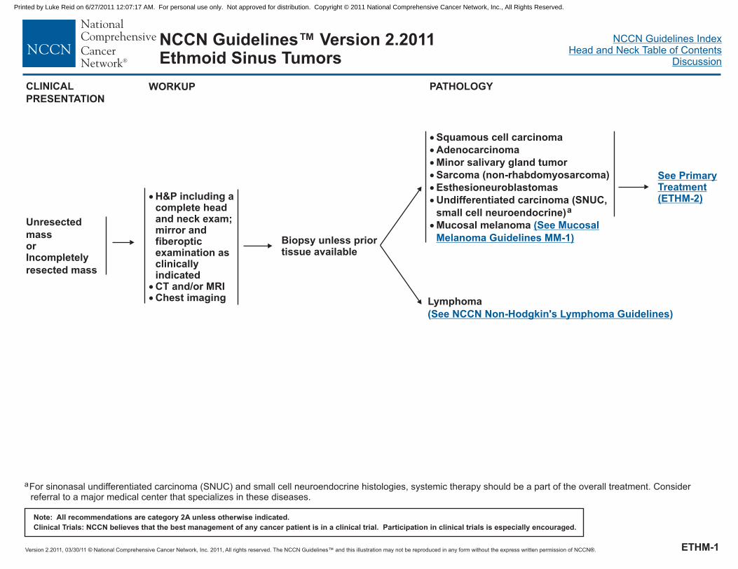
Version 2.2011, 03/30/11 © National Comprehensive Cancer Network, Inc. 2011, All rights reserved. The NCCN Guidelines™ and this illustration may not be reproduced in any form without the express written permission of NCCN®.
NCCN Guidelines IndexHead and Neck Table of Contents
Discussion
Note: All recommendations are category 2A unless otherwise indicated.
Clinical Trials: NCCN believes that the best management of any cancer patient is in a clinical trial. Participation in clinical trials is especially encouraged.
NCCN Guidelines™ Version 2.2011Ethmoid Sinus Tumors
WORKUP
�
�
�
�
�
�
�
Squamous cell carcinoma
Adenocarcinoma
Minor salivary gland tumor
Sarcoma (non-rhabdomyosarcoma)
Esthesioneuroblastomas
Undifferentiated carcinoma (SNUC,
small cell neuroendocrine)a
Mucosal melanoma (See Mucosal
Melanoma Guidelines MM-1)Biopsy unless priortissue available
�
��
H&P
CT and/or MRIChest imaging
including acomplete headand neck exam;mirror andfiberopticexamination asclinicallyindicated
Lymphoma
( )See NCCN Non-Hodgkin's Lymphoma Guidelines
See PrimaryTreatment(ETHM-2)
PATHOLOGY
aFor sinonasal undifferentiated carcinoma (SNUC) and small cell neuroendocrine histologies, systemic therapy should be a part of the overall treatment. Considerreferral to a major medical center that specializes in these diseases.
CLINICAL
PRESENTATION
Unresected
massorIncompletely
resected mass
ETHM-1
Printed by Luke Reid on 6/27/2011 12:07:17 AM. For personal use only. Not approved for distribution. Copyright © 2011 National Comprehensive Cancer Network, Inc., All Rights Reserved.

Version 2.2011, 03/30/11 © National Comprehensive Cancer Network, Inc. 2011, All rights reserved. The NCCN Guidelines™ and this illustration may not be reproduced in any form without the express written permission of NCCN®.
NCCN Guidelines IndexHead and Neck Table of Contents
Discussion
Note: All recommendations are category 2A unless otherwise indicated.
Clinical Trials: NCCN believes that the best management of any cancer patient is in a clinical trial. Participation in clinical trials is especially encouraged.
NCCN Guidelines™ Version 2.2011Ethmoid Sinus Tumors
PRIMARY TREATMENT FOLLOW-UP
b
c
d
.
For minor salivary gland tumors,see
See Principles of Surgery (SURG-A)
See Principles of (CHEM-A)
ee Principles of Radiation Therapy (ETHM-A)(SALI-A)
.
..
Systemic Therapy
S
Newly diagnosed, T4b or
patient declines surgery
Chemo/RT
or
RT
or
Clinical trial (preferred)
c,d
d
Surgical resection (preferred)b
or
Definitive RTd
RT
or
Observation for T1 only(category 2B)
or
Consider Chemo/RT (category 2B)
if adverse features
d
e
f
c,dNewly diagnosed;
T1, T2
Surgical resectionb (preferred)
or
Chemo/RTc,d
Newly diagnosed;
T3, T4a
Surgery (preferred), if feasibleorRTorChemo/RT
b
d
c,d
Diagnosed after incompleteexcision (eg, polypectomy,endoscopic procedure) andgross residual disease
Diagnosed after incompleteexcision
and noresidual disease on physicalexam, imaging, and/orendoscopy
(eg, polypectomy,endoscopic procedure)
RTd
or
Surgery, if feasible(See newly diagnosed T1,T2)
b
ADJUVANT TREATMENT
RecurrentorPersistentDisease(See ADV-2)
CLINICAL
PRESENTATION
RT
or
Consider chemo/RT (category 2B)
if adverse features
d
f
c,d
RT
or
Consider Chemo/RT (category 2B)
if adverse features
d
f
c,d
RT
or
d
Observation for T1 only
(category 2B)
e
Follow-up
(See FOLL-A)
e
f
Pathologic features: Negative margins, favorable histology, central tumors,low grade tumors.
Adverse features include positive margins and intracranial extension( ).See Discussion
ETHM-2
Printed by Luke Reid on 6/27/2011 12:07:17 AM. For personal use only. Not approved for distribution. Copyright © 2011 National Comprehensive Cancer Network, Inc., All Rights Reserved.

Version 2.2011, 03/30/11 © National Comprehensive Cancer Network, Inc. 2011, All rights reserved. The NCCN Guidelines™ and this illustration may not be reproduced in any form without the express written permission of NCCN®.
NCCN Guidelines IndexHead and Neck Table of Contents
Discussion
Note: All recommendations are category 2A unless otherwise indicated.
Clinical Trials: NCCN believes that the best management of any cancer patient is in a clinical trial. Participation in clinical trials is especially encouraged.
NCCN Guidelines™ Version 2.2011Ethmoid Sinus Tumors
PRINCIPLES OF RADIATION THERAPY1
DEFINITIVERT
Primary and gross adenopathy:
Conventional fractionation: 66-70 Gy
(2.0 Gy/fraction; daily Monday-Friday) in 7 weeks
6 fractions/week accelerated; 66-70 Gy to gross disease,
44-64 Gy to subclinical disease.Concomitant boost accelerated RT: 72 Gy/6 weeks (2 Gy once
daily and then 1.8 Gy/fraction, large field; 1.5 Gy boost as
second daily fraction during last 12 treatment days)Hyperfractionation: 81.6 Gy/7 weeks
(1.2 Gy/fraction, twice daily)
Uninvolved nodal stations: 44-64 Gy (1.6-2.0 Gy/fraction)
Concurrent chemoradiation
Primary and gross adenopathy: 70 Gy (2.0 Gy/fraction)
Neck:Uninvolved nodal stations: 44-64-Gy (1.6-2.0 Gy/fraction)
�
Altered fractionation:
Neck
�
�
�
�
�
� �
��
2
1
2Treatment to uninvolved nodal stations is not consistently performed at all institutions.
See Radiation Techniques (RAD-A) and Discussion.
POSTOPERATIVERT
Involved nodal stations: 60-66 Gy (2.0 Gy/fraction)Uninvolved nodal station: 44-64 Gy (1.6-2.0 Gy/fraction)
Primary and gross adenopathy:Preferred interval between resection and postoperative RT is
6 weeks
Concurrent single agent cisplatin at
100 mg/m every 3 wks x 3 doses is recommended.
��
�
�
Primary: 60-66 Gy (2.0 Gy/fraction)Neck�
�
�
Postoperative chemoradiation
�2
IMRT is a preferred technique for maxillary sinus or paranasal/ethmoid sinus tumors to minimize dose to critical structures.
ETHM-A
Printed by Luke Reid on 6/27/2011 12:07:17 AM. For personal use only. Not approved for distribution. Copyright © 2011 National Comprehensive Cancer Network, Inc., All Rights Reserved.

Version 2.2011, 03/30/11 © National Comprehensive Cancer Network, Inc. 2011, All rights reserved. The NCCN Guidelines™ and this illustration may not be reproduced in any form without the express written permission of NCCN®.
NCCN Guidelines IndexHead and Neck Table of Contents
Discussion
Note: All recommendations are category 2A unless otherwise indicated.
Clinical Trials: NCCN believes that the best management of any cancer patient is in a clinical trial. Participation in clinical trials is especially encouraged.
NCCN Guidelines™ Version 2.2011Maxillary Sinus Tumors
WORKUP PATHOLOGY
aBiopsy:
Preferred route is transnasal.
Needle biopsy may be acceptable.
Avoid canine fossa puncture or Caldwell-Luc approach.
�
�
�bFor sinonasal undifferentiated carcinoma (SNUC) and small cell neuroendocrine histologies, systemic therapy should be a part of the overall treatment. Considerreferral to a major medical center that specializes in these diseases.
�
�
�
�
H&P
Complete head andneck CT withcontrast and/or MRIDental/prostheticconsultation asindicatedChest imaging
including acomplete head andneck exam; mirrorand fiberopticexamination asclinically indicated
Biopsya
LymphomaSee NCCN Non-Hodgkin’s
Lymphoma Guidelines
�
�
Squamous cell carcinoma
Mucosal melanoma
���
��
AdenocarcinomaMinor salivary gland tumorSarcoma (non-rhabdomyosarcoma)EsthesioneuroblastomaUndifferentiated carcinoma(SNUC, small cellneuroendocrine)b
(See Mucosal Melanoma
Guidelines MM-1)
T1-2, N0
All histologies
T3-4, N0, Any T, N+
All histologies
See PrimaryTreatment (MAXI-2)
See PrimaryTreatment (MAXI-3)
MAXI-1
Printed by Luke Reid on 6/27/2011 12:07:17 AM. For personal use only. Not approved for distribution. Copyright © 2011 National Comprehensive Cancer Network, Inc., All Rights Reserved.

Version 2.2011, 03/30/11 © National Comprehensive Cancer Network, Inc. 2011, All rights reserved. The NCCN Guidelines™ and this illustration may not be reproduced in any form without the express written permission of NCCN®.
NCCN Guidelines IndexHead and Neck Table of Contents
Discussion
Note: All recommendations are category 2A unless otherwise indicated.
Clinical Trials: NCCN believes that the best management of any cancer patient is in a clinical trial. Participation in clinical trials is especially encouraged.
NCCN Guidelines™ Version 2.2011Maxillary Sinus Tumors
PRIMARY TREATMENT ADJUVANT TREATMENTSTAGING
T1-2, N0
Adenoid
cystic
Surgical
resectionc
RTf
T1-2, N0
All histologies
except
adenoid cystic
Surgical
resectionc
Margin
negative
Perineural
invasion
Consider RT
or
Consider chemo/RT
d
d,e
(category 2B)
Margin
positive
Surgical reresection,
if possibleChemo/RT
(category 2B)
d,e
FOLLOW-UP
Follow-up
(See FOLL-A)
c
d
e
f
.
For adenoid cystic tumors and minor salivary gland tumors, see .
See Principles of Surgery (SURG-A)
See Principles of (CHEM-A)
.
See Principles of Radiation Therapy (MAXI-A)
(SALI-A)
Systemic Therapy
.
Margin
negative
Margin
positive
Consider RTd
Suprastructurec
InfrastructurecObservation
or
RTf
RecurrentorPersistentDisease(See ADV-2)
MAXI-2
Printed by Luke Reid on 6/27/2011 12:07:17 AM. For personal use only. Not approved for distribution. Copyright © 2011 National Comprehensive Cancer Network, Inc., All Rights Reserved.

Version 2.2011, 03/30/11 © National Comprehensive Cancer Network, Inc. 2011, All rights reserved. The NCCN Guidelines™ and this illustration may not be reproduced in any form without the express written permission of NCCN®.
NCCN Guidelines IndexHead and Neck Table of Contents
Discussion
Note: All recommendations are category 2A unless otherwise indicated.
Clinical Trials: NCCN believes that the best management of any cancer patient is in a clinical trial. Participation in clinical trials is especially encouraged.
NCCN Guidelines™ Version 2.2011Maxillary Sinus Tumors
PRIMARY TREATMENT ADJUVANT TREATMENTSTAGING FOLLOW-UP
Follow-up
(See FOLL-A)
T1-T4a, N+
Surgical
excision
+ neck
dissectionc
RT to primary + neckd,f
T4b, Nany
Clinical trial
or
Definitive RT
or
Chemo/RT
f
d,e
T3-T4a, N0
Complete
surgical
resectioncRT to primary and neck (category 2B
for neck) (for squamous cell carcinoma
and undifferentiated tumors)
d,f
Adverse
featuresg
No adverse
featuresg
Chemo/RT to
primary and neck
(category 2B)
d,e
Adverse
featuresg
No adverse
featuresg
c
d
e
f
g
.
Adverse features include positive margins or extracapsular nodal spread .
For adenoid cystic tumors and minor salivary gland tumors, see .
See Principles of Surgery (SURG-A)
See Principles of (CHEM-A)
See Discussion
.
( )
See Principles of Radiation Therapy (MAXI-A)
(SALI-A)
.
Systemic Therapy
Chemo/RT to
primary and neck
(category 2B)
d,e
RecurrentorPersistentDisease(See ADV-2)
MAXI-3
Printed by Luke Reid on 6/27/2011 12:07:17 AM. For personal use only. Not approved for distribution. Copyright © 2011 National Comprehensive Cancer Network, Inc., All Rights Reserved.

Version 2.2011, 03/30/11 © National Comprehensive Cancer Network, Inc. 2011, All rights reserved. The NCCN Guidelines™ and this illustration may not be reproduced in any form without the express written permission of NCCN®.
NCCN Guidelines IndexHead and Neck Table of Contents
Discussion
Note: All recommendations are category 2A unless otherwise indicated.
Clinical Trials: NCCN believes that the best management of any cancer patient is in a clinical trial. Participation in clinical trials is especially encouraged.
NCCN Guidelines™ Version 2.2011Maxillary Sinus Tumors
PRINCIPLES OF RADIATION THERAPY1
RT
Conventional fractionation: 66-70 Gy (2.0 Gy/fraction; daily
Monday-Friday) in 7 weeks
6 fractions/week accelerated; 66-70 Gy to gross disease,
44-64 Gy to subclinical disease.Concomitant boost accelerated RT: 72 Gy/6 weeks (2 Gy once
daily and then 1.8 Gy/fraction, large field; 1.5 Gy boost as second
daily fraction during last 12 treatment days)Hyperfractionation: 81.6 Gy/7 weeks (1.2 Gy/fraction, twice daily)
Uninvolved nodal stations: 44-64 Gy (1.6-2.0 Gy/fraction)
Primary and gross adenopathy: 70 Gy (2.0 Gy/fraction)
Neck:Uninvolved nodal stations: 44-64-Gy (1.6-2.0 Gy/fraction)
DEFINITIVE
Concurrent chemoradiation
�
Altered fractionation:
Neck
�
�
�
�
�
� �
��
2
1
2Treatment to uninvolved nodal stations is not consistently performed at all institutions.
See Radiation Techniques (RAD-A) and Discussion.
IMRT is a preferred technique for maxillary sinus or paranasal/ethmoid sinus tumors to minimize dose to critical structures.
POSTOPERATIVERT
Involved nodal stations: 60-66 Gy (2.0 Gy/fraction)Uninvolved nodal station: 44-64 Gy (1.6-2.0 Gy/fraction)
Primary and gross adenopathy:Preferred interval between resection and postoperative RT
is 6 weeks
Postoperative chemoradiation
Concurrent single agent cisplatin at
100 mg/m every 3 wks x 3 doses is recommended.
��
�
�
�
Primary: 60-66 Gy (2.0 Gy/fraction)Neck�
�
�
2
MAXI-A
Printed by Luke Reid on 6/27/2011 12:07:17 AM. For personal use only. Not approved for distribution. Copyright © 2011 National Comprehensive Cancer Network, Inc., All Rights Reserved.

Version 2.2011, 03/30/11 © National Comprehensive Cancer Network, Inc. 2011, All rights reserved. The NCCN Guidelines™ and this illustration may not be reproduced in any form without the express written permission of NCCN®.
NCCN Guidelines IndexHead and Neck Table of Contents
Discussion
Note: All recommendations are category 2A unless otherwise indicated.
Clinical Trials: NCCN believes that the best management of any cancer patient is in a clinical trial. Participation in clinical trials is especially encouraged.
NCCN Guidelines™ Version 2.2011Very Advanced Head and Neck Cancer
TREATMENT OF HEAD AND NECK CANCER
Newly diagnosed (M0);T4b, any N,or unresectable nodaldisease
Clinical trial preferred
Standardtherapy
Concurrent cisplatinchemotherapy + RT (category 1)a,b
b
c
c
or
Induction chemotherapy followed byRT or chemoradiation (category 3)
Definitive RT± concurrent systemictherapy
c
b
PS 0-1
PS 2
a
b
c
d
The single-agent cisplatin or carboplatin chemoradiotherapy regimens have not been compared in randomized trials. Therefore, no optimal standard regimen isdefined. Combination chemotherapy regimens are more toxic and have not been directly compared to single-agent regimens.
.See Principles of Systemic Therapy (CHEM-A)
See Principles of Radiation Therapy (ADV-A).
See Principles of Surgery (SURG-A).
RT
or
Best supportive care
c
or
Single-agent
chemotherapybPS 3
Residual neck
disease + primary
site controlled:
Neck dissection,
if feasible
d
DIAGNOSIS
Follow-up(See FOLL-A)
RecurrentorPersistentDisease(See ADV-2)
PS = Performance Status (ECOG)
ADV-1
Printed by Luke Reid on 6/27/2011 12:07:17 AM. For personal use only. Not approved for distribution. Copyright © 2011 National Comprehensive Cancer Network, Inc., All Rights Reserved.

Version 2.2011, 03/30/11 © National Comprehensive Cancer Network, Inc. 2011, All rights reserved. The NCCN Guidelines™ and this illustration may not be reproduced in any form without the express written permission of NCCN®.
NCCN Guidelines IndexHead and Neck Table of Contents
Discussion
Note: All recommendations are category 2A unless otherwise indicated.
Clinical Trials: NCCN believes that the best management of any cancer patient is in a clinical trial. Participation in clinical trials is especially encouraged.
NCCN Guidelines™ Version 2.2011Very Advanced Head and Neck Cancer
TREATMENT OF HEAD AND NECK CANCERDIAGNOSIS
RecurrentorPersistentdisease
Standard
therapyb
Clinical trial preferred
PS 0-1
PS 2
PS 3 Best supportive care
Chemotherapy,
clinical trial preferred
or
Best supportive care
Best supportive care
Distantmetastases
Locoregionalrecurrence orSecond primarywith prior RT
Locoregionalrecurrencewithoutprior RT
Resectable
Unresectable
Surgeryclinical trial preferred
d ± reirradiation± chemotherapy,
Resectable
Unresectable
Surgeryd
or
Chemo/RTb,c
Reirradiation , clinical trial preferredorChemotherapy (see distant metastases pathway)
± chemotherapy
Combination chemotherapy
or
Single-agent chemotherapy
b
b
Single-agent chemotherapy
or
Best supportive care
b
b
c
d
e
.
.
Adverse features: extracapsular nodal spread, positive margins, pT3 or pT4 primary, N2 or N3 nodal disease, perineural invasion, vascular embolism .
See Principles of (CHEM-A)
(ADV-A)
See Principles of Surgery (SURG-A)
Systemic Therapy
See Principles of Radiation Therapy
See Discussion
.
( )
See Treatment of Very AdvancedHead and Neck Cancer (ADV-1)
No adverse
featurese
Adverse
featurese
Other risk
features
RTc
or
Consider chemo/RTb,c
Extracapsular
spread and/or
positive margin
Chemo/RTb,c
(category 1)
Observe
Salvage therapy for
persistent disease
as indicated
Follow-up(See FOLL-A)
PS = Performance Status (ECOG)
ADV-2
Printed by Luke Reid on 6/27/2011 12:07:17 AM. For personal use only. Not approved for distribution. Copyright © 2011 National Comprehensive Cancer Network, Inc., All Rights Reserved.

Version 2.2011, 03/30/11 © National Comprehensive Cancer Network, Inc. 2011, All rights reserved. The NCCN Guidelines™ and this illustration may not be reproduced in any form without the express written permission of NCCN®.
NCCN Guidelines IndexHead and Neck Table of Contents
Discussion
Note: All recommendations are category 2A unless otherwise indicated.
Clinical Trials: NCCN believes that the best management of any cancer patient is in a clinical trial. Participation in clinical trials is especially encouraged.
NCCN Guidelines™ Version 2.2011Very Advanced Head and Neck Cancer
PRINCIPLES OF RADIATION THERAPY1
Concurrent chemoradiation (preferred)
Chemoradiation
Conventional fractionation:
Primary and gross adenopathy: 70 Gy (2.0 Gy/ )
NeckUninvolved nodal stations:
44-64 Gy (1.6-2.0 Gy/fraction)
Based on published data, concurrent chemoradiation most commonly uses conventional fractionation at 2.0 Gy per fraction to 70 Gy
in 7 wks with single agent cisplatin given every 3 wks at 100 mg/m x 3 doses. Other fraction sizes (eg, 1.8 Gy, conventional), multiagent
chemotherapy, other dosing schedules of cisplatin; altered fractionation with chemotherapy are efficacious, and there is no consensus
on the optimal approach. In general, the use of concurrent chemoradiation carries a high toxicity burden; altered fractionation or
multiagent chemotherapy will likely further increase the toxicity burden. For any chemoradiation approach, close attention should be
paid to published reports for the specific chemotherapy agent, dose, and schedule of administration. Chemoradiation should be
performed by an experienced team and should include substantial supportive care.
Conventional fractionation:Primary and gross adenopathy: 70-74 Gy (2.0 Gy/fraction; daily Monday-Friday) in 7 weeksNeck
Uninvolved nodal stations:
44-64 Gy (1.6-2.0 Gy/fraction)
Altered fractionation:
6 fractions/week accelerated; 70 Gy to gross disease, 50 Gy to subclinical disease.Concomitant boost accelerated RT:
72 Gy/6 wks (1.8 Gy/fraction, large field; 1.5 Gy boost as second daily fraction during last 12 treatment days)Hyperfractionation:
81.6 Gy/7 wks (1.2 Gy/fraction, twice daily)
� �
�
�
�
fraction
�
2
Definitive RT
�
�
�
�
�
�
�
� Modified fractionation total dose > 70 Gy and treatment course < 7 wks
1See Radiation Techniques (RAD-A) and Discussion.
ADV-A
Printed by Luke Reid on 6/27/2011 12:07:17 AM. For personal use only. Not approved for distribution. Copyright © 2011 National Comprehensive Cancer Network, Inc., All Rights Reserved.

Version 2.2011, 03/30/11 © National Comprehensive Cancer Network, Inc. 2011, All rights reserved. The NCCN Guidelines™ and this illustration may not be reproduced in any form without the express written permission of NCCN®.
NCCN Guidelines IndexHead and Neck Table of Contents
Discussion
Note: All recommendations are category 2A unless otherwise indicated.
Clinical Trials: NCCN believes that the best management of any cancer patient is in a clinical trial. Participation in clinical trials is especially encouraged.
NCCN Guidelines™ Version 2.2011Occult Primary
WORKUPPRESENTATION
Neck
mass
Fine-needle
aspiration(preferred)
or
Open
biopsy
a
a
Lymphoma
Melanoma
See NCCN Non-Hodgkin’sLymphomas Guidelines
Squamous cell
carcinoma,
adenocarcinoma,
and anaplastic
epithelial tumorsb
�
�
�
�
�
Chest imaging
CT with contrast or MRI with
gadolinium (skull base through
thoracic inlet)
PET/CT scan (before biopsy)
HPV, EBV testing suggested for
squamous cell or undifferentiated
histology
Thyroglobulin and calcitonin
staining for adenocarcinoma and
anaplastic undifferentiated
tumors
c
Systemic work-up per
NCCN Melanoma Guidelines
� Skin exam, note regressing
lesions
See Workupand PrimaryTreatment(OCC-2)
See PrimaryTherapy forMelanoma(MM-4)
aRepeat FNA, core, or open biopsy may be necessary for uncertain or non-diagnostic histologies. Patient should be prepared for neck dissection at time of open biopsy,if indicated.
b
cDetermined with appropriate immunohistochemical stains.
Human papilloma virus (HPV) or Epstein-Barr virus (EBV) positive status may help to define the radiation fields .( )See Discussion
Thyroid See NCCN ThyroidCarcinoma Guidelines
Primary
found
Primary
not found
Treat asappropriate(SeeGuidelinesIndex)
H&P:Complete headand neck exam
with attention
to skin; mirror
and fiberoptic
examination as
indicated to
visualize
nasopharynx,
oropharynx,
hypopharynyx,
and larynx
OCC-1
Printed by Luke Reid on 6/27/2011 12:07:17 AM. For personal use only. Not approved for distribution. Copyright © 2011 National Comprehensive Cancer Network, Inc., All Rights Reserved.

Version 2.2011, 03/30/11 © National Comprehensive Cancer Network, Inc. 2011, All rights reserved. The NCCN Guidelines™ and this illustration may not be reproduced in any form without the express written permission of NCCN®.
NCCN Guidelines IndexHead and Neck Table of Contents
Discussion
Note: All recommendations are category 2A unless otherwise indicated.
Clinical Trials: NCCN believes that the best management of any cancer patient is in a clinical trial. Participation in clinical trials is especially encouraged.
NCCN Guidelines™ Version 2.2011Occult Primary
PATHOLOGIC
FINDINGS
Node level
I, II, III,
upper V
Node level
IV, lower V
�
�
EUA including direct
laryngoscopy,
esophagoscopy
Chest/abdominal/
pelvic CT
(or PET-CT if not
previously performed)
�
�
�
�
Examination under
anesthesia (EUA)
Palpation and inspection
Biopsy of areas of
clinical concern and
tonsillectomy
Direct laryngoscopy and
nasopharynx survey
Adenocarcinoma
of neck node,
thyroglobulin
negative,
calcitonin
negative
Poorly
differentiated or
nonkeratinizing
squamous cell or
NOS or anaplastic
(not thyroid) of
neck nodeorSquamous cell
carcinoma of
neck noded
Neck dissection
+ parotidectomy,
if indicatede
WORKUP DEFINITIVE TREATMENT
d
e
f
HPV, EBV testing suggested if not yet done.
.
See Principles of Surgery (SURG-A)
(OCC-A)
.
See Principles of Radiation Therapy
RT to neck
± parotid bed
f
Primary
foundTreat as appropriate(See Guidelines Index)
Levels IV, V Neck dissection,if indicated
eEvaluate for
infraclavicular
primary
Levels I-III
See DefinitiveTreatment (OCC-3)
OCC-2
Printed by Luke Reid on 6/27/2011 12:07:17 AM. For personal use only. Not approved for distribution. Copyright © 2011 National Comprehensive Cancer Network, Inc., All Rights Reserved.
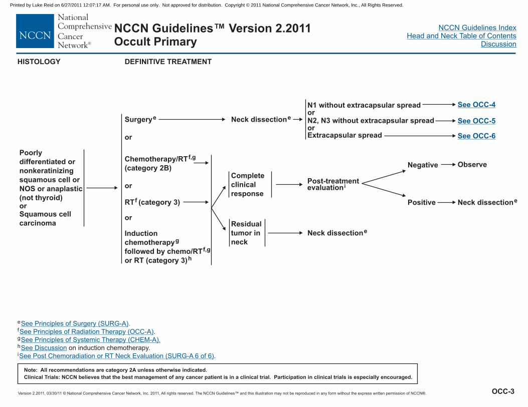
Version 2.2011, 03/30/11 © National Comprehensive Cancer Network, Inc. 2011, All rights reserved. The NCCN Guidelines™ and this illustration may not be reproduced in any form without the express written permission of NCCN®.
NCCN Guidelines IndexHead and Neck Table of Contents
Discussion
Note: All recommendations are category 2A unless otherwise indicated.
Clinical Trials: NCCN believes that the best management of any cancer patient is in a clinical trial. Participation in clinical trials is especially encouraged.
NCCN Guidelines™ Version 2.2011Occult Primary
Neck dissectione
Residual
tumor in
neck
Complete
clinical
response
Neck dissectione
N1 without extracapsular spreador
orExtracapsular spread
N2, N3 without extracapsular spread
See OCC-4
Surgerye
RT (category 3)f
or
or
Induction
chemotherapy
followed by chemo/RT
or RT (category 3)
g
f,g
h
Negative
Positive
Observe
Neck dissectione
e
h
i
f
g.
on induction chemotherapy.
See Principles of Surgery (SURG-A)
See Principles of Radiation Therapy
See Discussion
.
(OCC-A)
See Principles of Systemic Therapy (CHEM-A).
See Post Chemoradiation or RT Neck Evaluation (SURG-A 6 of 6).
Poorly
differentiated or
nonkeratinizing
squamous cell or
NOS or anaplastic
(not thyroid)orSquamous cell
carcinoma
DEFINITIVE TREATMENTHISTOLOGY
Post-treatmentevaluationi
See OCC-5
See OCC-6
Chemotherapy/RT
(category 2B)
f,g
or
OCC-3
Printed by Luke Reid on 6/27/2011 12:07:17 AM. For personal use only. Not approved for distribution. Copyright © 2011 National Comprehensive Cancer Network, Inc., All Rights Reserved.

Version 2.2011, 03/30/11 © National Comprehensive Cancer Network, Inc. 2011, All rights reserved. The NCCN Guidelines™ and this illustration may not be reproduced in any form without the express written permission of NCCN®.
NCCN Guidelines IndexHead and Neck Table of Contents
Discussion
Note: All recommendations are category 2A unless otherwise indicated.
Clinical Trials: NCCN believes that the best management of any cancer patient is in a clinical trial. Participation in clinical trials is especially encouraged.
NCCN Guidelines™ Version 2.2011Occult Primary
RT to oral cavity, Waldeyer’s ring, oropharynx,
bilateral neck (block RT to the larynx)
f
orObservation
RT to oropharynx and bilateral neckc,f
orObservation
RT to Waldeyer’s ring, larynx,
hypopharynx, bilateral neck
f
orObservation
RT to larynx, hypopharynx, bilateral neckf
orObservation
Level I only
Level II, III, upper level V
Level IV only
Lower level V
N1 without
extracapsular spread
Post neck
dissection
cHPV or EBV positive status may help to define the radiation fieldsf .
( ).See Discussion
See Principles of Radiation Therapy (OCC-A)
Follow-up(See FOLL-A)
RecurrentorPersistentDisease(See ADV-2)
TREATMENT
OCC-4
Printed by Luke Reid on 6/27/2011 12:07:17 AM. For personal use only. Not approved for distribution. Copyright © 2011 National Comprehensive Cancer Network, Inc., All Rights Reserved.

Version 2.2011, 03/30/11 © National Comprehensive Cancer Network, Inc. 2011, All rights reserved. The NCCN Guidelines™ and this illustration may not be reproduced in any form without the express written permission of NCCN®.
NCCN Guidelines IndexHead and Neck Table of Contents
Discussion
Note: All recommendations are category 2A unless otherwise indicated.
Clinical Trials: NCCN believes that the best management of any cancer patient is in a clinical trial. Participation in clinical trials is especially encouraged.
NCCN Guidelines™ Version 2.2011Occult Primary
extracapsular
spread
N2, N3 without
RT to oral cavity, Waldeyer’s ring, oropharynx,
both sides of the neck (block RT to the larynx)orChemotherapy/RT (category 2B)or
f
f,g
fRT to neck only (category 3)
RT to nasopharynx, both sides of the
neck, hypopharynx, larynx, oropharynx
c,f
orChemotherapy/RT (category 2B)orRT to neck only (category 3)
f,g
f
RT to Waldeyer’s ring, larynx,
hypopharynx, both sides of the neck
f
orChemotherapy/RT (category 2B)orRT to neck only (category 3)
f,g
f
RT to larynx, hypopharynx, both
sides of the neck
f
orChemotherapy/RT (category 2B)orRT to neck only (category 3)
f,g
f
Level I only
Level II, III, upper level V
Level IV only
Lower level V
cHPV or EBV positive status may help to define the radiation fieldsf
g.
.
( ).See Discussion
See Principles of Radiation Therapy
Systemic Therapy
(OCC-A)
See Principles of (CHEM-A)
Post neck
dissection
Follow-up(See FOLL-A)
RecurrentorPersistentDisease(See ADV-2)
TREATMENT
OCC-5
Printed by Luke Reid on 6/27/2011 12:07:17 AM. For personal use only. Not approved for distribution. Copyright © 2011 National Comprehensive Cancer Network, Inc., All Rights Reserved.

Version 2.2011, 03/30/11 © National Comprehensive Cancer Network, Inc. 2011, All rights reserved. The NCCN Guidelines™ and this illustration may not be reproduced in any form without the express written permission of NCCN®.
NCCN Guidelines IndexHead and Neck Table of Contents
Discussion
Note: All recommendations are category 2A unless otherwise indicated.
Clinical Trials: NCCN believes that the best management of any cancer patient is in a clinical trial. Participation in clinical trials is especially encouraged.
NCCN Guidelines™ Version 2.2011Occult Primary
Extracapsular
spread
RT to oral cavity, Waldeyer’s ring, oropharynx,
both sides of the neck (block RT to the larynx)or
Chemotherapy/RT (category 1)with
RT to neck only (category 3)
f,g
f
f
Chemotherapy/RT (category 1)with
orRT to neck only (category 3)
f,g
f
RT to nasopharynx, both sides of the
neck, hypopharynx, larynx, oropharynx
c,f
RT to Waldeyer’s ring, larynx,
hypopharynx, both sides of the neck
Chemotherapy/RT (category 1)with
orRT to neck only (category 3)
f,g
f
f
RT to larynx, hypopharynx, both
sides of the neck
Chemotherapy/RT (category 1)with
orRT to neck only (category 3)
f,g
f
f
Level I only
Level II, III, upper level V
Level IV only
Lower level V
Post neck
dissection
Follow-up(See FOLL-A)
RecurrentorPersistentDisease(See ADV-2)
cHPV or EBV positive status may help to define the radiation fieldsf
g.
.
( ).See Discussion
See Principles of Radiation Therapy
Systemic Therapy
(OCC-A)
See Principles of (CHEM-A)
TREATMENT
OCC-6
Printed by Luke Reid on 6/27/2011 12:07:17 AM. For personal use only. Not approved for distribution. Copyright © 2011 National Comprehensive Cancer Network, Inc., All Rights Reserved.

Version 2.2011, 03/30/11 © National Comprehensive Cancer Network, Inc. 2011, All rights reserved. The NCCN Guidelines™ and this illustration may not be reproduced in any form without the express written permission of NCCN®.
NCCN Guidelines IndexHead and Neck Table of Contents
Discussion
Note: All recommendations are category 2A unless otherwise indicated.
Clinical Trials: NCCN believes that the best management of any cancer patient is in a clinical trial. Participation in clinical trials is especially encouraged.
NCCN Guidelines™ Version 2.2011Occult Primary
PRINCIPLES OF RADIATION THERAPY1,2
Definitive RT:
Concurrent chemoradiation
�
�
Conventional fractionation:Gross Adenopathy: 66-74 Gy (2.0 Gy/fraction; daily Monday-Friday) in 7 weeksMucosal dosing: 50-66 Gy (2.0 Gy/ ) to putative mucosal sites, depending on field size and use of chemotherapy. Consider
higher dose to 60-66 Gy to particularly suspicious areasNeck:
:
Conventional fractionation:
Neck: Uninvolved nodal stations: 44-64 Gy (1.6-2.0 Gy/fraction)
�
�
�
�
fraction
44-64 Gy (1.6-2.0 Gy/fraction)Uninvolved nodal stations:
Gross adenopathy: 70 Gy (2.0 Gy/fraction)Mucosal dosing: 50-60 Gy (2.0 Gy/fraction) to putative mucosal primary sites. Consider higher dose to 60-66 Gy to particularly
suspicious areas
IMRT is a preferred technique when targeting the oropharynx to minimize the dose to critical structures, especially the parotid glands.
3
�
�
�
1
2
For squamous cell carcinoma, adenocarcinoma, and poorly differentiated carcinoma.
Based on published data, concurrent chemoradiation most commonly uses conventional fractionation at 2.0 Gy per fraction to 70 Gy in 7 wks with single agent
cisplatin given every 3 wks at 100 mg/m x 3 doses. Other fraction sizes (eg, 1.8 Gy, conventional), multiagent chemotherapy, other dosing schedules of cisplatin, or
altered fractionation with chemotherapy are efficacious, and there is no consensus on the optimal approach. In general, the use of concurrent chemoradiation carries a
high toxicity burden; altered fractionation or multiagent chemotherapy will likely further increase the toxicity burden. For any chemoradiation approach, close attention
should be paid to published reports for the specific chemotherapy agent, dose, and schedule of administration. Chemoradiation should be performed by an
experienced team and should include substantial supportive care.
2
3 �
See Radiation Techniques (RAD-A) and Discussion.
OCC-A
Printed by Luke Reid on 6/27/2011 12:07:17 AM. For personal use only. Not approved for distribution. Copyright © 2011 National Comprehensive Cancer Network, Inc., All Rights Reserved.

Version 2.2011, 03/30/11 © National Comprehensive Cancer Network, Inc. 2011, All rights reserved. The NCCN Guidelines™ and this illustration may not be reproduced in any form without the express written permission of NCCN®.
NCCN Guidelines IndexHead and Neck Table of Contents
Discussion
Note: All recommendations are category 2A unless otherwise indicated.
Clinical Trials: NCCN believes that the best management of any cancer patient is in a clinical trial. Participation in clinical trials is especially encouraged.
NCCN Guidelines™ Version 2.2011Salivary Gland Tumors
a
b
c
Site and stage determine therapeutic approaches.
For advanced cancer, includes CT/MRI: base of skull to clavicle.
Characteristics of benign tumor include mobile superficial lobe, slow growth, painless, VII intact, and no neck nodes.
Unresected
salivary gland
mass
ParotidSubmandibularMinor salivarygland
���
a
or
Incompletely
resected salivary
gland mass
CLINICAL
PRESENTATION
�
��
H&P
CT/MRI, if clinically indicatedChest imaging
or c
including a complete head andneck exam; mirror and fiberopticexamination as clinically indicated
Open biopsy
b
� onsider
fine-needle aspiration
(may not be necessary in incompletely
resected patients)
WORKUP
LymphomaSee NCCN Non-Hodgkin’s LymphomasGuidelines
Clinically benign
or
Carcinoma
c
See SALI-2
SALI-1
Printed by Luke Reid on 6/27/2011 12:07:17 AM. For personal use only. Not approved for distribution. Copyright © 2011 National Comprehensive Cancer Network, Inc., All Rights Reserved.

Version 2.2011, 03/30/11 © National Comprehensive Cancer Network, Inc. 2011, All rights reserved. The NCCN Guidelines™ and this illustration may not be reproduced in any form without the express written permission of NCCN®.
NCCN Guidelines IndexHead and Neck Table of Contents
Discussion
Note: All recommendations are category 2A unless otherwise indicated.
Clinical Trials: NCCN believes that the best management of any cancer patient is in a clinical trial. Participation in clinical trials is especially encouraged.
NCCN Guidelines™ Version 2.2011Salivary Gland Tumors
c
d
e
Characteristics of benign tumor include mobile superficial lobe, slow growth, painless, VII intact, and no neck nodes.
Surgical excision of clinically benign tumor: no enucleation of lateral lobe, intraoperative communication with pathologist if indicated.
See Principles of Radiation Therapy (SALI-A).
Complete
surgical
excisiond
Benign
or
Low grade
Adenoid cystic;Indeterminateor high grade
Consider RT
(category 2B for T1)
e
Benign
Surgical
evaluation
Cancer
site
Parotid
gland
Othersalivaryglands
T3, T4a
See Treatment(SALI-3)
Clinically benign or
carcinoma, T1, T2
c
T4b
PATHOLOGY
RESULT
No surgical resection
possible or surgical
resection not recommended
Definitive RT
or
Chemo/RT
(category 2B)
e
Follow-up(See FOLL-A)
Follow-up as
clinically indicated
Follow-up(See FOLL-A)
Follow-up as
clinically indicated
RecurrentorPersistentDisease(See SALI-4)
If tumor spillage,
consider RTe
RecurrentorPersistentDisease(See SALI-4)
SALI-2
Printed by Luke Reid on 6/27/2011 12:07:17 AM. For personal use only. Not approved for distribution. Copyright © 2011 National Comprehensive Cancer Network, Inc., All Rights Reserved.

Version 2.2011, 03/30/11 © National Comprehensive Cancer Network, Inc. 2011, All rights reserved. The NCCN Guidelines™ and this illustration may not be reproduced in any form without the express written permission of NCCN®.
NCCN Guidelines IndexHead and Neck Table of Contents
Discussion
Note: All recommendations are category 2A unless otherwise indicated.
Clinical Trials: NCCN believes that the best management of any cancer patient is in a clinical trial. Participation in clinical trials is especially encouraged.
NCCN Guidelines™ Version 2.2011Salivary Gland Tumors
Othersalivaryglandsites
Complete tumorresectiong
Completeand
lymph nodedissection
tumorresection
g
Parotid
gland
Clinical N0
Clinical N+
Completely
excised
Incompletely
excised, gross
residual disease
No adverse
features
Adjuvant RTe
or
Consider
chemo/RT
(category 2B)
Definitive RT
or
Chemo/RT
(category 2B)
e
Follow-up(See FOLL-A)
TREATMENTf
Clinical N0
Clinical N+
Parotidectomy
with complete
excision of tumor
neck dissection
for high-grade and
high-stage tumors
± g
Parotidectomy
+ neck dissectiong
Adverse features:
�
�
�
�
�
Intermediate or high grade
Close or positive margins
Neural/perineural invasion
Lymph node metastases
Lymphatic/vascular invasion
.
The facial nerve should be preserved if possible.
e
f
g
See Principles of Radiation Therapy (SALI-A)
See Principles of Surgery (SURG-A).
Adenoid cysticRT
(category 2B)
e
Surgical
resection,
if possible
g
CANCER SITE
No further surgical
resection possible
Recurrentor PersistentDisease(See SALI-4)
SALI-3
Printed by Luke Reid on 6/27/2011 12:07:17 AM. For personal use only. Not approved for distribution. Copyright © 2011 National Comprehensive Cancer Network, Inc., All Rights Reserved.

Version 2.2011, 03/30/11 © National Comprehensive Cancer Network, Inc. 2011, All rights reserved. The NCCN Guidelines™ and this illustration may not be reproduced in any form without the express written permission of NCCN®.
NCCN Guidelines IndexHead and Neck Table of Contents
Discussion
Note: All recommendations are category 2A unless otherwise indicated.
Clinical Trials: NCCN believes that the best management of any cancer patient is in a clinical trial. Participation in clinical trials is especially encouraged.
NCCN Guidelines™ Version 2.2011Salivary Gland Tumors
Follow-up
(See FOLL-A)
RECURRENCE
e
g
See Principles of Radiation Therapy (SALI-A)
See Principles of Surgery (SURG-A)
.
.
Standard
therapy
Distantmetastases
Locoregionalrecurrence orsecond primarywith prior RT
Locoregionalrecurrencewithoutprior RT
Resectable
Unresectable
Resectable
UnresectableRTorChemo/RT (category 2B)
e
Surgery (preferred)orReirradiation chemotherapy, clinical trial preferred
g
±
Reirradiation ± chemotherapy, clinical trial preferredorChemotherapy (see Distant metastases pathway below)
Clinical trial
preferred Chemotherapy
Expectant management (with slow growing disease)or
or
Selected metastasectomy (category 3)
Best supportive care
TREATMENT FOR RECURRENCE
Completely
excised
Adjuvant RTe
or
Consider chemo/RT
(category 2B)
Follow-up,(See FOLL-A)
Adverse features:
�
�
�
�
�
Intermediate or high grade
Close or positive margins
Neural/perineural invasion
Lymph node metastases
Lymphatic/vascular invasion
Adenoid cysticRT
(category 2B)
e
PS 3
PS 0-2
RTe
PS = Performance Status (ECOG)
SALI-4
Printed by Luke Reid on 6/27/2011 12:07:17 AM. For personal use only. Not approved for distribution. Copyright © 2011 National Comprehensive Cancer Network, Inc., All Rights Reserved.

Version 2.2011, 03/30/11 © National Comprehensive Cancer Network, Inc. 2011, All rights reserved. The NCCN Guidelines™ and this illustration may not be reproduced in any form without the express written permission of NCCN®.
NCCN Guidelines IndexHead and Neck Table of Contents
Discussion
Note: All recommendations are category 2A unless otherwise indicated.
Clinical Trials: NCCN believes that the best management of any cancer patient is in a clinical trial. Participation in clinical trials is especially encouraged.
NCCN Guidelines™ Version 2.2011Salivary Gland Tumors
Definitive RT
��
��
�
Photon or photon/electron therapy or neutron therapyDose
Photon or photon/electron therapy or neutron therapyDose
�
�
�
�
Primary and gross adenopathy:
66-74 Gy (1.8-2.0 Gy/fraction) or
19.2 nGy (1.2 nGy/fraction)Uninvolved nodal stations:
44-64 Gy (1.6-2.0 Gy/fraction) or
13.2 nGy (1.2 nGy/fraction)
Primary: 60 Gy (1.8-2.0 Gy/fraction)
or 18 nGy (1.2 nGy/fraction)Uninvolved nodal stations:
44-64 Gy (1.6-2.0 Gy/fraction)
or 13.2 nGy (1.2 nGy/fraction)
2
2
2
2
Postoperative RT
PRINCIPLES OF RADIATION THERAPY1
1
2Range based on grade/natural history of disease (eg, 1.8 Gy fraction may be used for slower growing tumors).
See Radiation Techniques (RAD-A) and Discussion).
SALI-A
Printed by Luke Reid on 6/27/2011 12:07:17 AM. For personal use only. Not approved for distribution. Copyright © 2011 National Comprehensive Cancer Network, Inc., All Rights Reserved.

Version 2.2011, 03/30/11 © National Comprehensive Cancer Network, Inc. 2011, All rights reserved. The NCCN Guidelines™ and this illustration may not be reproduced in any form without the express written permission of NCCN®.
NCCN Guidelines IndexHead and Neck Table of Contents
Discussion
Note: All recommendations are category 2A unless otherwise indicated.
Clinical Trials: NCCN believes that the best management of any cancer patient is in a clinical trial. Participation in clinical trials is especially encouraged.
NCCN Guidelines™ Version 2.2011Mucosal Melanoma
Biopsy
confirms
diagnosis of
mucosal
malignant
melanoma
PRESENTATION WORKUP
�
�
�
�
�
H&P including complete head and neck
exam; mirror and fiberoptic examination
as clinically indicated
Verification of pathology using
appropriate staining
(HMB-45, S-100, Melan-A)
CT and/or MRI to determine anatomic
extent of disease, particularly for sinus
disease
Chest imaging as indicated
Consider PET-CT scan to rule out
metastatic disease
TREATMENT
Sinus or nasal cavity
mucosal melanoma
Oral cavity, oropharynx,
larynx, or hypopharynx
mucosal melanoma
See Primary
Treatment (MM-2)
See Primary
Treatment (MM-3)
MM-1
Printed by Luke Reid on 6/27/2011 12:07:17 AM. For personal use only. Not approved for distribution. Copyright © 2011 National Comprehensive Cancer Network, Inc., All Rights Reserved.

Version 2.2011, 03/30/11 © National Comprehensive Cancer Network, Inc. 2011, All rights reserved. The NCCN Guidelines™ and this illustration may not be reproduced in any form without the express written permission of NCCN®.
NCCN Guidelines IndexHead and Neck Table of Contents
Discussion
Note: All recommendations are category 2A unless otherwise indicated.
Clinical Trials: NCCN believes that the best management of any cancer patient is in a clinical trial. Participation in clinical trials is especially encouraged.
NCCN Guidelines™ Version 2.2011Mucosal Melanoma
PRIMARY
TREATMENT
Stage III
T4a, N0
T3-T4a, N1
Stage IVB
Sinus or nasal cavity
mucosal melanoma
Stage IVC
Wide surgical resection of primarya
ADJUVANT
TREATMENT
Wide surgical resectiona Postoperative RT
to primary siteb
Wide surgical resection
+ neck dissection of positive necka
Postoperative RT
to primary site and
neckb
Strongly consider
postoperative RT
to primary siteb
Clinical trial (preferred)orPrimary RTorSystemic therapy
b
c
Clinical trial (preferred)orBest supportive careorPrimary RTorSystemic therapy
b
c
a
b
c
See Principles of Surgery (SURG-A).See Principles of Radiation Therapy (MM-A)See Principles of Systemic Therapy for Advanced or Metastatic Melanoma page ME-D from the NCCN Melanoma Guidelines
..
Follow-up
(See FOLL-A)
Recurrent
or
Persistent
Disease
See NCCN
Melanoma
Guidelines
MM-2
Printed by Luke Reid on 6/27/2011 12:07:17 AM. For personal use only. Not approved for distribution. Copyright © 2011 National Comprehensive Cancer Network, Inc., All Rights Reserved.
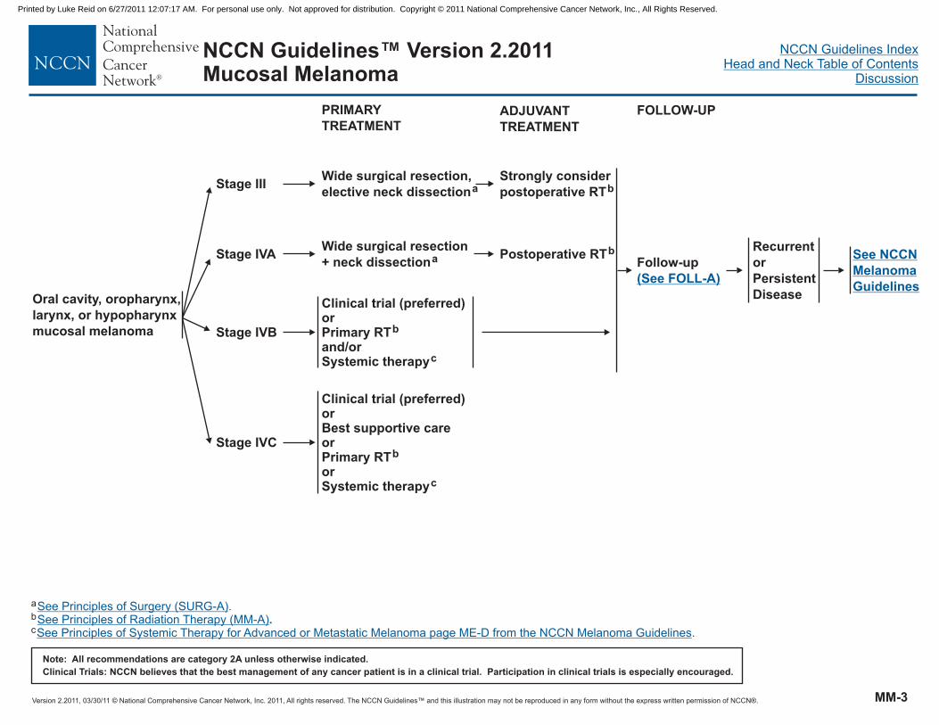
Version 2.2011, 03/30/11 © National Comprehensive Cancer Network, Inc. 2011, All rights reserved. The NCCN Guidelines™ and this illustration may not be reproduced in any form without the express written permission of NCCN®.
NCCN Guidelines IndexHead and Neck Table of Contents
Discussion
Note: All recommendations are category 2A unless otherwise indicated.
Clinical Trials: NCCN believes that the best management of any cancer patient is in a clinical trial. Participation in clinical trials is especially encouraged.
NCCN Guidelines™ Version 2.2011Mucosal Melanoma
PRIMARY
TREATMENT
Stage III
Stage IVA
Stage IVB
Stage IVC
Wide surgical resection,
elective neck dissectiona
ADJUVANT
TREATMENT
Wide surgical resection
+ neck dissectiona Postoperative RTb
Strongly consider
postoperative RTb
Clinical trial (preferred)orBest supportive careorPrimary RTorSystemic therapy
b
c
Oral cavity, oropharynx,
larynx, or hypopharynx
mucosal melanoma
Clinical trial (preferred)orPrimary RTand/orSystemic therapy
b
c
Follow-up
(See FOLL-A)
Recurrent
or
Persistent
Disease
See NCCN
Melanoma
Guidelines
FOLLOW-UP
a
b
c
See Principles of Surgery (SURG-A).See Principles of Radiation Therapy (MM-A)See Principles of Systemic Therapy for Advanced or Metastatic Melanoma page ME-D from the NCCN Melanoma Guidelines
..
MM-3
Printed by Luke Reid on 6/27/2011 12:07:17 AM. For personal use only. Not approved for distribution. Copyright © 2011 National Comprehensive Cancer Network, Inc., All Rights Reserved.

Version 2.2011, 03/30/11 © National Comprehensive Cancer Network, Inc. 2011, All rights reserved. The NCCN Guidelines™ and this illustration may not be reproduced in any form without the express written permission of NCCN®.
NCCN Guidelines IndexHead and Neck Table of Contents
Discussion
Note: All recommendations are category 2A unless otherwise indicated.
Clinical Trials: NCCN believes that the best management of any cancer patient is in a clinical trial. Participation in clinical trials is especially encouraged.
NCCN Guidelines™ Version 2.2011Mucosal Melanoma
Level V,
occipital node
All other
nodal sites
Posterior lateral
node dissection
Neck dissectiona
± RT to nodal bedd,e ± Adjuvant systemic therapy, per
NCCN Melanoma Guidelines
a
d
e
Adjuvant radiotherapy: 30 Gy/5 fx over 2.5 weeks (6.0 Gy/fx). Careful attention to dosimetry is necessary.(Ang KK, Peters LJ, Weber RS, et al. Postoperative radiotherapy for cutaneous melanoma of the head and neck region.International Journal of Radiation Oncology, Biology, Physics 30:795-798, 1994).
RT is indicated for satellitosis, positive nodes, or extracapsular spread.
See Principles of Surgery (SURG-A).
PRIMARY THERAPY FOR OCCULT PRIMARY- MELANOMA
MM-4
Printed by Luke Reid on 6/27/2011 12:07:17 AM. For personal use only. Not approved for distribution. Copyright © 2011 National Comprehensive Cancer Network, Inc., All Rights Reserved.

Version 2.2011, 03/30/11 © National Comprehensive Cancer Network, Inc. 2011, All rights reserved. The NCCN Guidelines™ and this illustration may not be reproduced in any form without the express written permission of NCCN®.
NCCN Guidelines IndexHead and Neck Table of Contents
Discussion
Note: All recommendations are category 2A unless otherwise indicated.
Clinical Trials: NCCN believes that the best management of any cancer patient is in a clinical trial. Participation in clinical trials is especially encouraged.
NCCN Guidelines™ Version 2.2011Mucosal Melanoma
RT for unresectable locally advanced melanoma:
66-74 Gy
Palliative RT dose and schedule may be considered
�
�
Postoperative RT
�
�
�
�
�
Primary site resection:Paranasal sites:
RT to primary site + 2-3 cm margins or to anatomic compartmentOral cavity, oropharynx, and hypopharynx sites:
RT to primary site (+ 2-3 cm margins or anatomic zone) and elective treatment to neck
(unless negative pathology findings of neck dissection)Also strongly consider radiation to primary site for any locally recurrent disease after
previous resection.
Neck/nodal basin dissection:High-risk features:
2 nodes
Single node 3 cmExtracapsular nodal diseaseNode excision (alone) with no further basin dissectionRecurrence in nodal basin after previous surgery.
Dose and Fractionation:Primary and neck (high-risk sites): 60-66 Gy (2.0 Gy/fraction) or 70 Gy for gross disease
�
�
�
�
�
�
�
�
�
�
�
�
� Low risk, undissected, or uninvolved portions of neck: 50-60 Gy (2 Gy/fraction)
PRINCIPLES OF RADIATION THERAPY1
1See Radiation Techniques (RAD-A) and Discussion.
MM-A
Printed by Luke Reid on 6/27/2011 12:07:17 AM. For personal use only. Not approved for distribution. Copyright © 2011 National Comprehensive Cancer Network, Inc., All Rights Reserved.

Version 2.2011, 03/30/11 © National Comprehensive Cancer Network, Inc. 2011, All rights reserved. The NCCN Guidelines™ and this illustration may not be reproduced in any form without the express written permission of NCCN®.
NCCN Guidelines IndexHead and Neck Table of Contents
Discussion
Note: All recommendations are category 2A unless otherwise indicated.
Clinical Trials: NCCN believes that the best management of any cancer patient is in a clinical trial. Participation in clinical trials is especially encouraged.
NCCN Guidelines™ Version 2.2011Head and Neck Cancers
FOLLOW-UP RECOMMENDATIONS
1
2For mucosal melanoma, physical exam should include endoscopic inspection for paranasal sinus disease.
For cancer of the oropharynx, hypopharynx, glottic larynx, supraglottic larynx, and nasopharynx: imaging recommended for T3-4 or N2-3 disease only.
�
�
History and physical exam :Year 1, every 1–3 moYear 2, every 2–4 moYears 3–5, every 4–6 mo> 5 years, every 6–12 mo
Post-treatment baseline imaging of primary (and neck if treated) recommended within 6 mo of treatment (category 2B)Further reimaging as indicated based on signs/symptoms; not routinely recommended for asymptomatic patients
Chest imaging as clinically indicated
Thyroid-stimulating hormone (TSH) every 6-12 mo if neck irradiated
Speech/hearing and swallowing evaluation and rehabilitation as clinically indicated
Smoking cessation and alcohol counseling as clinically indicated
Dental evaluation
Consider Epstein-Barr virus (EBV) monitoring for nasopharynx
1
2
�
�
�
�
�
�
�
�
�
�
�
�
�
�
Recommended for oral cavityAs indicated for oropharynx, hypopharynx, and nasopharynxAs indicated for other sites, if significant intraoral radiation
FOLL-A
Printed by Luke Reid on 6/27/2011 12:07:17 AM. For personal use only. Not approved for distribution. Copyright © 2011 National Comprehensive Cancer Network, Inc., All Rights Reserved.

Version 2.2011, 03/30/11 © National Comprehensive Cancer Network, Inc. 2011, All rights reserved. The NCCN Guidelines™ and this illustration may not be reproduced in any form without the express written permission of NCCN®.
NCCN Guidelines IndexHead and Neck Table of Contents
Discussion
Note: All recommendations are category 2A unless otherwise indicated.
Clinical Trials: NCCN believes that the best management of any cancer patient is in a clinical trial. Participation in clinical trials is especially encouraged.
NCCN Guidelines™ Version 2.2011Head and Neck Cancers
PRINCIPLES OF SURGERY
Evaluation
Integration of Therapy
Assessment of Resectability
All patients should be evaluated by a head and neck surgical oncologist prior to treatment to assure the following:
To review the adequacy of biopsy material, review staging and imaging to determine the extent of disease, exclude the presence of a
synchronous primary tumor, assess current functional status, and evaluate for potential surgical salvage if initial treatment is non-
surgical.
To participate in the multidisciplinary team discussions regarding patient treatment options with the goal of maximizing survival with
preservation of form and function.
To develop a prospective surveillance plan that includes adequate dental, nutritional, and health behavior evaluation and intervention and
any other ancillary evaluations that would provide for comprehensive rehabilitation.
For patients undergoing planned surgery, the surgical procedure, margins, and reconstructive plan should be developed and designed to
resect all gross tumor with adequate tumor free surgical margins. The surgical procedure should not be modified based upon any
response observed before therapy except in instances of tumor progression that mandates a more extensive procedure in order to
encompass the tumor at the time of definitive resection.
It is critical that multidisciplinary evaluation and treatment be coordinated and integrated prospectively by all modalities involved in
patient care.
Tumor involvement of the following sites is associated with poor prognosis or with T4b cancer (ie, unresectable based on technical ability
to obtain clear margins).
Involvement of the pterygoid muscles particularly when associated with severe trismus or pterygopalatine fossa involvement with cranial
neuropathy;
Gross extension of tumor to the skull base (ie, erosion of the pterygoid plates or sphenoid bone, widening of the foramen ovale, etc.);
Direct extension to superior nasopharynx or deep extension into the Eustachian tube and lateral nasopharyngeal walls;
Suspected invasion (encasement) of the common or internal carotid artery. Encasement is usually assessed radiographically and defined
as tumor surrounding the carotid artery 270 degrees or greater;
Direct extension of neck disease to involve the external skin;
Direct extension to mediastinal structures, prevertebral fascia or cervical vertebrae.
�
�
�
�
�
�
�
�
�
�
�
1
1
1
1
1In selected cases, surgery might still be considered.
Continued on next page
SURG-A1 of 6
Printed by Luke Reid on 6/27/2011 12:07:17 AM. For personal use only. Not approved for distribution. Copyright © 2011 National Comprehensive Cancer Network, Inc., All Rights Reserved.

Version 2.2011, 03/30/11 © National Comprehensive Cancer Network, Inc. 2011, All rights reserved. The NCCN Guidelines™ and this illustration may not be reproduced in any form without the express written permission of NCCN®.
NCCN Guidelines IndexHead and Neck Table of Contents
Discussion
Note: All recommendations are category 2A unless otherwise indicated.
Clinical Trials: NCCN believes that the best management of any cancer patient is in a clinical trial. Participation in clinical trials is especially encouraged.
NCCN Guidelines™ Version 2.2011Head and Neck Cancers
Primary Tumor Resection
The resection of advanced tumors of the oral cavity, oropharynx, hypopharynx, larynx, or paranasal sinus will vary in extent depending on
the structures involved. The primary tumor should be considered surgically curable by wide excision using accepted criteria for adequate
excision, depending on the region involved.
En bloc resection of the primary tumor should be attempted whenever feasible.
In continuity neck dissection is necessary when there is direct extension of the primary tumor into the neck.
Surgical resection should be planned based upon the extent of the primary tumor as ascertained by clinical examination and careful
interpretation of appropriate radiographic images.
Perineural invasion should be suspected when tumors are adjacent to motor or sensory nerves. When invasion is suspected, the nerve
should be dissected both proximally and distally and should be resected to obtain clearance of disease. Frozen section determination of
the proximal and distal nerve margins may prove helpful to facilitate tumor clearance.
Partial or segmental resection of the mandible may be necessary to encompass adequately the cancer with adequate tumor free margins.
Adequate resection may require partial, horizontal, or sagittal resection of the mandible for tumors involving or adherent to mandibular
periosteum. Segmental resection should be considered in tumors that grossly involve mandibular periosteum (as determined by tumor
fixation to the mandible) or show evidence of direct tumor involvement of the bone at the time of operation or through preoperative
imaging. The extent of mandibular resection will depend on the degree of involvement accessed clinically and in the operating room.
For tumors of the larynx, the decision to perform either total laryngectomy or conservation laryngeal surgery (ie, laser resection,
hemilaryngectomy, supraglottic laryngectomy, etc.) will be decided by the surgeon but should adhere to the principle of complete tumor
extirpation with curative intent.
�
�
�
�
�
�
�
�
For oral cavity cancers, as thickness of the lesion increases, so does the risk of regional metastases and the need for adjuvant elective
neck dissection.
For maxillary sinus tumors, note that “Ohngren's line" runs from the medial canthus of the eye to the angle of the mandible, helping to
define a plane passing through the maxillary sinus. Tumors "below" or "before" this line involve the maxillary infrastructure. Those
"above" or "behind" Ohngren's line involve the suprastructure.
PRINCIPLES OF SURGERY
Continued on next page
SURG-A2 of 6
Printed by Luke Reid on 6/27/2011 12:07:17 AM. For personal use only. Not approved for distribution. Copyright © 2011 National Comprehensive Cancer Network, Inc., All Rights Reserved.

Version 2.2011, 03/30/11 © National Comprehensive Cancer Network, Inc. 2011, All rights reserved. The NCCN Guidelines™ and this illustration may not be reproduced in any form without the express written permission of NCCN®.
NCCN Guidelines IndexHead and Neck Table of Contents
Discussion
Note: All recommendations are category 2A unless otherwise indicated.
Clinical Trials: NCCN believes that the best management of any cancer patient is in a clinical trial. Participation in clinical trials is especially encouraged.
NCCN Guidelines™ Version 2.2011Head and Neck Cancers
MarginsFrozen section margin assessment is always at the discretion of the surgeon and should be considered when it will facilitate complete
tumor removal. The achievement of adequate wide margins may require resection of an adjacent structure in the oral cavity or
laryngopharynx such as the base of tongue and/or anterior tongue, mandible, larynx, or portions of the cervical esophagus.
Adequate excision is defined as clear resection margins with at least enough clearance from gross tumor to obtain clear frozen section
and permanent margins (typically 1.5 - 2cm). In general, frozen section examination of the margins will usually be undertaken
intraoperatively if a margin has less than 2 cm clearance from the gross tumor, a line of resection has uncertain clearance because of
indistinct tumor margins, or there is suspected residual disease (ie, soft tissue, cartilage, carotid artery, or mucosal irregularity).
The details of resection margins should be included in the operative dictation. The margins may be assessed on the resected specimen
or alternatively from the surgical bed with proper orientation.
A clear margin is defined as the distance from the invasive tumor front that is 5 mm or more from the resected margin.
A close margin is defined as the distance from the invasive tumor front to the resected margin that is less than 5 mm.
The primary tumor should be marked in a fashion adequate for orientation by the surgical pathologist.
The neck dissection should be oriented or sectioned in order to identify levels of lymph nodes encompassed in the dissection.
Reconstruction of surgical defects should be performed using conventional techniques at the discretion of the surgeon. Primary closure
is recommended when appropriate but should not be pursued at the expense of obtaining wide, tumor free margins. Reconstructive
closure with local/regional flaps, free tissue transfer, or split thickness skin or other grafts with or without mandibular reconstruction is
performed at the discretion of the surgeon.
�
�
�
�
�
�
�
Surgical management of cranial nerves VII, X (including the recurrent laryngeal nerve), XI, and XIIOperative management of the facial nerve and other major cranial nerves during primary or regional node resection is influenced by the
preoperative clinical function of the nerve.
When the nerve is functioning, every attempt should be made to preserve the structure and function of the nerve (main trunk and/or
branches) even if wide tumor margins are not achieved recognizing that the surgeon should leave no gross residual disease.
Adjuvant postoperative radiation or chemoradiation is generally prescribed when microscopic residual or gross residual tumor is
suspected.
Direct nerve invasion by tumor and/or preoperative paralysis of the nerve may warrant segmental resection and nerve grafting at the
discretion of the surgeon if tumor free margins are assured throughout the remainder of the procedure.
�
�
�
PRINCIPLES OF SURGERY
Continued on next page
SURG-A3 of 6
Printed by Luke Reid on 6/27/2011 12:07:17 AM. For personal use only. Not approved for distribution. Copyright © 2011 National Comprehensive Cancer Network, Inc., All Rights Reserved.
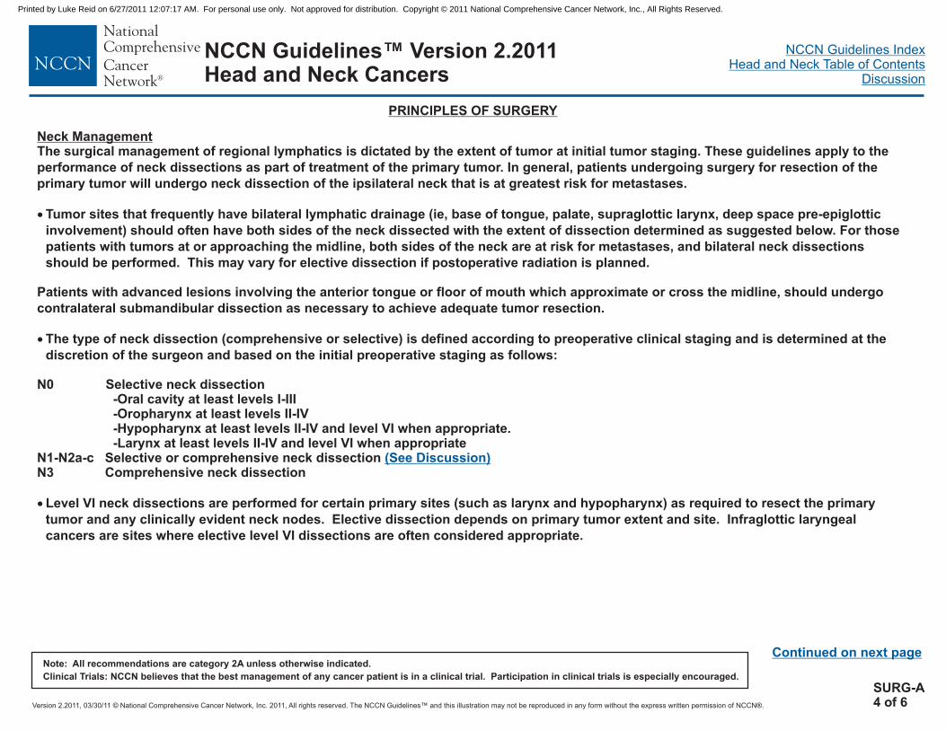
Version 2.2011, 03/30/11 © National Comprehensive Cancer Network, Inc. 2011, All rights reserved. The NCCN Guidelines™ and this illustration may not be reproduced in any form without the express written permission of NCCN®.
NCCN Guidelines IndexHead and Neck Table of Contents
Discussion
Note: All recommendations are category 2A unless otherwise indicated.
Clinical Trials: NCCN believes that the best management of any cancer patient is in a clinical trial. Participation in clinical trials is especially encouraged.
NCCN Guidelines™ Version 2.2011Head and Neck Cancers
Neck ManagementThe surgical management of regional lymphatics is dictated by the extent of tumor at initial tumor staging. These guidelines apply to the
performance of neck dissections as part of treatment of the primary tumor. In general, patients undergoing surgery for resection of the
primary tumor will undergo neck dissection of the ipsilateral neck that is at greatest risk for metastases.
Tumor sites that frequently have bilateral lymphatic drainage (ie, base of tongue, palate, supraglottic larynx, deep space pre-epiglottic
involvement) should often have both sides of the neck dissected with the extent of dissection determined as suggested below. For those
patients with tumors at or approaching the midline, both sides of the neck are at risk for metastases, and bilateral neck dissections
should be performed. This may vary for elective dissection if postoperative radiation is planned.
Patients with advanced lesions involving the anterior tongue or floor of mouth which approximate or cross the midline, should undergo
contralateral submandibular dissection as necessary to achieve adequate tumor resection.
The type of neck dissection (comprehensive or selective) is defined according to preoperative clinical staging and is determined at the
discretion of the surgeon and based on the initial preoperative staging as follows:
N0 Selective neck dissection-Oral cavity at least levels I-III-Oropharynx at least levels II-IV
-Larynx at least levels II-IV and level VI when appropriate
Level VI neck dissections are performed for certain primary sites (such as larynx and hypopharynx) as required to resect the primary
tumor and any clinically evident neck nodes. Elective dissection depends on primary tumor extent and site. Infraglottic laryngeal
cancers are sites where elective level VI dissections are often considered appropriate.
�
�
�
-Hypopharynx at least levels II-IV and level VI when appropriate.
N1-N2a-c Selective or comprehensive neck dissectionN3 Comprehensive neck dissection
(See Discussion)
PRINCIPLES OF SURGERY
Continued on next page
SURG-A4 of 6
Printed by Luke Reid on 6/27/2011 12:07:17 AM. For personal use only. Not approved for distribution. Copyright © 2011 National Comprehensive Cancer Network, Inc., All Rights Reserved.

Version 2.2011, 03/30/11 © National Comprehensive Cancer Network, Inc. 2011, All rights reserved. The NCCN Guidelines™ and this illustration may not be reproduced in any form without the express written permission of NCCN®.
NCCN Guidelines IndexHead and Neck Table of Contents
Discussion
Note: All recommendations are category 2A unless otherwise indicated.
Clinical Trials: NCCN believes that the best management of any cancer patient is in a clinical trial. Participation in clinical trials is especially encouraged.
NCCN Guidelines™ Version 2.2011Head and Neck Cancers
Management of Recurrences
Surveillance
Surgically resectable primary cancers should be re-resected with curative intent if feasible, and recurrences in a previously treated neck
should undergo surgical salvage, as well. Neck disease in an untreated neck should be addressed by formal neck dissection or
modification depending on the clinical situation. Non-surgical therapy may also be utilized as clinically appropriate.
All patients should have regular follow-up visits to assess for symptoms and possible tumor recurrence, health behaviors, nutrition, dental
health, and speech and swallowing function.
Tumor evaluations must be performed by specialists skilled in head and neck clinical examination.
The frequency of evaluation is summarized elsewhere in the NCCN guidelines
Post chemoradiation or RT neck evaluation
�
�
�
( .See Follow-up Recommendations [FOLL-A])
(See Principles of Surgery-Neck Evaluation SURG-A 6 of 6)
PRINCIPLES OF SURGERY
Continued on next page
SURG-A5 of 6
Printed by Luke Reid on 6/27/2011 12:07:17 AM. For personal use only. Not approved for distribution. Copyright © 2011 National Comprehensive Cancer Network, Inc., All Rights Reserved.
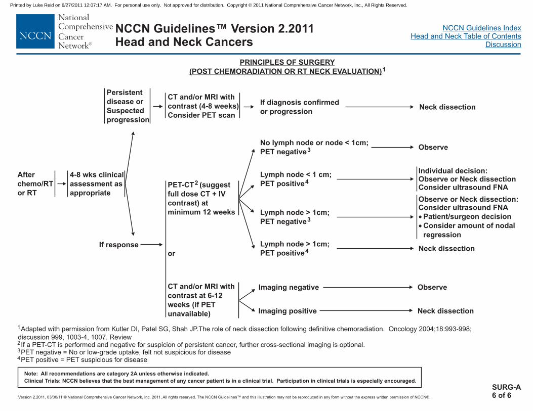
Version 2.2011, 03/30/11 © National Comprehensive Cancer Network, Inc. 2011, All rights reserved. The NCCN Guidelines™ and this illustration may not be reproduced in any form without the express written permission of NCCN®.
NCCN Guidelines IndexHead and Neck Table of Contents
Discussion
Note: All recommendations are category 2A unless otherwise indicated.
Clinical Trials: NCCN believes that the best management of any cancer patient is in a clinical trial. Participation in clinical trials is especially encouraged.
NCCN Guidelines™ Version 2.2011Head and Neck Cancers
PRINCIPLES OF SURGERY
(POST CHEMORADIATION OR RT NECK EVALUATION)1
After
chemo/RT
or RT
4-8 wks clinical
assessment as
appropriate
CT and/or MRI with
contrast (4-8 weeks)
Consider PET scan
PET-CT (suggest
full dose CT + IV
contrast) at
minimum 12 weeks
or
CT and/or MRI with
contrast at 6-12
weeks (if PET
unavailable)
2
No lymph node or node < 1cm;
PET negative3
Lymph node < 1 cm;
PET positive4
Lymph node > 1cm;
PET negative3
Lymph node > 1cm;
PET positive4
Observe
Individual decision:Observe or Neck dissectionConsider ultrasound FNA
Observe or Neck dissection:Consider ultrasound FNA
Patient/surgeon decision
Consider amount of nodal
regression
�
�
Neck dissection
1Adapted with permission from Kutler DI, Patel SG, Shah JP.The role of neck dissection following definitive chemoradiation. Oncology 2004;18:993-998;
discussion 999, 1003-4, 1007. Review
PET negative = No or low-grade uptake, felt not suspicious for diseasePET positive = PET suspicious for disease
2If a PET-CT is performed and negative for suspicion of persistent cancer, further cross-sectional imaging is optional.3
4
Neck dissection
Imaging negative
Imaging positive
Observe
Neck dissection
Persistent
disease or
Suspected
progression
If response
If diagnosis confirmed
or progression
SURG-A6 of 6
Printed by Luke Reid on 6/27/2011 12:07:17 AM. For personal use only. Not approved for distribution. Copyright © 2011 National Comprehensive Cancer Network, Inc., All Rights Reserved.

Version 2.2011, 03/30/11 © National Comprehensive Cancer Network, Inc. 2011, All rights reserved. The NCCN Guidelines™ and this illustration may not be reproduced in any form without the express written permission of NCCN®.
NCCN Guidelines IndexHead and Neck Table of Contents
Discussion
Note: All recommendations are category 2A unless otherwise indicated.
Clinical Trials: NCCN believes that the best management of any cancer patient is in a clinical trial. Participation in clinical trials is especially encouraged.
NCCN Guidelines™ Version 2.2011Head and Neck Cancers
Target delineation and optimal dose distribution require experience in head and neck imaging, and a thorough understanding of patterns ofdisease spread. Standards for target definition, dose specification, fractionation (with and without concurrent chemotherapy), and normal tissueconstraints are still evolving. IMRT, 3D, and 2D conformal techniques may be used as appropriate depending on the stage, tumor location,physician training/experience, and available physics support. Close interplay exists between radiation technology, techniques, fractionation, andchemotherapy options resulting in a large number of combinations that may impact toxicity or tumor control.
IMRT has been shown to be useful in reducing long-term toxicity in oropharyngeal, paranasal sinus, and nasopharyngeal cancers by reducing thedose to salivary glands, temporal lobes, auditory structures (including cochlea), and optic structures. The application of IMRT to other sites(eg, oral cavity, larynx, hypopharynx, salivary glands) is evolving and may be used at the discretion of treating physicians.
A number of ways exist to integrate IMRT, target volume dosing, and fractionation. The Simultaneous Integrated Boost (SIB) technique usesdifferential “dose painting” (66-74 Gy to gross disease; 50-60 Gy to subclinical disease) for each fraction of treatment throughout the entirecourse of radiation. SIB is commonly used in conventional (5 fractions/week) and the “6 fractions/week accelerated” schedule. The Sequential(SEQ) IMRT technique typically delivers the initial (lower dose) phase (weeks 1-5) followed by the high-dose boost volume phase (weeks 6-7)using 2-3 separate dose plans, and is commonly applied in standard fractionation and hyperfractionation. The Concomitant Boost Acceleratedschedule may utilize a “Modified SEQ” dose plan by delivering the dose to the subclinical targets once a day for 6 weeks, and a separate boostdose plan as a second daily fraction for the last 12 treatment days.
Close cooperation andinterdisciplinary management are critical to treatment planning and radiation targeting, especially in the postoperative setting or after inductionchemotherapy.9
Intensity-Modulated Radiotherapy (IMRT)
IMRT and Fractionation10,11
4 5
6
RADIATION TECHNIQUES1-8
1
2L
3
4
6
Dogan N, King S, Emami B, et al. Assessment of different IMRT boost delivery methods ontarget coverage and normal-tissue sparing. Int J Radiat Oncol Biol Phys 2003;57(5):1480-1491.
ee NY, de Arruda FF, Puri DR, et al. A comparison of intensity-modulated radiation therapyand concomitant boost radiotherapy in the setting of concurrent chemotherapy for locallyadvanced oropharyngeal carcinoma. Int J Radiat Oncol Biol Phys 2006;66(4):966-974.
Lee NY, O'Meara W, Chan K, et al. Concurrent chemotherapy and intensity-modulatedradiotherapy for locoregionally advanced laryngeal and hypopharyngeal cancers. Int JRadiat Oncol Biol Phys 2007;69(2):459-468. Epub 2007 May 9.
Mohan R, Wu Q, Morris M, et al. “Simultaneous Integrated Boost” (SIB) IMRT of advancedhead and neck squamous cell carcinomas—dosimetric analysis. Int J Radiat Oncol BiolPhys 2001;51(3):180–181.
5Overgaard J, Hansen HS, Specht L, et al. Five compared with six fractions per week ofconventional radiotherapy of squamous-cell carcinoma of head and neck: DAHANCA 6 and7 randomised controlled trial. Lancet 2003;362(9388):933-940.
Schoenfeld GO, Amdur RJ, Morris CG, et al. Patterns of failure and toxicity after intensity-modulated radiotherapy for head and neck cancer. Int J Radiat Oncol Biol Phys2008;71(2):377-385. Epub 2007 Dec 31.
7
8
9
10
11
Wolden SL, Chen WC, Pfister DG, et al. Intensity-modulated radiation therapy (IMRT) fornasopharynx cancer: update of the Memorial Sloan-Kettering experience. Int J Radiat OncolBiol Phys 2006;64(1):57-62. Epub 2005 Jun 2.
Wu Q, Manning M, Schmidt-Ullrich R, Mohan R. The potential for sparing of parotids andescalation of biologically effective dose with intensity-modulated radiation treatments ofhead and neck cancers: a treatment design study. Int J Radiat Oncol Biol Phys2000;46(1):195-205.
Salama JK, Haddad RI, Kies MS, et al. Clinical Practice Recommendations for RadiotherapyPlanning following Induction Chemotherapy in Locoregionally Advanced Head and NeckCancer. Int J Radiat Oncol Biol Phys. 75(3):725-733, 2009.
Hartford AC, Palisca MG, Eichler TJ, et al. American Society for Therapeutic Radiology andOncology (ASTRO) and American College of Radiology (ACR) practice guidelines forintensity-modulated radiation therapy (IMRT). Int J Radiat Oncol Biol Phys. 2009;73(1):9-14.
IMRT Documentation Working Group, Holmes T, Das R, Low D, et al. American Society ofRadiation Oncology recommendations for documenting intensity-modulated radiationtherapy treatments. Int J Radiat Oncol Biol Phys. 2009;74(5):1311-1318.
RAD-A
Printed by Luke Reid on 6/27/2011 12:07:17 AM. For personal use only. Not approved for distribution. Copyright © 2011 National Comprehensive Cancer Network, Inc., All Rights Reserved.
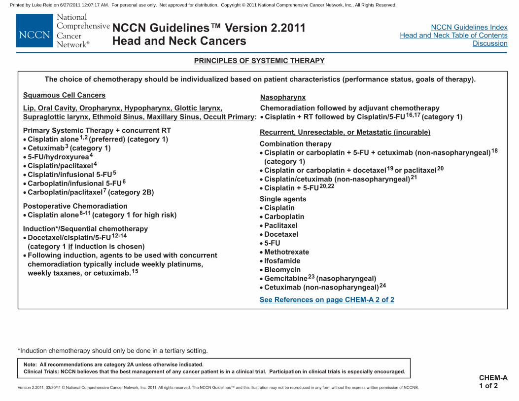
Version 2.2011, 03/30/11 © National Comprehensive Cancer Network, Inc. 2011, All rights reserved. The NCCN Guidelines™ and this illustration may not be reproduced in any form without the express written permission of NCCN®.
NCCN Guidelines IndexHead and Neck Table of Contents
Discussion
Note: All recommendations are category 2A unless otherwise indicated.
Clinical Trials: NCCN believes that the best management of any cancer patient is in a clinical trial. Participation in clinical trials is especially encouraged.
NCCN Guidelines™ Version 2.2011Head and Neck Cancers
PRINCIPLES OF SYSTEMIC THERAPY
The choice of chemotherapy should be individualized based on patient characteristics (performance status, goals of therapy).
Combination therapy
�
�
�
�
Cisplatin or carboplatin + 5-FU + cetuximab (non-nasopharyngeal)
(category 1)
Cisplatin or carboplatin + docetaxel or paclitaxel
Cisplatin/cetuximab (non-nasopharyngeal)
Cisplatin + 5-FU
18
19 20
21
20,22
Recurrent, Unresectable, or Metastatic (incurable)
Lip, Oral Cavity, Oropharynx, Hypopharynx, Glottic larynx,
Supraglottic larynx, Ethmoid Sinus, Maxillary Sinus, Occult Primary:
Nasopharynx
Chemoradiation followed by adjuvant chemotherapy
Cisplatin + RT followed by Cisplatin/5-FU (category 1)� 16,17
Induction*/Sequential chemotherapy
Docetaxel/cisplatin/5-FU
(category 1 induction is chosen)
Following induction, agents to be used with concurrent
chemoradiation typically include weekly platinums,
weekly taxanes, or cetuximab.
�
�
12-14
if
15
Primary Systemic Therapy + concurrent RT
Cisplatin alone (preferred) (category 1)
Cetuximab (category 1)
Carboplatin/paclitaxel (category 2B)
�
�
�
�
�
�
�
1,2
3
5-FU/hydroxyurea
Cisplatin/paclitaxel
Cisplatin/infusional 5-FU
Carboplatin/infusional 5-FU
4
4
5
6
7
See References on page CHEM-A 2 of 2
Squamous Cell Cancers
Postoperative Chemoradiation
Cisplatin alone� 8-11 (category 1 for high risk)
Single agents
�
�
�
�
�
�
�
�
�
�
Cisplatin
Carboplatin
Paclitaxel
Docetaxel
5-FU
Methotrexate
Ifosfamide
Bleomycin
Gemcitabine (nasopharyngeal)
Cetuximab (non-nasopharyngeal)
23
24
*Induction chemotherapy should only be done in a tertiary setting.
CHEM-A1 of 2
Printed by Luke Reid on 6/27/2011 12:07:17 AM. For personal use only. Not approved for distribution. Copyright © 2011 National Comprehensive Cancer Network, Inc., All Rights Reserved.

Version 2.2011, 03/30/11 © National Comprehensive Cancer Network, Inc. 2011, All rights reserved. The NCCN Guidelines™ and this illustration may not be reproduced in any form without the express written permission of NCCN®.
NCCN Guidelines IndexHead and Neck Table of Contents
Discussion
Note: All recommendations are category 2A unless otherwise indicated.
Clinical Trials: NCCN believes that the best management of any cancer patient is in a clinical trial. Participation in clinical trials is especially encouraged.
NCCN Guidelines™ Version 2.2011Head and Neck Cancers
PRINCIPLES OF SYSTEMIC THERAPY (REFERENCES)
1
2
3
4
5
6
7
8
9
10
11
12
13
14
15
16
17
18
19
20
21
22
23
24
Forastiere A, Maor M, Weber R, et al. Long term results of Intergroup RTOG 91-11: APhase III trial to preserve the larynx - Induction cisplatin/5-FU and radiation therapy versusconcurrent cisplatin and radiation therapy versus radiation therapy [abstract]. J Clin Oncol2006;24:Abstract 5517.
Adelstein DJ, Li Y, Adams GL, et al. An intergroup phase III comparison of standardradiation therapy and two schedules of concurrent chemoradiotherapy in patients withunresectable squamous cell head and neck cancer. J Clin Oncol 2003;21(1):92-98.
Bonner JA, Harari PM, Giralt J, et al. Radiotherapy plus cetuximab for locoregionallyadvanced head and neck cancer: 5-year survival data from a phase 3 randomised trial,and relation between cetuximab-induced rash and survival. Lancet Oncol 2010;11:21-28.Epub 2009 Nov 10.
Garden AS, Harris J, Vokes EE, et al. Preliminary results of Radiation Therapy OncologyGroup 97-03: A randomized phase II trial of concurrent radiation and chemotherapy foradvanced squamous cell carcinomas of the head and neck.J Clin Oncol 2004;22:2856-2864.
Taylor S, Murthy A, Vannetzel J, et al. Randomized comparison of neoadjuvant cisplatinand fluorouracil infusion followed by radiation versus concomitant treatment in advancedhead and neck cancer. J Clin Oncol 1994;12:385-395.
Denis F, Garaud P, Bardet E, et al. Final results of the 94-01 French Head and NeckOncology and Radiotherapy Group randomized trial comparing radiotherapy alone withconcomitant radiochemotherapy in advanced-stage oropharynx carcinoma. J Clin Oncol2004;22(1):69-76. Epub 2003 Dec 2.
Suntharalingam M, Haas ML, Conley BA, et al. The use of carboplatin and paclitaxel withdaily radiotherapy in patients with locally advanced squamous cell carcinomas of the headand neck. Int J Radiat Oncol Biol Phys 2000;47:49-56.
Cooper JS, Pajak TF, Forastiere AA, et al. Postoperative concurrent radiotherapy andchemotherapy for high-risk squamous-cell carcinoma of the head and neck. N Engl J Med2004;350:1937-44.
Bernier J, Domenge C, Ozsahin M, et al. Postoperative irradiation with or withoutconcomitant chemotherapy for locally advanced head and neck cancer. N Engl J Med2004;350:1945-1952.
Bernier J, Cooper JS, Pajak TF, et al. Defining risk levels in locally advanced head andneck cancers: A comparative analysis of concurrent postoperative radiation pluschemotherapy trials of the EORTC (#22931) and RTOG (# 9501). Head Neck2005;27:843-850.
Bachaud JM, Cohen-Jonathan E, Alzieu C, et al. Combined postoperative radiotherapyand weekly cisplatin infusion for locally advanced head and neck carcinoma: final report ofa randomized trial. Int J Radiat Oncol Biol Phys. 1996 Dec 1;36(5):999-1004.
Vermorken JB, Remenar E, van Herpen C, et al; EORTC 24971/TAX 323 Study Group.Cisplatin, fluorouracil, and docetaxel in unresectable head and neck cancer. N Engl J Med2007;357(17):1695-1704.
Posner MR, Hershock DM, Blajman CR, et al. Cisplatin and fluorouracil alone or withdocetaxel in head and neck cancer. N Engl J Med 2007;357(17):1705-1715.
Pointreau Y, Garaud P, Chapet S, et al. Randomized trial of induction chemotherapy withcisplatin and 5-fluorouracil with or without docetaxel for larynx preservation. J Natl CancerInst 2009;101:498-506.
Chitapanarux I, Lorvidhaya V, Kamnerdsupaphon P, et al. Chemoradiation comparingcisplatin versus carboplatin in locally advanced nasopharyngeal cancer: Randomised, non-inferiority, open trial. Eur J Cancer 2007;43:1399-1406.
Al-Sarraf M, LeBlanc M, Giri PG, et al. Chemoradiotherapy versus radiotherapy in patientswith advanced nasopharyngeal cancer: phase III randomized Intergroup study 0099. J ClinOncol 1998;16:1310-1317.
Chan AT, Leung SF, Ngan RK, et al. Overall survival after concurrent cisplatin-radiotherapycompared with radiotherapy alone in locoregionally advanced nasopharyngeal carcinoma. JNatl Cancer Inst 2005;97:536-539.
Vermorken JB, Mesia R, Rivera F, et al. Platinum-based chemotherapy plus cetuximab inhead and neck cancer. N Engl J Med 2008;359:1116-1127.
Samlowski WE, Moon J, Kuebler JP, et al. Evaluation of the combination ofdocetaxel/carboplatin in patients with metastatic or recurrent squamous cell carcinoma ofthe head and neck (SCCHN): a Southwest Oncology Group Phase II study. Cancer Invest2007;25:182-188.
Gibson MK, Li Y, Murphy B, et al. Randomized phase III evaluation of cisplatin plusfluorouracil versus cisplatin plus paclitaxel in advanced head and neck cancer (E1395): AnIntergroup Trial of the Eastern Cooperative Oncology Group. J Clin Oncol2005;23(15):3562-3567.
Burtness B, Goldwasser MA, Flood W, et al. Phase III randomized trial of cisplatin plusplacebo versus cisplatin plus cetuximab in metastatic/recurrent head and neck cancer: AnEastern Cooperative Oncology Group Study. J Clin Oncol 2005;23:8646-8654.
Forastiere AA, Metch B, Schuller DE, et al. Randomized comparison of cisplatin plusflurouracil and carboplatin plus fluorouracil versus methotrexate in advanced squamous cellcarcinoma of the head and neck: A Southwest Oncology Group Study. J Clin Oncol1992;10(8):1245-1251.
Zhang L, Zhang Y, Huang PY, et al. Phase II clinical study of gemcitabine in the treatmentof patients with advanced nasopharyngeal carcinoma after the failure of platinum-basedchemotherapy. Cancer Chemother Pharmacol 2008;61(1):33-38. Epub 2007 Mar 20.
Vermorken JB, Trigo J, Hitt R, et al. Open-label, uncontrolled, multicenter phase II study toevaluate the efficacy and toxicity of cetuximab as a single agent in patients with recurrentand/or metastatic squamous cell carcinoma of the head and neck who failed to respond toplatinum-based therapy. J Clin Oncol 2007;25(16):2171-2177.
CHEM-A2 of 2
Printed by Luke Reid on 6/27/2011 12:07:17 AM. For personal use only. Not approved for distribution. Copyright © 2011 National Comprehensive Cancer Network, Inc., All Rights Reserved.

Version 2.2011, 03/30/11 © National Comprehensive Cancer Network, Inc. 2011, All rights reserved. The NCCN Guidelines™ and this illustration may not be reproduced in any form without the express written permission of NCCN®.
NCCN Guidelines IndexHead and Neck Table of Contents
Discussion
NCCN Guidelines™ Version 2.2011 StagingHead and Neck Cancers
Table 1
American Joint Committee on Cancer (AJCC)
TNM Staging Classification for
Primary Tumor (T)TXT0TisT1T2
T3T4a
T4b
Regional Lymph Nodes (N)NXN0N1
N2
N2a
N2b
N2c
N3
Distant Metastasis (M)M0M1
the Lip and Oral Cavity
(Nonepithelial tumors such as those of lymphoid tissue, soft tissue,
bone, and cartilage are not included)
Primary tumor cannot be assessedNo evidence of primary tumorCarcinomaTumor 2 cm or less in greatest dimensionTumor more than 2 cm but not more than 4 cm in greatestdimensionTumor more than 4 cm in greatest dimension
Tumor invades adjacent structures (eg,through cortical bone [mandible or maxilla] into deep[extrinsic] muscle of tongue [genioglossus, hyoglossus,palatoglossus, and styloglossus], maxillary sinus, skin offace)Very advanced local diseaseTumor invades masticator space, pterygoid plates, or skullbase and/or encases internal carotid artery
Regional lymph nodes cannot be assessedNo regional lymph node metastasisMetastasis in a single ipsilateral lymph node, 3 cm orless in greatest dimensionMetastasis in a single ipsilateral lymph node, more than3 cm but not more than 6 cm in greatest dimension; or inmultiple ipsilateral lymph nodes, none more than 6 cm ingreatest dimension; or in bilateral or contralateral lymphnodes, none more than 6 cm in greatest dimensionMetastasis in single ipsilateral lymph node more than 3cm but not more than 6 cm in greatest dimensionMetastasis in multiple ipsilateral lymph nodes, nonemore than 6 cm in greatest dimensionMetastasis in bilateral or contralateral lymph nodes,none more than 6 cm in greatest dimensionMetastasis in a lymph node more than 6 cm in greatestdimension
No distant metastasisDistant metastasis
in situ
*Note: Superficial erosion alone of bone/tooth socket by gingival primary isnot sufficient to classify a tumor as T4.
(7th ed., 2010)
Moderately advanced local disease*(lip) Tumor invades through cortical bone, inferior alveolarnerve, floor of mouth, or skin of face, that is, chin or nose(oral cavity)
Histologic Grade (G)GXG1G2G3G4
Grade cannot be assessedWell differentiatedModerately differentiatedPoorly differentiatedUndifferentiated
Continued...
Used with the permission of the American Joint Committee on Cancer (AJCC), Chicago, Illinois. The original and primary source for this information is the AJCCCancer Staging Manual, Seventh Edition (2010) published by Springer Science and Business Media LLC (SBM). (For complete information and data supportingthe staging tables, visit .) Any citation or quotation of this material must be credited to the AJCC as its primary source. The inclusion of thisinformation herein does not authorize any reuse or further distribution without the expressed, written permission of Springer SBM, on behalf of the AJCC.
www.springer.com
ST-1
Printed by Luke Reid on 6/27/2011 12:07:17 AM. For personal use only. Not approved for distribution. Copyright © 2011 National Comprehensive Cancer Network, Inc., All Rights Reserved.

Version 2.2011, 03/30/11 © National Comprehensive Cancer Network, Inc. 2011, All rights reserved. The NCCN Guidelines™ and this illustration may not be reproduced in any form without the express written permission of NCCN®.
NCCN Guidelines IndexHead and Neck Table of Contents
Discussion
Used with the permission of the American Joint Committee on Cancer (AJCC), Chicago, Illinois. The original and primary source for this information is the AJCCCancer Staging Manual, Seventh Edition (2010) published by Springer Science and Business Media LLC (SBM). (For complete information and data supportingthe staging tables, visit .) Any citation or quotation of this material must be credited to the AJCC as its primary source. The inclusion of thisinformation herein does not authorize any reuse or further distribution without the expressed, written permission of Springer SBM, on behalf of the AJCC.
www.springer.com
NCCN Guidelines™ Version 2.2011 StagingHead and Neck Cancers
Table 1 - Continued
American Joint Committee on Cancer (AJCC)
TNM Staging Classification for
Anatomic Stage/Prognostic GroupsStage 0Stage IStage IIStage III
Stage IVA
Stage IVB
Stage IVC
the Lip and Oral Cavity
(Nonepithelial tumors such as those of lymphoid tissue, soft tissue,
bone, and cartilage are not included)
Tis N0 M0T1 N0 M0T2 N0 M0T3 N0 M0T1 N1 M0T2 N1 M0T3 N1 M0
T4a N0 M0T4a N1 M0T1 N2 M0T2 N2 M0T3 N2 M0
T4a N2 M0Any T N3 M0T4b Any N M0
Any T Any N M1
(7th ed., 2010)
ST-2
Printed by Luke Reid on 6/27/2011 12:07:17 AM. For personal use only. Not approved for distribution. Copyright © 2011 National Comprehensive Cancer Network, Inc., All Rights Reserved.

Version 2.2011, 03/30/11 © National Comprehensive Cancer Network, Inc. 2011, All rights reserved. The NCCN Guidelines™ and this illustration may not be reproduced in any form without the express written permission of NCCN®.
NCCN Guidelines IndexHead and Neck Table of Contents
Discussion
ST-3
NCCN Guidelines™ Version 2.2011 StagingHead and Neck Cancers
Table 2:
American Joint Committee on Cancer (AJCC)(7th ed., 2010)TNM Staging System for the Pharynx
(Nonepithelial tumors such as those of lymphoid tissue, soft tissue,bone, and cartilage are not included)
Primary Tumor (T)TXT0Tis
NasopharynxT1
T2T3
T4
OropharynxT1T2
T3
T4a
T4b
T2
T3
T4a
T4b
Primary tumor cannot be assessedNo evidence of primary tumorCarcinoma
Tumor confined to the nasopharynx, or tumor extends tooropharynx and/or nasal cavity without parapharyngealextension*Tumor with parapharyngeal extension*Tumor involves bony structures of skull base and/orparanasal sinusesTumor with intracranial extension and/or involvement ofcranial nerves, hypopharynx, orbit, or with extension tothe infratemporal fossa/masticator space
Tumor 2 cm or less in greatest dimensionTumor more than 2 cm but not more than 4 cm in greatestdimensionTumor more than 4 cm in greatest dimension or extensionto lingual surface of epiglottisModerately advanced local diseaseTumor invades the larynx, extrinsic muscle of tongue,medial pterygoid, hard palate, or mandible*Very advanced local diseaseTumor invades lateral pterygoid muscle, pterygoid plates,lateral nasopharynx, or skull base or en
mited to one subsite of hypopharynx and/or 2 cmor less in greatest dimensionTumor invades more than one subsite of hypopharynx oran adjacent site, or measures more than 2 cm but notmore than 4 cm in greatest diameter without fixation ofhemilarynxTumor more than 4 cm in greatest dimension or withfixation of hemilarynx or extension to esophagusModerately advanced local diseaseTumor invades thyroid/cricoid cartilage, hyoid bone,thyroid gland, or central compartment soft tissue**Very advanced local diseaseTumor invades prevertebral fascia, encases carotidartery, or involves mediastinal structures
in situ
*Note: Parapharyngeal extension denotes posterolateral infiltration oftumor.
cases carotidartery
Tumor li
*Note: Mucosal extension to lingual surface of epiglottis from primarytumors of the base of the tongue and vallecula does not constitute invasionof larynx.
**Note: Central compartment soft tissue includes prelaryngeal strapmuscles and subcutaneous fat.
HypopharynxT1
Continued...
Used with the permission of the American Joint Committee on Cancer (AJCC),Chicago, Illinois. The original and primary source for this information is theAJCC Cancer Staging Manual, Seventh Edition (2010) published by SpringerScience and Business Media LLC (SBM). (For complete information and datasupporting the staging tables, visit .) Any citation orquotation of this material must be credited to the AJCC as its primary source.The inclusion of this information herein does not authorize any reuse orfurther distribution without the expressed, written permission of SpringerSBM, on behalf of the AJCC.
www.springer.com
Printed by Luke Reid on 6/27/2011 12:07:17 AM. For personal use only. Not approved for distribution. Copyright © 2011 National Comprehensive Cancer Network, Inc., All Rights Reserved.

Version 2.2011, 03/30/11 © National Comprehensive Cancer Network, Inc. 2011, All rights reserved. The NCCN Guidelines™ and this illustration may not be reproduced in any form without the express written permission of NCCN®.
NCCN Guidelines IndexHead and Neck Table of Contents
Discussion
ST-4
Used with the permission of the American Joint Committee on Cancer (AJCC), Chicago, Illinois. The original and primary source for this information is the AJCCCancer Staging Manual, Seventh Edition (2010) published by Springer Science and Business Media LLC (SBM). (For complete information and data supportingthe staging tables, visit .) Any citation or quotation of this material must be credited to the AJCC as its primary source. The inclusion of thisinformation herein does not authorize any reuse or further distribution without the expressed, written permission of Springer SBM, on behalf of the AJCC.
www.springer.com
NCCN Guidelines™ Version 2.2011 StagingHead and Neck Cancers
Table 2 - Continued
American Joint Committee on Cancer (AJCC)
Regional Lymph Nodes (N) :
TNM Staging System for the Pharynx
Regional Lymph Nodes (N):
(Nonepithelial tumors such as those of lymphoid tissue, soft tissue,bone, and cartilage are not included)
Na Oropharynx and HypopharynxNX
N0
N1
N2
N2a
N2b
N2c
N3
Distant Metastasis (M)
M0
M1
**Supraclavicular zone or fossa is relevant to the staging ofnasopharyngeal carcinoma and is the triangular region originally describedby Ho. It is defined by three points: (1) the superior margin of the sternalend of the clavicle; (2) the superior margin of the lateral end of the clavicle,and (3) the point where the neck meets the shoulder. Note that this wouldinclude caudal portions of levels IV and VB. All cases with lymph nodes(whole or part) in the fossa are considered N3b.
Regional lymph nodes cannot be assessed
No regional lymph node metastasis
Metastasis in a single ipsilateral lymph node, 3 cm or lessin greatest dimension
Metastasis in a single ipsilateral lymph node, more than 3cm but not more than 6 cm in greatest dimension, or inmultiple ipsilateral lymph nodes, none more than 6 cm ingreatest dimension, or in bilateral or contralateral lymphnodes, none more than 6 cm in greatest dimension
Metastasis in a single ipsilateral lymph node more than 3cm but not more than 6 cm in greatest dimension
Metastasis in multiple ipsilateral lymph nodes, none morethan 6 cm in greatest dimension
Metastasis in bilateral or contralateral lymph nodes, nonemore than 6 cm in greatest dimension
Metastasis in a lymph node more than 6 cm in greatestdimension
No distant metastasis
Distant metastasis
Note: Metastases at level VII are considered regional lymph node
metastases.
(7th ed., 2010)
†
†
sopharynx
NXN0N1
N2
N3
N3aN3b
The distribution and the prognostic impact of regional lymph nodespread from nasopharynx cancer, particularly of the undifferentiatedtype, are different from those of other head and neck mucosalcancers and justify the use of a different N classification system.
Regional lymph nodes cannot be assessedNo regional lymph node metastasisUnilateral metastasis in cervical lymph node(s), 6 cm orless in greatest dimension, above the supraclavicularfossa, and/or unilateral or bilateral, retropharyngeallymph nodes, 6 cm or less, in greatest dimension*Bilateral metastasis in cervical lymph node(s), 6 cm orless in greatest dimension, above the supraclavicularfossa*Metastasis in a lymph node(s)* > 6 cm and/or tosupraclavicular fossaMore than 6 cm in dimensionExtension to the supraclavicular fossa**
*Note: Midline nodes are considered ipsilateral nodes.
Continued...
Printed by Luke Reid on 6/27/2011 12:07:17 AM. For personal use only. Not approved for distribution. Copyright © 2011 National Comprehensive Cancer Network, Inc., All Rights Reserved.

Version 2.2011, 03/30/11 © National Comprehensive Cancer Network, Inc. 2011, All rights reserved. The NCCN Guidelines™ and this illustration may not be reproduced in any form without the express written permission of NCCN®.
NCCN Guidelines IndexHead and Neck Table of Contents
Discussion
ST-5
Used with the permission of the American Joint Committee on Cancer (AJCC), Chicago, Illinois. The original and primary source for this information is the AJCCCancer Staging Manual, Seventh Edition (2010) published by Springer Science and Business Media LLC (SBM). (For complete information and data supportingthe staging tables, visit .) Any citation or quotation of this material must be credited to the AJCC as its primary source. The inclusion of thisinformation herein does not authorize any reuse or further distribution without the expressed, written permission of Springer SBM, on behalf of the AJCC.
www.springer.com
NCCN Guidelines™ Version 2.2011 StagingHead and Neck Cancers
Histologic Grade (G)GXG1G2G3G4
Grade cannot be assessedWell differentiatedModerately differentiatedPoorly differentiatedUndifferentiated
Anatomic Stage/Prognostic Groups: Nasopharynx
Stage 0
Stage I
Stage II
Stage III
Stage IVA
Stage IVB
Stage IVC
Tis N0 M0
T1 N0 M0
T1 N1 M0
T2 N0 M0
T2 N1 M0
T1 N2 M0
T2 N2 M0
T3 N0 M0
T3 N1 M0
T3 N2 M0
T4 N0 M0
T4 N1 M0
T4 N2 M0
Any T N3 M0
Any T Any N M1
Anatomic Stage/Prognostic Groups: Oropharynx, Hypopharynx
Stage 0
Stage I
Stage II
Stage III
Stage IVA
Stage IVB
Stage IVC
Tis N0 M0
T1 N0 M0
T2 N0 M0
T3 N0 M0
T1 N1 M0
T2 N1 M0
T3 N1 M0
T4a N0 M0
T4a N1 M0
T1 N2 M0
T2 N2 M0
T3 N2 M0
T4a N2 M0
T4b Any N M0
Any T N3 M0
Any T Any N M1
Table 2 - Continued
American Joint Committee on Cancer (AJCC)TNM Staging System for the Pharynx(Nonepithelial tumors such as those of lymphoid tissue, soft tissue, bone, and cartilage are not included)
(7th ed., 2010)
Printed by Luke Reid on 6/27/2011 12:07:17 AM. For personal use only. Not approved for distribution. Copyright © 2011 National Comprehensive Cancer Network, Inc., All Rights Reserved.

Version 2.2011, 03/30/11 © National Comprehensive Cancer Network, Inc. 2011, All rights reserved. The NCCN Guidelines™ and this illustration may not be reproduced in any form without the express written permission of NCCN®.
NCCN Guidelines IndexHead and Neck Table of Contents
Discussion
ST-6
Used with the permission of the American Joint Committee on Cancer (AJCC), Chicago, Illinois. The original and primary source for this information is the AJCCCancer Staging Manual, Seventh Edition (2010) published by Springer Science and Business Media LLC (SBM). (For complete information and data supportingthe staging tables, visit .) Any citation or quotation of this material must be credited to the AJCC as its primary source. The inclusion of thisinformation herein does not authorize any reuse or further distribution without the expressed, written permission of Springer SBM, on behalf of the AJCC.
www.springer.com
NCCN Guidelines™ Version 2.2011 StagingHead and Neck Cancers
Table 3
American Joint Committee on Cancer (AJCC) TNM Staging System for the Larynx (7th ed., 2010)
(Nonepithelial tumors such as those of lymphoid tissue, soft tissue, bone, and cartilage are not included)
GlottisT1
T1aT1b
T2
T3
T4a
T4b
SubglottisT1T2
T3T4a
T4b
Tumor limited to the vocal cord(s) (may involve anterior orposterior commissure) with normal mobilityTumor limited to one vocal cordTumor involves both vocal cordsTumor extends to supraglottis and/or subglottis, and/or withimpaired vocal cord mobilityTumor limited to the larynx with vocal cord fixation and/orinvasion of paraglottic space, and/or inner cortex of thethyroid cartilageModerately advanced local diseaseTumor invades through the outer cortex of the thyroidcartilage and/or invades tissues beyond the larynx (eg,trachea, soft tissues of neck including deep extrinsicmuscle of the tongue, strap muscles, thyroid, or esophagus)Very advanced local diseaseTumor invades prevertebral space, encases carotid artery,or invades mediastinal structures
Tumor limited to the subglottisTumor extends to vocal cord(s) with normal or impairedmobilityTumor limited to larynx with vocal cord fixationModerately advanced local diseaseTumor invades cricoid or thyroid cartilage and/or invadestissues beyond the larynx (eg, trachea, soft tissues of neckincluding deep extrinsic muscles of the tongue, strapmuscles, thyroid, or esophagus)Very advanced local diseaseTumor invades prevertebral space, encases carotid artery,or invades mediastinal structures
Primary Tumor (T)
TXT0Tis
Supraglottis
T1
T2
T3
T4a
T4b
Primary tumor cannot be assessedNo evidence of primary tumorCarcinoma
Tumor limited to one subsite of supraglottis with normalvocal cord mobilityTumor invades mucosa of more than one adjacent subsite ofsupraglottis or glottis or region outside the supraglottis (eg,mucosa of base of tongue, vallecula, medial wall of pyriformsinus) without fixation of the larynxTumor limited to larynx with vocal cord fixation and/orinvades any of the following: postcricoid area, pre-epiglotticspace, paraglottic space, and/or inner cortex of thyroidcartilageModerately advanced local diseaseTumor invades through the thyroid cartilage and/or invadestissues beyond the larynx (eg, trachea, soft tissues of neckincluding deep extrinsic muscle of the tongue, strapmuscles, thyroid, or esophagus)Very advanced local diseaseTumor invades prevertebral space, encases carotid artery,or invades mediastinal structures
in situ
Continued on next page
Printed by Luke Reid on 6/27/2011 12:07:17 AM. For personal use only. Not approved for distribution. Copyright © 2011 National Comprehensive Cancer Network, Inc., All Rights Reserved.

Version 2.2011, 03/30/11 © National Comprehensive Cancer Network, Inc. 2011, All rights reserved. The NCCN Guidelines™ and this illustration may not be reproduced in any form without the express written permission of NCCN®.
NCCN Guidelines IndexHead and Neck Table of Contents
Discussion
ST-7
Used with the permission of the American Joint Committee on Cancer (AJCC), Chicago, Illinois. The original and primary source for this information is the AJCCCancer Staging Manual, Seventh Edition (2010) published by Springer Science and Business Media LLC (SBM). (For complete information and data supportingthe staging tables, visit .) Any citation or quotation of this material must be credited to the AJCC as its primary source. The inclusion of thisinformation herein does not authorize any reuse or further distribution without the expressed, written permission of Springer SBM, on behalf of the AJCC.
www.springer.com
NCCN Guidelines™ Version 2.2011 StagingHead and Neck Cancers
Regional Lymph Nodes (N)*NX
N1
N2
N2a
N2b
N2c
N3
Distant Metastasis (M)M0M1
Regional lymph nodes cannot be assessed N0; noregional lymph node metastasisMetastasis in a single ipsilateral lymph node, 3 cm orless in greatest dimensionMetastasis in a single ipsilateral lymph node, more than3 cm but not more than 6 cm in greatest dimension; orin multiple ipsilateral lymph nodes, none more than 6cm in greatest dimension; or in bilateral or contralaterallymph nodes, none more than 6 cm in greatestdimensionMetastasis in a single ipsilateral lymph node, more than3 cm but not more than 6 cm in greatest dimensionMetastasis in multiple ipsilateral lymph nodes, nonemore than 6 cm in greatest dimensionMetastasis in bilateral or contralateral lymph nodes,none more than 6 cm in greatest dimensionMetastasis in a lymph node, more than 6 cm in greatestdimension
No distant metastasisDistant metastasis
*Note: Metastases at level VII are considered regional lymph node
metastases.
Table 3 - continued
American Joint Committee on Cancer (AJCC)TNM Staging System for the Larynx (7th ed., 2010)
(Nonepithelial tumors such as those of lymphoid tissue, soft tissue, bone, and cartilage are not included)
Anatomic Stage/Prognostic Groups
Stage 0
Stage I
Stage II
Stage III
Stage IVA
Stage IVB
Stage IVC
Tis N0 M0
T1 N0 M0
T2 N0 M0
T3 N0 M0
T1 N1 M0
T2 N1 M0
T3 N1 M0
T4a N0 M0
T4a N1 M0
T1 N2 M0
T2 N2 M0
T3 N2 M0
T4a N2 M0
T4b Any N M0
Any T N3 M0
Any T Any N M1
Histologic Grade (G)GXG1G2G3G4
Grade cannot be assessedWell differentiatedModerately differentiatedPoorly differentiatedUndifferentiated
Printed by Luke Reid on 6/27/2011 12:07:17 AM. For personal use only. Not approved for distribution. Copyright © 2011 National Comprehensive Cancer Network, Inc., All Rights Reserved.

Version 2.2011, 03/30/11 © National Comprehensive Cancer Network, Inc. 2011, All rights reserved. The NCCN Guidelines™ and this illustration may not be reproduced in any form without the express written permission of NCCN®.
NCCN Guidelines IndexHead and Neck Table of Contents
Discussion
ST-8
NCCN Guidelines™ Version 2.2011 StagingHead and Neck Cancers
Continued on next page
Table 4
American Joint Committee on Cancer (AJCC)
TNM Staging System for the Nasal Cavity and Paranasal Sinuses
(7th ed., 2010)
(Nonepithelial tumors such as those of lymphoid tissue, soft tissue, bone,
and cartilage are not included)Primary Tumor (T)TXT0Tis
Maxillary SinusT1
T2
T3
T4a
T4b
Nasal Cavity and Ethmoid SinusT1
T2
T3
T4a
T4b
Regional Lymph Nodes (N)NXN0N1
N2
N2a
N2b
N2c
N3
Distant Metastasis (M)
M0
M1
Primary tumor cannot be assessedNo evidence of primary tumorCarcinoma in situ
Tumor limited to maxillary sinus mucosa with no erosion ordestruction of boneTumor causing bone erosion or destruction includingextension into the hard palate and/or middle nasal meatus,except extension to posterior wall of maxillary sinus andpterygoid platesTumor invades any of the following: bone of the posteriorwall of maxillary sinus, subcutaneous tissues, floor ormedial wall of orbit, pterygoid fossa, ethmoid sinusesModerately advanced local diseaseTumor invades anterior orbital contents, skin of cheek,pterygoid plates, infratemporal fossa, cribriform plate,sphenoid or frontal sinusesVery advanced local diseaseTumor invades any of the following: orbital apex, dura,brain, middle cranial fossa, cranial nerves other thanmaxillary division of trigeminal nerve (V ), nasopharynx, orclivus
Tumor restricted to any one subsite, with or without bonyinvasionTumor invading two subsites in a single region or extendingto involve an adjacent region within the nasoethmoidalcomplex, with or without bony invasion
Tumor extends to invade the medial wall or floor of theorbit, maxillary sinus, palate, or cribriform plateModerately advanced local diseaseTumor invades any of the following: anterior orbitalcontents, skin of nose or cheek, minimal extension toanterior cranial fossa, pterygoid plates, sphenoid orfrontal sinusesVery advanced local diseaseTumor invades any of the following: orbital apex, dura,brain, middle cranial fossa, cranial nerves other than(V ), nasopharynx, or clivus
Regional lymph nodes cannot be assessedNo regional lymph node metastasisMetastasis in a single ipsilateral lymph node, 3 cm or lessin greatest dimensionMetastasis in a single ipsilateral lymph node, more than3 cm but not more than 6 cm in greatest dimension; or inmultiple ipsilateral lymph nodes, none more than 6 cm ingreatest dimension; or in bilateral or contralateral lymphnodes, none more than 6 cm in greatest dimensionMetastasis in a single ipsilateral lymph node, more than3 cm but not more than 6 cm in greatest dimensionMetastasis in multiple ipsilateral lymph nodes, none morethan 6 cm in greatest dimensionMetastasis in bilateral or contralateral lymph nodes, nonemore than 6 cm in greatest dimensionMetastasis in a lymph node, more than 6 cm in greatestdimension
No distant metastasis
Distant metastasis
2
2
Used with the permission of the American Joint Committee on Cancer (AJCC), Chicago, Illinois. The original and primary source for this information is the AJCC Cancer StagingManual, Seventh Edition (2010) published by Springer Science and Business Media LLC (SBM). (For complete information and data supporting the staging tables, visit
.) Any citation or quotation of this material must be credited to the AJCC as its primary source. The inclusion of this information herein does not authorize anyreuse or further distribution without the expressed, written permission of Springer SBM, on behalf of the AJCC.www.springer.com
Printed by Luke Reid on 6/27/2011 12:07:17 AM. For personal use only. Not approved for distribution. Copyright © 2011 National Comprehensive Cancer Network, Inc., All Rights Reserved.

Version 2.2011, 03/30/11 © National Comprehensive Cancer Network, Inc. 2011, All rights reserved. The NCCN Guidelines™ and this illustration may not be reproduced in any form without the express written permission of NCCN®.
NCCN Guidelines IndexHead and Neck Table of Contents
Discussion
ST-9
Used with the permission of the American Joint Committee on Cancer (AJCC), Chicago, Illinois. The original and primary source for this information is the AJCCCancer Staging Manual, Seventh Edition (2010) published by Springer Science and Business Media LLC (SBM). (For complete information and data supportingthe staging tables, visit .) Any citation or quotation of this material must be credited to the AJCC as its primary source. The inclusion of thisinformation herein does not authorize any reuse or further distribution without the expressed, written permission of Springer SBM, on behalf of the AJCC.
www.springer.com
NCCN Guidelines™ Version 2.2011 StagingHead and Neck Cancers
Histologic Grade (G)GXG1G2G3G4
Grade cannot be assessedWell differentiatedModerately differentiatedPoorly differentiatedUndifferentiated
Table - Continued
Anatomic Stage/Prognostic Groups
Stage 0Stage IStage IIStage III
Stage IVA
Stage IVB
Stage IVC
Tis N0 M0T1 N0 M0T2 N0 M0T3 N0 M0T1 N1 M0T2 N1 M0T3 N1 M0
T4a N0 M0T4a N1 M0T1 N2 M0T2 N2 M0T3 N2 M0
T4a N2 M0T4b Any N M0
Any T N3 M0Any T Any N M1
4
American Joint Committee on Cancer (AJCC)
TNM Staging System for the Nasal Cavity and Paranasal Sinuses (7th ed., 2010)
(Nonepithelial tumors such as those of lymphoid tissue, soft tissue, bone, and cartilage are not included)
Printed by Luke Reid on 6/27/2011 12:07:17 AM. For personal use only. Not approved for distribution. Copyright © 2011 National Comprehensive Cancer Network, Inc., All Rights Reserved.
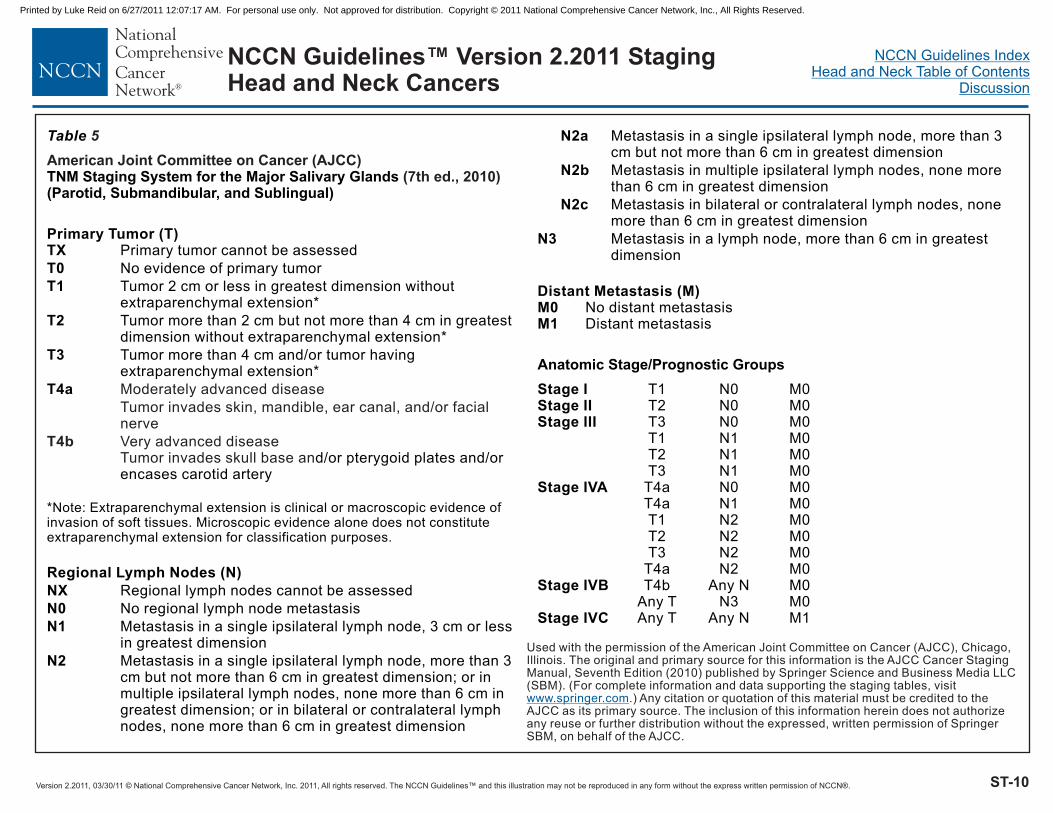
Version 2.2011, 03/30/11 © National Comprehensive Cancer Network, Inc. 2011, All rights reserved. The NCCN Guidelines™ and this illustration may not be reproduced in any form without the express written permission of NCCN®.
NCCN Guidelines IndexHead and Neck Table of Contents
Discussion
ST-10
NCCN Guidelines™ Version 2.2011 StagingHead and Neck Cancers
Table
TNM Staging System for the Major Salivary Glands(Parotid, Submandibular, and Sublingual)
Distant Metastasis (M)
Anatomic Stage/Prognostic Groups
Primary Tumor (T)TX
T0
T1
T2
T3
T4a
Regional Lymph Nodes (N)
NX
N0
N1
N2
N2a
N2b
N2c
N3
M0M1
Stage IStage IIStage III
Stage IVA
Stage IVB
Stage IVC
Primary tumor cannot be assessed
No evidence of primary tumor
Tumor 2 cm or less in greatest dimension withoutextraparenchymal extension*
Tumor more than 2 cm but not more than 4 cm in greatestdimension without extraparenchymal extension*
Tumor more than 4 cm and/or tumor havingextraparenchymal extension*
d/or pterygoid plates and/orencases carotid artery
Regional lymph nodes cannot be assessed
No regional lymph node metastasis
Metastasis in a single ipsilateral lymph node, 3 cm or lessin greatest dimension
Metastasis in a single ipsilateral lymph node, more than 3cm but not more than 6 cm in greatest dimension; or inmultiple ipsilateral lymph nodes, none more than 6 cm ingreatest dimension; or in bilateral or contralateral lymphnodes, none more than 6 cm in greatest dimension
Metastasis in a single ipsilateral lymph node, more than 3cm but not more than 6 cm in greatest dimension
Metastasis in multiple ipsilateral lymph nodes, none morethan 6 cm in greatest dimension
Metastasis in bilateral or contralateral lymph nodes, nonemore than 6 cm in greatest dimension
Metastasis in a lymph node, more than 6 cm in greatestdimension
No distant metastasisDistant metastasis
T1 N0 M0T2 N0 M0T3 N0 M0T1 N1 M0T2 N1 M0T3 N1 M0
T4a N0 M0T4a N1 M0T1 N2 M0T2 N2 M0T3 N2 M0
T4a N2 M0T4b Any N M0
Any T N3 M0Any T Any N M1
*Note: Extraparenchymal extension is clinical or macroscopic evidence ofinvasion of soft tissues. Microscopic evidence alone does not constituteextraparenchymal extension for classification purposes.
5
Moderately advanced disease
Tumor invades skin, mandible, ear canal, and/or facialnerve
Very advanced diseaseTumor invades skull base an
T4b
American Joint Committee on Cancer (AJCC)(7th ed., 2010)
Used with the permission of the American Joint Committee on Cancer (AJCC), Chicago,Illinois. The original and primary source for this information is the AJCC Cancer StagingManual, Seventh Edition (2010) published by Springer Science and Business Media LLC(SBM). (For complete information and data supporting the staging tables, visit
.) Any citation or quotation of this material must be credited to theAJCC as its primary source. The inclusion of this information herein does not authorizeany reuse or further distribution without the expressed, written permission of SpringerSBM, on behalf of the AJCC.
www.springer.com
Printed by Luke Reid on 6/27/2011 12:07:17 AM. For personal use only. Not approved for distribution. Copyright © 2011 National Comprehensive Cancer Network, Inc., All Rights Reserved.

Version 2.2011, 03/30/11 © National Comprehensive Cancer Network, Inc. 2011, All rights reserved. The NCCN Guidelines™ and this illustration may not be reproduced in any form without the express written permission of NCCN®.
NCCN Guidelines IndexHead and Neck Table of Contents
Discussion
ST-11
Used with the permission of the American Joint Committee on Cancer (AJCC), Chicago, Illinois. The original and primary source for this information is the AJCCCancer Staging Manual, Seventh Edition (2010) published by Springer Science and Business Media LLC (SBM). (For complete information and data supportingthe staging tables, visit .) Any citation or quotation of this material must be credited to the AJCC as its primary source. The inclusion of thisinformation herein does not authorize any reuse or further distribution without the expressed, written permission of Springer SBM, on behalf of the AJCC.
www.springer.com
NCCN Guidelines™ Version 2.2011 StagingHead and Neck Cancers
Table 6
American Joint Committee on Cancer (AJCC)
(7th ed., 2010)
Primary Tumor (T)T3
T4a
T4b
Regional Lymph Nodes (N)
NX
N0
N1
M0M1
Stage IIIStage IVA
Stage IVBStage IVC
TNM Staging System for Mucosal Melanoma of the Head and Neck
Distant Metastasis (M)
Anatomic Stage/Prognostic GroupsMucosal disease
Moderately advanced disease
Tumor involving deep soft tissue, cartilage, bone, oroverlying skin
Very advanced diseaseTumor involving brain, dura, skull base, lower cranialnerves (IX, X, XI, XII), masticator space, carotid artery,prevertebral space, or mediastinal structures
Regional lymph nodes cannot be assessed
No regional lymph node metastases
Regional lymph node metastases present
No distant metastasisDistant metastasis
T3 N0 M0T4a N0 M0
T3-T4a N1 M0T4b Any N M0
Any T Any N M1
Histologic Grade (G)GXG1G2G3G4
Grade cannot be assessedWell differentiatedModerately differentiatedPoorly differentiatedUndifferentiated
Printed by Luke Reid on 6/27/2011 12:07:17 AM. For personal use only. Not approved for distribution. Copyright © 2011 National Comprehensive Cancer Network, Inc., All Rights Reserved.

Version 2.2011, 03/30/11 © National Comprehensive Cancer Network, Inc. 2011, All rights reserved. The NCCN Guidelines™ and this illustration may not be reproduced in any form without the express written permission of NCCN®. MS-1
NCCN Guidelines IndexHead and Neck Table of Contents
Discussion
NCCN Guidelines™ Version 2.2011 Head and Neck Cancers
Discussion
NCCN Categories of Evidence and Consensus
Category 1: The recommendation is based on high-level evidence (e.g. randomized controlled trials) and there is uniform NCCN consensus.
Category 2A: The recommendation is based on lower-level evidence and there is uniform NCCN consensus.
Category 2B: The recommendation is based on lower-level evidence and there is nonuniform NCCN consensus (but no major disagreement).
Category 3: The recommendation is based on any level of evidence but reflects major disagreement.
All recommendations are category 2A unless otherwise noted.
Overview The NCCN Head and Neck (H&N) Cancers guidelines address tumors arising in the lip, oral cavity, pharynx, larynx, and paranasal sinuses (see Figure 1); occult primary cancer, salivary gland cancer, and mucosal melanoma are also addressed.1 A brief overview of the epidemiology and management of H&N cancer is provided.
By definition, the NCCN practice guidelines cannot incorporate all possible clinical variations and are not intended to replace good clinical judgment or individualization of treatments. Exceptions to the rule were discussed among the members of the panel while developing these guidelines. A 5% rule (omitting clinical scenarios that comprise less
than 5% of all cases) was used to eliminate uncommon clinical occurrences or conditions from these guidelines.
Incidence and Etiology It is estimated that about 49,260 new cases of oral cavity, pharyngeal, and laryngeal cancers occurred in 2010, which account for about 3% of new cancer cases in the United States. An estimated 11,480 deaths from H&N cancers occurred during the same time period.2, 3 Squamous cell carcinoma or a variant is the histologic type in over 90% of these tumors. Alcohol and tobacco abuse are common etiologic factors in cancers of the oral cavity, oropharynx, hypopharynx, and larynx. Because the entire aerodigestive tract epithelium may be exposed to these carcinogens, patients with H&N cancer are at risk for developing second primary neoplasms of the H&N, lung, esophagus, and other sites that share these risk factors.
Human papillomavirus (HPV) infection is now well accepted as a risk factor for the development of squamous cancers of the oropharynx (particularly cancers of the lingual and palatine tonsils, and base of tongue).4-10 The overall incidence of HPV positive H&N cancer is increasing in the United States, while the incidence of HPV negative (primarily tobacco- and alcohol-related) cancer is decreasing.11 A strong causal relationship has been established between the HPV type 16 and development of oropharyngeal cancer (see “HPV Testing” in the oropharyngeal section of this Discussion).4 It has not been demonstrated yet whether HPV vaccination will decrease the incidence of HPV positive oropharyngeal cancer.
Staging Stage at diagnosis predicts survival rates and guides management in patients with H&N cancer. The 2010 American Joint Committee on Cancer (AJCC) staging classification (7th edition) was used as a basis
Printed by Luke Reid on 6/27/2011 12:07:17 AM. For personal use only. Not approved for distribution. Copyright © 2011 National Comprehensive Cancer Network, Inc., All Rights Reserved.
Version 2.2011, 03/30/11 © National Comprehensive Cancer Network, Inc. 2011, All rights reserved. The NCCN Guidelines™ and this illustration may not be reproduced in any form without the express written permission of NCCN®. MS-2
NCCN Guidelines IndexHead and Neck Table of Contents
Discussion
NCCN Guidelines™ Version 2.2011 Head and Neck Cancers
for the NCCN's treatment recommendations for H&N cancer.12, 13 The 7th edition of the AJCC staging guidelines became effective January 1, 2010.12 The AJCC added staging criteria for mucosal melanoma in the 7th edition. However, no major changes in the T classification or stage groupings for the other sites have been made in the revisions for the 7th edition for H&N cancer; the minor changes are described below.
The TNM staging systems developed by the AJCC for the lip and oral cavity, pharynx (nasopharynx, oropharynx, and hypopharynx), larynx (glottis and supraglottis), paranasal sinuses (ethmoid and maxillary), major salivary glands (parotid, submandibular, and sublingual), and mucosal melanoma are shown in Tables 1-6, respectively.13 Definitions for regional lymph node (N) involvement and spread to distant metastatic sites (M) are uniform except for N staging of nasopharyngeal carcinoma (see Table 2). Definitions for staging the primary tumor (T), based on its size, are uniform for the lip, oral cavity, and oropharynx. In contrast, T stage is based on subsite involvement and is specific to each subsite for the glottic larynx, supraglottic larynx, hypopharynx, and nasopharynx.
In general, stage I or II disease defines a relatively small primary tumor with no nodal involvement. Stage III or IV cancers include larger primary tumors, which may invade underlying structures, and/or spread to regional nodes. Distant metastases are uncommon at presentation. More advanced TNM stages are associated with worse survival.
The anatomic criteria for definitions of T4a and T4b for the oropharynx, hypopharynx, larynx, nasal cavity, paranasal sinuses, and major salivary glands remain unchanged in the 7th edition of the AJCC staging manual. However, the words “resectable” (T4a) and “unresectable” (T4b) have been replaced by the terms “moderately advanced” (T4a) and “very advanced” (T4b).12 These changes were deemed necessary,
because a substantial proportion of advanced stage malignancies of the H&N, although resectable, are being treated non-surgically. Furthermore, a clear consensus in criteria for resectability can be difficult to obtain. For example, some tumors deemed unresectable are in fact anatomically resectable, but surgery is not pursued because of medical contraindications to surgery or because it is anticipated that surgery will not improve prognosis (see “Resectable versus Unresectable Disease” in the “Head and Neck Surgery” section of this Discussion).
This change in terminology allows revising of stage IV disease into moderately advanced local/regional disease (stage IVA), very advanced local/regional disease (stage IVB), and distant metastatic disease (stage IVC) for many sites (i.e., lip, oral cavity, oropharynx, hypopharynx, larynx, paranasal sinuses, major salivary glands, and mucosal melanoma). Of note, a designation of stage IV disease does not necessarily mean the disease is incurable, particularly in the absence of distant metastases.
An algorithm for mucosal melanoma was added to the NCCN 2010 H&N guidelines. Mucosal melanomas are rare, very aggressive tumors that mainly affect the nasal cavity and paranasal sinuses. Thus, melanomas confined to the mucosa only are T3; those with moderately advanced lesions (involving underlying cartilage or bone) are T4a, and very advanced primary tumors are T4b.
Minor changes were made in the T staging categories for the nasopharynx in the 7th edition of the AJCC (see Table 2).12 Thus, former T2a lesions are now designated T1; therefore, former stage IIA is now stage I. Lesions previously staged as T2b are now T2; therefore, former stage IIB is now stage II. Regardless of unilateral or bilateral location, retropharyngeal lymph nodes are considered N1.
Printed by Luke Reid on 6/27/2011 12:07:17 AM. For personal use only. Not approved for distribution. Copyright © 2011 National Comprehensive Cancer Network, Inc., All Rights Reserved.
Version 2.2011, 03/30/11 © National Comprehensive Cancer Network, Inc. 2011, All rights reserved. The NCCN Guidelines™ and this illustration may not be reproduced in any form without the express written permission of NCCN®. MS-3
NCCN Guidelines IndexHead and Neck Table of Contents
Discussion
NCCN Guidelines™ Version 2.2011 Head and Neck Cancers
Management Approaches Treating the patient with H&N cancer is complex. The specific site of disease, the extent of disease (stage), and the pathologic findings guide the appropriate surgical procedure, radiation targets, dose and fractionation, and indications for chemotherapy. Single-modality treatment with surgery or radiotherapy is generally recommended for the approximately 30%-40% of patients who present with early-stage disease (stage I or stage II). The 2 modalities result in similar survival in these individuals. In contrast, combined modality therapy is generally recommended for the approximately 60% of patients with locally or regionally advanced disease at diagnosis. The treatment of patients with locally advanced T4b or unresectable nodal disease, metastatic disease, or recurrent disease for the following sites (i.e., lip, oral cavity, pharynx, larynx, paranasal sinus, and occult primary cancer) is addressed in the “Very Advanced/Recurrent/Persistent H&N Cancers” section of the 2011 H&N algorithm.
Participation in clinical trials is a preferred or recommended treatment option in many situations. In formulating these H&N guidelines, the panel has tried to make them evidence based while providing a statement of consensus as to the acceptable range of treatment options.
Multidisciplinary Team Involvement The initial evaluation and development of a plan for treating the patient with H&N cancer require a multidisciplinary team of health care providers with expertise in caring for these patients. Similarly, managing and preventing sequelae of radical surgery, radiotherapy, and chemotherapy (e.g., pain, xerostomia, speech and swallowing problems, depression) require professionals familiar with the disease. Follow-up for these sequelae should include a comprehensive H&N
examination. Adequate nutritional support can help to prevent severe weight loss in patients receiving treatment for H&N cancer; therefore, patients should be encouraged to see a dietician.14 Patients should also be encouraged to stop smoking and to modify alcohol consumption if excessive, because these habits may decrease the efficacy of treatment and adversely affect other health outcomes.15, 16 Programs using behavioral counseling combined with medications that promote smoking cessation (approved by the FDA [Food and Drug Administration]) can be very useful (http://www.ahrq.gov/path/tobacco.htm). Specific components of patient support and follow-up are listed in the algorithm (see “Team Approach” in the NCCN 2011 H&N algorithm). The panel also recommends referring to the NCCN Guidelines for Palliative Care.
Comorbidity and Quality of Life Comorbidity Comorbidity refers to the presence of concomitant disease (in addition to H&N cancer) that may affect the diagnosis, treatment, and prognosis for the patient.17-19 Documentation of comorbidity is particularly important in oncology to facilitate optimal treatment selection and estimates of prognosis. Comorbidity is known to be a strong independent predictor for mortality in H&N cancer patients,19-26 and comorbidity also influences costs of care, utilization, and quality of life.27-29 Traditional indices of comorbidity include the Charlson index18 and the Kaplan-Feinstein index and its modifications.19, 30 The Adult Comorbidity Evaluation-27 (ACE-27) is specific for H&N cancer and has excellent emerging reliability and validity.31, 32
Quality of Life Health-related quality-of-life issues are paramount in H&N cancer. These tumors affect basic physiological functions (i.e., the ability to chew, swallow, and breathe), the senses (taste, smell, and hearing),
Printed by Luke Reid on 6/27/2011 12:07:17 AM. For personal use only. Not approved for distribution. Copyright © 2011 National Comprehensive Cancer Network, Inc., All Rights Reserved.
Version 2.2011, 03/30/11 © National Comprehensive Cancer Network, Inc. 2011, All rights reserved. The NCCN Guidelines™ and this illustration may not be reproduced in any form without the express written permission of NCCN®. MS-4
NCCN Guidelines IndexHead and Neck Table of Contents
Discussion
NCCN Guidelines™ Version 2.2011 Head and Neck Cancers
and uniquely human characteristics (i.e., appearance and voice). Health status describes an individual’s physical, emotional, and social capabilities and limitations. Function and performance refer to how well an individual is able to perform important roles, tasks, or activities. Quality of life differs, because the central focus is on the value (determined by the patient alone) that individuals place on their health status and function.33
A National Institutes of Health (NIH)–sponsored conference34 recommended the use of patient-completed scales to measure quality of life. For H&N cancer-specific issues, the 3 validated measures that have received the most widespread acceptance are: (1) the University of Washington Quality of Life scale (UW-QOL);35 (2) the European Organization for Research and Treatment of Cancer Quality of Life Questionnaire (EORTC-HN35);36 and (3) the Functional Assessment of Cancer Therapy Head and Neck module (FACT-HN).37 The Performance Status Scale for H&N cancer patients is a clinician-rated performance scale—with a narrower focus than the previously mentioned Quality of Life scales—that has also achieved widespread use.38
Head and Neck Surgery Principles of Surgery All patients should be evaluated by an H&N surgical oncologist before treatment. In addition, it is critical that multidisciplinary evaluation and treatment be well coordinated. Evaluation, integration of therapy, assessment of resectability, primary tumor resection, margins, surgical management of cranial nerves (VII, X-XII), neck management, management of recurrences, and surveillance (including post-treatment neck evaluation) are discussed in the “Principles of Surgery” in the 2011 H&N algorithm.1, 39 Resectable disease, neck dissection,
postoperative management, and salvage surgery of high-risk disease are discussed in the following sections.
Resectable Versus Unresectable Disease The term “unresectable” has resisted formal definition by H&N cancer specialists. The experience of the surgeon and the support available from reconstructive surgeons, physiatrists, and prosthodontists often strongly influence recommendations, especially in institutions where only a few patients with locally advanced H&N cancer are treated. The NCCN member institutions have teams experienced in the treatment of H&N cancer and maintain the multidisciplinary infrastructure needed for reconstruction and rehabilitation. A patient’s cancer is deemed unresectable if H&N surgeons at NCCN member institutions do not think they can remove all gross tumor on anatomic grounds or if they are certain local control will not be achieved after an operation (even with the addition of radiotherapy to the treatment approach). Typically, these unresectable tumors densely involve the cervical vertebrae, brachial plexus, deep muscles of the neck, or carotid artery (see “Principles of Surgery” in the NCCN 2011 H&N algorithm). Tumor involvement of certain sites is associated with poor prognosis (i.e., direct extension of neck disease to involve the external skin, direct extension to mediastinal structures, prevertebral fascia, or cervical vertebrae).
Unresectable tumors (i.e., those tumors that cannot be removed without causing unacceptable morbidity) should be distinguished from inoperable tumors in those patients whose constitutional state precludes an operation (even if the cancer could be readily resected with few sequelae). Additionally, a subgroup of patients will refuse surgical management, but these tumors should not be deemed unresectable. Although local and regional disease may be surgically treatable, patients with distant metastases are usually treated as
Printed by Luke Reid on 6/27/2011 12:07:17 AM. For personal use only. Not approved for distribution. Copyright © 2011 National Comprehensive Cancer Network, Inc., All Rights Reserved.

Version 2.2011, 03/30/11 © National Comprehensive Cancer Network, Inc. 2011, All rights reserved. The NCCN Guidelines™ and this illustration may not be reproduced in any form without the express written permission of NCCN®. MS-5
NCCN Guidelines IndexHead and Neck Table of Contents
Discussion
NCCN Guidelines™ Version 2.2011 Head and Neck Cancers
though the primary tumor was unresectable. Thus, patient choice or a physician’s expectations regarding cure and morbidity will influence or determine treatment.
Patients with resectable tumors who can also be adequately treated without surgery represent a very important group. Definitive treatment with radiation therapy (RT) alone or RT combined with chemotherapy may represent equivalent or preferable approaches to resection in these individuals. Although such patients may not undergo surgery, their tumors should not be labeled as unresectable. Their disease is usually far less extensive than disease that truly cannot be removed.
Neck Dissection Historically, cervical lymph node (i.e., neck) dissections have been classified as “radical” or “modified radical” procedures. The less radical procedures preserved the sternocleidomastoid muscle, jugular vein, spinal accessory nerve, or selective lymph node levels. The panel prefers to classify cervical lymphadenectomy using contemporary nomenclature, thus classifying cervical lymph node dissections as either “comprehensive” or “selective.”40 A comprehensive neck dissection is one that removes all lymph node groups that would be included in a classic radical neck dissection. Whether the sternocleidomastoid muscle, jugular vein, or spinal accessory nerve is preserved does not affect whether the dissection is classified as comprehensive. Depending on the site, comprehensive neck dissection is often recommended for N3 disease (see specific site guidelines and “Neck Management” in “Principles of Surgery” in the 2011 H&N algorithm).
Selective neck dissections have been developed based on an understanding of the common pathways for spread of H&N cancers to regional nodes (see Figure 2).41, 42 Depending on the site, selective
neck dissection is often recommended for N0 disease (see specific site guidelines and “Neck Management” in “Principles of Surgery” in the 2011 H&N algorithm). To remove the nodes most commonly involved with metastases from the oral cavity, a selective neck dissection is recommended which includes the nodes found above the omohyoid muscle (levels I-III and sometimes the superior parts of level V).40, 43 Similarly, to remove the nodes most commonly involved with metastases from the pharynx and larynx, a selective neck dissection is recommended which includes the nodes in levels II-IV and level VI when appropriate.40 Elective level VI dissections are often considered appropriate for infraglottic laryngeal cancers. H&N squamous cell cancer with no clinical nodal involvement rarely presents with nodal metastasis beyond the confines of an appropriate selective neck dissection (<10% of the time).44-46
The chief role of selective neck dissections in these NCCN H&N guidelines is to select patients for possible adjuvant therapy (i.e., chemo/RT or RT), although selective neck dissections may be used as treatment when neck tumor burden is low.47 In general, patients undergoing selective neck dissection should not have clinical nodal disease; however, selective neck dissection may prevent morbidity in patients with nodal disease and may be appropriate in certain patients with N1-N2 disease.48-50 In the guidelines, patients with cervical node metastases who undergo operations with therapeutic intent are generally treated with comprehensive neck dissections, because often they have disease outside the bounds of selective neck dissections. Determining whether an ipsilateral or bilateral neck dissection is needed depends on tumor thickness, the extent of the tumor, and the site of the tumor.39 For example, bilateral neck dissection is often recommended for tumors at or near the midline and/or for tumor sites with bilateral drainage.
Printed by Luke Reid on 6/27/2011 12:07:17 AM. For personal use only. Not approved for distribution. Copyright © 2011 National Comprehensive Cancer Network, Inc., All Rights Reserved.
Version 2.2011, 03/30/11 © National Comprehensive Cancer Network, Inc. 2011, All rights reserved. The NCCN Guidelines™ and this illustration may not be reproduced in any form without the express written permission of NCCN®. MS-6
NCCN Guidelines IndexHead and Neck Table of Contents
Discussion
NCCN Guidelines™ Version 2.2011 Head and Neck Cancers
It is particularly important for nonsurgically treated patients to have careful and regular follow-up examinations by a trained H&N surgical oncologist so that any local or regional recurrence is detected early, and salvage surgery (and neck dissection as indicated) is performed. After either RT or chemoradiation, post-treatment evaluation with imaging (i.e., CT and/or MRI with contrast, PET-CT) guides the use of neck dissection (see “Post Chemoradiation or RT Neck Evaluation” in the “Principles of Surgery” in the 2011 H&N algorithm).51-54 If PET-CT is used for follow-up, the first scan should be performed at approximately 12 weeks after treatment to reduce the false-positive rate.52, 55
Note that a complete clinical response (i.e., clinically negative) may be defined as no visible or palpable neck disease and no radiographic findings (i.e., the absence of either focally abnormal lymph nodes or large nodes [> 1.5 cm]); 51, 56 a complete pathologic response requires pathologic confirmation. If a complete clinical response has been achieved in patients who were N0 at initial staging, all of the panel members recommend observing the patient.51, 56, 57 In patients who have a clinically negative neck, a negative PET-CT is 90% reliable and further imaging is optional.58-60 Panelists also concur that any patient with residual disease or suspected progression in the neck after radiotherapy or chemoradiation should undergo a neck dissection.51
Postoperative Management of High-Risk Disease Many factors influence survival and locoregional tumor control in patients with H&N cancer. The role of chemotherapy in the postoperative management of the patient with adverse prognostic risk factors has been clarified by 2 separate multicenter randomized trials61,62 and a combined analysis of data from the 2 trials for patients with high-risk cancers of the oral cavity, oropharynx, larynx, or hypopharynx.63
The US Intergroup trial—the Radiation Therapy Oncology Group (RTOG) 95-01 trial—randomly assigned patients with 2 or more involved nodes, positive margins, or extracapsular nodal spread of tumor to receive standard postoperative radiotherapy or the same radiotherapy plus cisplatin 100 mg/m2 every 3 weeks for 3 doses.62 The European trial (i.e., European Organization for Research and Treatment of Cancer [EORTC] 22931 trial) was designed using the same chemotherapy treatment and similar RT dosing but also included as high-risk factors the presence of perineural or perivascular disease and nodal involvement at levels 4 and 5 from an oral cavity or oropharynx cancer.61 The RTOG trial demonstrated statistically significant improvement in locoregional control and disease-free survival but not overall survival, whereas the EORTC trial found significant improvement in survival and the other outcome parameters. A schedule using cisplatin at 50 mg IV weekly has also demonstrated improved survival in this setting in a randomized trial.64
To better define risk, a combined analysis of prognostic factors and outcome from the 2 trials was performed. This analysis demonstrated that patients in both trials with extracapsular nodal spread of tumor and/or positive resection margins benefited from the addition of cisplatin to postoperative radiotherapy. For those with multiple involved regional nodes without extracapsular spread, there was no survival advantage.63 The NCCN panel noted that the combined analysis was considered exploratory by the authors, because it was not part of the initial protocol design.63 These publications form the basis for the NCCN recommendations.
In NCCN member institutions, patients with extracapsular nodal spread and/or positive surgical margins receive adjuvant chemoradiotherapy after resection.64-70 The presence of other adverse risk factors—multiple positive nodes (without extracapsular nodal spread),
Printed by Luke Reid on 6/27/2011 12:07:17 AM. For personal use only. Not approved for distribution. Copyright © 2011 National Comprehensive Cancer Network, Inc., All Rights Reserved.

Version 2.2011, 03/30/11 © National Comprehensive Cancer Network, Inc. 2011, All rights reserved. The NCCN Guidelines™ and this illustration may not be reproduced in any form without the express written permission of NCCN®. MS-7
NCCN Guidelines IndexHead and Neck Table of Contents
Discussion
NCCN Guidelines™ Version 2.2011 Head and Neck Cancers
vascular/lymphatic/perineural invasion, pT3 or pT4 primary, and oral cavity or oropharynx primary cancers with positive level 4 or 5 nodes—are established indications for postoperative RT. Because patients with these other adverse features were also included in the EORTC 22931 trial that showed a survival advantage for patients receiving cisplatin concurrent with postoperative radiotherapy compared to radiotherapy alone, the panel added “consider chemoradiation” for these features.61
Salvage Surgery Patients with advanced carcinoma (any T, N2-3) who undergo nonsurgical treatment, such as concurrent chemotherapy and RT, need very close follow-up both to evaluate for local recurrence and to assess for ipsilateral or contralateral neck recurrence (see “Follow-up Recommendations” in the 2011 H&N algorithm). The patients who do not have a complete clinical response to chemotherapy/RT require salvage surgery plus neck dissection as indicated. However, all panelists emphasized that it may be difficult to detect local or regional recurrence due to radiation-related tissue changes, and this may result in a delayed diagnosis of persistent or recurrent disease.
The panelists also emphasized the increased risk of complications when salvage surgery is attempted. Some of these patients may require microvascular free flap reconstruction to cover the defects at the primary site. The patients undergoing neck dissection may develop complications related to delayed wound healing, skin necrosis, or carotid exposure. Laryngectomy may be required to obtain clear surgical margins or to prevent aspiration (e.g., in patients with advanced oropharyngeal cancer). The patients requiring salvage laryngectomy may have high incidence of pharyngocutaneous fistula and may require either a free flap reconstruction of the laryngopharyngeal defect, or a myocutaneous flap to buttress the suture line if the pharynx can be closed primarily.
Head and Neck Radiotherapy In the 2011 NCCN H&N guidelines, the radiotherapy guidelines were revised for each site (see “UPDATES” in the 2011 H&N algorithm). Radiotherapy for H&N cancer has grown increasingly complex. The availability and technical precision of intensity modulated radiotherapy has markedly increased, perhaps beyond our confidence in estimating location of small subsites of microscopic disease. A thorough understanding of natural history, anatomy, clinical circumstances, and imaging continue to guide the use of radiation as primary treatment or as an adjuvant. The NCCN radiotherapeutic guidelines are not all inclusive. Although technical guidelines are rapidly evolving and becoming more specific, advanced technologies provide much opportunity for variations and individualization in targeting and dose delivery, challenging traditional notions of “standard” fields and targets.
Radiation Doses Selection of radiation total dose depends on the primary tumor and neck node size, fractionation, and clinical circumstances, including whether to use concurrent chemotherapy (see “Radiation Techniques” in the 2011 H&N algorithm and the individual “Principles of Radiation Therapy” guidelines for each primary site). In general, the primary tumor and gross adenopathy require a total of 66-74 Gy (2.0 Gy/fraction), and up to 81.6 Gy (1.2 Gy/fraction) in hyperfractionation. External radiation doses exceeding 75 Gy using conventional fractionation (2.0 Gy/fraction) may lead to unacceptable rates of normal tissue injury.
In contrast, elective irradiation to low and intermediate risk nodal stations in the neck requires 44-64 Gy, depending on the estimated level of tumor burden, and fraction size. Postoperative irradiation is recommended based on stage, histology, and surgical-pathological
Printed by Luke Reid on 6/27/2011 12:07:17 AM. For personal use only. Not approved for distribution. Copyright © 2011 National Comprehensive Cancer Network, Inc., All Rights Reserved.

Version 2.2011, 03/30/11 © National Comprehensive Cancer Network, Inc. 2011, All rights reserved. The NCCN Guidelines™ and this illustration may not be reproduced in any form without the express written permission of NCCN®. MS-8
NCCN Guidelines IndexHead and Neck Table of Contents
Discussion
NCCN Guidelines™ Version 2.2011 Head and Neck Cancers
findings. In general, postoperative RT is recommended for selected risk factors, including advanced T-stage, depth of invasion, multiple positive nodes (without extracapsular nodal spread), or perineural/lymphatic/vascular invasion. Higher doses of radiation alone (60-66 Gy), or with chemotherapy, are recommended for the high-risk features of extracapsular disease and/or positive margins. The preferred interval between resection and commencement of postoperative RT is 6 weeks or less.
Fractionation in RT Alone No single fractionation schedule has proven to be best for all tumors. Data strongly indicate squamous cancers of the H&N can grow rapidly, and may compensate for radiotherapy-induced cell loss through the mechanism of accelerated repopulation.71-73 Especially in RT alone settings, schedules delivering at least 1000 cGy per week are recommended,74-78 with the exception of salivary gland tumors, which may have slower cell kinetics. Trials in early-stage glottic larynx cancer have shown higher recurrence rates with daily fraction sizes <200 cGy where the cumulative weekly dose is <1000 cGy.79, 80
Two large, randomized clinical trials from Europe have reported improved locoregional control using altered fractionation. The EORTC protocol 22791 compared hyperfractionation (1.15 Gy twice daily, or 80.5 Gy over 7 weeks) with conventional fractionation (2 Gy once daily, or 70 Gy over 7 weeks) in the treatment of T2, T3, N0-1 oropharyngeal carcinoma excluding base of tongue primaries. At 5 years, there was a statistically significant increase in local control in the hyperfractionation arm (38% versus 56%; P=.01) and no increase in late complications.81 A long-term follow-up analysis has also demonstrated a small survival advantage for hyperfractionation (P=.05).82 Another EORTC protocol (22851) compared accelerated fractionation (1.6 Gy 3 times daily, or 72 Gy over 5 weeks) with conventional fractionation (1.8-2.0 Gy once
daily, or 70 Gy over 7-8 weeks) in various intermediate to advanced H&N cancers (excluding cancers of the hypopharynx). Patients in the accelerated fractionation arm had significantly better locoregional control at 5 years (P=.02). Disease-specific survival showed a trend in favor of the accelerated fractionation arm (P=.06). Acute and late toxicity were increased with acceleration, however, raising questions about the net advantages of accelerated fractionation.83
The RTOG reported the initial 2-year results and subsequent mature results (after a median follow-up of 8.5 years) of a 4-armed phase III randomized clinical trial (protocol 90-03) comparing hyperfractionation and 2 variants of accelerated fractionation against standard fractionation.84, 85 After 2 years of follow-up, both accelerated fractionation with a concomitant boost (AFX-C) and hyperfractionation were associated with improved locoregional control and disease-free survival compared with standard fractionation. However, acute toxicity was increased. No significant difference was demonstrated in the frequency of grade 3 or worse late effects reported at 6 to 24 months after treatment start, among the various treatment groups. Long-term follow-up confirmed a statistically significant improvement in locoregional control with either AFX-C or hyperfractionation compared to standard fractionation. However, neither disease-free survival nor overall survival were significantly improved.
A meta-analysis of updated individual patient data from 15 randomized trials analyzing the effect of hyperfractionated or accelerated radiotherapy on survival of patients with H&N cancer has been published.86 Standard fractionation constituted the control arm in all of the trials in this meta-analysis. An absolute survival benefit of 3.4% at 5 years (HR 0.92; 95% CI, 0.86-0.97; P=.003) was reported. This benefit, however, was limited to patients younger than 60 years of age.86 Consensus regarding altered fractionation schedules with concomitant
Printed by Luke Reid on 6/27/2011 12:07:17 AM. For personal use only. Not approved for distribution. Copyright © 2011 National Comprehensive Cancer Network, Inc., All Rights Reserved.

Version 2.2011, 03/30/11 © National Comprehensive Cancer Network, Inc. 2011, All rights reserved. The NCCN Guidelines™ and this illustration may not be reproduced in any form without the express written permission of NCCN®. MS-9
NCCN Guidelines IndexHead and Neck Table of Contents
Discussion
NCCN Guidelines™ Version 2.2011 Head and Neck Cancers
boost or hyperfractionation for stage III or IV oral cavity, oropharynx, supraglottic larynx, and hypopharyngeal squamous cell cancers has not yet emerged among NCCN member institutions.85-88
Fractionation in Concurrent Chemoradiation There is no consensus regarding the optimal radiation dose fractionation scheme when administered with concurrent chemotherapy. Most published studies have used conventional fractionation at 2.0 Gy per fraction to 70 Gy or more in 7 weeks with single-agent cisplatin given every 3 weeks at 100 mg/m2. Other fraction sizes (e.g., 1.8 Gy, conventional), other dosing schedules of cisplatin, other single agents, multiagent chemotherapy, and altered fractionation with chemotherapy have been evaluated alone or in combination. Numerous trials have shown that modified fractionation and concurrent chemotherapy are more efficacious than modified fractionation alone.88-91 However, whether modified fractionation and concurrent chemotherapy is superior to standard fractionation and concurrent chemotherapy is unknown at this time. RTOG 0129 is assessing accelerated fractionation versus standard fractionation with concurrent cisplatin. Preliminary results suggest that accelerated fractionation does not improve survival over standard fractionation.92
Concurrent chemoradiation increases acute toxicity compared to radiation alone, although an increase in late toxicity beyond that caused by radiotherapy alone is less clear.93-95 Altered fractionation and/or multiagent chemotherapy may further increase the toxicity burden.96 For any chemotherapeutic approach, close attention should be paid to published reports for the specific chemotherapy agent, dose, and schedule of administration. Chemoradiation should be performed by an experienced team and should include substantial supportive care.
Radiation Techniques and IMRT The intensity of the radiation beam can be modulated in order to decrease doses to normal structures without compromising the doses to the cancer targets http://www.icru.org/index.php?option=com_content&task=view&id=171.97, 98 Intensity-modulated radiation therapy (IMRT) is an advanced form of conformal RT permitting more precise cancer targeting while reducing dose to normal tissues.99-102 Xerostomia is a common long-term side effect of RT, which can be reduced with use of IMRT, drug therapy (e.g., pilocarpine, cevimeline), and other novel approaches (e.g., acupuncture).103-107
IMRT dose painting refers to the method of assigning different dose levels to different structures within the same treatment fraction (e.g., 2.0 to gross tumor, 1.7 to microscopic tumor, and <1.0 Gy to parotid gland) resulting in different total doses to different targets (e.g., 70 Gy, 56 Gy, <26 Gy).108 Although dose painting has been used to simplify radiation planning, hot spots associated with higher toxicity can occur.108, 109 Alternatively, separate dose plans for the low versus higher dose targets can be delivered sequentially (“reduce target size and boost”) or on the same day as separate fractions in twice a day schemas (see ”Radiation Techniques” in the in the 2011 H&N algorithm).101, 110
IMRT is now widely used in H&N cancer and is the predominant technique used at NCCN centers. It is useful in reducing long-term toxicity in oropharyngeal, paranasal sinus, and nasopharyngeal cancers by reducing the dose to one or more major salivary glands, temporal lobes, mandible, auditory structures (including cochlea), and optic structures.103, 104, 111-117 However, overall survival is similar between patients treated with IMRT and those receiving conventional RT.111,118,119 In-field recurrences, low-grade mucositis in areas away from the cancer targets, and posterior neck hair loss can occur with
Printed by Luke Reid on 6/27/2011 12:07:17 AM. For personal use only. Not approved for distribution. Copyright © 2011 National Comprehensive Cancer Network, Inc., All Rights Reserved.

Version 2.2011, 03/30/11 © National Comprehensive Cancer Network, Inc. 2011, All rights reserved. The NCCN Guidelines™ and this illustration may not be reproduced in any form without the express written permission of NCCN®. MS-10
NCCN Guidelines IndexHead and Neck Table of Contents
Discussion
NCCN Guidelines™ Version 2.2011 Head and Neck Cancers
IMRT.120, 121 The application of IMRT to other sites (e.g., oral cavity, larynx, hypopharynx, salivary glands) is evolving and may be used at the discretion of treating physicians.122, 123
Numerous phase II studies show a decrease in late toxicity (xerostomia) without compromising tumor control for nasopharyngeal, sinonasal, and other sites. More recently, 3 randomized trials have supported the clinical benefits of IMRT in H&N cancer with regard to the reduction in xerostomia. Pow and colleagues evaluated treatment of early-stage nasopharyngeal carcinoma with conventional radiotherapy techniques versus with IMRT.103 The results showed a statistical improvement in salivary flow and in patient-reported quality of life parameters.103 In the study by Kam and colleagues, patients with nasopharyngeal carcinoma were randomized to either IMRT or conventional 2-D radiotherapy.104 At 1 year after treatment, patients in the IMRT arm had significantly lower rates of clinician-rated severe xerostomia than patients in the 2-D RT arm (39.3% versus 82.1%; P=.001). Salivary flow rates were also higher with IMRT. The mean parotid dose was 32 Gy in the IMRT group and 62 Gy in the conventional group. Although a trend for improvement in patient-reported dry mouth was observed after IMRT, recovery was incomplete and there was no significant difference in patient-reported outcomes between the 2 arms. The authors concluded that other salivary glands may also be important and merit protection.
Recent data from the PARSPORT phase III randomized trial indicate that IMRT decreases xerostomia when compared with conventional RT in patients with non-nasopharyngeal carcinoma.124, 125 In this trial, patients with T1-T4, N0-N3, M0 disease were treated to a total dose of 60 or 65 Gy in 30 fractions either with conventional RT (i.e., parallel opposed technique) or with IMRT; 80 patients with oropharyngeal and 14 patients with hypopharyngeal tumors were included. Grade 2 or
worse (LENT-SOMA scale) xerostomia 1 year after treatment was seen in 74% of patients receiving conventional RT versus 38% of patients in the IMRT group (P = .003). No differences were seen in the rates of locoregional control or survival.
Follow-up After RT For patients whose cancer has been treated with RT, the recommended follow-up (see “Follow-up Recommendations” in the 2011 H&N algorithm) includes an assessment of thyroid function (i.e., the thyroid stimulating hormone [TSH] level should be determined every 6 to 12 months). Increased TSH levels have been detected in 20% to 25% of patients who received neck irradiation.126
Brachytherapy Brachytherapy is used less often in recent years because of improved local control obtained with concurrent chemoradiation. However, brachytherapy still has a role for lip cancer (see “Principles of Radiation Therapy” for “Cancer of the Lip” in the 2011 H&N algorithm).127
Cancer of the Lip The guidelines for squamous cell carcinoma of the lip generally follow accepted clinical practice patterns established over several decades. No randomized clinical trials have been conducted that can be used to direct therapy. The incidence of lymph node metastases, especially in early-stage lower lip cancer, is low, averaging less than 10%. The risk of lymph node metastases is related to the location, size, and grade of the primary tumor. Elective neck dissection or neck irradiation can be avoided in patients with early-stage disease and a clinically negative neck. Treatment recommendations are based on clinical stage, medical status of the patient, anticipated functional and cosmetic results, and patient preference.
Printed by Luke Reid on 6/27/2011 12:07:17 AM. For personal use only. Not approved for distribution. Copyright © 2011 National Comprehensive Cancer Network, Inc., All Rights Reserved.

Version 2.2011, 03/30/11 © National Comprehensive Cancer Network, Inc. 2011, All rights reserved. The NCCN Guidelines™ and this illustration may not be reproduced in any form without the express written permission of NCCN®. MS-11
NCCN Guidelines IndexHead and Neck Table of Contents
Discussion
NCCN Guidelines™ Version 2.2011 Head and Neck Cancers
Workup and Staging The workup for patients with squamous cell carcinoma of the lip consists of a complete H&N examination and other appropriate studies (see “Workup” in “Cancer of the Lip” in the 2011 H&N algorithm). A dental Panorex (panoramic x-ray), computerized tomographic (CT) scan, or magnetic resonance imaging (MRI) are done as indicated to better assess soft tissue or nodal spread or if bone invasion is suspected.
The AJCC TNM staging system reflects tumor size, extension, and nodal disease (see Table 1).12 This system does predict the risk for local recurrence. The location of the primary tumor also is predictive. Tumors in the upper lip and commissural areas have a higher incidence of lymph node metastases at the time of diagnosis. Systemic dissemination is rare, occurring in approximately 10% to 15% of patients, most often in those with uncontrolled locoregional disease.
Treatment of the Primary The treatment of lip cancer is governed by the stage of the disease. The choice of a local treatment modality is based on the expected functional and cosmetic outcome. In early-stage cancers (T1-2, N0), surgery is preferred and radiation is an option in terms of local control (see “Cancer of the Lip” in the 2011 H&N algorithm).128-130 Some very small or superficial cancers are managed more expeditiously with a surgical excision without resultant functional deformity or an undesired cosmetic result. A superficial cancer that occupies most of the lower lip, however, would be best managed with RT.131 Some advanced lip cancers can cause a great deal of tissue destruction and secondary deformity; surgery is preferred in this clinical setting. Surgery is also the local modality of choice for advanced cancers with extension into the bone.
Patients with resectable T3-,T4a, N0; or any T, N1-3 disease who are a poor surgical risk can be treated with definitive RT (with or without brachytherapy) or with chemotherapy/RT.131 In patients who appear to have a complete response after either RT or chemoradiation, post-treatment evaluation with imaging can be used to guide the use of neck dissection (see “Cancer of the Lip” in the 2011 H&N algorithm).
Management of the Neck The management of the neck is also governed by stage, but the location of the tumor should also be taken into account. For example, the lymphatics of the upper lip are very extensive. Thus, tumors in this location are more apt to spread to deep superior jugular nodes. The position of the tumor along the lip also can be helpful in predicting the pattern of lymph node spread. A midline location can place a patient at higher risk for contralateral disease. For patients with advanced disease (T3, T4a) and an N0 neck, an ipsilateral or bilateral selective neck dissection is recommended (see “Cancer of the Lip” in the 2011 H&N algorithm). When a patient presents with palpable disease, care is taken to ensure all appropriate nodal levels are dissected.
Radiation Therapy Radiotherapy, when used as definitive treatment, may consist of external-beam RT with or without brachytherapy, depending on the size of the tumor. The dose required also depends on tumor size, but doses of 66-74 Gy are adequate to control the disease (see “Principles of Radiation Therapy” for “Cancer of the Lip” in the 2011 H&N algorithm). In the adjuvant setting, doses of 60-66 Gy are required, depending on the pathologic features. In both definitive and adjuvant settings, the neck is treated with doses that address adverse features, such as positive margins or invasion (perineural, vascular, and/or lymphatic).132
Printed by Luke Reid on 6/27/2011 12:07:17 AM. For personal use only. Not approved for distribution. Copyright © 2011 National Comprehensive Cancer Network, Inc., All Rights Reserved.

Version 2.2011, 03/30/11 © National Comprehensive Cancer Network, Inc. 2011, All rights reserved. The NCCN Guidelines™ and this illustration may not be reproduced in any form without the express written permission of NCCN®. MS-12
NCCN Guidelines IndexHead and Neck Table of Contents
Discussion
NCCN Guidelines™ Version 2.2011 Head and Neck Cancers
Follow-up/Surveillance Recommendations for surveillance are provided in the algorithm (see “Follow-up Recommendations” in the 2011 H&N algorithm).
Cancer of the Oral Cavity The oral cavity includes the following subsites: buccal mucosa, upper and lower alveolar ridge, retromolar trigone, floor of the mouth, hard palate, and anterior two thirds of the tongue. There is a rich lymphatic supply to the area, and initial regional node dissemination is to nodal groups at levels I-III.
Regional node involvement at presentation is evident in approximately 30% of patients, but the risk varies according to subsite. For example, primaries of the alveolar ridge and hard palate infrequently involve the neck, whereas occult neck metastasis is common (50% to 60%) in patients with anterior tongue cancers. In general, all patients undergo either ipsilateral or bilateral selective neck dissection, which is guided by tumor thickness. If definitive RT is chosen for treatment of T1-2, N0 disease, at least 44-64 Gy is given to the neck (see “Principles of Radiation Therapy” for “Cancer of the Oral Cavity” in the 2011 H&N algorithm).
Workup and Staging Imaging studies to evaluate mandibular involvement and a careful dental evaluation (including Panorex, as indicated) are particularly important for staging (see Table 1) and planning therapy for oral cavity cancers in addition to a complete H&N examination, biopsy, and other appropriate studies (see “Workup” in Cancer of the Oral Cavity” in the 2011 H&N algorithm). For patients who appear to have stage III-IV disease, PET-CT may alter management by upstaging patients.133
Treatment Surgery and RT represent the standards of care for early-stage and locally advanced resectable lesions in the oral cavity. The specific treatment is dictated by the TN stage and, if N0 at diagnosis, by the risk of nodal involvement (see “Cancer of the Oral Cavity” in the 2011 H&N algorithm). Multidisciplinary team involvement is particularly important for this site because of the critical physiologic functions of mastication, deglutition, and articulation of speech, which may be affected. Most panelists prefer surgical therapy for resectable oral cavity tumors, even for more advanced tumors. The concept of organ preservation using chemotherapy in the initial management of these patients has received less attention in the management of oral cavity cancers, because the functional outcome after primary surgical management is often quite good, given advances in reconstruction using microvascular techniques. Primary RT may be offered to select patients who are medically inoperable or refuse surgery.
Postoperative chemotherapy/RT (preferred, category 1) or re-excision of positive margins (if technically feasible) is recommended for all patients with resected oral cavity cancers with the adverse pathologic features of extracapsular nodal spread and/or a positive mucosal margin.61-64 For other risk features—such as pT3 or pT4 primary, N2 or N3 nodal disease, nodal disease in levels IV or V, or perineural invasion or vascular tumor embolism—clinical judgment should be utilized in the consideration of adding chemotherapy to RT or treating with RT alone.
Follow-up/Surveillance Recommendations for surveillance are provided in the algorithm (see “Follow-up Recommendations” in the 2011 H&N algorithm).
Printed by Luke Reid on 6/27/2011 12:07:17 AM. For personal use only. Not approved for distribution. Copyright © 2011 National Comprehensive Cancer Network, Inc., All Rights Reserved.

Version 2.2011, 03/30/11 © National Comprehensive Cancer Network, Inc. 2011, All rights reserved. The NCCN Guidelines™ and this illustration may not be reproduced in any form without the express written permission of NCCN®. MS-13
NCCN Guidelines IndexHead and Neck Table of Contents
Discussion
NCCN Guidelines™ Version 2.2011 Head and Neck Cancers
Cancer of the Oropharynx The oropharynx includes the base of the tongue, tonsils, soft palate, and posterior pharyngeal wall. The oropharynx is extremely rich in lymphatics. Depending on the subsite involved, 15% to 75% of patients present with lymph node involvement. Efforts to improve the outcome of patients with locally advanced disease are ongoing. Participation in clinical trials is strongly recommended.
Workup and Staging A multidisciplinary consultation is encouraged. Accurate staging (see Table 2) depends on complete H&N examination coupled with appropriate imaging studies (see “Workup” in “Cancer of the Oropharynx” in the 2011 H&N algorithm).12, 134 Tumor HPV testing is suggested for cancers of the oropharynx given the established relationship between prior HPV infection and the development of a significant proportion of oropharyngeal cancers (see next section on “HPV Testing”).
HPV Testing A number of studies have recently documented an increase in the incidence of HPV-related cancer, now estimated to comprise up to 60% to 70% of newly diagnosed cancers of the oropharynx in the United States and parts of the European Union.11, 135, 136 A strong causal relationship has been established particularly between HPV type 16 and development of oropharyngeal cancer.4 Prospective and retrospective analyses of clinical trials indicate that patients with HPV-positive cancers have improved response to treatment and improved survival and progression-free survival when compared with HPV-negative tumors.137-142 How this information should be used in routine clinical decision-making and in the design of clinical trials is currently a matter of intense investigation among NCCN centers. There
is growing consensus that HPV status should be used as a stratification factor or should be addressed in separate trials (HPV related versus unrelated disease) for which oropharynx patients are eligible.
With the exception of cancers of unknown primary (see “Occult Primary Cancer” in this Discussion and the 2011 NCCN Occult Primary Cancer algorithm), the panel believes that HPV status should not be a routine consideration in treatment selection at this time. Additional studies are needed to better understand the effect of HPV status on response to different therapies, treatment outcome, and patterns of failure and in relation to other prognostic or predictive factors such as smoking history and stage. A number of clinical trial groups are reporting retrospective analyses of response to therapy in HPV-related versus HPV-unrelated oropharynx cancers.137-139, 141 The panel strongly urges that, where available, patients with HPV-related cancers be enrolled in clinical trials evaluating biological and treatment-related questions.
HPV testing options in a clinical setting include HPV in situ hybridization [ISH]) and a surrogate marker, p16 immunohistochemistry (which is a more widely available test that has been shown in several studies to strongly correlate with HPV status and is similarly associated with improved prognosis).139-141, 143 Sufficient pathologic material for HPV testing can be obtained by fine-needle aspiration (FNA).144 The panel notes that HPV testing may prompt questions about prognosis (i.e., a favorable, or a less favorable forecast) and sexual history that the clinician should be prepared to address. Thus, without a specific reason for testing, HPV information may add anxiety and stress for some patients. Alternatively, gaining an understanding of the etiology for one’s cancer can result in reduced anxiety for some patients.
Printed by Luke Reid on 6/27/2011 12:07:17 AM. For personal use only. Not approved for distribution. Copyright © 2011 National Comprehensive Cancer Network, Inc., All Rights Reserved.

Version 2.2011, 03/30/11 © National Comprehensive Cancer Network, Inc. 2011, All rights reserved. The NCCN Guidelines™ and this illustration may not be reproduced in any form without the express written permission of NCCN®. MS-14
NCCN Guidelines IndexHead and Neck Table of Contents
Discussion
NCCN Guidelines™ Version 2.2011 Head and Neck Cancers
Treatment The treatment algorithm has been divided into 3 staging categories: (1) T1-2, N0-1; (2) T3-4a, N0-1; and (3) any T, N2-3. Of note, T4b, N any, or unresectable nodal disease is treated as advanced cancer (see “Very Advanced/Recurrent/Persistent H&N Cancers” section of the 2011 H&N algorithm).
Early-stage (T1-2, N0-1) oropharyngeal cancers may be treated with primary surgery including neck dissection, as indicated, or with definitive radiotherapy. The panel members felt that the third option of RT plus systemic therapy (category 2B for systemic therapy) was only appropriate for T2, N1 (see “Cancer of the Oropharynx” in the 2011 H&N algorithm). Adjuvant chemotherapy/RT is recommended (category 1) for adverse pathologic features of extracapsular nodal spread and/or positive mucosal margin.61-63
For locally advanced resectable disease (T3-4a, N0-1; or any T, N2-3), there are 3 treatment approaches in the algorithms (see “Cancer of the Oropharynx” in the 2011 H&N algorithm), in addition to enrollment in a multimodality clinical trial that includes function evaluation. The 3 approaches are: (1) concurrent systemic therapy/RT cisplatin (category 1) (salvage surgery is used for managing residual or recurrent disease);93 (2) surgery with appropriate adjuvant therapy (chemo/RT or RT); or (3) induction chemotherapy followed by RT or chemo/RT for which there was major disagreement among panel members.
Concurrent systemic therapy/RT with cisplatin alone (category 1) is preferred for treatment of locally or regionally advanced (T3-4a, N0-1, or any T, N2-3) cancer of the oropharynx. Panel members differed in their opinion as to whether induction chemotherapy should be considered a standard treatment option for T3-4a, N0-1 disease. This disagreement is reflected by a category 3 recommendation in the
algorithms (see next section on “The Induction Chemotherapy Controversy”).93, 145-154 Of note, for patients with any T, N2-3 disease, the category designation is 2B for induction chemotherapy because of the increased risk of distant metastases in patients with more advanced neck disease (see “Cancer of the Oropharynx” in the NCCN 2011 H&N algorithm).
The Induction Chemotherapy Controversy Defining the optimal role of induction chemotherapy in the management of locally or regionally advanced H&N cancer has generated considerable discussion within the NCCN H&N Cancer Guidelines panel in recent years. The algorithm for the management of advanced oropharynx cancer (see “Cancer of the Oropharynx” in the 2011 H&N algorithm) illustrates well the lack of consensus among member institutions despite the extensive discussion. Thus, induction chemotherapy has a category 3 (“major disagreement”) designation for the management of T3-4a, N0-1 oropharyngeal disease. In addition, induction chemotherapy has a category 2B (“non-uniform consensus, no major disagreement’) for any T, N2-3 oropharyngeal disease. However, the lack of consensus is not unique to the oropharyngeal cancer algorithm; it is also apparent in other algorithms (see “Cancer of the Glottic Larynx,” “Cancer of the Supraglottic Larynx,” “Cancer of the Hypopharynx,” “Cancer of the Nasopharynx,” “Occult Primary Cancer,” and “Very Advanced H&N Cancer” in the 2011 H&N algorithm) where no better than a category 2B designation occurs and category 3 designations are common. Only for hypopharyngeal cancers less than T4a in extent (which if managed surgically would required total laryngectomy) is the use of induction chemotherapy—utilized here as part of a larynx preservation strategy—associated with a higher level of panel consensus (i.e., category 2A).
Printed by Luke Reid on 6/27/2011 12:07:17 AM. For personal use only. Not approved for distribution. Copyright © 2011 National Comprehensive Cancer Network, Inc., All Rights Reserved.

Version 2.2011, 03/30/11 © National Comprehensive Cancer Network, Inc. 2011, All rights reserved. The NCCN Guidelines™ and this illustration may not be reproduced in any form without the express written permission of NCCN®. MS-15
NCCN Guidelines IndexHead and Neck Table of Contents
Discussion
NCCN Guidelines™ Version 2.2011 Head and Neck Cancers
A brief review of the available data helps provide some perspective on the NCCN panel’s deliberations. Most randomized trials of induction chemotherapy followed by radiotherapy and/or surgery compared to locoregional treatment alone published in the 1980s and 1990s did not demonstrate an improvement in overall survival with the incorporation of chemotherapy.150 However, a change in the pattern of failure with less distant metastases was noted in some studies;155 also, there appeared to be a correlation between response to induction chemotherapy and subsequent durable response to radiation.155, 156 Thus, the concept developed that in selected patients, induction chemotherapy could facilitate organ preservation, avoid morbid surgery, and improve overall quality of life of the patient even though overall survival was not improved. Because total laryngectomy is among the procedures most feared by patients,157 “larynx preservation” was the focus of initial studies.
Two randomized studies—the Veterans Affairs (VA) Laryngeal Cancer Study Group trial in advanced larynx cancer and the EORTC trial predominantly in advanced hypopharynx cancer—established the role of induction cisplatin/5-FU chemotherapy followed by definitive RT in responding patients as an alternative treatment to primary total laryngectomy and postoperative radiation, offering potential larynx preservation without compromise in survival (see Discussion, “Cancer of the Larynx” and “Cancer of the Hypopharynx”).155, 156 Yet even in this setting, the role of induction chemotherapy decreased with time. Randomized trials and related meta-analyses indicated that concurrent chemoradiotherapy (with cisplatin being the best studied agent) offered superior locoregional tumor control and survival compared to radiation alone,158-168 and shorter duration of therapy compared to induction therapy followed by radiation. Meta-analyses reported that concurrent chemoradiotherapy was more efficacious than an induction
chemotherapy strategy.150, 154 In the larynx preservation setting, Intergroup 91-11 compared radiation alone, concurrent cisplatin/radiation, and induction cisplatin/5-FU followed by radiation, all with surgery for salvage. The concurrent arm had the highest larynx preservation rate (see Discussion, Cancer of the Larynx).169
Nonetheless, there has been renewed interest in the role of induction chemotherapy for a few reasons. Given improvements in local/regional control now achieved with advances in surgery, RT, and concurrent chemotherapy/RT, the role of distant metastases as a source of treatment failure has increased and induction chemotherapy allows greater drug delivery for this purpose.170 There has been growing concern regarding the long-term morbidity of concurrent chemoradiotherapy and related increasing interest in exploring alternative approaches that might have a different and hopefully more favorable side-effect profile. Finally, a more effective triplet chemotherapy regimen has been identified compared to the standard cisplatin/5-FU used in induction trials of the 1980s and 1990s, and the related meta-analyses. Results from 3 phase III trials—which compared induction cisplatin plus infusional 5-FU with or without the addition of a taxane (docetaxel or paclitaxel) followed by the same locoregional treatment—showed significantly improved outcomes (response rates, disease-free survival, or overall survival depending on the trial) for patients in the 3-drug induction group compared to those receiving 2 drugs (cisplatin plus 5-FU).147, 149, 152, 153 A randomized trial in the larynx preservation setting similarly demonstrated superior larynx preservation outcome when induction docetaxel/cisplatin/5-FU (TPF) and cisplatin/5-FU were compared.171
However, a clear advantage in overall survival from the addition of induction chemotherapy to concurrent chemoradiation has not been demonstrated yet. A randomized phase II study in patients with stage III
Printed by Luke Reid on 6/27/2011 12:07:17 AM. For personal use only. Not approved for distribution. Copyright © 2011 National Comprehensive Cancer Network, Inc., All Rights Reserved.

Version 2.2011, 03/30/11 © National Comprehensive Cancer Network, Inc. 2011, All rights reserved. The NCCN Guidelines™ and this illustration may not be reproduced in any form without the express written permission of NCCN®. MS-16
NCCN Guidelines IndexHead and Neck Table of Contents
Discussion
NCCN Guidelines™ Version 2.2011 Head and Neck Cancers
or IV squamous cell H&N cancer of induction TPF followed by concurrent cisplatin/5-FU with RT versus concurrent cisplatin/5-FU with RT alone did report a higher radiologic complete response rate with the incorporation of induction chemotherapy.172 However, a randomized 3-arm study comparing concurrent cisplatin/RT versus induction chemotherapy with TPF or cisplatin/5-FU followed by concurrent cisplatin/RT reported a decrease in time to treatment failure with the incorporation of induction therapy, but no difference in survival. Furthermore, approximately 3 times as many patients were not included in the efficacy assessments on the induction arms suggesting potential toxicity concerns.173
There also remains considerable uncertainty and disagreement among panel members concerning which radiation or chemoradiation plan should follow induction.174 Panel members agree that high-dose cisplatin (100 mg/m2 every 21 days × 3) may not be feasible for many patients in this setting,173, 175 raising concerns that any efficacy gains of induction may be offset by the use of better tolerated but potentially less effective concurrent programs or poorer patient compliance with the radiation-based part of treatment. There is no one preferred concurrent chemotherapy regimen to use. Panel members agreed that many different alternatives are reasonable (including concurrent low-dose weekly cisplatin, weekly taxanes, cetuximab, or combinations thereof), but are inadequately studied, to be specifically recommended.176
After induction chemotherapy, the use of cetuximab is supported by data from the TREMPLIN study, in which patients with advanced laryngeal or hypopharyngeal cancer who had a major response to induction TPF were randomized to high-dose cisplatin for 3 cycles versus weekly cetuximab concurrent with RT. Patients on the cetuximab arm tolerated therapy better, had better compliance with
drug delivery, and 3-month laryngeal preservation rates were similar to those observed on the cisplatin arm.175 Some panel members specifically considered exclusive use of low-dose weekly carboplatin in this setting to be inadequate.177 There is some evidence suggesting that chemotherapy with weekly carboplatin might be equivalent to cisplatin; however, data are from the nasopharyngeal setting.178 Other sequential induction-concurrent regimens, using less aggressive induction or less intensive concurrent chemotherapy, appear to have higher compliance rates.149, 179 However, a definitive study has not been done comparing these newer strategies to concurrent chemoradiotherapy alone.
Because of these uncertainties, enrollment of patients in appropriate clinical trials is particularly encouraged. Outside of a clinical trial, proceeding directly to concurrent chemoradiotherapy, cisplatin preferred, remains a standard treatment option for these patients, and is the preferred approach from the panel’s perspective in several settings as indicated. When induction chemotherapy is used, randomized data have clearly proven that the addition of a taxane to cisplatin/5-FU, of which TPF is the most extensively studied, is more efficacious than cisplatin/5-FU.
Radiation Therapy Fractionation Standard conventional fractionation is preferred when radiotherapy is used definitively for T1-2, N0 tumors. Altered fractionation is appropriate for selected T1-2, N1 tumors, particularly if concurrent chemotherapy is not used. The recommended schedules are shown in the “Cancer of the Oropharynx” section of the 2011 H&N algorithm.
Follow-up/Surveillance Recommendations for surveillance are provided in the algorithm (see “Follow-up Recommendations” in the 2011 H&N algorithm).
Printed by Luke Reid on 6/27/2011 12:07:17 AM. For personal use only. Not approved for distribution. Copyright © 2011 National Comprehensive Cancer Network, Inc., All Rights Reserved.

Version 2.2011, 03/30/11 © National Comprehensive Cancer Network, Inc. 2011, All rights reserved. The NCCN Guidelines™ and this illustration may not be reproduced in any form without the express written permission of NCCN®. MS-17
NCCN Guidelines IndexHead and Neck Table of Contents
Discussion
NCCN Guidelines™ Version 2.2011 Head and Neck Cancers
Cancer of the Hypopharynx The hypopharynx extends from the superior border of the hyoid bone to the lower border of the cricoid cartilage and is essentially a muscular, lined tube extending from the oropharynx to the cervical esophagus. For staging purposes, the hypopharynx is divided into 3 areas: (1) the pyriform sinus (the most common site of cancer in the hypopharynx); (2) the lateral and posterior pharyngeal walls; and (3) the postcricoid area.
Workup and Staging A multidisciplinary consultation is encouraged. Accurate staging (see Table 2) depends on a complete H&N examination coupled with appropriate studies (see “Workup” in the “Cancer of the Hypopharynx” in the 2011 H&N algorithm).12
At the time of diagnosis, approximately 60% of patients with cancer of the hypopharynx have locally advanced disease with spread to regional nodes. Furthermore, autopsy series have shown a high rate of distant metastases (60%) involving virtually every organ.180 Thus, the prognosis for patients with cancer of the hypopharynx can be quite poor despite aggressive combined modality treatment.
Treatment Patients with resectable disease are divided into 2 groups: 1) those patients with early-stage cancer (most T1, N0; selected T2, N0) who do not require a total laryngectomy; and 2) those patients with advanced resectable cancer (T1, N+; T2-4a, any N) who do require laryngectomy. The surgery and radiotherapy options for the former group (see “Cancer of the Hypopharynx” in the 2011 H&N algorithm) represent a consensus among the panel members.
Patients with more advanced disease (defined as T1, N+; T2-3, any N) requiring total laryngectomy and partial or total pharyngectomy may be managed with 3 approaches (see “Cancer of the Hypopharynx” in the 2011 H&N algorithm) in addition to enrollment in multimodality clinical trials: (1) induction chemotherapy followed by definitive RT if a complete response was achieved at the primary site155 or followed by other options depending on the response (see “Cancer of the Hypopharynx” in the 2011 H&N algorithm); (2) surgery with neck dissection and postoperative radiation or chemoradiation as dictated by pathologic risk features; or (3) concurrent chemotherapy/RT, cisplatin preferred. Given the functional loss resulting from this surgery and the poor prognosis, participation in clinical trials is emphasized.
The recommendation of the induction chemotherapy/definitive radiotherapy option is based on the results of an EORTC randomized trial.155 This trial enrolled 194 eligible patients with stage II, stage III, or stage IV resectable squamous cell carcinoma of the pyriform sinus (152 patients) and aryepiglottic fold (42 patients), excluding patients with T1 or N2c disease. Patients were randomly assigned either to laryngopharyngectomy and postoperative radiotherapy, or to chemotherapy with cisplatin and 5-FU for a maximum of 3 cycles, followed by definitive radiotherapy. In contrast to a similar approach used for laryngeal cancer, a complete response to induction chemotherapy was required in order to proceed with definitive radiotherapy. The published results showed equivalent survival, with median survival duration and 3-year survival rate of 25 months and 43%, respectively, for the surgery group versus 44 months and 57%, respectively, for the induction chemotherapy group.155 A functioning larynx was preserved in 42% of patients who did not undergo surgery. Local or regional failure rates did not differ between the surgery-treated patients and chemotherapy-treated patients, although the
Printed by Luke Reid on 6/27/2011 12:07:17 AM. For personal use only. Not approved for distribution. Copyright © 2011 National Comprehensive Cancer Network, Inc., All Rights Reserved.

Version 2.2011, 03/30/11 © National Comprehensive Cancer Network, Inc. 2011, All rights reserved. The NCCN Guidelines™ and this illustration may not be reproduced in any form without the express written permission of NCCN®. MS-18
NCCN Guidelines IndexHead and Neck Table of Contents
Discussion
NCCN Guidelines™ Version 2.2011 Head and Neck Cancers
chemotherapy recipients did demonstrate a significant reduction in distant metastases as a site of first failure (P=.041). Adherence to the requirements for complete response to chemotherapy and for inclusion of only patients with the specified TN-stage is emphasized.
A recently published randomized trial demonstrated that an alternating program of cisplatin/5-FU with RT yielded larynx preservation, progression-free interval, and overall survival rates equivalent to those obtained with induction platinum/5-FU followed by RT.181 Given available randomized data demonstrating the superiority of TPF compared with PF for induction chemoradiation, the triplet is now recommended as induction for this approach.149,154,171
As noted in the algorithm, surgery is recommended if less than a partial response occurs after induction chemotherapy (see “Cancer of the Hypopharynx” in the 2011 H&N algorithm). In this situation, or when primary surgery is the selected management path, postoperative chemotherapy/RT is recommended (category 1) for the adverse pathologic features of extracapsular nodal spread and/or positive mucosal margin. For other risk features, clinical judgment should be utilized when deciding to use RT alone or when considering adding chemotherapy to RT (see “Cancer of the Hypopharynx” in the 2011 H&N algorithm).
Options for patients with T4a, any N disease (see “Cancer of the Hypopharynx” in the 2011 H&N algorithm) include surgery plus neck dissection (preferred) followed by adjuvant chemotherapy/RT or RT, multimodality clinical trials, or category 3 recommendations.
Follow-up/Surveillance Recommendations for surveillance are provided in the algorithm (see “Follow-up Recommendations” in the 2011 H&N algorithm).
Cancer of the Nasopharynx Carcinoma of the nasopharynx is uncommon in the United States. Among H&N cancers, it has among the highest propensity to metastasize to distant sites. Nasopharyngeal cancer also poses a significant risk for isolated local recurrences after definitive radiation (without chemotherapy) for locally advanced disease.182-185 Regional recurrences are uncommon in this disease, occurring in only 10% to 19% of patients.185, 186
The NCCN H&N guidelines for the evaluation and management of carcinoma of the nasopharynx attempt to address risk for both local and distant disease. Stage is accepted as prognostically important. The prognostic significance of histology is still controversial. RT was the standard treatment for all stages of this disease, until the mid-1990s, when trial data showed improved survival for locally advanced tumors treated with concurrent RT and cisplatin.187
Workup and Staging The workup of nasopharyngeal cancer includes a complete H&N examination and other studies (see “Cancer of the Nasopharynx” in the 2011 H&N algorithm). These studies are important to determine the full extent of tumor in order to assign stage appropriately and to design radiation ports that will encompass all the disease with appropriate doses. Multidisciplinary consultation is encouraged. The 2010 AJCC staging classification (7th edition) is used as the basis for treatment recommendations (see Table 2).12
Treatment Patients with T1, N0, M0 nasopharyngeal tumors may be treated with definitive RT alone (see “Cancer of the Nasopharynx” in the 2011 H&N algorithm). For early-stage cancer in this setting, radiation doses of
Printed by Luke Reid on 6/27/2011 12:07:17 AM. For personal use only. Not approved for distribution. Copyright © 2011 National Comprehensive Cancer Network, Inc., All Rights Reserved.

Version 2.2011, 03/30/11 © National Comprehensive Cancer Network, Inc. 2011, All rights reserved. The NCCN Guidelines™ and this illustration may not be reproduced in any form without the express written permission of NCCN®. MS-19
NCCN Guidelines IndexHead and Neck Table of Contents
Discussion
NCCN Guidelines™ Version 2.2011 Head and Neck Cancers
66-70 Gy given with standard fractions are necessary for control of gross tumor (see “Principles of Radiation Therapy” in the 2011 Cancer of the Nasopharynx algorithm). The local control rate for these tumors ranges from 80% to 90%, whereas T3-4 tumors have a control rate of 30% to 65% with RT alone.188, 189
The combination of RT and concurrent platinum-based chemotherapy followed by adjuvant cisplatin/5-FU has been shown to increase the local control rate from 54% to 78%. The Intergroup trial 0099, which randomly assigned patients to chemotherapy plus external-beam RT versus external radiation alone, closed early when an interim analysis disclosed a significant survival and progression-free survival advantage favoring the combined chemotherapy and radiation group.187 The addition of chemotherapy also decreased local, regional, and distant recurrence rates.
A similar randomized study conducted in Singapore, which was modeled after the Intergroup treatment regimen, continued to show the benefit of the addition of chemotherapy to RT. Adjuvant chemotherapy after combined chemotherapy and radiation was also given in this trial.190 In addition, the administration of the cisplatin dose was spread out over several days, and this regimen appeared to reduce toxicity while still providing a beneficial antitumor effect.
Another phase III randomized trial showed that concurrent chemo/RT (using weekly cisplatin) increased survival when compared with RT alone.191 Five-year overall survival was 70% for the chemo/RT group versus 59% for the RT group. A randomized trial compared chemo/RT using cisplatin versus carboplatin and found that the 3-year overall survival rates were similar (78% versus 79%).178 However, the NCCN guidelines recommend cisplatin for chemo/RT in patients who do not
have a contraindication to the drug, because there are more randomized data supporting the use of cisplatin in this setting.
The guidelines recommend concurrent chemotherapy (cisplatin) plus radiotherapy (category 1) followed by adjuvant cisplatin/5-FU for T1, N1-3; and for T2-T4, any N lesions (see “Cancer of the Nasopharynx” in the 2011 H&N algorithm). Although an unusual occurrence, a patient with residual disease in the neck and a complete response at the primary should undergo a neck dissection. Initial therapy for patients who present with metastatic disease should consist of a platinum-based combination chemotherapy regimen (see “Cancer of the Nasopharynx” in the 2011 H&N algorithm).
The management of patients with recurrent or persistent nasopharyngeal cancer is described in the 2011 H&N algorithm (see “Very Advanced Head and Neck Cancer”). When chemotherapy is indicated, commonly used active agents alone or in combination include gemcitabine, paclitaxel, docetaxel, cisplatin, or carboplatin.192-196 Cetuximab plus carboplatin has been studied for patients with recurrent or metastatic nasopharyngeal cancer who have failed platinum-based therapy;193 however, this regimen is not currently recommended in the NCCN guidelines.
Follow-up/Surveillance Recommendations for surveillance are provided in the algorithm (see “Follow-up Recommendations” in the 2011 H&N algorithm).
Cancer of the Larynx The larynx is divided into 3 regions: supraglottis, glottis, and subglottis. The distribution of cancers is as follows: 30% to 35% in the supraglottic region, 60% to 65% in the glottic region, and 5% in the subglottic region.
Printed by Luke Reid on 6/27/2011 12:07:17 AM. For personal use only. Not approved for distribution. Copyright © 2011 National Comprehensive Cancer Network, Inc., All Rights Reserved.
Version 2.2011, 03/30/11 © National Comprehensive Cancer Network, Inc. 2011, All rights reserved. The NCCN Guidelines™ and this illustration may not be reproduced in any form without the express written permission of NCCN®. MS-20
NCCN Guidelines IndexHead and Neck Table of Contents
Discussion
NCCN Guidelines™ Version 2.2011 Head and Neck Cancers
The incidence and pattern of metastatic spread to regional nodes varies with the primary region. More than 50% of patients with supraglottic primaries present with spread to regional nodes because of an abundant lymphatic network that crosses the midline. Bilateral adenopathy is not uncommon with early-stage primaries. Thus, supraglottic cancer is often locally advanced at diagnosis. In contrast, the lymphatic drainage of the glottis is sparse and early-stage primaries rarely spread to regional nodes. Because hoarseness is an early symptom, most glottic cancers are in an early stage at diagnosis. Thus, glottic cancers have an excellent cure rate—in the range of 80% to 90%. Nodal involvement adversely affects survival rates.
Workup and Staging The evaluation of the patient to determine tumor stage is similar for glottic and supraglottic primaries (see “Cancer of the Glottic Larynx” and “Cancer of the Supraglottic Larynx” in the 2011 H&N algorithm). Multidisciplinary consultation is critical for both sites because of the potential for loss of speech and, in some instances, for swallowing dysfunction. The 2010 AJCC staging classification (7th edition) for laryngeal primary tumors is determined by the number of subsites involved, vocal cord mobility, and the presence of metastases (see Table 3).12
Treatment In the NCCN guidelines, the treatment of patients with laryngeal cancer is divided into 2 categories: (1) tumors of the glottic larynx; or (2) tumors of the supraglottic larynx. Subglottic cancers are not discussed, because they are so uncommon.
For patients with carcinoma in situ of the larynx, recommended treatment options include endoscopic removal (stripping, laser) or RT.197, 198 For early-stage glottic or supraglottic cancers, surgery (partial
laryngectomy through either endoscopic or open approaches) and radiotherapy have similar effectiveness (see “Cancer of the Glottic Larynx” and “Cancer of the Supraglottic Larynx” in the 2011 H&N algorithm).199 The choice of treatment modality depends on anticipated functional outcome, the patient’s wishes, reliability of follow-up, and general medical condition. Management of the neck is dictated by the risk of occult nodal spread.
Resectable, advanced-stage glottic and supraglottic primaries are usually managed with a combined modality approach (see “Cancer of the Glottic Larynx” and “Cancer of the Supraglottic Larynx” in the 2011 H&N algorithm). If treated with primary surgery, total laryngectomy is typically required, although selected cases can be managed with conservation surgical techniques that preserve vocal function.
If surgical management would require totally laryngectomy but laryngeal preservation is desired, the preferred approach is concurrent chemotherapy (consisting of cisplatin [preferred] 100 mg/m2 on days 1, 22, and 43) and radiotherapy (category 1 for cisplatin).169 Induction chemotherapy with management based on response is an option for all but T3, N0-1 glottic cancers, with panel consensus being category 2B or 3, depending on the setting. Definitive RT (without chemotherapy) is an option for patients who are medically unfit or refuse chemotherapy (see “Cancer of the Glottic Larynx” and “Cancer of the Supraglottic Larynx” in the 2011 H&N algorithm). Surgery is reserved for managing the neck as indicated, for those patients whose disease persists after chemo/RT or radiotherapy, or those patients who develop a subsequent locoregional recurrence (see “Post-chemoradiation or RT Neck Evaluation” in the “Principles of Surgery” section of the H&N algorithm).
The NCCN recommendations for managing locally advanced, resectable glottic and supraglottic cancers requiring laryngectomy with
Printed by Luke Reid on 6/27/2011 12:07:17 AM. For personal use only. Not approved for distribution. Copyright © 2011 National Comprehensive Cancer Network, Inc., All Rights Reserved.

Version 2.2011, 03/30/11 © National Comprehensive Cancer Network, Inc. 2011, All rights reserved. The NCCN Guidelines™ and this illustration may not be reproduced in any form without the express written permission of NCCN®. MS-21
NCCN Guidelines IndexHead and Neck Table of Contents
Discussion
NCCN Guidelines™ Version 2.2011 Head and Neck Cancers
concurrent cisplatin and radiation reflect the results of Intergroup trial R91-11.169 Before 2002, either induction chemotherapy with cisplatin/5-FU followed by radiotherapy (based on the results of the VA Laryngeal Cancer Study Group trial published in 1991156) or definitive radiotherapy alone (without chemotherapy) were the standard of care options recommended in the NCCN H&N guidelines. Currently, concurrent radiotherapy and cisplatin 100 mg/m2 is the recommended option for achieving laryngeal preservation.169
R91-11 was a successor trial to the VA trial and compared 3 non-surgical regimens: (1) induction cisplatin/5-FU followed by RT (control arm and identical to that in the VA trial); (2) concurrent RT and cisplatin 100 mg/m2 days 1, 22, and 43; and (3) RT alone. Radiotherapy was uniform in all 3 arms (70 Gy/7 weeks, 2 Gy/fraction), as was the option of surgery including total laryngectomy to salvage treatment failures in all arms. Stage III and IV (M0) patients were eligible, excluding T1 primaries and high-volume T4 primaries (tumor extending more than 1 cm into the base of tongue or tumor penetrating through cartilage). The key findings of the R91-11 trial were 1) a statistically significant higher 2-year laryngeal preservation (local control) rate of 88% for concurrent RT with cisplatin, compared to 74% for induction chemotherapy and to 69% for RT alone; 2) no significant difference in laryngeal preservation between induction and RT alone treatments; and 3) similar survival for all treatment groups. These R91-11 results changed the preferred standard of care to concurrent RT and cisplatin (category 1) for achieving laryngeal preservation for T3, N0 and T4a, N0 supraglottic cancers and for most T3, any N glottic cancers.169
For patients with glottic and supraglottic T4a tumors, the standard approach is a laryngectomy with ipsilateral thyroidectomy and neck dissection as indicated (see “Cancer of the Glottic Larynx” and “Cancer of the Supraglottic Larynx” in the 2011 H&N algorithm). For selected
patients with T4a tumors who decline surgery, the panel recommends (1) considering concurrent chemoradiation; (2) clinical trials; or (3) induction chemotherapy followed by chemo/RT (category 2B).169
Follow-up/Surveillance Recommendations for surveillance are provided in the algorithm (see “Follow-up Recommendations” in the 2011 H&N algorithm). Follow-up examinations in many of these patients may need to be supplemented with serial endoscopy or high-resolution, advanced radiologic imaging techniques because of the scarring, edema, and fibrosis that occur in the laryngeal tissues and neck after high-dose radiation.
Paranasal Tumors (Maxillary and Ethmoid Sinus Tumors) Tumors of the paranasal sinuses are rare, and patients are often asymptomatic until late in the course of their disease. Tumors of the maxillary sinus are more common than those of the ethmoid sinus or nasal cavity.12 Although the most common histology for these tumors is squamous cell carcinoma, multiple histologies have been reported including adenocarcinoma, esthesioneuroblastoma (also known as olfactory neuroblastoma), sarcoma, and undifferentiated carcinoma (sinonasal undifferentiated carcinoma [SNUC], small cell neuroendocrine).200-203 Locoregional control and incidence of distant metastasis are dependent on T stage, N stage, and tumor histology.204 However, T stage remains the most reliable predictor of survival and local regional control (see Table 4).12 Note that mucosal melanoma also occurs in the paranasal sinus region, nasal cavity, and oral cavity (see “Mucosal Melanoma of the Head and Neck” at the end of this Discussion and in the NCCN 2011 H&N algorithm).
Printed by Luke Reid on 6/27/2011 12:07:17 AM. For personal use only. Not approved for distribution. Copyright © 2011 National Comprehensive Cancer Network, Inc., All Rights Reserved.

Version 2.2011, 03/30/11 © National Comprehensive Cancer Network, Inc. 2011, All rights reserved. The NCCN Guidelines™ and this illustration may not be reproduced in any form without the express written permission of NCCN®. MS-22
NCCN Guidelines IndexHead and Neck Table of Contents
Discussion
NCCN Guidelines™ Version 2.2011 Head and Neck Cancers
Management of Ethmoid Sinus Cancer Patients with early-stage cancer of the ethmoid sinus are typically asymptomatic. These neoplasms are often found after a routine nasal polypectomy or during the course of a nasal endoscopic procedure. For a patient with gross residual disease who has had a nasal endoscopic surgical procedure, the preferred treatment is complete surgical excision of the residual tumor. This procedure often entails an anterior craniofacial resection to remove the cribriform plate and to ensure clear surgical margins.
Most patients affected by ethmoid sinus cancer present after having had an incomplete excision. The patient who is diagnosed after incomplete excision (e.g., polypectomy, endoscopic surgical procedure)—and has no documented residual disease on physical examination, imaging, and/or endoscopy—should be treated with surgical resection if feasible (see “Ethmoid Sinus Tumors” in the 2011 H&N algorithm). If no adverse pathologic factors are found, this treatment may obviate the need for postoperative radiotherapy in T1 patients only (category 2B). However, RT may be used as definitive treatment in patients if pre-biopsy imaging studies and nasal endoscopy demonstrate that the superior extent of the disease does not involve the skull base. Systemic therapy should be part of the overall treatment for patients with SNUC or small cell neuroendocrine histologies.205-212 Surgery and RT have been used to treat patients with esthesioneuroblastomas; chemotherapy has also been incorporated into the local/regional treatment.211-214 Long-term follow-up is necessary for esthesioneuroblastomas, because recurrence can even occur after 15 years.211, 215
Treatment of Maxillary Sinus Tumors Complete surgical resection for all T stages (except T4b, any N) followed by postoperative therapy remains a cornerstone of treatment for maxillary sinus tumors (see “Maxillary Sinus Tumors” in the 2011 H&N algorithm).216-219
Recent studies using IMRT have shown that it reduces the incidence of complications, such as radiation-induced ophthalmologic toxicity; however, the 5-year overall survival rate has not improved.116, 218, 220-222 Participation in clinical trials is recommended for patients with malignant tumors of the paranasal sinuses.
Follow-up Recommendations for surveillance are provided in the algorithm (see “Follow-up Recommendations” in the 2011 H&N algorithm).
Very Advanced Head and Neck Cancers Very advanced H&N cancers include newly diagnosed locally advanced T4b or unresectable nodal disease, metastatic disease, or recurrent disease. The treatment goal for patients with newly diagnosed but unresectable disease is cure (see discussion about unresectable disease in the “Head and Neck Surgery” section of this manuscript). For the recurrent disease group, the goal is cure (if surgery or radiation remains feasible) or palliation (if the patient has received previous radiotherapy and the disease is unresectable). The goal for patients with metastatic disease is palliation or prolongation of life.
Treatment Participation in clinical trials is preferred for all patients with very advanced H&N cancers.
Printed by Luke Reid on 6/27/2011 12:07:17 AM. For personal use only. Not approved for distribution. Copyright © 2011 National Comprehensive Cancer Network, Inc., All Rights Reserved.

Version 2.2011, 03/30/11 © National Comprehensive Cancer Network, Inc. 2011, All rights reserved. The NCCN Guidelines™ and this illustration may not be reproduced in any form without the express written permission of NCCN®. MS-23
NCCN Guidelines IndexHead and Neck Table of Contents
Discussion
NCCN Guidelines™ Version 2.2011 Head and Neck Cancers
Newly Diagnosed Advanced Disease For patients with a performance status (PS) of 0 or 1, the standard treatment of newly diagnosed, very advanced disease is concurrent cisplatin chemotherapy and radiotherapy (category 1).158 The panel had a major disagreement regarding whether induction chemotherapy (TPF) followed by RT or chemoradiation should be used for patients with a PS of 0 or 1, which is reflected in the category 3 recommendation (see also discussion about “The Induction Chemotherapy Controversy” in this manuscript).149, 153 Other options for patients with PS 2-3 are described in the algorithm (see the “Very Advanced/Recurrent/Persistent H&N Cancers” section of the 2011 H&N algorithm).
Many randomized trials64, 90, 91, 158-164 and meta-analyses of clinical trials150, 165-168 demonstrate significantly improved overall survival, disease-free survival, and local control when a concomitant or alternating chemotherapy and radiation regimen is compared with radiotherapy alone for advanced disease. All combined chemoradiotherapy regimens are associated with various degrees of enhanced mucosal toxicities, which require close patient monitoring, ideally provided by a team experienced in treating H&N cancer patients. Limited data are available comparing the efficacy of different chemoradiotherapy regimens. Single-agent cisplatin plus RT is effective and relatively easy to administer and typically uses conventional fractionation at 2.0 Gy per fraction to 70 Gy or more in 7 weeks with single-agent cisplatin given every 3 weeks at 100 mg/m2 (see the “Principles of Radiation Therapy” in the “Very Advanced/Recurrent/Persistent H&N Cancers” section of the 2011 H&N algorithm).158
Bonner and colleagues randomly assigned 424 patients with locally advanced and measurable stage III/IV squamous cell carcinomas of the H&N to receive definitive radiotherapy with or without cetuximab.223
Locoregional control and median overall survival (49 months versus 29.3 months, P=.03) were significantly improved in patients treated with radiotherapy and cetuximab compared to radiotherapy alone. Radiotherapy and cetuximab may provide a therapeutic option for patients not considered medically fit for standard chemoradiotherapy regimens.
Other preferred chemoradiation options were also identified by the panel (see the “Principles of Chemotherapy” section of the 2011 H&N algorithm).224, 225 Limited data are available comparing combination chemoradiation versus using a single agent concurrently with RT.
Recurrent or Persistent Disease Surgery is recommended for resectable recurrent or persistent locoregional disease; adjuvant therapy depends on the risk factors (see the “Very Advanced/Recurrent/Persistent H&N Cancers” section of the 2011 H&N algorithm). If the recurrence is unresectable and the patient did not have prior RT, then radiotherapy with concurrent systemic therapy is recommended, depending on the PS (see the “Very Advanced/Recurrent/Persistent H&N Cancers” section of the 2011 H&N algorithm). For patients with recurrent disease not amenable to curative-intent radiation or surgery, the treatment approach is the same as that for patients with metastatic disease; enrollment in a clinical trial is preferred.
Metastatic Disease Palliative adjunctive measures include radiotherapy to areas of symptomatic disease, analgesics, and other measures to control other manifestations of disease spread (e.g., hypercalcemia). Single agents and combination systemic chemotherapy regimens are both used (see the “Principles of Chemotherapy” section of the 2011 H&N algorithm). Response rates to single agents range from 15% to 35%. The most
Printed by Luke Reid on 6/27/2011 12:07:17 AM. For personal use only. Not approved for distribution. Copyright © 2011 National Comprehensive Cancer Network, Inc., All Rights Reserved.

Version 2.2011, 03/30/11 © National Comprehensive Cancer Network, Inc. 2011, All rights reserved. The NCCN Guidelines™ and this illustration may not be reproduced in any form without the express written permission of NCCN®. MS-24
NCCN Guidelines IndexHead and Neck Table of Contents
Discussion
NCCN Guidelines™ Version 2.2011 Head and Neck Cancers
active single agents include cisplatin, carboplatin, paclitaxel, docetaxel, 5-FU, methotrexate, ifosfamide, bleomycin, gemcitabine (for nasopharyngeal cancer), and cetuximab (for non-nasopharyngeal cancer).194, 226-228 Active combination regimens include (1) cisplatin or carboplatin, plus 5-FU229, 230 with cetuximab (for non-nasopharyngeal cancer);231 (2) cisplatin or carboplatin, plus a taxane;229, 232 (3) cisplatin with cetuximab (for non-nasopharyngeal cancer),233 or 4) cisplatin with 5-FU.229, 230 These regimens, on average, result in a doubling of response rates compared to single agents.
Randomized trials assessing a cisplatin-based combination regimen (such as cisplatin plus 5-FU) versus single-agent therapy with cisplatin, 5-FU, or methotrexate have demonstrated significantly higher response rates for the combination regimen. No difference in overall survival, however, is demonstrable.229, 230, 234-236 Historically, the median survival with chemotherapy is approximately 6 months, and the 1-year survival rate is approximately 20%. Achievement of a complete response is associated with longer survival and, although infrequent, has been reported more often with combination regimens.230 A randomized phase III trial in patients with metastatic or recurrent H&N cancer found no significant difference in survival when comparing cisplatin plus 5-FU with cisplatin plus paclitaxel.229
The epidermal growth factor receptor (EGFR) is a trans-membrane glycoprotein; activation of EGFR triggers a cascade of downstream intracellular signaling events important for regulation of epithelial cell growth. Overexpression of EGFR and/or common ligands has been observed in greater than 90% of squamous cell carcinomas of the H&N. This finding has led to the development of EGFR inhibitors, such as the monoclonal antibody cetuximab and small molecule tyrosine kinase inhibitors (i.e., erlotinib and gefitinib).
Data from phase II studies indicate that in the cisplatin-refractory setting, the single-agent response rate of cetuximab is about 12% to 14%. Burtness and colleagues233 compared cisplatin plus cetuximab versus cisplatin plus placebo as first-line treatment of recurrent disease; they reported a significant improvement in response rate with cetuximab (26% versus 10%, respectively). Of note, a phase III randomized trial (EXTREME) of 442 patients with recurrent or metastatic squamous cell carcinoma found that cetuximab plus cisplatin/5-FU or carboplatin/5-FU improved median survival when compared to the standard chemotherapy doublet (10.1 months versus 7.4 months, P=.04).231 The response rate was also improved with cetuximab (20% to 36% [P<.001]). Available data for tyrosine kinase inhibitors (such as erlotinib and gefitinib) have not established them as treatment options for recurrent or metastatic H&N cancer outside of a clinical trial. In one randomized trial, treatment with 2 different dosing schedules of gefitinib offered no survival advantage compared to treatment with methotrexate.237
The standard treatment of patients with incurable, recurrent, or metastatic H&N cancer should be dictated, in large part, by the patient’s PS (see the “Very Advanced/Recurrent/Persistent Head and Neck Cancers” section of the 2011 H&N algorithm). Patients should be fully informed about the goals of treatment, cost of combination chemotherapy, and potential for added toxicity.
Occult Primary Cancer When patients present with metastatic tumor in a neck node and no primary site can be identified after appropriate investigation, the tumor is defined as an “occult” or unknown primary cancer; this is an uncommon disease, accounting for about 5% of patients presenting to referral centers. Although patients with very small tonsil and tongue
Printed by Luke Reid on 6/27/2011 12:07:17 AM. For personal use only. Not approved for distribution. Copyright © 2011 National Comprehensive Cancer Network, Inc., All Rights Reserved.

Version 2.2011, 03/30/11 © National Comprehensive Cancer Network, Inc. 2011, All rights reserved. The NCCN Guidelines™ and this illustration may not be reproduced in any form without the express written permission of NCCN®. MS-25
NCCN Guidelines IndexHead and Neck Table of Contents
Discussion
NCCN Guidelines™ Version 2.2011 Head and Neck Cancers
base cancers frequently present with enlarged neck nodes and are classified as an “unknown primary,” most will be diagnosed by directed biopsy and tonsillectomy. H&N cancer of unknown primary site is a highly curable disease. After appropriate evaluation and treatment, most patients experience low morbidity and many will be cured. The primary tumor becomes apparent on follow-up only in a few cases. Patients and oncologists are often concerned when the primary cancer cannot be found. This concern may lead to intensive, fruitless, and costly diagnostic maneuvers.
Most patients older than 40 years who present with a neck mass, prove to have metastatic cancer. The source of the lymphadenopathy is almost always discovered in the course of a complete H&N examination, which should be performed on all patients with neck masses before other studies are initiated. The following should be assessed during office evaluation: 1) risk factors (e.g., tobacco or alcohol use); 2) antecedent history of malignancy; and 3) prior excision, destruction, or regression of cutaneous lesions.
Workup When patients present with a neck mass, they should have a complete H&N examination. FNA is preferred (over open biopsy), which generally guides management and treatment planning. Unless FNA is inconclusive, core or open biopsy should be avoided because it may alter or interfere with subsequent treatment. Open biopsy should not be performed unless the patient is prepared for definitive surgical management of the malignancy as indicated, if documented in the operating room. This management may entail a formal neck dissection. Therefore, an open biopsy of an undiagnosed neck mass should not be undertaken lightly, and patients should be thoroughly apprised of treatment decisions and related sequelae.
When a needle biopsy demonstrates squamous cell carcinoma, adenocarcinoma, or anaplastic epithelial cancer and no primary site has been found, additional studies are needed (see the “Occult Primary” section of the 2011 H&N algorithm). A PET/CT scan should only be done (before biopsy) if other tests do not reveal a primary.
HPV-16 and Epstein Barr Virus (EBV) testing are suggested for squamous cell or undifferentiated histology.194, 238-241 HPV testing can be useful in workup and management of cancers of the neck of unknown primary. An HPV-positive test strongly suggests an occult primary is located in the tonsil or base of tongue regions, permitting one to customize radiation targets to these mucosal regions.144
When the imaging studies and a complete H&N examination do not reveal a primary tumor, then an examination under anesthesia should be performed. Mucosal sites should be inspected and examined. Appropriate endoscopies with directed biopsies of likely primary sites are recommended, but they seldom disclose a primary cancer. Many primary cancers are identified after tonsillectomy. However, the therapeutic benefit of this surgery is uncertain, because when patients have been treated without tonsillectomy, only a few develop a clinically significant primary tumor.
Treatment Neck dissection is recommended for all patients with thyroglobulin-negative and calcitonin-negative adenocarcinoma (see the “Occult Primary” section of the 2011 H&N algorithm). If the metastatic adenocarcinoma presents high in the neck, parotidectomy may be included with the neck dissection. After neck dissection, management depends on the findings (i.e., N1 without extracapsular spread, N2 or N3 without extracapsular spread, or extracapsular spread) (see the “Occult Primary” section of the 2011 H&N algorithm).
Printed by Luke Reid on 6/27/2011 12:07:17 AM. For personal use only. Not approved for distribution. Copyright © 2011 National Comprehensive Cancer Network, Inc., All Rights Reserved.

Version 2.2011, 03/30/11 © National Comprehensive Cancer Network, Inc. 2011, All rights reserved. The NCCN Guidelines™ and this illustration may not be reproduced in any form without the express written permission of NCCN®. MS-26
NCCN Guidelines IndexHead and Neck Table of Contents
Discussion
NCCN Guidelines™ Version 2.2011 Head and Neck Cancers
There is significant variation among NCCN member institutions regarding the management of patients with squamous cell carcinoma, poorly differentiated or nonkeratinizing squamous cell, anaplastic cancer (not thyroid) of unknown primary site, or other uncommon histologies. Most members believe such patients should be managed with surgery and neck dissection (levels I-V) followed by RT or chemo/RT. Others believe the following options can be used: (1) chemoradiation (category 2B); (2) primary RT (category 3); or (3) induction chemotherapy followed by chemoradiation or RT (category 3) (see the “Occult Primary” section of the 2011 H&N algorithm). However, a neck dissection may be recommended after treatment, depending on the clinical response.
After a neck dissection, NCCN institutions recommend either radiation that encompasses the potential primary sites as determined by the neck node levels involved for N1 disease without extracapsular spread or observation (see the “Occult Primary” section of the 2011 H&N algorithm). Postoperative radiation or concurrent chemoradiation (category 2B for chemoradiation) is recommended for N2 or N3 disease without extracapsular spread (see the “Occult Primary” section of the 2011 H&N algorithm). Although some NCCN institutions would radiate the neck only (category 3), most institutions would also radiate the likely occult primary sites based on the level of nodes involved. Extending the radiation field to encompass all possible mucosal primary sites is controversial and the source of disagreement. Little evidence supports a survival benefit from radiation to all possible primary sites. For extracapsular spread, concurrent chemoradiation is a category 1 recommendation (see the “Occult Primary” section of the 2011 H&N algorithm).61, 62
Salivary Gland Tumors Salivary gland tumors can arise in the major salivary glands (parotid, submandibular, sublingual) or in one of the minor salivary glands, which are widely spread throughout the aerodigestive tract.242 Many minor salivary gland tumors are located on the hard palate. Approximately 20% of the parotid gland tumors are malignant; the incidence of malignancy in submandibular and minor salivary gland tumors is approximately 50% and 80%, respectively. These malignant tumors constitute a broad spectrum of histologic types, including mucoepidermoid, acinic, adenocarcinoma, adenoid cystic carcinoma, malignant myoepithelial tumors, and squamous carcinoma. The primary diagnosis of squamous carcinoma of the parotid gland is rare; however, the parotid is a frequent site of metastasis from skin cancer.243 Prognosis and tendency to metastasize vary among these histologic types. Major prognostic factors are histologic grade, tumor size, and local invasion. Staging is done using the AJCC Cancer Staging Manual (7th edition) (see Table 5).12
Treatment The major therapeutic approach for salivary gland tumors is adequate and appropriate surgical resection.244-247 Surgical intervention requires careful planning and execution, particularly in parotid tumor surgery because the facial nerve is in the gland, which should be preserved if the nerve is not directly involved by the tumor. Most parotid gland tumors are located in the superficial lobe, and if the facial nerve is functioning preoperatively, the nerve can be preserved in most patients. The facial nerve should be sacrificed if there is preoperative facial nerve involvement with facial palsy or if there is direct invasion of the tumor into the nerve where the tumor cannot be separated from the nerve. Malignant deep lobe parotid tumors are quite rare; however, they are generally a challenge for the surgeon where the patient may require
Printed by Luke Reid on 6/27/2011 12:07:17 AM. For personal use only. Not approved for distribution. Copyright © 2011 National Comprehensive Cancer Network, Inc., All Rights Reserved.

Version 2.2011, 03/30/11 © National Comprehensive Cancer Network, Inc. 2011, All rights reserved. The NCCN Guidelines™ and this illustration may not be reproduced in any form without the express written permission of NCCN®. MS-27
NCCN Guidelines IndexHead and Neck Table of Contents
Discussion
NCCN Guidelines™ Version 2.2011 Head and Neck Cancers
superficial parotidectomy and identification and retraction of the facial nerve to remove the deep lobe parotid tumor.
Most malignant deep lobe parotid tumors will require postoperative RT because of adverse features such as the limitations of surgical margins in the resection of these tumors (see the “Salivary Gland Tumors” section of the 2011 H&N algorithm).244, 246, 248 RT is also used in an adjuvant setting for tumors with other adverse features (e.g., intermediate or high grade);245 chemotherapy/RT (category 2B) can also be considered (see the “Salivary Gland Tumors” section of the 2011 H&N algorithm).249 Efficacy data for chemo/RT in this setting are limited. However, extensive safety data are available from the management of squamous cell H&N cancers. With regard to unresectable salivary gland tumors, the panel was not in agreement regarding chemoradiation, because there are limited published trials of this approach. However, there are data to support the use of neutron therapy.250 Chemotherapy may be used for palliation in advanced disease. Various agents alone or in combination (e.g., cisplatin, cyclophosphamide, doxorubicin, mitoxantrone; carboplatin and vinorelbine) and combinations of these have been shown in small series to be active for some salivary gland malignant histologies.251-254
Follow-up Recommendations for surveillance are provided in the algorithm (see “Follow-up Recommendations” in the 2011 H&N algorithm).
Mucosal Melanoma of the Head and Neck Mucosal melanoma (MM) is a rare but highly aggressive neoplasm with a poor prognosis. It may occur throughout the upper aerodigestive tract. Most MM (70%-80%) occurs in the nasal cavity or paranasal sinus region, and most of the remainder develops in the oral cavity.255
Sinonasal MM is typically confined to the primary site at presentation.256 Oral cavity MM more frequently presents with clinically apparent lymph node metastasis.257 No etiological risk factors are yet apparent.
Workup and Staging Workup for MM should include clinical examination and CT and/or MRI for paranasal sinus disease and appropriate imaging for other mucosal sites. PET-CT scanning may be considered to define the presence of distant disease in more advanced situations.
The AJCC Cancer Staging Manual (7th edition) includes a staging system for MM (see Table 6);12 previous editions have not had a classification for MM. The AJCC staging recognizes 2 key factors specific to MM: 1) the poor prognosis of the disease even with a limited primary burden of disease; and 2) there is still some gradation of survival based on the burden of disease as reflected in local, regional, and distant extent. Thus, the AJCC staging system for MM begins with stage III disease as the most limited form of disease (similar to anaplastic thyroid carcinoma), and it breaks the disease down into stages reflecting local burden of disease, as well as regional and distant extent. In addition, the AJCC staging system reflects the fact that MM occurs at all mucosal sites in the H&N. Therefore, rules for classifying, staging, and surgical principles should be based on the appropriate anatomic site of origin.
Treatment of the Primary Although limited data exist on treatment options, primary treatment should be surgical for stage III-IVA disease; however, surgery is not recommended for stage IVB-C disease.258 Adjuvant radiation appears effective in improving local control and survival in most case series.259 Radiation is clearly indicated in more advanced cases as an adjunct to surgery.260 The role of radiation in stage III disease is not clear, but it
Printed by Luke Reid on 6/27/2011 12:07:17 AM. For personal use only. Not approved for distribution. Copyright © 2011 National Comprehensive Cancer Network, Inc., All Rights Reserved.

Version 2.2011, 03/30/11 © National Comprehensive Cancer Network, Inc. 2011, All rights reserved. The NCCN Guidelines™ and this illustration may not be reproduced in any form without the express written permission of NCCN®. MS-28
NCCN Guidelines IndexHead and Neck Table of Contents
Discussion
NCCN Guidelines™ Version 2.2011 Head and Neck Cancers
can be considered and should be determined on an individual basis by the treating clinicians. The NCCN strongly encourages clinical trials for all patients with MM to better define treatment choices at all stages of the disease.
Treatment of the Neck Neck dissection and postoperative radiation are recommended for clinical nodal disease.261, 262 The role of elective neck treatment is unclear. The extension of elective treatment to the neck seems unwarranted in most cases of N0 paranasal sinus MM (see the “Mucosal Melanoma” section of the NCCN 2011 H&N algorithm). However, for oral cavity disease, the likelihood of positive disease is significantly higher and the treatment can be better localized to the ipsilateral neck with both surgery and radiation (see “Mucosal Melanoma” section of the 2011 H&N algorithm). Therefore, elective treatment to the neck for oral cavity MM appears justifiable.
Radiation Therapy Prospective trials evaluating the role of radiotherapy in MM are lacking. However, recently reported results of a randomized trial in cutaneous melanoma are considered relevant to MM in the postoperative setting after neck dissection (see third paragraph in this section).263 Retrospective studies in MM have shown local recurrence to be common after surgery alone.264 After using postoperative radiation, lower rates of local and neck recurrence have been seen in historical comparison series.259, 265 Reasonable local control outcomes using radiotherapy alone in unresectable or medically inoperable cases have been reported in small cohort series of MMs.266-268
Radiotherapy is often recommended in the postoperative management of MMs. Primary size or thickness is not used as a risk factor when considering radiotherapy to the primary site; all invasive primaries are
considered at high risk for local recurrence. For sinonasal primary sites, target volumes may include the primary site without elective treatment of the neck (see the “Mucosal Melanoma” section of the 2011 H&N algorithm). Because oral cavity primary sites are felt to be at a higher risk for failure in the neck, elective management with neck dissection and RT may be applied (see the “Mucosal Melanoma” section of the 2011 H&N algorithm).
Indications for postoperative radiation to the neck are generally extrapolated from cutaneous melanoma. Recently, an Australian-New Zealand consortium reported on a randomized trial (250 patients) of postoperative radiotherapy versus observation in patients with palpable adenopathy from cutaneous primaries. Postoperative radiotherapy was associated with a significant reduction in relapse in the nodal basin (19% versus 31%) and a significant improvement in lymph node field control.263 Only 20 patients relapsed who received RT, whereas 34 patients relapsed who received observation only (P = .04).
Considering this trial and retrospective studies in MM, the NCCN panel recommends postoperative radiotherapy for the following high-risk features: extracapsular disease, involvement of 2 or more neck or intraparotid nodes, any node 3 cm or greater, neck excision (alone) with no further basin dissection, or recurrence in the neck or soft tissue after initial surgical resection.269, 270 Conventional fractionation is recommended (at 2 Gy per fraction to a total postoperative dose of 60-66 Gy, or to 70 Gy for gross disease). While the Australian-New Zealand randomized trial used 48 Gy in 20 fractions (240 cGy/fraction) to neck, axilla, or groin,263 the panel prefers conventional fractionation to somewhat higher total doses (60-66 Gy) in the neck because of concerns about late effects from larger dose per fraction, which may not be fully expressed for many years after treatment.
Printed by Luke Reid on 6/27/2011 12:07:17 AM. For personal use only. Not approved for distribution. Copyright © 2011 National Comprehensive Cancer Network, Inc., All Rights Reserved.

Version 2.2011, 03/30/11 © National Comprehensive Cancer Network, Inc. 2011, All rights reserved. The NCCN Guidelines™ and this illustration may not be reproduced in any form without the express written permission of NCCN®. MS-29
NCCN Guidelines IndexHead and Neck Table of Contents
Discussion
NCCN Guidelines™ Version 2.2011 Head and Neck Cancers
IMRT may be very useful in helping to achieve homogenous dose distributions and sparing of critical organs, especially in paranasal sinus sites.116, 220 There are reports of good outcomes with the use of hypofractionation in cutaneous melanomas which carries the advantage of convenience, but no clear cancer control advantage. There is little experience using large dose per fraction in mucosal sites. Due to the close proximity of neural structures and risk of late effects, hypofractionation (if used) must be carefully planned and delivered.
Systemic Therapy The role of systemic therapy is discussed in the NCCN [cutaneous] Melanoma Guidelines and should be consulted for management recommendations for systemic disease.
Follow-up Recommendations for surveillance are provided in the algorithm (see “Follow-up Recommendations” in the 2011 H&N algorithm). Note that physical examination should include endoscopic inspection for paranasal sinus disease.
Recommended Reading List Adelstein DJ, Li Y, Adams GL, et al. An intergroup phase III comparison of standard radiation therapy and two schedules of concurrent chemoradiotherapy in patients with unresectable squamous cell head and neck cancer. J Clin Oncol. 2003;21:92-98.
Adelstein DJ, Ridge JA, Gillison ML, et al. Head and neck squamous cell cancer and the human papillomavirus: summary of a National Cancer Institute State of the Science Meeting, November 9-10, 2008, Washington, D.C. Head Neck. 2009;31:1393-1422.
Al-Sarraf M, LeBlanc M, Giri PG, et al. Chemoradiotherapy versus radiotherapy in patients with advanced nasopharyngeal cancer: phase III randomized Intergroup study 0099. J Clin Oncol. 1998;16:1310-1317.
Bernier J, Cooper JS, Pajak TF, et al. Defining risk levels in locally advanced head and neck cancers: a comparative analysis of concurrent postoperative radiation plus chemotherapy trials of the EORTC (#22931) and RTOG (# 9501). Head Neck. 2005;27:843-850.
Bernier J, Domenge C, Ozsahin M, et al. Postoperative irradiation with or without concomitant chemotherapy for locally advanced head and neck cancer. N Engl J Med. 2004;350:1945-1952.
Bourhis J, Overgaard J, Audry H, et al. Hyperfractionated or accelerated radiotherapy in head and neck cancer: a meta-analysis. Lancet. 2006;368:843-854.
Brizel DM, Albers ME, Fisher SR, et al. Hyperfractionated irradiation with or without concurrent chemotherapy for locally advanced head and neck cancer. N Engl J Med. 1998;338:1798-1804.
Colevas AD. Chemotherapy options for patients with metastatic or recurrent squamous cell carcinoma of the head and neck. J Clin Oncol. 2006;24:2644-2652.
Cooper JS, Pajak TF, Forastiere AA, et al. Postoperative concurrent radiotherapy and chemotherapy for high-risk squamous-cell carcinoma of the head and neck. N Engl J Med. 2004;350:1937-1944.
DeVita Jr. VT, Lawrence TS, Rosenberg SA, eds. Cancer: Principles & Practice of Oncology, 8th edition. Philadelphia: Lippincott Williams & Wilkins; 2008.
Fu KK, Pajak TF, Trotti A, et al. A Radiation Therapy Oncology Group (RTOG) phase III randomized study to compare hyperfractionation and two variants of accelerated fractionation to standard fractionation radiotherapy for head and neck squamous cell carcinomas: first report of RTOG 9003. Int J Radiat Oncol Biol Phys. 2000;48:7-16.
Furniss CS, McClean MD, Smith JF, et al. Human papillomavirus 16 and head and neck squamous cell carcinoma. Int J Cancer. 2007;120:2386-2392.
Gillison ML, Koch WM, Capone RB, et al. Evidence for a causal association between human papillomavirus and a subset of head and neck cancers. J Natl Cancer Inst. 2000;92:709-720.
Kutler DI, Patel SG, Shah JP. The role of neck dissection following definitive chemoradiation. Oncology (Williston Park). 2004;18:993-998; discussion 999, 1003-1004, 1007.
Laurie SA, Licitra L. Systemic therapy in the palliative management of advanced salivary gland cancers. J Clin Oncol. 2006;24:2673-2678.
Lefebvre JL, Chevalier D, Luboinski B, Kirkpatrick A, Collette L, Sahmoud T. Larynx preservation in pyriform sinus cancer: preliminary results of a European Organization for Research and Treatment of Cancer phase III trial. EORTC Head and Neck Cancer Cooperative Group. J Natl Cancer Inst. 1996;88:890-899.
Piccirillo JF. Importance of comorbidity in head and neck cancer. Laryngoscope. 2000;110:593-602.
Pignon JP, Bourhis J, Domenge C, et al on behalf of the MACH-NC Collaborative Group. Chemotherapy added to locoregional treatment for head and neck squamous-cell carcinoma: Three meta-analyses of updated individual data. Lancet 2000;355:949-955.
Rosenthal DI, Trotti A. Strategies for managing radiation-induced mucositis in head and neck cancer. Semin Radiat Oncol. 2009;19:29-34.
Vermorken JB, Mesia R, Rivera F, et al. Platinum-based chemotherapy plus cetuximab in head and neck cancer. N Engl J Med. 2008;359:1116-1127.
Printed by Luke Reid on 6/27/2011 12:07:17 AM. For personal use only. Not approved for distribution. Copyright © 2011 National Comprehensive Cancer Network, Inc., All Rights Reserved.

Version 2.2011, 03/30/11 © National Comprehensive Cancer Network, Inc. 2011, All rights reserved. The NCCN Guidelines™ and this illustration may not be reproduced in any form without the express written permission of NCCN®. MS-30
NCCN Guidelines IndexHead and Neck Table of Contents
Discussion
NCCN Guidelines™ Version 2.2011 Head and Neck Cancers
Figure 1 Anatomic sites and subsites of the head and neck
Reprinted with permission, from CMP Healthcare Media. Source: Cancer Management: A Multidisciplinary Approach, 9th ed. Pazdur R, Coia L, Hoskins W, et al (eds), Chapter 4. Copyright 2005, All rights reserved.
Figure 2 Level designation for cervical lymphatics in the right neck
Reprinted with permission, from CMP Healthcare Media. Source: Cancer Management: A Multidisciplinary Approach, 9th ed. Pazdur R, Coia L, Hoskins W, et al (eds), Chapter 4. Copyright 2005, All rights reserved.
Printed by Luke Reid on 6/27/2011 12:07:17 AM. For personal use only. Not approved for distribution. Copyright © 2011 National Comprehensive Cancer Network, Inc., All Rights Reserved.

Version 2.2011, 03/30/11 © National Comprehensive Cancer Network, Inc. 2011, All rights reserved. The NCCN Guidelines™ and this illustration may not be reproduced in any form without the express written permission of NCCN®. REF-1
NCCN Guidelines IndexHead and Neck Table of Contents
Discussion
NCCN Guidelines™ Version 2.2011 Head and Neck Cancers
References 1. DeVita Jr. V, Lawrence T, Rosenberg S, eds. Cancer: Principles & Practice of Oncology, 8th edition. Philadelphia: Lippincott Williams & Wilkins; 2008.
2. Jemal A, Siegel R, Xu J, Ward E. Cancer statistics, 2010. CA Cancer J Clin 2010;60:277-300. Available at: http://www.ncbi.nlm.nih.gov/pubmed/20610543.
3. American Cancer Society. Cancer Facts & Figures 2010. Atlanta: American Cancer Society; 2010. Available at: http://www.cancer.org/downloads/STT/Cancer_Facts_and_Figures_2010.pdf.
4. Gillison ML, Koch WM, Capone RB, et al. Evidence for a causal association between human papillomavirus and a subset of head and neck cancers. J Natl Cancer Inst 2000;92:709-720. Available at: http://www.ncbi.nlm.nih.gov/pubmed/10793107.
5. Kreimer AR, Clifford GM, Boyle P, Franceschi S. Human papillomavirus types in head and neck squamous cell carcinomas worldwide: a systematic review. Cancer Epidemiol Biomarkers Prev 2005;14:467-475. Available at: http://www.ncbi.nlm.nih.gov/pubmed/15734974.
6. Applebaum KM, Furniss CS, Zeka A, et al. Lack of association of alcohol and tobacco with HPV16-associated head and neck cancer. J Natl Cancer Inst 2007;99:1801-1810. Available at: http://www.ncbi.nlm.nih.gov/pubmed/18042931.
7. D'Souza G, Kreimer AR, Viscidi R, et al. Case-control study of human papillomavirus and oropharyngeal cancer. N Engl J Med 2007;356:1944-1956. Available at: http://www.ncbi.nlm.nih.gov/pubmed/17494927.
8. Schlecht NF, Burk RD, Adrien L, et al. Gene expression profiles in HPV-infected head and neck cancer. J Pathol 2007;213:283-293. Available at: http://www.ncbi.nlm.nih.gov/pubmed/17893858.
9. Sturgis EM, Cinciripini PM. Trends in head and neck cancer incidence in relation to smoking prevalence: an emerging epidemic of human papillomavirus-associated cancers? Cancer 2007;110:1429-1435. Available at: http://www.ncbi.nlm.nih.gov/pubmed/17724670.
10. Adelstein DJ, Ridge JA, Gillison ML, et al. Head and neck squamous cell cancer and the human papillomavirus: summary of a National Cancer Institute State of the Science Meeting, November 9-10, 2008, Washington, D.C. Head Neck 2009;31:1393-1422. Available at: http://www.ncbi.nlm.nih.gov/pubmed/19787782.
11. Chaturvedi AK, Engels EA, Anderson WF, Gillison ML. Incidence trends for human papillomavirus-related and -unrelated oral squamous cell carcinomas in the United States. J Clin Oncol 2008;26:612-619. Available at: http://www.ncbi.nlm.nih.gov/pubmed/18235120.
12. Edge S, Byrd D, Compton C, et al. AJCC Cancer Staging Manual, 7th ed. New York: Springer; 2010.
13. Greene F, Page D, Fleming I, et al. AJCC Cancer Staging Manual, 6th ed. New York: Springer-Verlag; 2002.
14. Colasanto JM, Prasad P, Nash MA, et al. Nutritional support of patients undergoing radiation therapy for head and neck cancer. Oncology (Williston Park) 2005;19:371-379. Available at: http://www.ncbi.nlm.nih.gov/pubmed/15828552.
15. Schnoll RA, Zhang B, Rue M, et al. Brief physician-initiated quit-smoking strategies for clinical oncology settings: a trial coordinated by the Eastern Cooperative Oncology Group. J Clin Oncol 2003;21:355-365. Available at: http://www.ncbi.nlm.nih.gov/pubmed/12525530.
16. Gritz ER, Carr CR, Rapkin D, et al. Predictors of long-term smoking cessation in head and neck cancer patients. Cancer Epidemiol
Printed by Luke Reid on 6/27/2011 12:07:17 AM. For personal use only. Not approved for distribution. Copyright © 2011 National Comprehensive Cancer Network, Inc., All Rights Reserved.

Version 2.2011, 03/30/11 © National Comprehensive Cancer Network, Inc. 2011, All rights reserved. The NCCN Guidelines™ and this illustration may not be reproduced in any form without the express written permission of NCCN®. REF-2
NCCN Guidelines IndexHead and Neck Table of Contents
Discussion
NCCN Guidelines™ Version 2.2011 Head and Neck Cancers
Biomarkers Prev 1993;2:261-270. Available at: http://www.ncbi.nlm.nih.gov/pubmed/8318879.
17. Feinstein AR. The pre-therapeutic classification of co-morbidity in chronic disease. Journal of Chronic Diseases 1970;23:455-468. Available at: http://www.sciencedirect.com/science/article/B7GH4-4C11F3X-9S/2/93279d36e5705e1516636407be4c3a2f.
18. Charlson ME, Pompei P, Ales KL, MacKenzie CR. A new method of classifying prognostic comorbidity in longitudinal studies: development and validation. J Chronic Dis 1987;40:373-383. Available at: http://www.ncbi.nlm.nih.gov/pubmed/3558716.
19. Piccirillo JF. Importance of comorbidity in head and neck cancer. Laryngoscope 2000;110:593-602. Available at: http://www.ncbi.nlm.nih.gov/pubmed/10764003.
20. Piccirillo JF, Lacy PD, Basu A, Spitznagel EL. Development of a new head and neck cancer-specific comorbidity index. Arch Otolaryngol Head Neck Surg 2002;128:1172-1179. Available at: http://www.ncbi.nlm.nih.gov/pubmed/12365889.
21. Piccirillo JF. Impact of comorbidity and symptoms on the prognosis of patients with oral carcinoma. Arch Otolaryngol Head Neck Surg 2000;126:1086-1088. Available at: http://www.ncbi.nlm.nih.gov/pubmed/10979121.
22. Chen AY, Matson LK, Roberts D, Goepfert H. The significance of comorbidity in advanced laryngeal cancer. Head Neck 2001;23:566-572. Available at: http://www.ncbi.nlm.nih.gov/pubmed/11400245.
23. Singh B, Bhaya M, Stern J, et al. Validation of the Charlson comorbidity index in patients with head and neck cancer: a multi-institutional study. Laryngoscope 1997;107:1469-1475. Available at: http://www.ncbi.nlm.nih.gov/pubmed/9369392.
24. Hall SF, Rochon PA, Streiner DL, et al. Measuring comorbidity in patients with head and neck cancer. Laryngoscope 2002;112:1988-1996. Available at: http://www.ncbi.nlm.nih.gov/pubmed/12439168.
25. Hall SF, Groome PA, Rothwell D. The impact of comorbidity on the survival of patients with squamous cell carcinoma of the head and neck. Head Neck 2000;22:317-322. Available at: http://www.ncbi.nlm.nih.gov/pubmed/10862012.
26. Ribeiro KC, Kowalski LP, Latorre MR. Impact of comorbidity, symptoms, and patients' characteristics on the prognosis of oral carcinomas. Arch Otolaryngol Head Neck Surg 2000;126:1079-1085. Available at: http://www.ncbi.nlm.nih.gov/pubmed/10979120.
27. de Graeff A, de Leeuw JR, Ros WJ, et al. Pretreatment factors predicting quality of life after treatment for head and neck cancer. Head Neck 2000;22:398-407. Available at: http://www.ncbi.nlm.nih.gov/pubmed/10862025.
28. Funk GF, Karnell LH, Whitehead S, et al. Free tissue transfer versus pedicled flap cost in head and neck cancer. Otolaryngol Head Neck Surg 2002;127:205-212. Available at: http://www.ncbi.nlm.nih.gov/pubmed/12297811.
29. Farwell DG, Reilly DF, Weymuller EA, et al. Predictors of perioperative complications in head and neck patients. Arch Otolaryngol Head Neck Surg 2002;128:505-511. Available at: http://www.ncbi.nlm.nih.gov/pubmed/12003580.
30. Kaplan MH, Feinstein AR. The importance of classifying initial co-morbidity in evaluatin the outcome of diabetes mellitus. J Chronic Dis 1974;27:387-404. Available at: http://www.ncbi.nlm.nih.gov/pubmed/4436428.
31. Bang D, Piccirillo J, Littenberg B, al e. The Adult Comorbidity Evaluation-27 (ACE-27) test: a new comorbidity index for patients with cancer [abstract]. J Clin Oncol 2000. Available at:
Printed by Luke Reid on 6/27/2011 12:07:17 AM. For personal use only. Not approved for distribution. Copyright © 2011 National Comprehensive Cancer Network, Inc., All Rights Reserved.
Version 2.2011, 03/30/11 © National Comprehensive Cancer Network, Inc. 2011, All rights reserved. The NCCN Guidelines™ and this illustration may not be reproduced in any form without the express written permission of NCCN®. REF-3
NCCN Guidelines IndexHead and Neck Table of Contents
Discussion
NCCN Guidelines™ Version 2.2011 Head and Neck Cancers
http://www.asco.org/ASCOv2/Meetings/Abstracts?&vmview=abst_detail_view&confID=2&abstractID=367.
32. Piccirillo J, Costas I, Claybour P, al e. The measurement of comorbidity by cancer registries. J Registry Manag 2003;30:8-14. Available at: http://oto2.wustl.edu/clinepi/PDF/Measurement_Comorbidity_Cancer_Registries.pdf.
33. Patrick D, Erickson P. Health status and health policy: quality of life in health care evaluation and resource allocation. New York: Oxford University Press; 1993.
34. Yueh B. Measuring and Reporting Quality of Life in Head and Neck Cancer. McLean, Virginia; 2002.
35. Rogers SN, Gwanne S, Lowe D, et al. The addition of mood and anxiety domains to the University of Washington quality of life scale. Head Neck 2002;24:521-529. Available at: http://www.ncbi.nlm.nih.gov/pubmed/12112548.
36. Bjordal K, Hammerlid E, Ahlner-Elmqvist M, et al. Quality of life in head and neck cancer patients: validation of the European Organization for Research and Treatment of Cancer Quality of Life Questionnaire-H&N35. J Clin Oncol 1999;17:1008-1019. Available at: http://www.ncbi.nlm.nih.gov/pubmed/10071296.
37. Cella D. Manual for the Functional Assessment of Cancer Therapy (FACT) Measurement System (version 4). Chicago: Rush Medical Center; 1997.
38. List MA, D'Antonio LL, Cella DF, et al. The Performance Status Scale for Head and Neck Cancer Patients and the Functional Assessment of Cancer Therapy-Head and Neck Scale. A study of utility and validity. Cancer 1996;77:2294-2301. Available at: http://www.ncbi.nlm.nih.gov/pubmed/8635098.
39. Harrison L, Sessions R, Hong W. Head and Neck Cancer: A Multidisciplinary Approach, 3rd edition. Philadelphia, PA: Lippincott Williams & Wilkins; 2009.
40. Robbins KT, Shaha AR, Medina JE, et al. Consensus statement on the classification and terminology of neck dissection. Arch Otolaryngol Head Neck Surg 2008;134:536-538. Available at: http://www.ncbi.nlm.nih.gov/pubmed/18490577.
41. Byers RM. Neck dissection: concepts, controversies, and technique. Semin Surg Oncol 1991;7:9-13. Available at: http://www.ncbi.nlm.nih.gov/pubmed/2003186.
42. Stringer SP. Current concepts in surgical management of neck metastases from head and neck cancer. Oncology (Williston Park) 1995;9:547-554. Available at: http://www.ncbi.nlm.nih.gov/pubmed/8719100.
43. Robbins KT, Clayman G, Levine PA, et al. Neck dissection classification update: revisions proposed by the American Head and Neck Society and the American Academy of Otolaryngology-Head and Neck Surgery. Arch Otolaryngol Head Neck Surg 2002;128:751-758. Available at: http://www.ncbi.nlm.nih.gov/pubmed/12117328.
44. Candela FC, Kothari K, Shah JP. Patterns of cervical node metastases from squamous carcinoma of the oropharynx and hypopharynx. Head Neck 1990;12:197-203. Available at: http://www.ncbi.nlm.nih.gov/pubmed/2358329.
45. Candela FC, Shah J, Jaques DP, Shah JP. Patterns of cervical node metastases from squamous carcinoma of the larynx. Arch Otolaryngol Head Neck Surg 1990;116:432-435. Available at: http://www.ncbi.nlm.nih.gov/pubmed/2317325.
46. Shah JP, Candela FC, Poddar AK. The patterns of cervical lymph node metastases from squamous carcinoma of the oral cavity. Cancer 1990;66:109-113. Available at: http://www.ncbi.nlm.nih.gov/pubmed/2354399.
Printed by Luke Reid on 6/27/2011 12:07:17 AM. For personal use only. Not approved for distribution. Copyright © 2011 National Comprehensive Cancer Network, Inc., All Rights Reserved.

Version 2.2011, 03/30/11 © National Comprehensive Cancer Network, Inc. 2011, All rights reserved. The NCCN Guidelines™ and this illustration may not be reproduced in any form without the express written permission of NCCN®. REF-4
NCCN Guidelines IndexHead and Neck Table of Contents
Discussion
NCCN Guidelines™ Version 2.2011 Head and Neck Cancers
47. Ferlito A, Rinaldo A, Silver CE, et al. Elective and therapeutic selective neck dissection. Oral Oncol 2006;42:14-25. Available at: http://www.ncbi.nlm.nih.gov/pubmed/15979381.
48. Schmitz S, Machiels JP, Weynand B, et al. Results of selective neck dissection in the primary management of head and neck squamous cell carcinoma. Eur Arch Otorhinolaryngol 2009;266:437-443. Available at: http://www.ncbi.nlm.nih.gov/pubmed/18648835.
49. Patel RS, Clark J, Wyten R, et al. Squamous cell carcinoma from an unknown head and neck primary site: a "selective treatment" approach. Arch Otolaryngol Head Neck Surg 2007;133:1282-1287. Available at: http://www.ncbi.nlm.nih.gov/pubmed/18086973.
50. Sivanandan R, Kaplan MJ, Lee KJ, et al. Long-term results of 100 consecutive comprehensive neck dissections: implications for selective neck dissections. Arch Otolaryngol Head Neck Surg 2004;130:1369-1373. Available at: http://www.ncbi.nlm.nih.gov/pubmed/15611394.
51. Liauw SL, Mancuso AA, Amdur RJ, et al. Postradiotherapy neck dissection for lymph node-positive head and neck cancer: the use of computed tomography to manage the neck. J Clin Oncol 2006;24:1421-1427. Available at: http://www.ncbi.nlm.nih.gov/pubmed/16549836.
52. Porceddu SV, Jarmolowski E, Hicks RJ, et al. Utility of positron emission tomography for the detection of disease in residual neck nodes after (chemo)radiotherapy in head and neck cancer. Head Neck 2005;27:175-181. Available at: http://www.ncbi.nlm.nih.gov/pubmed/15627258.
53. Yao M, Smith RB, Hoffman HT, et al. Clinical significance of postradiotherapy [18F]-fluorodeoxyglucose positron emission tomography imaging in management of head-and-neck cancer-a long-term outcome report. Int J Radiat Oncol Biol Phys 2009;74:9-14. Available at: http://www.ncbi.nlm.nih.gov/pubmed/18930358.
54. Lango MN, Myers JN, Garden AS. Controversies in surgical management of the node-positive neck after chemoradiation. Semin
Radiat Oncol 2009;19:24-28. Available at: http://www.ncbi.nlm.nih.gov/pubmed/19028342.
55. Isles MG, McConkey C, Mehanna HM. A systematic review and meta-analysis of the role of positron emission tomography in the follow up of head and neck squamous cell carcinoma following radiotherapy or chemoradiotherapy. Clin Otolaryngol 2008;33:210-222. Available at: http://www.ncbi.nlm.nih.gov/pubmed/18559026.
56. Corry J, Peters L, Fisher R, et al. N2-N3 neck nodal control without planned neck dissection for clinical/radiologic complete responders-results of Trans Tasman Radiation Oncology Group Study 98.02. Head Neck 2008;30:737-742. Available at: http://www.ncbi.nlm.nih.gov/pubmed/18286488.
57. Lau H, Phan T, Mackinnon J, Matthews TW. Absence of planned neck dissection for the N2-N3 neck after chemoradiation for locally advanced squamous cell carcinoma of the head and neck. Arch Otolaryngol Head Neck Surg 2008;134:257-261. Available at: http://www.ncbi.nlm.nih.gov/pubmed/18347249.
58. Ong SC, Schoder H, Lee NY, et al. Clinical utility of 18F-FDG PET/CT in assessing the neck after concurrent chemoradiotherapy for Locoregional advanced head and neck cancer. J Nucl Med 2008;49:532-540. Available at: http://www.ncbi.nlm.nih.gov/pubmed/18344440.
59. Nayak JV, Walvekar RR, Andrade RS, et al. Deferring planned neck dissection following chemoradiation for stage IV head and neck cancer: the utility of PET-CT. Laryngoscope 2007;117:2129-2134. Available at: http://www.ncbi.nlm.nih.gov/pubmed/17921898.
60. Abgral R, Querellou S, Potard G, et al. Does 18F-FDG PET/CT improve the detection of posttreatment recurrence of head and neck squamous cell carcinoma in patients negative for disease on clinical follow-up? J Nucl Med 2009;50:24-29. Available at: http://www.ncbi.nlm.nih.gov/pubmed/19091901.
Printed by Luke Reid on 6/27/2011 12:07:17 AM. For personal use only. Not approved for distribution. Copyright © 2011 National Comprehensive Cancer Network, Inc., All Rights Reserved.

Version 2.2011, 03/30/11 © National Comprehensive Cancer Network, Inc. 2011, All rights reserved. The NCCN Guidelines™ and this illustration may not be reproduced in any form without the express written permission of NCCN®. REF-5
NCCN Guidelines IndexHead and Neck Table of Contents
Discussion
NCCN Guidelines™ Version 2.2011 Head and Neck Cancers
61. Bernier J, Domenge C, Ozsahin M, et al. Postoperative irradiation with or without concomitant chemotherapy for locally advanced head and neck cancer. N Engl J Med 2004;350:1945-1952. Available at: http://www.ncbi.nlm.nih.gov/pubmed/15128894.
62. Cooper JS, Pajak TF, Forastiere AA, et al. Postoperative concurrent radiotherapy and chemotherapy for high-risk squamous-cell carcinoma of the head and neck. N Engl J Med 2004;350:1937-1944. Available at: http://www.ncbi.nlm.nih.gov/pubmed/15128893.
63. Bernier J, Cooper JS, Pajak TF, et al. Defining risk levels in locally advanced head and neck cancers: a comparative analysis of concurrent postoperative radiation plus chemotherapy trials of the EORTC (#22931) and RTOG (# 9501). Head Neck 2005;27:843-850. Available at: http://www.ncbi.nlm.nih.gov/pubmed/16161069.
64. Bachaud JM, Cohen-Jonathan E, Alzieu C, et al. Combined postoperative radiotherapy and weekly cisplatin infusion for locally advanced head and neck carcinoma: final report of a randomized trial. Int J Radiat Oncol Biol Phys 1996;36:999-104. Available at: http://www.ncbi.nlm.nih.gov/pubmed/8985019.
65. Shah JP, Cendon RA, Farr HW, Strong EW. Carcinoma of the oral cavity. factors affecting treatment failure at the primary site and neck. Am J Surg 1976;132:504-507. Available at: http://www.ncbi.nlm.nih.gov/pubmed/1015542.
66. Looser KG, Shah JP, Strong EW. The significance of "positive" margins in surgically resected epidermoid carcinomas. Head Neck Surg 1978;1:107-111. Available at: http://www.ncbi.nlm.nih.gov/pubmed/755803.
67. Johnson JT, Barnes EL, Myers EN, et al. The extracapsular spread of tumors in cervical node metastasis. Arch Otolaryngol 1981;107:725-729. Available at: http://www.ncbi.nlm.nih.gov/pubmed/7316852.
68. Feldman M, Fletcher GH. Analysis of the parameters relating to failures above the clavicles in patients treated by postoperative
irradiation for squamous cell carcinomas of the oral cavity or oropharynx. Int J Radiat Oncol Biol Phys 1982;8:27-30. Available at: http://www.ncbi.nlm.nih.gov/pubmed/7061253.
69. Mirimanoff RO, Wang CC, Doppke KP. Combined surgery and postoperative radiation therapy for advanced laryngeal and hypopharyngeal carcinomas. Int J Radiat Oncol Biol Phys 1985;11:499-504. Available at: http://www.ncbi.nlm.nih.gov/pubmed/3972662.
70. Peters LJ, Goepfert H, Ang KK, et al. Evaluation of the dose for postoperative radiation therapy of head and neck cancer: first report of a prospective randomized trial. Int J Radiat Oncol Biol Phys 1993;26:3-11. Available at: http://www.ncbi.nlm.nih.gov/pubmed/8482629.
71. Thames HD, Jr., Withers HR, Peters LJ, Fletcher GH. Changes in early and late radiation responses with altered dose fractionation: implications for dose-survival relationships. Int J Radiat Oncol Biol Phys 1982;8:219-226. Available at: http://www.ncbi.nlm.nih.gov/pubmed/7085377.
72. Withers H, Thames H, Peters L. Differences in the fractionation response of acutely and late-responding tissues In: Kaercher K, Kogelnik H, Reinartz G, eds, eds. Progress in Radio-Oncology II. Vol. 11. New York: Raven Press; 1982:287-296.
73. Withers HR, Taylor JM, Maciejewski B. The hazard of accelerated tumor clonogen repopulation during radiotherapy. Acta Oncol 1988;27:131-146. Available at: http://www.ncbi.nlm.nih.gov/pubmed/3390344.
74. Harwood AR, Beale FA, Cummings BJ, et al. T4NOMO glottic cancer: an analysis of dose-time volume factors. Int J Radiat Oncol Biol Phys 1981;7:1507-1512. Available at: http://www.ncbi.nlm.nih.gov/pubmed/7333899.
75. Amornmarn R, Prempree T, Viravathana T, et al. A therapeutic approach to early vocal cord carcinoma. Acta Radiol Oncol
Printed by Luke Reid on 6/27/2011 12:07:17 AM. For personal use only. Not approved for distribution. Copyright © 2011 National Comprehensive Cancer Network, Inc., All Rights Reserved.

Version 2.2011, 03/30/11 © National Comprehensive Cancer Network, Inc. 2011, All rights reserved. The NCCN Guidelines™ and this illustration may not be reproduced in any form without the express written permission of NCCN®. REF-6
NCCN Guidelines IndexHead and Neck Table of Contents
Discussion
NCCN Guidelines™ Version 2.2011 Head and Neck Cancers
1985;24:321-325. Available at: http://www.ncbi.nlm.nih.gov/pubmed/2994388.
76. Schwaibold F, Scariato A, Nunno M, et al. The effect of fraction size on control of early glottic cancer. Int J Radiat Oncol Biol Phys 1988;14:451-454. Available at: http://www.ncbi.nlm.nih.gov/pubmed/3343152.
77. Kim RY, Marks ME, Salter MM. Early-stage glottic cancer: importance of dose fractionation in radiation therapy. Radiology 1992;182:273-275. Available at: http://www.ncbi.nlm.nih.gov/pubmed/1727295.
78. Parson J. Time-dose-volume relationships in radiation therapy. In: Million R, Cassisi N, eds. Management of Head and Neck Cancer: A Multidisciplinary Approach, 2nd ed. Philadelphia: Lippincott Williams & Wilkins; 1994:203-243.
79. Yamazaki H, Nishiyama K, Tanaka E, et al. Radiotherapy for early glottic carcinoma (T1N0M0): results of prospective randomized study of radiation fraction size and overall treatment time. Int J Radiat Oncol Biol Phys 2006;64:77-82. Available at: http://www.ncbi.nlm.nih.gov/pubmed/16169681.
80. Yu E, Shenouda G, Beaudet MP, Black MJ. Impact of radiation therapy fraction size on local control of early glottic carcinoma. Int J Radiat Oncol Biol Phys 1997;37:587-591. Available at: http://www.ncbi.nlm.nih.gov/pubmed/9112457.
81. Horiot JC, Le Fur R, N'Guyen T, et al. Hyperfractionation versus conventional fractionation in oropharyngeal carcinoma: final analysis of a randomized trial of the EORTC cooperative group of radiotherapy. Radiother Oncol 1992;25:231-241. Available at: http://www.ncbi.nlm.nih.gov/pubmed/1480768.
82. Horiot JC. [Controlled clinical trials of hyperfractionated and accelerated radiotherapy in otorhinolaryngologic cancers]. Bull Acad
Natl Med 1998;182:1247-1260; discussion 1261. Available at: http://www.ncbi.nlm.nih.gov/pubmed/9812410.
83. Horiot JC, Bontemps P, van den Bogaert W, et al. Accelerated fractionation (AF) compared to conventional fractionation (CF) improves loco-regional control in the radiotherapy of advanced head and neck cancers: results of the EORTC 22851 randomized trial. Radiother Oncol 1997;44:111-121. Available at: http://www.ncbi.nlm.nih.gov/pubmed/9288839.
84. Fu KK, Pajak TF, Trotti A, et al. A Radiation Therapy Oncology Group (RTOG) phase III randomized study to compare hyperfractionation and two variants of accelerated fractionation to standard fractionation radiotherapy for head and neck squamous cell carcinomas: first report of RTOG 9003. Int J Radiat Oncol Biol Phys 2000;48:7-16. Available at: http://www.ncbi.nlm.nih.gov/pubmed/10924966.
85. Trotti A, Fu K, Pajak T, et al. Long term outcomes of RTOG 90-03: A comparison of hyperfractionation and two variants of accelerated fractionation to standard fractionation radiotherapy for head and neck squamous cell carcinoma [Abstract]. Int J Radiat Oncol Biol Phys 2005;63:S70-S71. Available at: http://www.oncolink.org/conferences/article.cfm?c=3&s=33&ss=197&id=1290.
86. Bourhis J, Overgaard J, Audry H, et al. Hyperfractionated or accelerated radiotherapy in head and neck cancer: a meta-analysis. Lancet 2006;368:843-854. Available at: http://www.ncbi.nlm.nih.gov/pubmed/16950362.
87. Budach V, Stuschke M, Budach W, et al. Hyperfractionated accelerated chemoradiation with concurrent fluorouracil-mitomycin is more effective than dose-escalated hyperfractionated accelerated radiation therapy alone in locally advanced head and neck cancer: final results of the radiotherapy cooperative clinical trials group of the German Cancer Society 95-06 Prospective Randomized Trial. J Clin
Printed by Luke Reid on 6/27/2011 12:07:17 AM. For personal use only. Not approved for distribution. Copyright © 2011 National Comprehensive Cancer Network, Inc., All Rights Reserved.

Version 2.2011, 03/30/11 © National Comprehensive Cancer Network, Inc. 2011, All rights reserved. The NCCN Guidelines™ and this illustration may not be reproduced in any form without the express written permission of NCCN®. REF-7
NCCN Guidelines IndexHead and Neck Table of Contents
Discussion
NCCN Guidelines™ Version 2.2011 Head and Neck Cancers
Oncol 2005;23:1125-1135. Available at: http://www.ncbi.nlm.nih.gov/pubmed/15718308.
88. Budach W, Hehr T, Budach V, et al. A meta-analysis of hyperfractionated and accelerated radiotherapy and combined chemotherapy and radiotherapy regimens in unresected locally advanced squamous cell carcinoma of the head and neck. BMC Cancer 2006;6:28-28. Available at: http://www.ncbi.nlm.nih.gov/pubmed/16448551.
89. Bensadoun R-J, Benezery K, Dassonville O, et al. French multicenter phase III randomized study testing concurrent twice-a-day radiotherapy and cisplatin/5-fluorouracil chemotherapy (BiRCF) in unresectable pharyngeal carcinoma: Results at 2 years (FNCLCC-GORTEC). Int J Radiat Oncol Biol Phys 2006;64:983-994. Available at: http://www.ncbi.nlm.nih.gov/pubmed/16376489.
90. Brizel DM, Albers ME, Fisher SR, et al. Hyperfractionated irradiation with or without concurrent chemotherapy for locally advanced head and neck cancer. N Engl J Med 1998;338:1798-1804. Available at: http://www.ncbi.nlm.nih.gov/pubmed/9632446.
91. Jeremic B, Shibamoto Y, Milicic B, et al. Hyperfractionated radiation therapy with or without concurrent low-dose daily cisplatin in locally advanced squamous cell carcinoma of the head and neck: a prospective randomized trial. J Clin Oncol 2000;18:1458-1464. Available at: http://www.ncbi.nlm.nih.gov/pubmed/10735893.
92. Ang K, Zhang Q, Wheeler RH, et al. A phase III trial (RTOG 0129) of two radiation-cisplatin regimens for head and neck carcinomas (HNC): Impact of radiation and cisplatin intensity on outcome [abstract]. J Clin Oncol 2010;28(Suppl 15):Abstract 5507. Available at: http://meeting.ascopubs.org/cgi/content/abstract/28/15_suppl/5507.
93. Denis F, Garaud P, Bardet E, et al. Final results of the 94-01 French Head and Neck Oncology and Radiotherapy Group randomized trial comparing radiotherapy alone with concomitant radiochemotherapy
in advanced-stage oropharynx carcinoma. J Clin Oncol 2004;22:69-76. Available at: http://www.ncbi.nlm.nih.gov/pubmed/14657228.
94. Denis F, Garaud P, Bardet E, et al. Late toxicity results of the GORTEC 94-01 randomized trial comparing radiotherapy with concomitant radiochemotherapy for advanced-stage oropharynx carcinoma: comparison of LENT/SOMA, RTOG/EORTC, and NCI-CTC scoring systems. Int J Radiat Oncol Biol Phys 2003;55:93-98. Available at: http://www.ncbi.nlm.nih.gov/pubmed/12504040.
95. Bourhis J, Calais G, Lapeyre M, et al. Concomitant radiochemotherapy or accelerated radiotherapy: analysis of two randomized trials of the French Head and Neck Cancer Group (GORTEC). Semin Oncol 2004;31:822-826. Available at: http://www.ncbi.nlm.nih.gov/pubmed/15599861.
96. Machtay M, Moughan J, Trotti A, et al. Factors associated with severe late toxicity after concurrent chemoradiation for locally advanced head and neck cancer: an RTOG analysis. J Clin Oncol 2008;26:3582-3589. Available at: http://www.ncbi.nlm.nih.gov/pubmed/18559875.
97. Hartford AC, Palisca MG, Eichler TJ, et al. American Society for Therapeutic Radiology and Oncology (ASTRO) and American College of Radiology (ACR) Practice Guidelines for Intensity-Modulated Radiation Therapy (IMRT). Int J Radiat Oncol Biol Phys 2009;73:9-14. Available at: http://www.ncbi.nlm.nih.gov/pubmed/19100920.
98. Holmes T, Das R, Low D, et al. American Society of Radiation Oncology recommendations for documenting intensity-modulated radiation therapy treatments. Int J Radiat Oncol Biol Phys 2009;74:1311-1318. Available at: http://www.ncbi.nlm.nih.gov/pubmed/19616738.
99. Wu Q, Manning M, Schmidt-Ullrich R, Mohan R. The potential for sparing of parotids and escalation of biologically effective dose with intensity-modulated radiation treatments of head and neck cancers: a treatment design study. Int J Radiat Oncol Biol Phys 2000;46:195-205. Available at: http://www.ncbi.nlm.nih.gov/pubmed/10656393.
Printed by Luke Reid on 6/27/2011 12:07:17 AM. For personal use only. Not approved for distribution. Copyright © 2011 National Comprehensive Cancer Network, Inc., All Rights Reserved.
Version 2.2011, 03/30/11 © National Comprehensive Cancer Network, Inc. 2011, All rights reserved. The NCCN Guidelines™ and this illustration may not be reproduced in any form without the express written permission of NCCN®. REF-8
NCCN Guidelines IndexHead and Neck Table of Contents
Discussion
NCCN Guidelines™ Version 2.2011 Head and Neck Cancers
100. Chao KS, Majhail N, Huang CJ, et al. Intensity-modulated radiation therapy reduces late salivary toxicity without compromising tumor control in patients with oropharyngeal carcinoma: a comparison with conventional techniques. Radiother Oncol 2001;61:275-280. Available at: http://www.ncbi.nlm.nih.gov/pubmed/11730997.
101. Dogan N, King S, Emami B, et al. Assessment of different IMRT boost delivery methods on target coverage and normal-tissue sparing. Int J Radiat Oncol Biol Phys 2003;57:1480-1491. Available at: http://www.ncbi.nlm.nih.gov/pubmed/14630288.
102. Li Y, Taylor JMG, Ten Haken RK, Eisbruch A. The impact of dose on parotid salivary recovery in head and neck cancer patients treated with radiation therapy. Int J Radiat Oncol Biol Phys 2007;67:660-669. Available at: http://www.ncbi.nlm.nih.gov/pubmed/17141973.
103. Pow EHN, Kwong DLW, McMillan AS, et al. Xerostomia and quality of life after intensity-modulated radiotherapy vs. conventional radiotherapy for early-stage nasopharyngeal carcinoma: initial report on a randomized controlled clinical trial. Int J Radiat Oncol Biol Phys 2006;66:981-991. Available at: http://www.ncbi.nlm.nih.gov/pubmed/17145528.
104. Kam MKM, Leung S-F, Zee B, et al. Prospective randomized study of intensity-modulated radiotherapy on salivary gland function in early-stage nasopharyngeal carcinoma patients. J Clin Oncol 2007;25:4873-4879. Available at: http://www.ncbi.nlm.nih.gov/pubmed/17971582.
105. Pfister D, Cassileth B, Deng G, et al. Acupuncture for pain and dysfunction after neck dissection: Results of a randomized controlled trial. J Clin Oncol 2010;28:2565-2570. Available at: http://www.ncbi.nlm.nih.gov/pubmed/20406930.
106. Scarantino C, LeVeque F, Swann RS, et al. Effect of pilocarpine during radiation therapy: results of RTOG 97-09, a phase III randomized study in head and neck cancer patients. J Support Oncol 2006;4:252-258. Available at: http://www.ncbi.nlm.nih.gov/pubmed/16724649.
107. Petrone D, Condemi JJ, Fife R, et al. A double-blind, randomized, placebo-controlled study of cevimeline in Sjogren's syndrome patients with xerostomia and keratoconjunctivitis sicca. Arthritis Rheum 2002;46:748-754. Available at: http://www.ncbi.nlm.nih.gov/pubmed/11920411.
108. Galvin JM, De Neve W. Intensity modulating and other radiation therapy devices for dose painting. J Clin Oncol 2007;25:924-930. Available at: http://www.ncbi.nlm.nih.gov/pubmed/17350940.
109. Lauve A, Morris M, Schmidt-Ullrich R, et al. Simultaneous integrated boost intensity-modulated radiotherapy for locally advanced head-and-neck squamous cell carcinomas: II--clinical results. Int J Radiat Oncol Biol Phys 2004;60:374-387. Available at: http://www.ncbi.nlm.nih.gov/pubmed/15380569.
110. Schoenfeld GO, Amdur RJ, Morris CG, et al. Patterns of failure and toxicity after intensity-modulated radiotherapy for head and neck cancer. Int J Radiat Oncol Biol Phys 2008;71:377-385. Available at: http://www.ncbi.nlm.nih.gov/pubmed/18164838.
111. Lee NY, de Arruda FF, Puri DR, et al. A comparison of intensity-modulated radiation therapy and concomitant boost radiotherapy in the setting of concurrent chemotherapy for locally advanced oropharyngeal carcinoma. Int J Radiat Oncol Biol Phys 2006;66:966-974. Available at: http://www.ncbi.nlm.nih.gov/pubmed/17145527.
112. de Arruda FF, Puri DR, Zhung J, et al. Intensity-modulated radiation therapy for the treatment of oropharyngeal carcinoma: the Memorial Sloan-Kettering Cancer Center experience. Int J Radiat Oncol Biol Phys 2006;64:363-373. Available at: http://www.ncbi.nlm.nih.gov/pubmed/15925451.
113. Garden AS, Morrison WH, Wong P-F, et al. Disease-control rates following intensity-modulated radiation therapy for small primary oropharyngeal carcinoma. Int J Radiat Oncol Biol Phys 2007;67:438-444. Available at: http://www.ncbi.nlm.nih.gov/pubmed/17141972.
Printed by Luke Reid on 6/27/2011 12:07:17 AM. For personal use only. Not approved for distribution. Copyright © 2011 National Comprehensive Cancer Network, Inc., All Rights Reserved.

Version 2.2011, 03/30/11 © National Comprehensive Cancer Network, Inc. 2011, All rights reserved. The NCCN Guidelines™ and this illustration may not be reproduced in any form without the express written permission of NCCN®. REF-9
NCCN Guidelines IndexHead and Neck Table of Contents
Discussion
NCCN Guidelines™ Version 2.2011 Head and Neck Cancers
114. Eisbruch A, Levendag PC, Feng FY, et al. Can IMRT or brachytherapy reduce dysphagia associated with chemoradiotherapy of head and neck cancer? The Michigan and Rotterdam experiences. Int J Radiat Oncol Biol Phys 2007;69:S40-42. Available at: http://www.ncbi.nlm.nih.gov/pubmed/17848291.
115. Wolden SL, Chen WC, Pfister DG, et al. Intensity-modulated radiation therapy (IMRT) for nasopharynx cancer: update of the Memorial Sloan-Kettering experience. Int J Radiat Oncol Biol Phys 2006;64:57-62. Available at: http://www.ncbi.nlm.nih.gov/pubmed/15936155.
116. Madani I, Bonte K, Vakaet L, et al. Intensity-modulated radiotherapy for sinonasal tumors: Ghent University Hospital update. Int J Radiat Oncol Biol Phys 2009;73:424-432. Available at: http://www.ncbi.nlm.nih.gov/pubmed/18755554.
117. Eisbruch A. Reducing xerostomia by IMRT: what may, and may not, be achieved. J Clin Oncol 2007;25:4863-4864. Available at: http://www.ncbi.nlm.nih.gov/pubmed/17971579.
118. Hodge CW, Bentzen SM, Wong G, et al. Are we influencing outcome in oropharynx cancer with intensity-modulated radiotherapy? An inter-era comparison. Int J Radiat Oncol Biol Phys 2007;69:1032-1041. Available at: http://www.ncbi.nlm.nih.gov/pubmed/17967300.
119. Veldeman L, Madani I, Hulstaert F, et al. Evidence behind use of intensity-modulated radiotherapy: a systematic review of comparative clinical studies. Lancet Oncol 2008;9:367-375. Available at: http://www.ncbi.nlm.nih.gov/pubmed/18374290.
120. Eisbruch A, Marsh LH, Dawson LA, et al. Recurrences near base of skull after IMRT for head-and-neck cancer: implications for target delineation in high neck and for parotid gland sparing. Int J Radiat Oncol Biol Phys 2004;59:28-42. Available at: http://www.ncbi.nlm.nih.gov/pubmed/15093896.
121. Rosenthal DI, Trotti A. Strategies for managing radiation-induced mucositis in head and neck cancer. Semin Radiat Oncol 2009;19:29-34. Available at: http://www.ncbi.nlm.nih.gov/pubmed/19028343.
122. Gomez DR, Zhung JE, Gomez J, et al. Intensity-modulated radiotherapy in postoperative treatment of oral cavity cancers. Int J Radiat Oncol Biol Phys 2009;73:1096-1103. Available at: http://www.ncbi.nlm.nih.gov/pubmed/18707827.
123. Lee NY, O'Meara W, Chan K, et al. Concurrent chemotherapy and intensity-modulated radiotherapy for locoregionally advanced laryngeal and hypopharyngeal cancers. Int J Radiat Oncol Biol Phys 2007;69:459-468. Available at: http://www.ncbi.nlm.nih.gov/pubmed/17493769.
124. Nutting CM, Morden JP, Harrington KJ, et al. Parotid-sparing intensity modulated versus conventional radiotherapy in head and neck cancer (PARSPORT): a phase 3 multicentre randomised controlled trial. Lancet Oncol 2011. Available at: http://www.ncbi.nlm.nih.gov/pubmed/21236730.
125. Nutting C, A'Hern R, Rogers MS, et al. First results of a phase III multicenter randomized controlled trial of intensity modulated (IMRT) versus conventional radiotherapy (RT) in head and neck cancer (PARSPORT: ISRCTN48243537; CRUK/03/005) [abstract]. J Clin Oncol 2009;27(Suppl 18):Abstract LBA6006. Available at: http://meeting.ascopubs.org/cgi/content/abstract/27/18S/LBA6006.
126. Posner MR, Ervin TJ, Miller D, et al. Incidence of hypothyroidism following multimodality treatment for advanced squamous cell cancer of the head and neck. Laryngoscope 1984;94:451-454. Available at: http://www.ncbi.nlm.nih.gov/pubmed/6708688.
127. Pigneux J, Richaud PM, Lagarde C. The place of interstitial therapy using 192 iridium in the management of carcinoma of the lip. Cancer 1979;43:1073-1077. Available at: http://www.ncbi.nlm.nih.gov/pubmed/427714.
Printed by Luke Reid on 6/27/2011 12:07:17 AM. For personal use only. Not approved for distribution. Copyright © 2011 National Comprehensive Cancer Network, Inc., All Rights Reserved.

Version 2.2011, 03/30/11 © National Comprehensive Cancer Network, Inc. 2011, All rights reserved. The NCCN Guidelines™ and this illustration may not be reproduced in any form without the express written permission of NCCN®.REF-10
NCCN Guidelines IndexHead and Neck Table of Contents
Discussion
NCCN Guidelines™ Version 2.2011 Head and Neck Cancers
128. McCombe D, MacGill K, Ainslie J, et al. Squamous cell carcinoma of the lip: a retrospective review of the Peter MacCallum Cancer Institute experience 1979-88. Aust N Z J Surg 2000;70:358-361. Available at: http://www.ncbi.nlm.nih.gov/pubmed/10830600.
129. de Visscher JG, van den Elsaker K, Grond AJ, et al. Surgical treatment of squamous cell carcinoma of the lower lip: evaluation of long-term results and prognostic factors--a retrospective analysis of 184 patients. J Oral Maxillofac Surg 1998;56:814-820. Available at: http://www.ncbi.nlm.nih.gov/pubmed/9663570.
130. de Visscher JG, Botke G, Schakenraad JA, van der Waal I. A comparison of results after radiotherapy and surgery for stage I squamous cell carcinoma of the lower lip. Head Neck 1999;21:526-530. Available at: http://www.ncbi.nlm.nih.gov/pubmed/10449668.
131. de Visscher JG, Grond AJ, Botke G, van der Waal I. Results of radiotherapy for squamous cell carcinoma of the vermilion border of the lower lip. A retrospective analysis of 108 patients. Radiother Oncol 1996;39:9-14. Available at: http://www.ncbi.nlm.nih.gov/pubmed/8735488.
132. Babington S, Veness MJ, Cakir B, et al. Squamous cell carcinoma of the lip: is there a role for adjuvant radiotherapy in improving local control following incomplete or inadequate excision? ANZ J Surg 2003;73:621-625. Available at: http://www.ncbi.nlm.nih.gov/pubmed/12887533.
133. Fleming AJ, Jr., Smith SP, Jr., Paul CM, et al. Impact of [18F]-2-fluorodeoxyglucose-positron emission tomography/computed tomography on previously untreated head and neck cancer patients. Laryngoscope 2007;117:1173-1179. Available at: http://www.ncbi.nlm.nih.gov/pubmed/17603315.
134. Branstetter BF, Blodgett TM, Zimmer LA, et al. Head and neck malignancy: is PET/CT more accurate than PET or CT alone? Radiology 2005;235:580-586. Available at: http://www.ncbi.nlm.nih.gov/pubmed/15858097.
135. Nasman A, Attner P, Hammarstedt L, et al. Incidence of human papillomavirus (HPV) positive tonsillar carcinoma in Stockholm, Sweden: an epidemic of viral-induced carcinoma? Int J Cancer 2009;125:362-366. Available at: http://www.ncbi.nlm.nih.gov/pubmed/19330833.
136. Hammarstedt L, Lindquist D, Dahlstrand H, et al. Human papillomavirus as a risk factor for the increase in incidence of tonsillar cancer. Int J Cancer 2006;119:2620-2623. Available at: http://www.ncbi.nlm.nih.gov/pubmed/16991119.
137. Ang KK, Harris J, Wheeler R, et al. Human papillomavirus and survival of patients with oropharyngeal cancer. N Engl J Med 2010;363:24-35. Available at: http://www.ncbi.nlm.nih.gov/pubmed/.
138. Fakhry C, Westra WH, Li S, et al. Improved survival of patients with human papillomavirus-positive head and neck squamous cell carcinoma in a prospective clinical trial. J Natl Cancer Inst 2008;100:261-269. Available at: http://www.ncbi.nlm.nih.gov/pubmed/18270337.
139. Gillison ML, Harris J, Westra W, et al. Survival outcomes by tumor human papillomavirus (HPV) status in stage III-IV oropharyngeal cancer (OPC) in RTOG 0129 [abstract]. J Clin Oncol 2009;27(Suppl 15):Abstract 6003. Available at: http://meeting.ascopubs.org/cgi/content/abstract/27/15S/6003.
140. Rischin D, Young R, Fisher R, et al. Prognostic significance of HPV and p16 status in patients with oropharyngeal cancer treated on a large international phase III trial [abstract]. J Clin Oncol 2009;27(Suppl 15):Abstract 6004. Available at: http://meeting.ascopubs.org/cgi/content/abstract/27/15S/6004.
141. Lassen P, Eriksen JG, Hamilton-Dutoit S, et al. Effect of HPV-associated p16INK4A expression on response to radiotherapy and survival in squamous cell carcinoma of the head and neck. J Clin Oncol 2009;27:1992-1998. Available at: http://www.ncbi.nlm.nih.gov/pubmed/19289615.
Printed by Luke Reid on 6/27/2011 12:07:17 AM. For personal use only. Not approved for distribution. Copyright © 2011 National Comprehensive Cancer Network, Inc., All Rights Reserved.
Version 2.2011, 03/30/11 © National Comprehensive Cancer Network, Inc. 2011, All rights reserved. The NCCN Guidelines™ and this illustration may not be reproduced in any form without the express written permission of NCCN®.REF-11
NCCN Guidelines IndexHead and Neck Table of Contents
Discussion
NCCN Guidelines™ Version 2.2011 Head and Neck Cancers
142. Ragin CCR, Taioli E. Survival of squamous cell carcinoma of the head and neck in relation to human papillomavirus infection: review and meta-analysis. Int J Cancer 2007;121:1813-1820. Available at: http://www.ncbi.nlm.nih.gov/pubmed/17546592.
143. Rischin D, Young RJ, Fisher R, et al. Prognostic significance of p16INK4A and human papillomavirus in patients with oropharyngeal cancer treated on TROG 02.02 phase III trial. J Clin Oncol 2010;28:4142-4148. Available at: http://www.ncbi.nlm.nih.gov/pubmed/20697079.
144. Begum S, Gillison ML, Nicol TL, Westra WH. Detection of human papillomavirus-16 in fine-needle aspirates to determine tumor origin in patients with metastatic squamous cell carcinoma of the head and neck. Clin Cancer Res 2007;13:1186-1191. Available at: http://www.ncbi.nlm.nih.gov/pubmed/17317828.
145. Vokes EE, Stenson K, Rosen FR, et al. Weekly carboplatin and paclitaxel followed by concomitant paclitaxel, fluorouracil, and hydroxyurea chemoradiotherapy: curative and organ-preserving therapy for advanced head and neck cancer. J Clin Oncol 2003;21:320-326. Available at: http://www.ncbi.nlm.nih.gov/pubmed/12525525.
146. Hitt R, Grau J, Lopez-Pousa A, et al. Phase II/III trial of induction chemotherapy (ICT) with cisplatin/5-fluorouracil (PF) vs. docetaxel (T) plus PF (TPF) followed by chemoradiotherapy (CRT) vs. CRT for unresectable locally advanced head and neck cancer (LAHNC) [abstract]. J Clin Oncol 2005;23(Suppl 16):Abstract 5578. Available at: http://meeting.ascopubs.org/cgi/content/abstract/23/16_suppl/5578.
147. Hitt R, Lopez-Pousa A, Martinez-Trufero J, et al. Phase III study comparing cisplatin plus fluorouracil to paclitaxel, cisplatin, and fluorouracil induction chemotherapy followed by chemoradiotherapy in locally advanced head and neck cancer. J Clin Oncol 2005;23:8636-8645. Available at: http://www.ncbi.nlm.nih.gov/pubmed/16275937.
148. Hitt R, Grau J, Lopez-Pousa A, et al. Randomized phase II/III clinical trial of induction chemotherapy (ICT) with either cisplatin/5-
fluorouracil (PF) or docetaxel/cisplatin/5-fluorouracil (TPF) followed by chemoradiotherapy (CRT) vs. crt alone for patients (pts) with unresectable locally advanced head and neck cancer (LAHNC) [abstract]. J Clin Oncol 2006;24(Suppl 18):Abstract 5515. Available at: http://meeting.ascopubs.org/cgi/content/abstract/24/18_suppl/5515.
149. Posner MR, Hershock DM, Blajman CR, et al. Cisplatin and fluorouracil alone or with docetaxel in head and neck cancer. N Engl J Med 2007;357:1705-1715. Available at: http://www.ncbi.nlm.nih.gov/pubmed/17960013.
150. Pignon JP, Bourhis J, Domenge C, Designe L. Chemotherapy added to locoregional treatment for head and neck squamous-cell carcinoma: three meta-analyses of updated individual data. MACH-NC Collaborative Group. Meta-Analysis of Chemotherapy on Head and Neck Cancer. Lancet 2000;355:949-955. Available at: http://www.ncbi.nlm.nih.gov/pubmed/10768432.
151. Paccagnella A, Orlando A, Marchiori C, et al. Phase III trial of initial chemotherapy in stage III or IV head and neck cancers: a study by the Gruppo di Studio sui Tumori della Testa e del Collo. J Natl Cancer Inst 1994;86:265-272. Available at: http://www.ncbi.nlm.nih.gov/pubmed/8158680.
152. Lorch JH, Goloubeva O, Haddad RI, et al. Induction chemotherapy with cisplatin and fluorouracil alone or in combination with docetaxel in locally advanced squamous-cell cancer of the head and neck: long-term results of the TAX 324 randomised phase 3 trial. Lancet Oncol 2011. Available at: http://www.ncbi.nlm.nih.gov/pubmed/21233014.
153. Vermorken JB, Remenar E, van Herpen C, et al. Cisplatin, fluorouracil, and docetaxel in unresectable head and neck cancer. N Engl J Med 2007;357:1695-1704. Available at: http://www.ncbi.nlm.nih.gov/pubmed/17960012.
154. Pignon J-P, le Maitre A, Maillard E, Bourhis J. Meta-analysis of chemotherapy in head and neck cancer (MACH-NC): an update on 93
Printed by Luke Reid on 6/27/2011 12:07:17 AM. For personal use only. Not approved for distribution. Copyright © 2011 National Comprehensive Cancer Network, Inc., All Rights Reserved.

Version 2.2011, 03/30/11 © National Comprehensive Cancer Network, Inc. 2011, All rights reserved. The NCCN Guidelines™ and this illustration may not be reproduced in any form without the express written permission of NCCN®.REF-12
NCCN Guidelines IndexHead and Neck Table of Contents
Discussion
NCCN Guidelines™ Version 2.2011 Head and Neck Cancers
randomised trials and 17,346 patients. Radiother Oncol 2009;92:4-14. Available at: http://www.ncbi.nlm.nih.gov/pubmed/19446902.
155. Lefebvre JL, Chevalier D, Luboinski B, et al. Larynx preservation in pyriform sinus cancer: preliminary results of a European Organization for Research and Treatment of Cancer phase III trial. EORTC Head and Neck Cancer Cooperative Group. J Natl Cancer Inst 1996;88:890-899. Available at: http://www.ncbi.nlm.nih.gov/pubmed/8656441.
156. Induction chemotherapy plus radiation compared with surgery plus radiation in patients with advanced laryngeal cancer. The Department of Veterans Affairs Laryngeal Cancer Study Group. N Engl J Med 1991;324:1685-1690. Available at: http://www.ncbi.nlm.nih.gov/pubmed/2034244.
157. McNeil BJ, Weichselbaum R, Pauker SG. Speech and survival: tradeoffs between quality and quantity of life in laryngeal cancer. N Engl J Med 1981;305:982-987. Available at: http://www.ncbi.nlm.nih.gov/pubmed/7278922.
158. Adelstein DJ, Li Y, Adams GL, et al. An intergroup phase III comparison of standard radiation therapy and two schedules of concurrent chemoradiotherapy in patients with unresectable squamous cell head and neck cancer. J Clin Oncol 2003;21:92-98. Available at: http://www.ncbi.nlm.nih.gov/pubmed/12506176.
159. Lo TC, Wiley AL, Jr., Ansfield FJ, et al. Combined radiation therapy and 5-fluorouracil for advanced squamous cell carcinoma of the oral cavity and oropharynx: a randomized study. AJR Am J Roentgenol 1976;126:229-235. Available at: http://www.ncbi.nlm.nih.gov/pubmed/175693.
160. Sanchiz F, Milla A, Torner J, et al. Single fraction per day versus two fractions per day versus radiochemotherapy in the treatment of head and neck cancer. Int J Radiat Oncol Biol Phys 1990;19:1347-1350. Available at: http://www.ncbi.nlm.nih.gov/pubmed/2262356.
161. Browman GP, Cripps C, Hodson DI, et al. Placebo-controlled randomized trial of infusional fluorouracil during standard radiotherapy in locally advanced head and neck cancer. J Clin Oncol 1994;12:2648-2653. Available at: http://www.ncbi.nlm.nih.gov/pubmed/7989940.
162. Smid L, Lesnicar H, Zakotnik B, et al. Radiotherapy, combined with simultaneous chemotherapy with mitomycin C and bleomycin for inoperable head and neck cancer--preliminary report. Int J Radiat Oncol Biol Phys 1995;32:769-775. Available at: http://www.ncbi.nlm.nih.gov/pubmed/7540606.
163. Merlano M, Benasso M, Corvo R, et al. Five-year update of a randomized trial of alternating radiotherapy and chemotherapy compared with radiotherapy alone in treatment of unresectable squamous cell carcinoma of the head and neck. J Natl Cancer Inst 1996;88:583-589. Available at: http://www.ncbi.nlm.nih.gov/pubmed/8609658.
164. Wendt TG, Grabenbauer GG, Rodel CM, et al. Simultaneous radiochemotherapy versus radiotherapy alone in advanced head and neck cancer: a randomized multicenter study. J Clin Oncol 1998;16:1318-1324. Available at: http://www.ncbi.nlm.nih.gov/pubmed/9552032.
165. Munro AJ. An overview of randomised controlled trials of adjuvant chemotherapy in head and neck cancer. Br J Cancer 1995;71:83-91. Available at: http://www.ncbi.nlm.nih.gov/pubmed/7819055.
166. El-Sayed S, Nelson N. Adjuvant and adjunctive chemotherapy in the management of squamous cell carcinoma of the head and neck region. A meta-analysis of prospective and randomized trials. J Clin Oncol 1996;14:838-847. Available at: http://www.ncbi.nlm.nih.gov/pubmed/8622032.
167. Bourhis J, Amand C, Pignon J-P. Update of MACH-NC (Meta-Analysis of Chemotherapy in Head & Neck Cancer) database focused on concomitant chemoradiotherapy [abstract]. J Clin Oncol
Printed by Luke Reid on 6/27/2011 12:07:17 AM. For personal use only. Not approved for distribution. Copyright © 2011 National Comprehensive Cancer Network, Inc., All Rights Reserved.

Version 2.2011, 03/30/11 © National Comprehensive Cancer Network, Inc. 2011, All rights reserved. The NCCN Guidelines™ and this illustration may not be reproduced in any form without the express written permission of NCCN®.REF-13
NCCN Guidelines IndexHead and Neck Table of Contents
Discussion
NCCN Guidelines™ Version 2.2011 Head and Neck Cancers
2004;22(Suppl 14):Abstract 5505. Available at: http://meeting.ascopubs.org/cgi/content/abstract/22/14_suppl/5505.
168. Pignon JP, le Maitre A, Bourhis J. Meta-Analyses of Chemotherapy in Head and Neck Cancer (MACH-NC): an update. Int J Radiat Oncol Biol Phys 2007;69:S112-114. Available at: http://www.ncbi.nlm.nih.gov/pubmed/17848275.
169. Forastiere AA, Goepfert H, Maor M, et al. Concurrent chemotherapy and radiotherapy for organ preservation in advanced laryngeal cancer. N Engl J Med 2003;349:2091-2098. Available at: http://www.ncbi.nlm.nih.gov/pubmed/14645636.
170. Argiris A, Haraf DJ, Kies MS, Vokes EE. Intensive concurrent chemoradiotherapy for head and neck cancer with 5-Fluorouracil- and hydroxyurea-based regimens: reversing a pattern of failure. Oncologist 2003;8:350-360. Available at: http://www.ncbi.nlm.nih.gov/pubmed/12897332.
171. Pointreau Y, Garaud P, Chapet S, et al. Randomized trial of induction chemotherapy with cisplatin and 5-fluorouracil with or without docetaxel for larynx preservation. J Natl Cancer Inst 2009;101:498-506. Available at: http://www.ncbi.nlm.nih.gov/pubmed/19318632.
172. Paccagnella A, Ghi MG, Loreggian L, et al. Concomitant chemoradiotherapy versus induction docetaxel, cisplatin and 5 fluorouracil (TPF) followed by concomitant chemoradiotherapy in locally advanced head and neck cancer: a phase II randomized study. Ann Oncol 2010;21:1515-1522. Available at: http://www.ncbi.nlm.nih.gov/pubmed/20032123.
173. Hitt R, Grau JJ, Lopez-Pousa A, et al. Final results of a randomized phase III trial comparing induction chemotherapy with cisplatin/5-FU or docetaxel/cisplatin/5-FU follow by chemoradiotherapy (CRT) versus CRT alone as first-line treatment of unresectable locally advanced head and neck cancer (LAHNC) [abstract]. J Clin Oncol 2009;27(Suppl 15):Abstract 6009. Available at: http://meeting.ascopubs.org/cgi/content/abstract/27/15S/6009.
174. Salama JK, Haddad RI, Kies MS, et al. Clinical practice guidance for radiotherapy planning after induction chemotherapy in locoregionally advanced head-and-neck cancer. Int J Radiat Oncol Biol Phys 2009;75:725-733. Available at: http://www.ncbi.nlm.nih.gov/pubmed/19362781.
175. Lefebvre J, Pointreau Y, Rolland F, et al. Sequential chemoradiotherapy (SCRT) for larynx preservation (LP): Preliminary results of the randomized phase II TREMPLIN study [abstract]. J Clin Oncol 2009;27(Suppl 15):Abstract 6010. Available at: http://meeting.ascopubs.org/cgi/content/abstract/27/15S/6010.
176. Buiret G, Combe C, Favrel V, et al. A retrospective, multicenter study of the tolerance of induction chemotherapy with docetaxel, Cisplatin, and 5-Fluorouracil followed by radiotherapy with concomitant cetuximab in 46 cases of squamous cell carcinoma of the head and neck. Int J Radiat Oncol Biol Phys 2010;77:430-437. Available at: http://www.ncbi.nlm.nih.gov/pubmed/19775831.
177. Nutting CM, Bhide SA, Harrington KJ. Treatment of head and neck cancer. N Engl J Med 2008;358:1076-1077; author reply 1077-1078. Available at: http://www.ncbi.nlm.nih.gov/pubmed/18326077.
178. Chitapanarux I, Lorvidhaya V, Kamnerdsupaphon P, et al. Chemoradiation comparing cisplatin versus carboplatin in locally advanced nasopharyngeal cancer: randomised, non-inferiority, open trial. Eur J Cancer 2007;43:1399-1406. Available at: http://www.ncbi.nlm.nih.gov/pubmed/17467265.
179. Kies MS, Holsinger FC, Lee JJ, et al. Induction chemotherapy and cetuximab for locally advanced squamous cell carcinoma of the head and neck: results from a phase II prospective trial. J Clin Oncol 2010;28:8-14. Available at: http://www.ncbi.nlm.nih.gov/pubmed/19917840.
180. Kotwall C, Sako K, Razack MS, et al. Metastatic patterns in squamous cell cancer of the head and neck. Am J Surg 1987;154:439-442. Available at: http://www.ncbi.nlm.nih.gov/pubmed/3661849.
Printed by Luke Reid on 6/27/2011 12:07:17 AM. For personal use only. Not approved for distribution. Copyright © 2011 National Comprehensive Cancer Network, Inc., All Rights Reserved.

Version 2.2011, 03/30/11 © National Comprehensive Cancer Network, Inc. 2011, All rights reserved. The NCCN Guidelines™ and this illustration may not be reproduced in any form without the express written permission of NCCN®.REF-14
NCCN Guidelines IndexHead and Neck Table of Contents
Discussion
NCCN Guidelines™ Version 2.2011 Head and Neck Cancers
181. Lefebvre JL, Rolland F, Tesselaar M, et al. Phase 3 randomized trial on larynx preservation comparing sequential vs alternating chemotherapy and radiotherapy. J Natl Cancer Inst 2009;101:142-152. Available at: http://www.ncbi.nlm.nih.gov/pubmed/19176454.
182. Cooper JS, del Rowe J, Newall J. Regional Stage IV carcinoma of the nasopharynx treated by aggressive radiotherapy. Int J Radiat Oncol Biol Phys 1983;9:1737-1745. Available at: http://www.ncbi.nlm.nih.gov/pubmed/6417075.
183. Bailet JW, Mark RJ, Abemayor E, et al. Nasopharyngeal carcinoma: treatment results with primary radiation therapy. Laryngoscope 1992;102:965-972. Available at: http://www.ncbi.nlm.nih.gov/pubmed/1518360.
184. Johansen LV, Mestre M, Overgaard J. Carcinoma of the nasopharynx: analysis of treatment results in 167 consecutively admitted patients. Head Neck 1992;14:200-207. Available at: http://www.ncbi.nlm.nih.gov/pubmed/1587737.
185. Sanguineti G, Geara FB, Garden AS, et al. Carcinoma of the nasopharynx treated by radiotherapy alone: determinants of local and regional control. Int J Radiat Oncol Biol Phys 1997;37:985-996. Available at: http://www.ncbi.nlm.nih.gov/pubmed/9169804.
186. Wang C. Radiation Therapy for Head and Neck Neoplasms, 3rd ed. New York: Wiley-Liss; 1997.
187. Al-Sarraf M, LeBlanc M, Giri PG, et al. Chemoradiotherapy versus radiotherapy in patients with advanced nasopharyngeal cancer: phase III randomized Intergroup study 0099. J Clin Oncol 1998;16:1310-1317. Available at: http://www.ncbi.nlm.nih.gov/pubmed/9552031.
188. Mesic JB, Fletcher GH, Goepfert H. Megavoltage irradiation of epithelial tumors of the nasopharynx. Int J Radiat Oncol Biol Phys 1981;7:447-453. Available at: http://www.ncbi.nlm.nih.gov/pubmed/6788731.
189. Hoppe RT, Goffinet DR, Bagshaw MA. Carcinoma of the nasopharynx. Eighteen years' experience with megavoltage radiation therapy. Cancer 1976;37:2605-2612. Available at: http://www.ncbi.nlm.nih.gov/pubmed/820419.
190. Wee J, Tan EH, Tai BC, et al. Randomized trial of radiotherapy versus concurrent chemoradiotherapy followed by adjuvant chemotherapy in patients with American Joint Committee on Cancer/International Union against cancer stage III and IV nasopharyngeal cancer of the endemic variety. J Clin Oncol 2005;23:6730-6738. Available at: http://www.ncbi.nlm.nih.gov/pubmed/16170180.
191. Chan AT, Leung SF, Ngan RK, et al. Overall survival after concurrent cisplatin-radiotherapy compared with radiotherapy alone in locoregionally advanced nasopharyngeal carcinoma. J Natl Cancer Inst 2005;97:536-539. Available at: http://www.ncbi.nlm.nih.gov/pubmed/15812080.
192. Leong S-S, Wee J, Tay MH, et al. Paclitaxel, carboplatin, and gemcitabine in metastatic nasopharyngeal carcinoma: a Phase II trial using a triplet combination. Cancer 2005;103:569-575. Available at: http://www.ncbi.nlm.nih.gov/pubmed/15611975.
193. Chan ATC, Hsu M-M, Goh BC, et al. Multicenter, phase II study of cetuximab in combination with carboplatin in patients with recurrent or metastatic nasopharyngeal carcinoma. J Clin Oncol 2005;23:3568-3576. Available at: http://www.ncbi.nlm.nih.gov/pubmed/15809453.
194. Zhang L, Zhang Y, Huang P-Y, et al. Phase II clinical study of gemcitabine in the treatment of patients with advanced nasopharyngeal carcinoma after the failure of platinum-based chemotherapy. Cancer Chemother Pharmacol 2008;61:33-38. Available at: http://www.ncbi.nlm.nih.gov/pubmed/17909810.
195. Ngan RKC, Yiu HHY, Lau WH, et al. Combination gemcitabine and cisplatin chemotherapy for metastatic or recurrent nasopharyngeal
Printed by Luke Reid on 6/27/2011 12:07:17 AM. For personal use only. Not approved for distribution. Copyright © 2011 National Comprehensive Cancer Network, Inc., All Rights Reserved.

Version 2.2011, 03/30/11 © National Comprehensive Cancer Network, Inc. 2011, All rights reserved. The NCCN Guidelines™ and this illustration may not be reproduced in any form without the express written permission of NCCN®.REF-15
NCCN Guidelines IndexHead and Neck Table of Contents
Discussion
NCCN Guidelines™ Version 2.2011 Head and Neck Cancers
carcinoma: report of a phase II study. Ann Oncol 2002;13:1252-1258. Available at: http://www.ncbi.nlm.nih.gov/pubmed/12181249.
196. Ma BBY, Tannock IF, Pond GR, et al. Chemotherapy with gemcitabine-containing regimens for locally recurrent or metastatic nasopharyngeal carcinoma. Cancer 2002;95:2516-2523. Available at: http://www.ncbi.nlm.nih.gov/pubmed/12467065.
197. Rodel RM, Steiner W, Muller RM, et al. Endoscopic laser surgery of early glottic cancer: involvement of the anterior commissure. Head Neck 2009;31:583-592. Available at: http://www.ncbi.nlm.nih.gov/pubmed/19132720.
198. Zouhair A, Azria D, Coucke P, et al. Decreased local control following radiation therapy alone in early-stage glottic carcinoma with anterior commissure extension. Strahlenther Onkol 2004;180:84-90. Available at: http://www.ncbi.nlm.nih.gov/pubmed/14762660.
199. Silver CE, Beitler JJ, Shaha AR, et al. Current trends in initial management of laryngeal cancer: the declining use of open surgery. Eur Arch Otorhinolaryngol 2009;266:1333-1352. Available at: http://www.ncbi.nlm.nih.gov/pubmed/19597837.
200. Katz TS, Mendenhall WM, Morris CG, et al. Malignant tumors of the nasal cavity and paranasal sinuses. Head Neck 2002;24:821-829. Available at: http://www.ncbi.nlm.nih.gov/pubmed/12211046.
201. Cohen ZR, Marmor E, Fuller GN, DeMonte F. Misdiagnosis of olfactory neuroblastoma. Neurosurg Focus 2002;12:e3. Available at: http://www.ncbi.nlm.nih.gov/pubmed/16119901.
202. Ejaz A, Wenig BM. Sinonasal undifferentiated carcinoma: clinical and pathologic features and a discussion on classification, cellular differentiation, and differential diagnosis. Adv Anat Pathol 2005;12:134-143. Available at: http://www.ncbi.nlm.nih.gov/pubmed/15900114.
203. Iezzoni JC, Mills SE. "Undifferentiated" small round cell tumors of the sinonasal tract: differential diagnosis update. Am J Clin Pathol
2005;124 Suppl:110-121. Available at: http://www.ncbi.nlm.nih.gov/pubmed/16468421.
204. Dulguerov P, Jacobsen MS, Allal AS, et al. Nasal and paranasal sinus carcinoma: are we making progress? A series of 220 patients and a systematic review. Cancer 2001;92:3012-3029. Available at: http://www.ncbi.nlm.nih.gov/pubmed/11753979.
205. Lin EM, Sparano A, Spalding A, et al. Sinonasal undifferentiated carcinoma: a 13-year experience at a single institution. Skull Base 2010;20:61-67. Available at: http://www.ncbi.nlm.nih.gov/pubmed/20808529.
206. Babin E, Rouleau V, Vedrine PO, et al. Small cell neuroendocrine carcinoma of the nasal cavity and paranasal sinuses. J Laryngol Otol 2006;120:289-297. Available at: http://www.ncbi.nlm.nih.gov/pubmed/16526967.
207. Chen AM, Daly ME, El-Sayed I, et al. Patterns of failure after combined-modality approaches incorporating radiotherapy for sinonasal undifferentiated carcinoma of the head and neck. Int J Radiat Oncol Biol Phys 2008;70:338-343. Available at: http://www.ncbi.nlm.nih.gov/pubmed/18207030.
208. Mendenhall WM, Mendenhall CM, Riggs CE, Jr., et al. Sinonasal undifferentiated carcinoma. Am J Clin Oncol 2006;29:27-31. Available at: http://www.ncbi.nlm.nih.gov/pubmed/16462499.
209. Kim BS, Vongtama R, Juillard G. Sinonasal undifferentiated carcinoma: case series and literature review. Am J Otolaryngol 2004;25:162-166. Available at: http://www.ncbi.nlm.nih.gov/pubmed/15124164.
210. Smith SR, Som P, Fahmy A, et al. A clinicopathological study of sinonasal neuroendocrine carcinoma and sinonasal undifferentiated carcinoma. Laryngoscope 2000;110:1617-1622. Available at: http://www.ncbi.nlm.nih.gov/pubmed/11037813.
Printed by Luke Reid on 6/27/2011 12:07:17 AM. For personal use only. Not approved for distribution. Copyright © 2011 National Comprehensive Cancer Network, Inc., All Rights Reserved.

Version 2.2011, 03/30/11 © National Comprehensive Cancer Network, Inc. 2011, All rights reserved. The NCCN Guidelines™ and this illustration may not be reproduced in any form without the express written permission of NCCN®.REF-16
NCCN Guidelines IndexHead and Neck Table of Contents
Discussion
NCCN Guidelines™ Version 2.2011 Head and Neck Cancers
211. Diaz EM, Johnigan RH, Pero C, et al. Olfactory neuroblastoma: the 22-year experience at one comprehensive cancer center. Head Neck 2005;27:138-149. Available at: http://www.ncbi.nlm.nih.gov/pubmed/15654688.
212. McLean JN, Nunley SR, Klass C, et al. Combined modality therapy of esthesioneuroblastoma. Otolaryngol Head Neck Surg 2007;136:998-1002. Available at: http://www.ncbi.nlm.nih.gov/pubmed/17547995.
213. Ozsahin M, Gruber G, Olszyk O, et al. Outcome and prognostic factors in olfactory neuroblastoma: a rare cancer network study. Int J Radiat Oncol Biol Phys 2010;78:992-997. Available at: http://www.ncbi.nlm.nih.gov/pubmed/20231062.
214. Sohrabi S, Drabick JJ, Crist H, et al. Neoadjuvant Concurrent Chemoradiation for Advanced Esthesioneuroblastoma: A Case Series and Review of the Literature. J Clin Oncol 2011. Available at: http://jco.ascopubs.org/content/early/2011/01/25/JCO.2010.30.9278.short.
215. Bachar G, Goldstein DP, Shah M, et al. Esthesioneuroblastoma: The Princess Margaret Hospital experience. Head Neck 2008;30:1607-1614. Available at: http://www.ncbi.nlm.nih.gov/pubmed/18798301.
216. Dirix P, Nuyts S, Geussens Y, et al. Malignancies of the nasal cavity and paranasal sinuses: long-term outcome with conventional or three-dimensional conformal radiotherapy. Int J Radiat Oncol Biol Phys 2007;69:1042-1050. Available at: http://www.ncbi.nlm.nih.gov/pubmed/17570610.
217. Hoppe BS, Stegman LD, Zelefsky MJ, et al. Treatment of nasal cavity and paranasal sinus cancer with modern radiotherapy techniques in the postoperative setting--the MSKCC experience. Int J Radiat Oncol Biol Phys 2007;67:691-702. Available at: http://www.ncbi.nlm.nih.gov/pubmed/17161557.
218. Chen AM, Daly ME, Bucci MK, et al. Carcinomas of the paranasal sinuses and nasal cavity treated with radiotherapy at a single institution over five decades: are we making improvement? Int J Radiat Oncol Biol Phys 2007;69:141-147. Available at: http://www.ncbi.nlm.nih.gov/pubmed/17459609.
219. Porceddu S, Martin J, Shanker G, et al. Paranasal sinus tumors: Peter MacCallum Cancer Institute experience. Head Neck 2004;26:322-330. Available at: http://www.ncbi.nlm.nih.gov/pubmed/15054735.
220. Dirix P, Vanstraelen B, Jorissen M, et al. Intensity-modulated radiotherapy for sinonasal cancer: improved outcome compared to conventional radiotherapy. Int J Radiat Oncol Biol Phys 2010;78:998-1004. Available at: http://www.ncbi.nlm.nih.gov/pubmed/20338694.
221. Hoppe BS, Nelson CJ, Gomez DR, et al. Unresectable carcinoma of the paranasal sinuses: outcomes and toxicities. Int J Radiat Oncol Biol Phys 2008;72:763-769. Available at: http://www.ncbi.nlm.nih.gov/pubmed/18395361.
222. Hoppe BS, Wolden SL, Zelefsky MJ, et al. Postoperative intensity-modulated radiation therapy for cancers of the paranasal sinuses, nasal cavity, and lacrimal glands: technique, early outcomes, and toxicity. Head Neck 2008;30:925-932. Available at: http://www.ncbi.nlm.nih.gov/pubmed/18302261.
223. Bonner JA, Harari PM, Giralt J, et al. Radiotherapy plus cetuximab for squamous-cell carcinoma of the head and neck. N Engl J Med 2006;354:567-578. Available at: http://www.ncbi.nlm.nih.gov/pubmed/16467544.
224. Garden AS, Harris J, Vokes EE, et al. Preliminary results of Radiation Therapy Oncology Group 97-03: a randomized phase II trial of concurrent radiation and chemotherapy for advanced squamous cell carcinomas of the head and neck. J Clin Oncol 2004;22:2856-2864. Available at: http://www.ncbi.nlm.nih.gov/pubmed/15254053.
Printed by Luke Reid on 6/27/2011 12:07:17 AM. For personal use only. Not approved for distribution. Copyright © 2011 National Comprehensive Cancer Network, Inc., All Rights Reserved.

Version 2.2011, 03/30/11 © National Comprehensive Cancer Network, Inc. 2011, All rights reserved. The NCCN Guidelines™ and this illustration may not be reproduced in any form without the express written permission of NCCN®.REF-17
NCCN Guidelines IndexHead and Neck Table of Contents
Discussion
NCCN Guidelines™ Version 2.2011 Head and Neck Cancers
225. Garden AS, Harris J, Vokes EE, et al. Results of Radiation Therapy Oncology Group 97-03—A randomized phase II trial of concurrent radiation and chemotherapy for advanced squamous cell carcinomas of the head and neck: Long-term results and late toxicities [abstract]. Int J Radiat Oncol Biol Phys 2007;69:S140. Available at: http://linkinghub.elsevier.com/retrieve/pii/S036030160701019X?showall=true.
226. Vermorken JB, Trigo J, Hitt R, et al. Open-label, uncontrolled, multicenter phase II study to evaluate the efficacy and toxicity of cetuximab as a single agent in patients with recurrent and/or metastatic squamous cell carcinoma of the head and neck who failed to respond to platinum-based therapy. J Clin Oncol 2007;25:2171-2177. Available at: http://www.ncbi.nlm.nih.gov/pubmed/17538161.
227. Colevas AD. Chemotherapy options for patients with metastatic or recurrent squamous cell carcinoma of the head and neck. J Clin Oncol 2006;24:2644-2652. Available at: http://www.ncbi.nlm.nih.gov/pubmed/16763278.
228. Forastiere AA, Shank D, Neuberg D, et al. Final report of a phase II evaluation of paclitaxel in patients with advanced squamous cell carcinoma of the head and neck: an Eastern Cooperative Oncology Group trial (PA390). Cancer 1998;82:2270-2274. Available at: http://www.ncbi.nlm.nih.gov/pubmed/9610709.
229. Gibson MK, Li Y, Murphy B, et al. Randomized phase III evaluation of cisplatin plus fluorouracil versus cisplatin plus paclitaxel in advanced head and neck cancer (E1395): an intergroup trial of the Eastern Cooperative Oncology Group. J Clin Oncol 2005;23:3562-3567. Available at: http://www.ncbi.nlm.nih.gov/pubmed/15908667.
230. Forastiere AA, Metch B, Schuller DE, et al. Randomized comparison of cisplatin plus fluorouracil and carboplatin plus fluorouracil versus methotrexate in advanced squamous-cell carcinoma of the head and neck: a Southwest Oncology Group study. J Clin Oncol 1992;10:1245-1251. Available at: http://www.ncbi.nlm.nih.gov/pubmed/1634913.
231. Vermorken JB, Mesia R, Rivera F, et al. Platinum-based chemotherapy plus cetuximab in head and neck cancer. N Engl J Med 2008;359:1116-1127. Available at: http://www.ncbi.nlm.nih.gov/pubmed/18784101.
232. Samlowski WE, Moon J, Kuebler JP, et al. Evaluation of the combination of docetaxel/carboplatin in patients with metastatic or recurrent squamous cell carcinoma of the head and neck (SCCHN): a Southwest Oncology Group Phase II study. Cancer Invest 2007;25:182-188. Available at: http://www.ncbi.nlm.nih.gov/pubmed/17530488.
233. Burtness B, Goldwasser MA, Flood W, et al. Phase III randomized trial of cisplatin plus placebo compared with cisplatin plus cetuximab in metastatic/recurrent head and neck cancer: an Eastern Cooperative Oncology Group study. J Clin Oncol 2005;23:8646-8654. Available at: http://www.ncbi.nlm.nih.gov/pubmed/16314626.
234. Jacobs C, Lyman G, Velez-Garcia E, et al. A phase III randomized study comparing cisplatin and fluorouracil as single agents and in combination for advanced squamous cell carcinoma of the head and neck. J Clin Oncol 1992;10:257-263. Available at: http://www.ncbi.nlm.nih.gov/pubmed/1732427.
235. Browman GP, Cronin L. Standard chemotherapy in squamous cell head and neck cancer: what we have learned from randomized trials. Semin Oncol 1994;21:311-319. Available at: http://www.ncbi.nlm.nih.gov/pubmed/7516093.
236. Clavel M, Vermorken JB, Cognetti F, et al. Randomized comparison of cisplatin, methotrexate, bleomycin and vincristine (CABO) versus cisplatin and 5-fluorouracil (CF) versus cisplatin (C) in recurrent or metastatic squamous cell carcinoma of the head and neck. A phase III study of the EORTC Head and Neck Cancer Cooperative Group. Ann Oncol 1994;5:521-526. Available at: http://www.ncbi.nlm.nih.gov/pubmed/7522527.
237. Stewart JS, Cohen EE, Licitra L, et al. Phase III study of gefitinib compared with intravenous methotrexate for recurrent squamous cell
Printed by Luke Reid on 6/27/2011 12:07:17 AM. For personal use only. Not approved for distribution. Copyright © 2011 National Comprehensive Cancer Network, Inc., All Rights Reserved.

Version 2.2011, 03/30/11 © National Comprehensive Cancer Network, Inc. 2011, All rights reserved. The NCCN Guidelines™ and this illustration may not be reproduced in any form without the express written permission of NCCN®.REF-18
NCCN Guidelines IndexHead and Neck Table of Contents
Discussion
NCCN Guidelines™ Version 2.2011 Head and Neck Cancers
carcinoma of the head and neck [corrected]. J Clin Oncol 2009;27:1864-1871. Available at: http://www.ncbi.nlm.nih.gov/pubmed/19289630.
238. Furniss CS, McClean MD, Smith JF, et al. Human papillomavirus 16 and head and neck squamous cell carcinoma. Int J Cancer 2007;120:2386-2392. Available at: http://www.ncbi.nlm.nih.gov/pubmed/17315185.
239. Fakhry C, Gillison ML. Clinical implications of human papillomavirus in head and neck cancers. J Clin Oncol 2006;24:2606-2611. Available at: http://www.ncbi.nlm.nih.gov/pubmed/16763272.
240. Loughrey M, Trivett M, Lade S, et al. Diagnostic application of Epstein-Barr virus-encoded RNA in situ hybridisation. Pathology 2004;36:301-308. Available at: http://www.ncbi.nlm.nih.gov/pubmed/15370127.
241. Yap Y-Y, Hassan S, Chan M, et al. Epstein-Barr virus DNA detection in the diagnosis of nasopharyngeal carcinoma. Otolaryngol Head Neck Surg 2007;136:986-991. Available at: http://www.ncbi.nlm.nih.gov/pubmed/17547993.
242. Spiro RH. Salivary neoplasms: overview of a 35-year experience with 2,807 patients. Head Neck Surg 1986;8:177-184. Available at: http://www.ncbi.nlm.nih.gov/pubmed/3744850.
243. Bron LP, Traynor SJ, McNeil EB, O'Brien CJ. Primary and metastatic cancer of the parotid: comparison of clinical behavior in 232 cases. Laryngoscope 2003;113:1070-1075. Available at: http://www.ncbi.nlm.nih.gov/pubmed/12782825.
244. Nagliati M, Bolner A, Vanoni V, et al. Surgery and radiotherapy in the treatment of malignant parotid tumors: a retrospective multicenter study. Tumori 2009;95:442-448. Available at: http://www.ncbi.nlm.nih.gov/pubmed/19856654.
245. Garden AS, Weber RS, Morrison WH, et al. The influence of positive margins and nerve invasion in adenoid cystic carcinoma of the head and neck treated with surgery and radiation. Int J Radiat Oncol Biol Phys 1995;32:619-626. Available at: http://www.ncbi.nlm.nih.gov/pubmed/7790247.
246. Bell RB, Dierks EJ, Homer L, Potter BE. Management and outcome of patients with malignant salivary gland tumors. J Oral Maxillofac Surg 2005;63:917-928. Available at: http://www.ncbi.nlm.nih.gov/pubmed/16003616.
247. Copelli C, Bianchi B, Ferrari S, et al. Malignant tumors of intraoral minor salivary glands. Oral Oncol 2008;44:658-663. Available at: http://www.ncbi.nlm.nih.gov/pubmed/17996484.
248. Cederblad L, Johansson S, Enblad G, et al. Cancer of the parotid gland; long-term follow-up. A single centre experience on recurrence and survival. Acta Oncol 2009;48:549-555. Available at: http://www.ncbi.nlm.nih.gov/pubmed/19140053.
249. Tanvetyanon T, Qin D, Padhya T, et al. Outcomes of postoperative concurrent chemoradiotherapy for locally advanced major salivary gland carcinoma. Arch Otolaryngol Head Neck Surg 2009;135:687-692. Available at: http://www.ncbi.nlm.nih.gov/pubmed/19620591.
250. Laramore GE, Krall JM, Griffin TW, et al. Neutron versus photon irradiation for unresectable salivary gland tumors: final report of an RTOG-MRC randomized clinical trial. Radiation Therapy Oncology Group. Medical Research Council. Int J Radiat Oncol Biol Phys 1993;27:235-240. Available at: http://www.ncbi.nlm.nih.gov/pubmed/8407397.
251. Laurie SA, Ho AL, Fury MG, et al. Systemic therapy in the management of metastatic or locally recurrent adenoid cystic carcinoma of the salivary glands: a systematic review. Lancet Oncol 2010. Available at: http://www.ncbi.nlm.nih.gov/pubmed/21147032.
Printed by Luke Reid on 6/27/2011 12:07:17 AM. For personal use only. Not approved for distribution. Copyright © 2011 National Comprehensive Cancer Network, Inc., All Rights Reserved.

Version 2.2011, 03/30/11 © National Comprehensive Cancer Network, Inc. 2011, All rights reserved. The NCCN Guidelines™ and this illustration may not be reproduced in any form without the express written permission of NCCN®.REF-19
NCCN Guidelines IndexHead and Neck Table of Contents
Discussion
NCCN Guidelines™ Version 2.2011 Head and Neck Cancers
252. Laurie SA, Licitra L. Systemic therapy in the palliative management of advanced salivary gland cancers. J Clin Oncol 2006;24:2673-2678. Available at: http://www.ncbi.nlm.nih.gov/pubmed/16763282.
253. Gilbert J, Li Y, Pinto HA, et al. Phase II trial of taxol in salivary gland malignancies (E1394): a trial of the Eastern Cooperative Oncology Group. Head Neck 2006;28:197-204. Available at: http://www.ncbi.nlm.nih.gov/pubmed/16470745.
254. Airoldi M, Pedani F, Succo G, et al. Phase II randomized trial comparing vinorelbine versus vinorelbine plus cisplatin in patients with recurrent salivary gland malignancies. Cancer 2001;91:541-547. Available at: http://www.ncbi.nlm.nih.gov/pubmed/11169936.
255. Bachar G, Loh KS, O'Sullivan B, et al. Mucosal melanomas of the head and neck: experience of the Princess Margaret Hospital. Head Neck 2008;30:1325-1331. Available at: http://www.ncbi.nlm.nih.gov/pubmed/18704964.
256. McLean N, Tighiouart M, Muller S. Primary mucosal melanoma of the head and neck. Comparison of clinical presentation and histopathologic features of oral and sinonasal melanoma. Oral Oncol 2008;44:1039-1046. Available at: http://www.ncbi.nlm.nih.gov/pubmed/18396446.
257. Patel SG, Prasad ML, Escrig M, et al. Primary mucosal malignant melanoma of the head and neck. Head Neck 2002;24:247-257. Available at: http://www.ncbi.nlm.nih.gov/pubmed/11891956.
258. Meleti M, Leemans CR, de Bree R, et al. Head and neck mucosal melanoma: experience with 42 patients, with emphasis on the role of postoperative radiotherapy. Head Neck 2008;30:1543-1551. Available at: http://www.ncbi.nlm.nih.gov/pubmed/18704960.
259. Temam S, Mamelle G, Marandas P, et al. Postoperative radiotherapy for primary mucosal melanoma of the head and neck.
Cancer 2005;103:313-319. Available at: http://www.ncbi.nlm.nih.gov/pubmed/15578718.
260. Trotti A, Peters LJ. Role of radiotherapy in the primary management of mucosal melanoma of the head and neck. Semin Surg Oncol 1993;9:246-250. Available at: http://www.ncbi.nlm.nih.gov/pubmed/8516612.
261. Ang KK, Peters LJ, Weber RS, et al. Postoperative radiotherapy for cutaneous melanoma of the head and neck region. Int J Radiat Oncol Biol Phys 1994;30:795-798. Available at: http://www.ncbi.nlm.nih.gov/pubmed/7960981.
262. Agrawal S, Kane JM, 3rd, Guadagnolo BA, et al. The benefits of adjuvant radiation therapy after therapeutic lymphadenectomy for clinically advanced, high-risk, lymph node-metastatic melanoma. Cancer 2009;115:5836-5844. Available at: http://www.ncbi.nlm.nih.gov/pubmed/19701906.
263. Burmeister B, Henderson M, Thompson J, et al. Adjuvant radiotherapy improves regional (lymph node field) control in melanoma patients after lymphadenectomy: Results of an Intergroup Randomized Trial (TROG 02.01/ANZMTG 01.02) [abstract]. Int J Radiat Oncol Biol Phys 2009;75:S2. Available at: http://www.redjournal.org/article/S0360-3016(09)01073-6/fulltext.
264. Moore ES, Martin H. Melanoma of the upper respiratory tract and oral cavity. Cancer 1955;8:1167-1176. Available at: http://www.ncbi.nlm.nih.gov/pubmed/13270234.
265. Owens JM, Roberts DB, Myers JN. The role of postoperative adjuvant radiation therapy in the treatment of mucosal melanomas of the head and neck region. Arch Otolaryngol Head Neck Surg 2003;129:864-868. Available at: http://www.ncbi.nlm.nih.gov/pubmed/12925346.
266. Gilligan D, Slevin NJ. Radical radiotherapy for 28 cases of mucosal melanoma in the nasal cavity and sinuses. Br J Radiol
Printed by Luke Reid on 6/27/2011 12:07:17 AM. For personal use only. Not approved for distribution. Copyright © 2011 National Comprehensive Cancer Network, Inc., All Rights Reserved.
Version 2.2011, 03/30/11 © National Comprehensive Cancer Network, Inc. 2011, All rights reserved. The NCCN Guidelines™ and this illustration may not be reproduced in any form without the express written permission of NCCN®.REF-20
NCCN Guidelines IndexHead and Neck Table of Contents
Discussion
NCCN Guidelines™ Version 2.2011 Head and Neck Cancers
1991;64:1147-1150. Available at: http://www.ncbi.nlm.nih.gov/pubmed/1773274.
267. Shibuya H, Takeda M, Matsumoto S, et al. The efficacy of radiation therapy for a malignant melanoma in the mucosa of the upper jaw: an analytic study. Int J Radiat Oncol Biol Phys 1993;25:35-39. Available at: http://www.ncbi.nlm.nih.gov/pubmed/8416880.
268. Wada H, Nemoto K, Ogawa Y, et al. A multi-institutional retrospective analysis of external radiotherapy for mucosal melanoma of the head and neck in Northern Japan. Int J Radiat Oncol Biol Phys 2004;59:495-500. Available at: http://www.ncbi.nlm.nih.gov/pubmed/15145168.
269. Bonnen MD, Ballo MT, Myers JN, et al. Elective radiotherapy provides regional control for patients with cutaneous melanoma of the head and neck. Cancer 2004;100:383-389. Available at: http://www.ncbi.nlm.nih.gov/pubmed/14716775.
270. Ballo MT, Bonnen MD, Garden AS, et al. Adjuvant irradiation for cervical lymph node metastases from melanoma. Cancer 2003;97:1789-1796. Available at: http://www.ncbi.nlm.nih.gov/pubmed/12655537.
Printed by Luke Reid on 6/27/2011 12:07:17 AM. For personal use only. Not approved for distribution. Copyright © 2011 National Comprehensive Cancer Network, Inc., All Rights Reserved.