He 20111
8
International Journal of Pharmaceutics 416 (2011) 69–76 Contents lists available at ScienceDirect International Journal of Pharmaceutics jo ur nal homep a ge: www.elsevier.com/locate/ijpharm Stabilization and encapsulation of recombinant human erythropoietin into PLGA microspheres using human serum albumin as a stabilizer Jintian He a,∗ , Meiyan Feng a , Xianglian Zhou a , Shufen Ma a,b , Yang Jiang b , Ying Wang b , Hongxia Zhang b a College of Life Science, Hebei Normal University, 113 Yuhua East Road, Shijiazhuang City, Hebei Province 050016, PR China b NCPC GeneTech Biotechnology Development Co., Ltd., Shijiazhuang, Hebei Province, 050000, PR China a r t i c l e i n f o Article history: Received 28 January 2011 Received in revised form 24 May 2011 Accepted 6 June 2011 Available online 14 June 2011 Keywords: Recombinant human erythropoietin Protein stabilization Microspheres Solid-in-oil-in-water (s/o/w) emulsion method Human serum albumin a b s t r a c t The aim of this study was to prepare recombinant human erythropoietin (rhEPO) loaded poly(lactic- co-glycolic acid) (PLGA) microspheres using human serum albumin (HSA) as a stabilizer. Prior to encapsulation, the rhEPO–HSA mixture microparticles were fabricated using a modified freezing-induced phase separation method. The microparticles were subsequently encapsulated into PLGA microspheres. Process optimization revealed that the polymer concentration in the organic phase and the sodium chloride (NaCl) concentration in the outer water phase of the s/o/w emulsion played critical roles in determining the properties of the resultant microspheres. An in vitro release test showed that rhEPO was released from PLGA microspheres in a sustained manner up to 30 days. A single injection of rhEPO- loaded PLGA microspheres in Sprague–Dawley rats resulted in elevated hemoglobin and red blood cell concentrations for about 33 days. The stability of the rhEPO within the PLGA microspheres was systemat- ically investigated by size-exclusion high-performance liquid chromatography (SEC-HPLC), SDS–PAGE, western blot and in vivo biological activity assay. The stability of rhEPO released from rhEPO-loaded microspheres was also examined by western blot. The results suggested that the integrity of rhEPO was successfully protected during the encapsulation process and the release period from polymeric matrices. © 2011 Elsevier B.V. All rights reserved. 1. Introduction Erythropoietin (EPO) is a hormone primarily produced by kid- ney cells and is the main regulator of red blood cell production in mammals. Recombinant EPO is used to treat anemia resulting from renal failure, zidovudine treatment for HIV infection, bone marrow transplantation, and cancer chemotherapy (Eschbach et al., 1987; Markham et al., 1995; Spivak, 1994). Recombinant EPO is usually administered via two to three intravenous or subcutaneous injections per week for years. Except for these parenteral routes of administration, no other patient-friendly routes exist. To improve patients’ compliance and therapeutic efficacy, a sustained-release delivery system that allows EPO administration once or twice a month is desirable. Of the diverse drug delivery systems, biodegradable poly(dl- lactic-co-glycolic acid) (PLGA) microspheres are a promising depot formulation of protein and peptide drugs (Mundargi et al., 2008; Pisal et al., 2010). Over the last decade, many attempts have been made to develop biodegradable microsphere systems for recombinant human erythropoietin (rhEPO). Morlock et al. devel- oped PLGA or PLGA–PEO–PLGA tri-block copolymer microspheres ∗ Corresponding author. Tel.: +86 311 85895276; fax: +86 311 86268313. E-mail address: he [email protected] (J. He). using a water-in-oil-in-water (w/o/w) double emulsion micro- encapsulation process (Morlock et al., 1997, 1998). With l-arginine, bovine serum albumin (BSA), or hydroxypropyl--cyclodextrin used as protein stabilizers, aggregated rhEPO was reduced to 3–5%. Pistel et al. prepared various PLGA and poly(ethylene oxide) copolymer microspheres using a modified w/o/w double-emulsion technique (Pistel et al., 1999). The amount of rhEPO aggregated inside the star-shaped block copolymers was found to be in the range of 3–10%. These results indicated that rhEPO was suscepti- ble to micro-encapsulation processes and easily aggregated. The amount of aggregated rhEPO within the microspheres prepared using the w/o/w double-emulsion technique was higher than safety standards allow (≤2%) (for a solution from EPO) (Villalobos et al., 2005). To reduce rhEPO aggregation, a solid-in-oil-in-water (s/o/w) emulsion method has been investigated to develop rhEPO-loaded microspheres. Burke et al. encapsulated a rhEPO variant, Darbe- poetin alfa, into a PLGA microsphere by spray-freeze drying and spray drying (Burke et al., 2004). While low levels of aggregation was achieved (≤2%), cumulative protein recovery over four weeks was very low (<30%). Moreover, covalent dimer (<6.5%) and high molecular weight aggregates (<2.3%) were recovered. Geng et al. reported a novel method to prepare erythropoietin-loaded PLGA microspheres (Geng et al., 2008). EPO was first formulated together with dextran to form EPO-dextran glassy particles. The mixture particles were subsequently encapsulated into PLGA microspheres 0378-5173/$ – see front matter © 2011 Elsevier B.V. All rights reserved. doi:10.1016/j.ijpharm.2011.06.008
description
bioteknologi
Transcript of He 20111

Sm
Ja
b
a
ARRAA
KRPMSmH
1
nifmauiapdm
lfPbro
0d
International Journal of Pharmaceutics 416 (2011) 69– 76
Contents lists available at ScienceDirect
International Journal of Pharmaceutics
jo ur nal homep a ge: www.elsev ier .com/ locate / i jpharm
tabilization and encapsulation of recombinant human erythropoietin into PLGAicrospheres using human serum albumin as a stabilizer
intian Hea,∗, Meiyan Fenga, Xianglian Zhoua, Shufen Maa,b, Yang Jiangb, Ying Wangb, Hongxia Zhangb
College of Life Science, Hebei Normal University, 113 Yuhua East Road, Shijiazhuang City, Hebei Province 050016, PR ChinaNCPC GeneTech Biotechnology Development Co., Ltd., Shijiazhuang, Hebei Province, 050000, PR China
r t i c l e i n f o
rticle history:eceived 28 January 2011eceived in revised form 24 May 2011ccepted 6 June 2011vailable online 14 June 2011
eywords:ecombinant human erythropoietinrotein stabilizationicrospheres
a b s t r a c t
The aim of this study was to prepare recombinant human erythropoietin (rhEPO) loaded poly(lactic-co-glycolic acid) (PLGA) microspheres using human serum albumin (HSA) as a stabilizer. Prior toencapsulation, the rhEPO–HSA mixture microparticles were fabricated using a modified freezing-inducedphase separation method. The microparticles were subsequently encapsulated into PLGA microspheres.Process optimization revealed that the polymer concentration in the organic phase and the sodiumchloride (NaCl) concentration in the outer water phase of the s/o/w emulsion played critical roles indetermining the properties of the resultant microspheres. An in vitro release test showed that rhEPOwas released from PLGA microspheres in a sustained manner up to 30 days. A single injection of rhEPO-loaded PLGA microspheres in Sprague–Dawley rats resulted in elevated hemoglobin and red blood cell
olid-in-oil-in-water (s/o/w) emulsionethoduman serum albumin
concentrations for about 33 days. The stability of the rhEPO within the PLGA microspheres was systemat-ically investigated by size-exclusion high-performance liquid chromatography (SEC-HPLC), SDS–PAGE,western blot and in vivo biological activity assay. The stability of rhEPO released from rhEPO-loadedmicrospheres was also examined by western blot. The results suggested that the integrity of rhEPO wassuccessfully protected during the encapsulation process and the release period from polymeric matrices.
. Introduction
Erythropoietin (EPO) is a hormone primarily produced by kid-ey cells and is the main regulator of red blood cell production
n mammals. Recombinant EPO is used to treat anemia resultingrom renal failure, zidovudine treatment for HIV infection, bone
arrow transplantation, and cancer chemotherapy (Eschbach etl., 1987; Markham et al., 1995; Spivak, 1994). Recombinant EPO issually administered via two to three intravenous or subcutaneous
njections per week for years. Except for these parenteral routes ofdministration, no other patient-friendly routes exist. To improveatients’ compliance and therapeutic efficacy, a sustained-releaseelivery system that allows EPO administration once or twice aonth is desirable.Of the diverse drug delivery systems, biodegradable poly(dl-
actic-co-glycolic acid) (PLGA) microspheres are a promising depotormulation of protein and peptide drugs (Mundargi et al., 2008;isal et al., 2010). Over the last decade, many attempts have
een made to develop biodegradable microsphere systems forecombinant human erythropoietin (rhEPO). Morlock et al. devel-ped PLGA or PLGA–PEO–PLGA tri-block copolymer microspheres∗ Corresponding author. Tel.: +86 311 85895276; fax: +86 311 86268313.E-mail address: he [email protected] (J. He).
378-5173/$ – see front matter © 2011 Elsevier B.V. All rights reserved.oi:10.1016/j.ijpharm.2011.06.008
© 2011 Elsevier B.V. All rights reserved.
using a water-in-oil-in-water (w/o/w) double emulsion micro-encapsulation process (Morlock et al., 1997, 1998). With l-arginine,bovine serum albumin (BSA), or hydroxypropyl-�-cyclodextrinused as protein stabilizers, aggregated rhEPO was reduced to3–5%. Pistel et al. prepared various PLGA and poly(ethylene oxide)copolymer microspheres using a modified w/o/w double-emulsiontechnique (Pistel et al., 1999). The amount of rhEPO aggregatedinside the star-shaped block copolymers was found to be in therange of 3–10%. These results indicated that rhEPO was suscepti-ble to micro-encapsulation processes and easily aggregated. Theamount of aggregated rhEPO within the microspheres preparedusing the w/o/w double-emulsion technique was higher than safetystandards allow (≤2%) (for a solution from EPO) (Villalobos et al.,2005). To reduce rhEPO aggregation, a solid-in-oil-in-water (s/o/w)emulsion method has been investigated to develop rhEPO-loadedmicrospheres. Burke et al. encapsulated a rhEPO variant, Darbe-poetin alfa, into a PLGA microsphere by spray-freeze drying andspray drying (Burke et al., 2004). While low levels of aggregationwas achieved (≤2%), cumulative protein recovery over four weekswas very low (<30%). Moreover, covalent dimer (<6.5%) and highmolecular weight aggregates (<2.3%) were recovered. Geng et al.
reported a novel method to prepare erythropoietin-loaded PLGAmicrospheres (Geng et al., 2008). EPO was first formulated togetherwith dextran to form EPO-dextran glassy particles. The mixtureparticles were subsequently encapsulated into PLGA microspheres
7 l of Ph
bscEscEAgiipms
sotagiptrbHTr
2
2
ng(fmAammoC
2
aeHfa55tpp
2
mto
0 J. He et al. / International Journa
y a solid-in-oil-in-water (S/O/W) double emulsion method. Thetability of EPO was preserved effectively during preparation pro-ess (aggregation of EPO <2%). In vitro release study showed thatPO could release from the composite PLGA microspheres in austained-release manner up to 60 days. However, in vivo effi-acy of EPO maintained only about 30 days. In vitro release ofPO-loaded microsphere last far longer than the in vivo efficacy.lthough the reason was not clearly illustrated, the protein aggre-ation and denaturation might be the important factors for thenefficacy of EPO during the late release period. Therefore, preserv-ng rhEPO stability during the encapsulation process and releaseeriod from polymeric matrices remains a challenging issue thatust be addressed in the development of rhEPO-loaded micro-
pheres as drug delivery systems.The aim of this study was to fabricate rhEPO-loaded PLGA micro-
pheres using a modified s/o/w emulsion method with the purposef preserving rhEPO stability during the encapsulation process andhe sustained-release period. A large amount of HSA was used as
stabilizer to protect the rhEPO from denaturation and aggre-ation. Process optimization revealed that polymer concentrationn the organic phase and NaCl concentration in the outer waterhase of the s/o/w emulsion played critical roles in determininghe properties of the resultant microspheres. The stability of thehEPO within the PLGA microspheres was systematically examinedy size-exclusion high-performance liquid chromatography (SEC-PLC), SDS–PAGE, Western blot and in vivo biological activity assay.he in vitro release profile and in vivo pharmacodynamics of thehEPO-loaded PLGA microspheres were also investigated.
. Materials and methods
.1. Materials
The rhEPO solution was obtained from NCPC GeneTech Biotech-ology Development Co., Ltd. (Shijiazhuang, China). Polyethylenelycol, 4000, 6000, and 8000 Da in average molecular weightcalled PEG or PEG 4000, 6000, 8000 hereafter), was purchasedrom Sigma (St. Louis, MO, USA). Polyvinyl alcohol (PVA) with a
olecular weight range of 31,000–50,000 Da was obtained fromldrich Chemical Company Inc. (USA). Poly (dl-lactic-co-glycoliccid) (PLGA, a copolymer with a ratio of 75:25 and with an averageolecular weight of 23 kDa) was purchased from Jinan Daigang Bio-aterial Co., Ltd. (Jinan, China). Human serum albumin (HSA) was
btained from Shanghai RAAS Blood Products Co., Ltd (Shanghai,hina). All other chemicals used were of analytical grade.
.2. Preparation of the rhEPO–HSA mixture microparticles
The rhEPO–HSA mixture microparticles were prepared using modified freezing-induced phase separation method (Moritat al., 2000a). In brief, a solution containing rhEPO (0.1%, w/w),SA (1%, w/w), PEG (5–20%) and 0–0.2 M sodium phosphate was
rozen at −80 ◦C overnight and subsequently lyophilized using Christ ALPHA 1-2 plus freeze-dryer operating at a pressure of.0 × 10−3 Pa for 20 h. The lyophilized powders were dispersed in
ml of dichloromethane at a high agitation speed, followed by cen-rifugation at 8000 rpm for 10 min to remove the PEG within thearticles. This operation was repeated three times and the finalrecipitates were dried under vacuum overnight.
.3. Preparation of the rhEPO-loaded PLGA microspheres
The rhEPO-loaded PLGA microspheres were prepared using aodified s/o/w method (Morita et al., 2000b). Briefly, 10 mg of
he rhEPO–HSA mixture microparticles was suspended in 2.5 mlf dichloromethane solution containing 60–240 mg/ml PLGA using
armaceutics 416 (2011) 69– 76
a magnetic stirrer at 20,000 rpm for 1.5 min. The resulting solid-in-oil (s/o) suspension was then added to 75 ml of 2% w/v aqueousPVA solution containing 0–5% NaCl and homogenized at 600 rpmfor 1 min to form the s/o/w emulsion. For solvent extraction, thes/o/w emulsion was immediately diluted with 225 ml of 20 mMPB at pH 7.4 containing 0–5% NaCl and was stirred with a magneticstirrer at 300 rpm for 6 h. The resulting rhEPO-loaded microsphereswere collected by filtration and were washed three times with dis-tilled water to remove the PVA and NaCl. The microspheres werethen lyophilized using a Christ ALPHA 1-2 plus freeze-dryer oper-ating at a pressure of 5.0 × 10⁻³ Pa overnight and stored at −20 ◦Cfor use.
2.4. Particle size and morphology of the rhEPO–HSA mixturemicroparticles and the rhEPO-loaded PLGA microspheres
The rhEPO–HSA mixture microparticles and the rhEPO-loadedPLGA microspheres were examined for morphology and sizeusing scanning electron microscopy (SEM, Hitachi S-520). TherhEPO–HSA mixture microparticles and the rhEPO-loaded PLGAmicrospheres were mounted onto metal stubs using double-sidedadhesive tape and vacuum-coated with a thin layer of gold. Thenthe samples were examined by SEM. One hundred PLGA micro-spheres were randomly selected and measured for diameter andthe results were given as an average value. To determine the meanparticle diameter and size distribution, the rhEPO–HSA mixturemicroparticles were dispersed in dichloromethane and subjectedto a Malvern Nano-S90 particle size analyzer. The mean diameterof the rhEPO–HSA mixture particles was calculated from the threetime measurements.
2.5. Determination of encapsulation efficiency by size-exclusionhigh-performance liquid chromatography (SEC-HPLC)
The protein content in the microspheres was determined usingan extraction method (Zhu et al., 2000). Thirty mg of dried micro-spheres were dissolved in methylene chloride. After centrifugationat 10,000 rpm for 15 min and the removal of the polymer solu-tion, the remaining protein pellet was dissolved in 0.2 ml of 20 mMphosphate buffer (pH 7.4). After centrifugation at 10,000 rpm for15 min, the concentrations of protein in the supernatant and pre-cipitate were determined, respectively. The water-soluble proteinwas assayed by applying the samples to a Thermo Scientific BioBa-sic SEC-300 size-exclusion column (7.8 mm diameter × 150 mmheight) using a Shimadzu LC-10Avp HPLC instrument (Shimadzu,Kyoto, Japan). 0.05 mol/L phosphate buffer with 100 mM sodiumchloride, pH 7.4, was used as the mobile phase running at the flowrate of 0.5 ml/min. Absorbance was recorded at 280 nm. The rhEPOcontent in the supernatant was determined using a Quantikinein vitro diagnostic rhEPO enzyme-linked immunosorbent assay kitpurchased from R&D Systems (Minneapolis, USA). The precipitatewere incubated in phosphate buffered saline (PBS) containing 6 Murea at 37 ◦C for 30 min for denaturation and the water-insolubleprotein percentage was then determined by Bradford protein assaykit (Bio-Rad Laboratories, USA). The amount of protein from boththe water-soluble and insoluble parts was taken into account inthe calculation of the actual protein loading (mg of encapsulatedprotein per 100 mg of microspheres). The encapsulation efficiencyof protein in the microspheres was calculated as the ratio of actualand theoretical protein loadings.
2.6. Dyeing of the rhEPO-loaded PLGA microspheres
To confirm the existence of proteins on the surface of the rhEPO-loaded PLGA microspheres, Coomassie brilliant blue G-250 (CBBG-250) was used to dye the microspheres. CBB G-250 could selec-

l of Ph
tmaqf
2
mrgp
ip4idCctow
2a
Peectcbmraj(
2
(iewtkQbTi
2m
s1Ewmm
J. He et al. / International Journa
ively stain the protein, and PLGA almost cannot be colored. PLGAicrosphere with and without rhEPO were dipped in CBB G-250
queous solution for several minutes. All samples were subse-uently washed using deionized water for several times, and thenreeze-dried overnight.
.7. Biological activity assay of rhEPO
In vivo biological assay of rhEPO was studied with BALB/Couse (6–8 weeks of age) (Ramos et al., 2003). The animals were
andomized into sample and standard group, with 8 mice eachroup. Standard and test samples were diluted appropriately inhosphate-buffered saline containing 0.1% bovine serum albumin.
A single dose of 10, 20 or 40 IU rhEPO/0.4 ml per mouse wasnjected subcutaneously on day 1. Then, 150–200 �l of blood sam-le was taken from the orbital venous sinus of each mouse on day
and immediately mixed with 0.2 mL of dilution solution contain-ng EDTA-K2 anticoagulant. Then, the number of reticulocytes wasetected by an automated reticulocyte analyzer R-3500 (Sysmexorporation, Kobe, Japan) and reported as a percentage of total redells. The activity of rhEPO in plasma sample was calculated byhe correlation between reticulocyte percentage and the activityf rhEPO. The results were expressed as specific activity (IU/mg)hich is biological activity per milligram of rhEPO.
.8. Western blot analysis of rhEPO during encapsulation processnd release period
Western blot assay of rhEPO recovered and released fromLGA microspheres was performed as described by Sambrookt al. (1989). Briefly, sodium dodecyl sulfate polyacrylamide gellectrophoresis (SDS–PAGE) was carried out under non-reducingonditions using 12% polyacrylamide gels. Then, the proteins wereransferred from the SDS–PAGE gels onto PVDF membranes at aonstant voltage of 70 V for 4 h. After the PVDF membranes werelocked with 5% (w/v) dried skim milk powder overnight at 4 ◦C, theembranes were incubated with rabbit polyclonal antisera against
hEPO for 2 h at room temperature. The membranes were washednd incubated with goat anti-rabbit IgG-alkaline phosphatase con-ugate for 1 h. Blots were then visualized by chemiluminescenceWest Pico SuperSignal substrate; Pierce).
.9. In vitro release of proteins from PLGA microspheres
Thirty mg of dried microspheres was suspended in 1 ml of PBSpH7.4) containing 0.05% (w/v) sodium azide and placed in a rock-ng incubator (SKY-211D, China) operating at 150 rpm and 37 ◦C. Atach sampling time, the supernatant was withdrawn and replacedith the same volume of fresh PBS solution. The total protein con-
ent in the supernatant was assayed using a Bradford Protein Assayit. The rhEPO content in the supernatant was determined using auantikine in vitro diagnostic rhEPO enzyme-linked immunosor-ent assay kit purchased from R&D Systems (Minneapolis, USA).he amount of protein released within 24 h was defined as thenitial burst.
.10. Pharmacodynamic studies of rhEPO-loaded PLGAicrospheres
The pharmacodynamics of the rhEPO-loaded PLGA micro-pheres were evaluated in male Sprague–Dawley (SD) rats (male,80–210 g, Grade II, Certificate No. 06057) purchased from the
xperimental Animal Center of Hebei Province in China. The ratsere housed under conventional laboratory conditions in a roomaintained at temperature of 24 ± 1 ◦C and were fed with com-ercial rat food and water ad libitum. Animals (five per group)armaceutics 416 (2011) 69– 76 71
were injected subcutaneously at the nape of the neck with 11 �g/kgrhEPO in microsphere form or the equivalent amount of blankmicrospheres (without rhEPO). Microspheres were dispersed in asterile vehicle containing 2% sodium carboxylmethylcellulose, 0.9%sodium chloride, and 2% polysorbate 20 in 20 mM PB at pH 7.4. Then,20 �l of blood samples were taken from the rats (at the tail vein)about twice per week after starting treatment, and immediatelymixed with 0.5 mL of dilution buffer provided by the manufacturer.The hemoglobin (HGB) concentration and red blood cell number(RBC) in the blood samples was determined using an XE-2100 auto-mated hematology analyzer (Sysmex, Japan).
3. Results and discussion
3.1. The formation of the rhEPO–HSA mixture microparticles
Although Morita et al. have prepared BSA microparticles using afreezing-induced phase separation method, the obtained micropar-ticles were in the lyophilized mixture of protein and PEG (Moritaet al., 2000a). Thus, the influence of formulation parameters forthe freezing-induced phase separation process on the propertiesof protein microparticles was not indicated clearly. In the presentwork, the protein microparticles were firstly isolated from thelyophilized mixture of protein and PEG. Then the influence ofPEG molecular weight, the ratio of PEG to proteins, and sodiumphosphate concentration on the particle size and recovery of pro-teins were investigated in detail. Sodium phosphate is addedbecause it is usually used as buffer to stabilize solution pH. Theresults are summarized in Table 1. The formation of microparticleswas significantly affected by the sodium phosphate concentra-tion. When no sodium phosphate was added, proteins aggregatedtogether and no rhEPO–HSA microparticles were obtained (Fig. 1A).When the concentration of sodium phosphate in the mixturesolution was 0.02 M, rhEPO–HSA microparticles formed no mat-ter what kind of PEG was selected. The results suggested sodiumphosphate facilitated the freezing-induced phase separation ofthe PEG/protein system. This phenomenon has been observed inother combinations of protein and nonionic polymers (Izutsu andKojima, 2000). It has been conjectured that salts could alter themolecular interactions between polymers and proteins by cover-ing the electrostatic effect and/or changing their hydration state(Tolstoguzov, 2000). Then, the miscibility between proteins andnonionic polymers in the frozen solutions were changed, whichfurther enhanced the phase separation between proteins and poly-mers.
The obtained rhEPO–HSA mixture microparticles possessed asmooth surface and the microparticle diameters were smallerthan 500 nm (Table 1 and Fig. 1B). The sizes of mixture parti-cles were suitable for encapsulation within PLGA microspheres(Cleland and Jones, 1996). When the sodium phosphate con-centration increased more than 0.1 M, rhEPO–HSA microparticles(∼500 nm) and large, irregular particles (>10 �m) were simultane-ously obtained (Fig. 1C). These large particles were crystal grainsof sodium phosphate and were not suitable for encapsulationwithin PLGA microspheres because they would cause severe burstrelease (Cleland and Jones, 1996). When the ratio of PEG to pro-teins increased from 5:1 to 10:1, the recovery efficiency of proteinsdecreased remarkably from 93.7% to 77%. Moreover, the molecularweight of PEG and the ratio of PEG to proteins have some influenceon the size and formation of rhEPO–HSA microparticles (Table 1).It has been reported that PEG could interact and formed complex
with protein (Furness et al., 1998). When PEG was removed usingdichloromethane, some protein might also be removed. Thus, withthe increase of the ratio of PEG to proteins, the recovery efficiencyof proteins decreased.
72 J. He et al. / International Journal of Pharmaceutics 416 (2011) 69– 76
Table 1Characterization of the rhEPO–HSA mixture microparticles.
Code Concentration ofsodium phosphate
Molecular weightof PEG
Ratio of PEG toproteins
Mean particle size(�m)
Recovery ofproteins (%)a
A 0 6000 5:1 NFb –B 0.02 6000 5:1 0.43 ± 0.04 95.7 ± 3.1C 0.02 6000 10:1 0.32 ± 0.04 77.0 ± 2.1D 0.02 6000 20:1 NF b –E 0.02 4000 5:1 0.52 ± 0.06 98.8 ± 4.2F 0.02 8000 5:1 0.43 ± 0.03 93.7 ± 3.3G 0.1 6000 5:1 >10 100.0 ± 2.6H 0.2 6000 5:1 >10 98.5 ± 2.1
3o
cwfvtsttct
taf8spassdprdrtF
Fw(
a Mean ± S.D., n = 3.b NF: no microparticle formed.
.2. The influence of PLGA in the organic phase and NaCl in theuter water phase on the properties of microspheres
The influences of experimental factors on encapsulation effi-iency, initial burst release, and particle size of PLGA microspheresere investigated using a single-factor experiment design. These
actors included the PLGA concentration in dichloromethane, theolume of the external aqueous phase, the initial homogenizingime, the initial homogenizing speed, the second homogenizingpeed, the second homogenizing time, and PVA and NaCl concen-ration in the external aqueous phase of the s/o/w emulsion. Ofhese factors, the PLGA concentration in dichloromethane and NaCloncentration in the external phase of the s/o/w emulsion affectedhe characteristics of the resultant microspheres remarkably.
The influences of NaCl on the properties of PLGA microspheres inhe external water phase were investigated in detail and the resultsre summarized in Table 2. When NaCl concentration increasedrom 0 to 5%, encapsulation efficiency increased from 64.1% to5.3%. How did NaCl concentration in the outer phase affect encap-ulation efficiency? It has been reported that NaCl in the outerhase could balance the osmotic pressure between the inner phasend the external aqueous phase and then affect properties of micro-phere (Han et al., 2001). Although the initial inner phase was inolid state, water could diffuse through the oil layer and rehy-rate solid microparticles to form protein solution in the innerhase. The unbalanced osmotic pressure would enhance the mate-ial exchange between the inner phase and the outer water phase
uring the solidification of the microspheres (Zhou et al., 2010). Thisesulted in the decreased encapsulation efficiency and the forma-ion of holes on the surface of the obtained microspheres (Table 2,ig. 2A). When NaCl was added to the outer water phase, theig. 1. Scanning electron microscopic images of freeze-dried rhEPO–HSA mixture micropere prepared from the solutions in which the concentration of rhEPO was 1 mg/ml, HSA
B), or 0.2 M (C), respectively.
osmotic pressure between the inner phase and the external aque-ous phase was balanced. The material exchange between the innerphase and the outer phase was inhibited (Han et al., 2001; Zhouet al., 2010). It could be seen from Fig. 2 that no pores formed onthe surface of the microspheres (Fig. 2B and C). The encapsulationefficiency increased significantly with the increase of NaCl concen-tration (Table 2). Moreover, with the influx of water from the outerphase into the inner phase being retarded, the resulting micro-spheres were denser and smaller. The sizes of PLGA microspheresdecreased from 107 �m to 82 �m when NaCl concentrations in theexternal phase increased from 0% to 5% (Table 2). These resultsfurther confirmed our speculations.
NaCl in the outer water phase also plays a critical role forinitial burst of rhEPO-loaded PLGA microsphere. When NaCl con-centration increased from 0 to 5%, initial burst release decreasedsignificantly from 73.3% to 19.9% (Table 2). It has been reportedthat initial burst was ordinarily attributed to rapid release of theprotein adsorbed on the surface of the microspheres (Huang andBrazel, 2001). To investigate the protein distribution on the surfaceof rhEPO-loaded PLGA microspheres, microspheres were coloredby CBB G-250. CBB G-250 could selectively stain the protein andPLGA almost cannot be colored (Fig. 3A). PLGA microsphere withthe addition of 5% NaCl in the outer water phase (Fig. 3C) was almostnot dyed, while PLGA microsphere prepared without addition ofNaCl presented an obvious blue color (Fig. 3B). This indicated thatthe proteins were almost not present on the surface of PLGA micro-spheres when 5% NaCl was added in the outer water phase. Then
the initial burst significantly reduced.When PLGA concentrations in the organic phase were increasedfrom 4% to 18%, the encapsulation efficiency increased remark-ably from 12.3% to 79.3% (Fig. 4). When the PLGA concentration
articles corresponding to formulations A, B, and H in Table 1; these microparticles was 10 mg/ml, PEG6000 was 55 mg/ml, and sodium phosphate was 0 M (A), 0.02 M

J. He et al. / International Journal of Pharmaceutics 416 (2011) 69– 76 73
Table 2Characterization of rhEPO-loaded PLGA microspheres.a
Code NaCl Concentr-ation (%) Microsphere diameter (�m) Yield efficiency (%) Encapsulation Efficiency (%)b Initial burst release (%)b
B-A 0 104.0 83.0 64.1 ± 7.6 73.3 ± 4.6B-B 1 87.2 83.7 70.5 ± 4.8 72.3 ± 3.2B-C 2 83.3 85.0 75.0 ± 4.8 67.0 ± 3.3B-D 3 81.3 91.7 78.6 ± 7.1 40.3 ± 10.3B-E 4 69.7 89.0 83.2 ± 5.7 24.4 ± 2.52
9.5
e 1 wa
faPelsPctcsatsprmttt
3
rarSortsbbt
F(
B-F 5 63.0 8
a The rhEPO–HSA mixture microparticles corresponding to formulation B in Tablb Mean ± S.D., n = 3.
urther increased from 16% to 24%, encapsulation efficiency virtu-lly did not change. It has been reported that the thickness of theLGA layer would increase with increased PLGA concentration (Liut al., 2007; Xie et al., 2008). Increasing the thickness of the PLGAayer would help prevent protein leakage from the s/o/w emul-ion during solidification of the microspheres. As a result, increasedLGA concentration would result in improved encapsulation effi-iency. Increased thickness of the PLGA layer would also hinderhe leakage of drugs from PLGA microspheres. The increased PLGAoncentration should also be helpful in reducing the rapid diffu-ion of drugs adjacent to the surface of the microspheres (Huangnd Brazel, 2001). When PLGA concentration increased from 6%o 18%, the initial burst release of PLGA microspheres decreasedignificantly from 81% to 26% (Fig. 4). The experimental results sup-ort the above conjecture. The increased PLGA concentration alsoesults in a more viscous (concentrated) polymer solution, whichakes it difficult to form small s/o/w emulsion droplets and leads
o increased particle size. Analysis of PLGA microspheres confirmedhat the microspheres increased in size from 56 �m to 93 �m whenhe PLGA concentration increased from 6% to 18%.
.3. The stability of rhEPO during formulation processes
The protein stability within rhEPO–HSA microparticles andhEPO-loaded microspheres were firstly examined by SEC-HPLCnd the results were shown in Fig. 5. The rhEPO, HSA andhEPO–HSA microparticles only displayed one elution peak onEC-HPLC (Fig. 5A–C). The elution peak of HSA was completelyverlapped with that of rhEPO. Clearly, the process for fabricatinghEPO–HSA microparticles did not cause obvious protein aggrega-ion. Proteins recovered from the rhEPO-loaded PLGA microspheres
howed one main elution peak and two minor peaks elutedefore the main component. Obviously, the minor peaks shoulde related to protein aggregates (Fig. 5D). To elucidate the pro-ein aggregation during encapsulation process, SDS–PAGE andig. 2. Scanning electron microscopic images of microspheres corresponding to formulatB), and 5.0% NaCl (C) in the external water phase of the s/o/w emulsion.
85.3 ± 9.5 19.9 ± 1.5
s used to fabricate the rhEPO-loaded PLGA microspheres.
Western blot was further used to analyze proteins recoveredfrom rhEPO–HSA microparticles and rhEPO-loaded microspheres.SDS–PAGE analysis indicated that a small amount of proteinsrecovered from rhEPO–HSA microparticles and rhEPO-loadedmicrospheres formed dimmers or polymers (Fig. 6A). However,No blot of aggregated rhEPO was developed with rabbit poly-clonal antisera against rhEPO (Fig. 6B). These results suggested thatthe aggregated proteins were obviously not from rhEPO but HSA.In vivo biological activity assay was further used to assess integrityof rhEPO recovered from rhEPO–HSA microparticles and rhEPO-loaded microspheres. The results showed that the specific activityof rhEPO recovered from rhEPO–HSA mixture microparticles andrhEPO-loaded PLGA microspheres was comparable to that of freshrhEPO solution (Fig. 7). These results suggested that the integrityof rhEPO was successfully protected during the encapsulationprocess.
To examine water-insoluble protein aggregates, the extractsrecovered from rhEPO-loaded PLGA microspheres were washedin phosphate buffer to remove soluble part. The water-insolubleaggregates were then determined. The results showed that thecontent of water-insoluble aggregates was less than 0.3%. Thewater-insoluble aggregates were also analyzed by Western-blot(Section 2.8). The content of rhEPO in the samples was below thedetection limit of Western-blot (about 20 pg). The results suggestedthat water-insoluble rhEPO aggregates formed during encapsula-tion process were not significant.
3.4. In vitro release of proteins from PLGA microspheres
In vitro release profiles of the total proteins and rhEPO fromthe PLGA microspheres were very similar (Fig. 8A). Both in vitro
release profiles presented a biphasic pattern: (1) an initial burstrelease of about 20% of the total proteins or rhEPO occurred withinone day; (2) after the initial burst, protein release profiles displayeda sustained release at a near zero-order release kinetic stage. Theions B-A, B-C, and B-F in Table 2, which were prepared with 0% NaCl (A), 2.0% NaCl

74 J. He et al. / International Journal of Pharmaceutics 416 (2011) 69– 76
Fig. 3. The protein distribution on the surface of blank microspheres (without pro-teins) (A), rhEPO-loaded PLGA microspheres prepared with 0% NaCl (B), and 5.0%NaCl (C) in the external water phase of the s/o/w emulsion. Coomassie brilliant bluewas used as the dye which could selectively color only proteins.
0
20
40
60
80
100
24.020.016.012.08.04.0PLGA concentration in dichloromethane (%,w/v)
Enc
apsu
latio
n ef
ficie
ncy
(%) a
ndin
itial
bur
st r
elea
se (%
)
Fig. 4. The influence of the PLGA concentration in the dichloromethane on encap-sulation efficiency (�) and initial release (�). The mean ± SD was derived from threeseparate experiments.
Fig. 5. SEC-HPLC plots of rhEPO recovered from various formulations. (A) HSA
solution; (B) Fresh rhEPO solution; (C) rhEPO recovered from rhEPO–HSA mix-ture microparticles; (D) rhEPO recovered from rhEPO-loaded PLGA microspherescorresponding to formulation B-F in Table 2.total amount of released proteins or rhEPO over a period of 30 dayswas more than 70%. The cumulative release of proteins was almostcomplete (more than 90%).
Western blot was subsequently used to detect aggregation ofin vitro released rhEPO from PLGA microspheres. No blot of aggre-
gated rhEPO was developed with rabbit polyclonal antisera againstrhEPO (Fig. 8B). These results suggested that the released rhEPOshould be in active state and not in polymeric state.Fig. 6. SDS/PAGE (A) and Western-blot (B) analysis of rhEPO recovered from variousformulations. M, molecular-mass marker proteins; lane 1, Fresh rhEPO solution;lane 2, rhEPO recovered from rhEPO–HSA mixture microparticles; lane 3, rhEPOrecovered from rhEPO-loaded PLGA microspheres corresponding to formulation B-Fin Table 2.
J. He et al. / International Journal of Ph
0
40000
80000
120000
160000
200000
rHEPO-HSArHEPOmicroparticles
rHEPO-loaded microspheres
Spec
ific
activ
ity o
f rhE
PO (
IU/m
g)
Ft
3m
waosbaFc
Fws
F(
ig. 7. In vivo biological activity assay of rhEPO recovered from various formula-ions.
.5. In vivo pharmacodynamics of rhEPO-loaded PLGAicrospheres
In vivo pharmacodynamics of rhEPO-loaded PLGA microspheresere examined based on elevated hemoglobin (HGB) concentration
nd red blood cells (RBCs) in male SD rats receiving a single injectionf the rhEPO-loaded PLGA microsphere formulation or blank micro-pheres (without rhEPO). The HGB concentration and RBCs in the
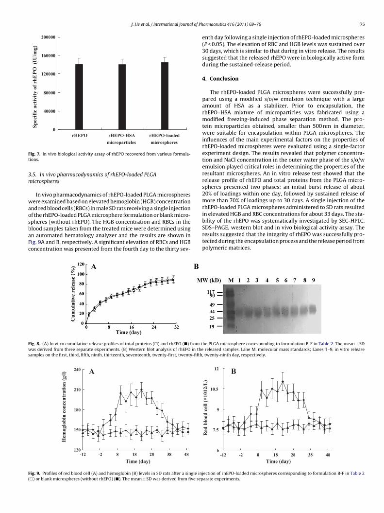
lood samples taken from the treated mice were determined usingn automated hematology analyzer and the results are shown inig. 9A and B, respectively. A significant elevation of RBCs and HGBoncentration was presented from the fourth day to the thirty sev-ig. 8. (A) In vitro cumulative release profiles of total proteins (�) and rhEPO (�) from tas derived from three separate experiments. (B) Western blot analysis of rhEPO in the
amples on the first, third, fifth, ninth, thirteenth, seventeenth, twenty-first, twenty-fifth
A
120
150
180
210
240
483828188-2-12Time (day)
Hem
oglo
bin
conc
entr
atio
n (g
/l)
ig. 9. Profiles of red blood cell (A) and hemoglobin (B) levels in SD rats after a single inj�) or blank microspheres (without rhEPO) (�). The mean ± SD was derived from five sep
armaceutics 416 (2011) 69– 76 75
enth day following a single injection of rhEPO-loaded microspheres(P < 0.05). The elevation of RBC and HGB levels was sustained over30 days, which is similar to that during in vitro release. The resultssuggested that the released rhEPO were in biologically active formduring the sustained-release period.
4. Conclusion
The rhEPO-loaded PLGA microspheres were successfully pre-pared using a modified s/o/w emulsion technique with a largeamount of HSA as a stabilizer. Prior to encapsulation, therhEPO–HSA mixture of microparticles was fabricated using amodified freezing-induced phase separation method. The pro-tein microparticles obtained, smaller than 500 nm in diameter,were suitable for encapsulation within PLGA microspheres. Theinfluences of the main experimental factors on the properties ofrhEPO-loaded microspheres were evaluated using a single-factorexperiment design. The results revealed that polymer concentra-tion and NaCl concentration in the outer water phase of the s/o/wemulsion played critical roles in determining the properties of theresultant microspheres. An in vitro release test showed that therelease profile of rhEPO and total proteins from the PLGA micro-spheres presented two phases: an initial burst release of about20% of loadings within one day, followed by sustained release ofmore than 70% of loadings up to 30 days. A single injection of therhEPO-loaded PLGA microspheres administered to SD rats resultedin elevated HGB and RBC concentrations for about 33 days. The sta-bility of the rhEPO was systematically investigated by SEC-HPLC,
SDS–PAGE, western blot and in vivo biological activity assay. Theresults suggested that the integrity of rhEPO was successfully pro-tected during the encapsulation process and the release period frompolymeric matrices.he PLGA microsphere corresponding to formulation B-F in Table 2. The mean ± SD released samples. Lane M, molecular mass standards; Lanes 1–9, in vitro release, twenty-ninth day, respectively.
B
6
7.5
9
10.5
12
483828188-2-12Time (day)
Red
blo
od c
ell (
×101
2/L
)
ection of rhEPO-loaded microspheres corresponding to formulation B-F in Table 2arate experiments.

7 l of Ph
A
PCPF
R
B
C
E
F
G
H
H
I
L
M
M
6 J. He et al. / International Journa
cknowledgements
This work is partially supported by research grants from therovincial Science & Technology Department of Hebei Province inhina (09276418D-3) and the Science & Technology project of therovincial Education Department of Hebei (200645) and Doctoroundation of Hebei Normal University (L2005B23).
eferences
urke, P.A., Klumb, L.A., Herberger, J.D., Nguyen, X.C., Harrell, R.A., Zordich, M.,2004. Poly(Lactide-Co-Glycolide) microsphere Formulations of darbepoetinalfa: spray drying is an alternative to encapsulation by spray-freeze drying.Pharm. Res. 21, 500–506.
leland, J.L., Jones, J.S., 1996. Stable formulations of recombinant human growthhormone and interferon-gama for microencapsulation in biodegradable micro-spheres. Pharm. Res. 13, 1464–1475.
schbach, J.W., Egrie, J.C., Downing, M.R., Browne, J.K., Adamson, J.W., 1987. Cor-rection of the anemia of end-stage renal disease with recombinant humanerythropoietin. N. Engl. J. Med. 316, 73–78.
urness, E.L., Ross, A., Davis, T.P., King, G.C., 1998. A hydrophobic interaction sitefor lysozyme binding to polyethylene glycol and model contact lens polymers.Biomaterials 19, 1361–1369.
eng, Y., Yuan, W., Wu, F., Chen, J., He, M., Jin, T., 2008. Formulating erythropoietin-loaded sustained-release PLGA microspheres without protein. J. Control. Release130, 259–265.
an, K., Lee, K.D., Gao, Zh.G., Park, J.S., 2001. Preparation and evaluation of poly(l-lactic acid) microspheres containing rhEGF for chronic gastric ulcer healing. J.Control. Release 75, 259–269.
uang, X., Brazel, C.S., 2001. On the importance and mechanisms of burst release inmatrix-controlled drug delivery systems. J. Control. Release 73, 121–136.
zutsu, K., Kojima, S., 2000. Freeze-concentration separates proteins and polymerexcipients into different amorphous phases. Pharm. Res. 17, 1316–1322.
iu, J., Gong, T., Wang, C.G., Zhong, Z.R., Zhang, Z.R., 2007. Solid lipid nanoparti-cles loaded with insulin by sodium cholate-phosphatidylcholine-based mixedmicelles: preparation and characterization. Int. J. Pharm. 340, 153–162.
arkham, A., Bryson, H., Epoetin, A., 1995. A review of its pharmacodynamicand pharmacokinetic properties and therapeutic use in non-renal applications.Drugs 49, 232–254.
orita, T., Sakamura, Y., Horikiri, Y., Suzuki, T., Yoshino, H., 2000b. Protein encapsu-lation into biodegradable microspheres by a novel s/o/w emulsion method using
armaceutics 416 (2011) 69– 76
poly(ethylene glycol) as a protein micronization adjuvant. J. Control. Release 69,435–444.
Morita, T., Horikiri, Y., Yamahara, H., Suzuki, T., Yoshino, H., 2000a. Formationand isolation of spherical fine protein microparticles through lyophilization ofprotein-poly(ethylene glycol) aqueous mixture. Pharm. Res. 17, 1367–1373.
Morlock, M., Koll, H., Winter, G., Kissel, T., 1997. Microencapsulation of rh-erythropoietin, using biodegradable poly(dl,-lactide-co-glycolide): proteinstability and the effects of stabilizing excipients. Eur. J. Pharm. Biopharm. 43,29–36.
Morlock, M., Kissel, T., Li, Y.X., Koll, H., Winter, G., 1998. Erythropoietin loadedmicrospheres prepared from biodegradable LPLG-PEO-LPLG triblock copoly-mers: protein stabilization and in-vitro release properties. J. Control. Release56, 105–115.
Mundargi, R.C., Babu, V.R., Rangaswamy, V., Patel, P., Aminabhavi, T.M., 2008.Nano/micro technologies for delivering macromolecular therapeutics usingpoly(d,l-lactide-co-glycolide) and its derivatives. J. Control. Release 125,193–209.
Pisal, D.S., Kosloski, M.P., Balu-Iyer, S.V., 2010. Delivery of therapeutic proteins. J.Pharm. Sci. 99, 2557–2575.
Pistel, K.F., Bittner, B., Koll, H., Winter, G., Kissel, T., 1999. Biodegradable recombinanthuman erythropoietin loaded microspheres prepared from linear and star-branched block copolymers: Influence of encapsulation technique and polymercomposition on particle characteristics. J. Control. Release 59, 309–325.
Ramos, A.S., Schmidt, C.A., Andrade, S.S., Fronza, M., Rafferty, B., Dalmora, S.L., 2003.Biological evaluation of recombinant human erythropoietin in pharmaceuticalproducts. Braz. J. Med. Biol. Res. 36, 1561–1569.
Sambrook, J., Fritsch, E.F., Maniatis, T., 1989. Molecular cloning: a laboratory manual,2nd ed. Cold Spring Harbor Laboratory Press, New York.
Tolstoguzov, V., 2000. Compositions and phase diagrams for aqueous systems basedon proteins and polysaccharides. Int. Rev. Cytol. 192, 3–31.
Spivak, J.L., 1994. Recombinant human erythropoietin and the anaemia of cancer.Blood 84, 997–1004.
Villalobos, A.P., Gunturi, S.R., Heavner, G.A., 2005. Interaction of polysorbate 80 witherythropoietin: a case study in protein–surfactant interactions. Pharm. Res. 22,1186–1194.
Xie, Sh.Y., Wang, S.L., Zhao, B.K., Han, Ch., Wang, M., Zhou, Zh.W., 2008. Effect ofPLGA as a polymeric emulsifier on preparation of hydrophilic protein-loadedsolid lipid nanoparticles. Collid Surf. B: Biointerfaces 67, 199–204.
Zhou, X., He, J., Zhou, Z., Ma, S., Jiang, Y., Wang, Y., 2010. Effect of NaCl in outer water
phase on the characteristics of BSA-loaded PLGA sustained-release microspheresfabricated by a solid-in-oil-in-water emulsion technique. Acta Pharmacol. Sin.45, 1057–1063.Zhu, G., Mallery, S.R., Schwendeman, S.P., 2000. Stabilization of proteins encapsu-lated in injectable poly(lactide-co-glycolide). Nat. Biotechnol. 18, 52–57.