Hassan˜Marzban Editor Development of the Cerebellum from ...
30
123 Contemporary Clinical Neuroscience Hassan Marzban Editor Development of the Cerebellum from Molecular Aspects to Diseases
Transcript of Hassan˜Marzban Editor Development of the Cerebellum from ...

123
Contemporary Clinical Neuroscience
Hassan Marzban Editor
Development of the Cerebellum from Molecular Aspects to Diseases

Contemporary Clinical Neuroscience
Series Editor
Mario Manto, Unité d’Etude du Mouvement (UEM), FNRS, Neurologie ULB-Erasme, Bruxelles, Belgium
More information about this series at http://www.springer.com/series/7678

Hassan MarzbanEditor
Development of the Cerebellum from Molecular Aspects to Diseases

Contemporary Clinical NeuroscienceISBN 978-3-319-59748-5 ISBN 978-3-319-59749-2 (eBook)DOI 10.1007/978-3-319-59749-2
Library of Congress Control Number: 2017946076
© Springer International Publishing AG 2017, corrected publication 2019This work is subject to copyright. All rights are reserved by the Publisher, whether the whole or part of the material is concerned, specifically the rights of translation, reprinting, reuse of illustrations, recitation, broadcasting, reproduction on microfilms or in any other physical way, and transmission or information storage and retrieval, electronic adaptation, computer software, or by similar or dissimilar methodology now known or hereafter developed.The use of general descriptive names, registered names, trademarks, service marks, etc. in this publication does not imply, even in the absence of a specific statement, that such names are exempt from the relevant protective laws and regulations and therefore free for general use.The publisher, the authors and the editors are safe to assume that the advice and information in this book are believed to be true and accurate at the date of publication. Neither the publisher nor the authors or the editors give a warranty, express or implied, with respect to the material contained herein or for any errors or omissions that may have been made. The publisher remains neutral with regard to jurisdictional claims in published maps and institutional affiliations.
Printed on acid-free paper
This Springer imprint is published by Springer NatureThe registered company is Springer International Publishing AGThe registered company address is: Gewerbestrasse 11, 6330 Cham, Switzerland
EditorHassan MarzbanDepartment of Human Anatomy and Cell ScienceThe Children’s Hospital Research Institute of Manitoba (CHRIM)Max Rady College of Medicine, Rady Faculty of Health ScienceUniversity of ManitobaWinnipeg, MB, Canada

v
Preface
For the past centuries, scientists have worked hard to understand the development of the cerebellum and its disorders. Cerebellar neurodevelopmental disorders are impairments of the growth and development of the cerebellum that result in birth defects that may have a severe impact on patients’ lives. A narrower use of the term refers to a disorder that unfolds during individual development to affect motor func-tion, emotion, learning ability, self-control, and memory. Knowledge obtained from advanced neuroimaging techniques and from molecular and genetic studies has pro-vided rapidly growing evidence that the cerebellum is a brain region that is highly impacted by developmental defects.
Advanced technologies for research have changed our views of cerebellar devel-opment. This has led us to identify many of the biological, genetic, and environmen-tal factors that contribute to the development and progression of cerebellar disorders. We believe that basic knowledge of the cerebellum, one of the most complex struc-tures in the brain, will empower cerebellar researchers, neuroscientists, neurolo-gists, and neurosurgeons every day.
This book covers diverse aspects of cerebellar development from different points of view including the epidemiology of cerebellar genetic disorders, developmental anatomy, cell biology, genetics, epigenetics, infectious diseases, and mechanisms involved in regulation of cell fate, while focusing on information that is relevant to clinicians serving patients with cerebellar disorders. This book consists of chapters written by experts in the field of cerebellar development, and it covers diseases related to the cerebellum, along with their epidemiology, clinical features, assess-ment, and management. To the extent that the book is a useful reference for neuro-scientists and clinicians, we will have succeeded.
Winnipeg, MB, Canada Hassan Marzban, PhD

vii
Contents
The Development of the Cerebellum: From the Beginnings . . . . . . . . . . . . . . 1Jan Voogd
The Embryology and Anatomy of the Cerebellum . . . . . . . . . . . . . . . . . . . 33Maryam Rahimi Balaei, Niloufar Ashtari, and Hugo Bergen
Cellular and Genetic Programs Underlying Cerebellum Development . . . . . . . . . . . . . . . . . . . . . . . . . . . . . . . . . . . . . . . . 45Alexandra L. Joyner, Ryan Willett, and Andrew Lawton
Early Purkinje Cell Development and the Origins of Cerebellar Patterning . . . . . . . . . . . . . . . . . . . . . . . . . . . . . . . . . . . . . . . . . 67Filippo Casoni, Laura Croci, Ottavio Cremona, Richard Hawkes, and G. Giacomo Consalez
Cerebellar Developmental Disorders and Cerebellar Nuclei . . . . . . . . . . . 87Hong-Ting Prekop, Alessio Delogu, and Richard J.T. Wingate
Motor Circuit Abnormalities During Cerebellar Development . . . . . . . . . 105Elizabeth P. Lackey and Roy V. Sillitoe
Developmental Disorders of the Cerebellum and Neurotrophic Factors . . . . . . . . . . . . . . . . . . . . . . . . . . . . . . . . . . . . . . . . . 129Leila Pirmoradi, Ali Akbar Owji, and Shahla Shojaei
Apoptosis, Autophagy, and Unfolded Protein Response and Cerebellar Development . . . . . . . . . . . . . . . . . . . . . . . . . . . . . . . . . . . . . 153Mohammad Amin Moosavi, Marveh Rahmati, Niloufar Ashtari, Javad Alizadeh, Mohammad Hashemi, Seyedeh Zahra Bathaei, and Saeid Ghavami
The Ubiquitin Proteasome System and Cerebellar Developmental Disease . . . . . . . . . . . . . . . . . . . . . . . . . . . . . . . . . . . . . . . . 179Jerry Vriend and Xiaodan Jiao
viii
Epigenetics and Cerebellar Neurodevelopmental Disorders . . . . . . . . . . . 197Mojgan Rastegar
Hormonal Regulation of Cerebellar Development and Its Disorders . . . . 219Noriyuki Koibuchi
Infections of the Cerebellum . . . . . . . . . . . . . . . . . . . . . . . . . . . . . . . . . . . . . 237Kevin M. Coombs
Neuroimmune Mechanisms of Cerebellar Development and Its Developmental Disorders: Bidirectional Link Between the Immune System and Nervous System . . . . . . . . . . . . . . . . . . . . . . . . . 255Nour Eissa, Laëtitia Kermarrec, and Jean-Eric Ghia
Teratogenic Influences on Cerebellar Development . . . . . . . . . . . . . . . . . . 275Albert E. Chudley
Primary Pediatric Brain Tumors of the Posterior Fossa: Part I . . . . . . . . . 301Kathleen Felton, Amanda Hogg, Lisa Liang, Christopher Aiken, Thomas Klonisch, Frank van Landeghem, Tamra E. Werbowetski-Ogilvie, and David D. Eisenstat
Primary Pediatric Brain Tumors of the Posterior Fossa Part II: A Comprehensive Overview of Medulloblastoma . . . . . . . . . . . . . . . . . . . . 327Lisa Liang, Christopher Aiken, Kathleen Felton, Amanda Hogg, Frank van Landeghem, T. Klonisch, David D. Eisenstat, and Tamra E. Werbowetski-Ogilvie
Can Cerebellar Neurodevelopmental Disorders Affect Behavioral Disorders or Vice Versa? . . . . . . . . . . . . . . . . . . . . . . . . . . . . . 353Seyed Soheil Saeedi Saravi and Ahmad Reza Dehpour
Neurodevelopmental Disorders of the Cerebellum: Autism Spectrum Disorder. . . . . . . . . . . . . . . . . . . . . . . . . . . . . . . . . . . . . . . 369Mehnosh Toback, Kambiz Zangeneh, Tabrez J. Siddiqui, and Hassan Marzban
Clinical Aspects of the Inherited Cerebellar Malformations . . . . . . . . . . . 389Asghar Marzban, Mohammad Vafaee-shahi, and Kamran Azarkhish
Clinical Features, Assessment, and Management of Patients with Developmental and Other Cerebellar Disorders . . . . . . . . . . . . . . . . . 407Michael S. Salman
Contents

ix
Epidemiology of Cerebellar Disorders . . . . . . . . . . . . . . . . . . . . . . . . . . . . . 423S. Shooshtari, B.M. Stoesz, P. Rad, and S. Khoeiniha
Cerebellar Transplantation: A Potential Model to Study Repair and Development of Neurons and Circuits in the Cerebellum . . . . . . . . . . 465Constantino Sotelo
Correction to: Primary Pediatric Brain Tumors of the Posterior Fossa Part II: A Comprehensive Overview of Medulloblastoma . . . . . . . . . . . . . . . . . . . . . . . . . . . . . . . . . . . . C1
Index . . . . . . . . . . . . . . . . . . . . . . . . . . . . . . . . . . . . . . . . . . . . . . . . . . . . . . . . . 495
Contents
1© Springer International Publishing AG 2017 H. Marzban (ed.), Development of the Cerebellum from Molecular Aspects to Diseases, Contemporary Clinical Neuroscience, DOI 10.1007/978-3-319-59749-2_1
The Development of the Cerebellum: From the Beginnings
Jan Voogd
Abstract Sotelo stated in his introduction for a consensus paper on cerebellar development (Leto et al., Cerebellum 15:789–828, 2015) that “The work done in the late nineteenth century until the late 1970s provided substantial and significant information; however, it was only descriptive and barely addressed the mechanisms involved.” Observations and their description, the nomenclature that evolved from these studies, and the ideas they fostered, indeed, formed the basis for our under-standing of the mechanisms that underlie the complex development of the cerebel-lum, to be reviewed in this volume. This chapter will highlight some of these early contributions to the origin of the cerebellum, its histogenesis, the migration of its neurons, the development of the longitudinal Purkinje cell zones, their target nuclei and their connections, and the folial pattern of the cerebellum.
The Origin of the Cerebellum
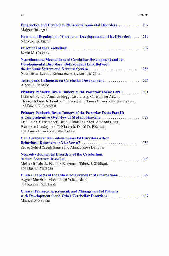
The study of cerebellar embryology begins with His’ [52, 53] description of his Rautenlippe (rhombic lip) in a human embryo. In the fifth week the “dorsal rim (of the rhombencephalic alar plate) curves laterally and forms a fold which surrounds the entire rhombic cavity …” (Fig. 1). His divided the rhombencephalon and its rhombic lip, into rostral and caudal portions. The rostral (upper) rhombic lip will give rise to the cerebellum, the caudal (lower) rhombic lip to several precerebellar nuclei. The upper rhombic lip develops in two, bilateral swellings connected by a thin midline portion (Fig. 2). At the midline, the cerebellum increases in bulk by the development of the cerebellar commissures and possibly by fusion of the intraven-tricular bulges. More recently the term “upper rhombic lip” is restricted to the pos-terior rim or germinal trigone of the cerebellar plate, with its attachment of the epithelial roof of the fourth ventricle, that should be distinguished from the ven-tricular zone, the neuroepithelium that covers its ventricular surface (Fig. 3).
J. Voogd (*) Department Neuroscience, Erasmus Medical Center, Rotterdam, The Netherlandse-mail: [email protected]
2
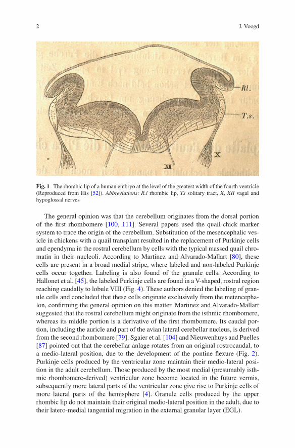
The general opinion was that the cerebellum originates from the dorsal portion of the first rhombomere [100, 111]. Several papers used the quail-chick marker system to trace the origin of the cerebellum. Substitution of the mesencephalic ves-icle in chickens with a quail transplant resulted in the replacement of Purkinje cells and ependyma in the rostral cerebellum by cells with the typical massed quail chro-matin in their nucleoli. According to Martinez and Alvarado-Mallart [80], these cells are present in a broad medial stripe, where labeled and non-labeled Purkinje cells occur together. Labeling is also found of the granule cells. According to Hallonet et al. [45], the labeled Purkinje cells are found in a V-shaped, rostral region reaching caudally to lobule VIII (Fig. 4). These authors denied the labeling of gran-ule cells and concluded that these cells originate exclusively from the metencepha-lon, confirming the general opinion on this matter. Martinez and Alvarado-Mallart suggested that the rostral cerebellum might originate from the isthmic rhombomere, whereas its middle portion is a derivative of the first rhombomere. Its caudal por-tion, including the auricle and part of the avian lateral cerebellar nucleus, is derived from the second rhombomere [79]. Sgaier et al. [104] and Nieuwenhuys and Puelles [87] pointed out that the cerebellar anlage rotates from an original rostrocaudal, to a medio-lateral position, due to the development of the pontine flexure (Fig. 2). Purkinje cells produced by the ventricular zone maintain their medio-lateral posi-tion in the adult cerebellum. Those produced by the most medial (presumably isth-mic rhombomere-derived) ventricular zone become located in the future vermis, subsequently more lateral parts of the ventricular zone give rise to Purkinje cells of more lateral parts of the hemisphere [4]. Granule cells produced by the upper rhombic lip do not maintain their original medio-lateral position in the adult, due to their latero-medial tangential migration in the external granular layer (EGL).
Fig. 1 The rhombic lip of a human embryo at the level of the greatest width of the fourth ventricle (Reproduced from His [52]). Abbreviations: R.l rhombic lip, Ts solitary tract, X, XII vagal and hypoglossal nerves
J. Voogd

3
Fig. 2 Cerebellum of a 15-week human embryo (Reproduced from Nieuwenhuys et al. [88]). Red arrow indicates position of rostrocaudal axis of the cerebellar anlage after the rotation of the cer-ebellar anlage due to the pontine flexure
Fig. 3 Diagram of a sagittal section, showing the division of the germinal zone of the early cerebellar plate into the ventricular zone that will give rise to inhibitory neurons and the upper rhombic lip that produces the excitatory neurons of the cerebellum (Redrawn from Goldowitz and Hamre [40])
The Development of the Cerebellum: From the Beginnings
4
Histogenesis
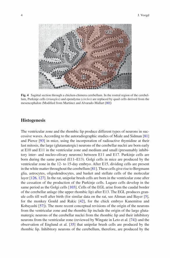
The ventricular zone and the rhombic lip produce different types of neurons in suc-cessive waves. According to the autoradiographic studies of Miale and Sidman [81] and Pierce [93] in mice, using the incorporation of radioactive thymidine at their last mitosis, the large (glutamatergic) neurons of the cerebellar nuclei are born early at E10 and E11 in the ventricular zone and medium and small (presumably inhibi-tory inter- and nucleo-olivary neurons) between E11 and E17. Purkinje cells are born during the same period (E11–E13). Golgi cells in mice are produced by the ventricular zone in the 12- to 15-day embryo. After E15, dividing cells are present in the white matter throughout the cerebellum [81]. These cells give rise to Bergmann glia, astrocytes, oligodendrocytes, and basket and stellate cells of the molecular layer [126, 127]. In the rat, unipolar brush cells are born in the ventricular zone after the cessation of the production of the Purkinje cells. Lugaro cells develop in the same period as the Golgi cells [103]. Cells of the EGL arise from the caudal border of the cerebellar anlage (the upper rhombic lip) after E13. The EGL produces gran-ule cells till well after birth (for similar data on the rat, see Altman and Bayer [5], for the monkey Gould and Rakic [42], for the chick embryo Kanemitsu and Kobayashi [57]). The more recent conceptual revisions of the origin of the neurons from the ventricular zone and the rhombic lip include the origin of the large gluta-matergic neurons of the cerebellar nuclei from the rhombic lip and their inhibitory neurons from the ventricular zone (reviewed by Wingate in Leto et al. [74]) and the observation of Englund et al. [35] that unipolar brush cells are produced by the rhombic lip. Inhibitory neurons of the cerebellum, therefore, are produced by the
Fig. 4 Sagittal section through a chicken-chimera cerebellum. In the rostral region of the cerebel-lum, Purkinje cells (triangles) and ependyma (circles) are replaced by quail cells derived from the mesencephalon (Modified from Martinez and Alvarado-Mallart [80])
J. Voogd

5
ventricular zone, excitatory neurons by the rhombic lip. Glutamatergic nuclear neu-rons and cells of the EGL are produced sequentially by the rhombic lip.
In their migration to the meningeal surface of the cerebellum Purkinje cells are supposed to use the processes of the neuroepithelial cells whose conical endfeet form the external limiting membrane (Fig. 5). A map of these processes that would predict the paths of migrating Purkinje cells is not available. In mice, migrating Purkinje cells at E15 avoid the cerebellar nuclei; at E17 they pass across them [125]. In the rat, all Purkinje cells migrate through the more superficially located transitory nuclear layer [3]. The clustering of the migrated Purkinje cells that will lead to the develop-ment of longitudinal Purkinje cell zones will be considered in another paragraph.
In his Golgi studies, Cajal [22, 23, 25] and his followers [10, 76, 95] distin-guished different phases in the development of the Purkinje cells (Fig. 6a–d). In the first phase of the “disoriented dendrites,” multiple processes arise from all over the cell body. The axon, first devoid of collaterals, enters the white matter. In the next stage, oriented and regular dendrites arise as a flattened tree from the upper pole of the cell. The axon emits multiple collaterals. Finally the processes of the cell body are resorbed, the dendritic tree acquires its definite shape, and many of the axonal collaterals are resorbed. Differentiation of the Purkinje cells is more advanced in the apices of the lobule. Purkinje cells of the rat mature early in lobules I and II and proximal V and VI and IX and X and late in distal VI, VII, and VIII [2, 41].
Fig. 5 Neuroepithelial cells (Popoff’s spongioblasts) in a 1–1/4 cm cat embryo. Golgi method (Reproduced from Popoff [95]). Abbreviations: e ventricular zone, ext external limiting membrane
The Development of the Cerebellum: From the Beginnings

6
The development of the climbing fibers closely follows that of the Purkinje cell [22, 25, 95]. Cajal distinguished an early pericellular nest stage where the climbing fiber forms an infracellular plexus, (Fig. 6e) followed by outgrowth over the emerg-ing dendrites, the place of the supranuclear capuchon, the stage of the young climb-ing fiber arborization, and finally its adult form (Fig. 6h). The shift of the climbing fiber synaptic connections with the filopodia of the Purkinje cell soma, to their posi-tion on stubby spines on the smooth proximal dendrites of the Purkinje cells and their replacement by the inhibitory synapses of the basket cell axons, was documented in the electron microscopic studies of Larramendi [69] and Morara
Fig. 6 Stages in the development of Purkinje cells (a–d) and climbing fibers (e–h) (Reproduced from Athias [10])
J. Voogd

7
et al. [82]. Multiple innervation by climbing fibers of the Purkinje cell was noticed by Cajal and others (Fig. 6e, h). The elimination of redundant climbing fibers was shown much later in physiological studies, reviewed by Hashimoto and Kano [46].
The external granular layer (EGL) of the cerebellar anlage gives rise to the gran-ule cells, although, during its history of more than 150 years, it was supposed to contribute to each cell type of the cerebellum. The first description of the EGL dates from Hess [50], who illustrated it as the stratum granulosum periphericum in the cortex of a neonate dog (Fig. 7). Its cells are provided with radially oriented filiform processes. In due time, the layer disappears, leaving only a few cells near the pia mater. Obersteiner [90] distinguished a superficial, tightly packed, and a deep layer with loosely arranged rounded cells in the EGL (Fig. 8). Like Hess, radial processes in the molecular layer were found to originate from these cells. Later authors often referred to the EGL as “Obersteiner’s layer.” Schaper [102] in fish and Herrick [49] in mice and guinea pig observed the origin of the EGL from the ventricular matrix next to the caudal attachment of the roof plate of the fourth ventricle and its rostral migration over the cerebellar surface. They observed mito-ses in the superficial EGL and identified it as a secondary matrix. Miale and Sidman [81] dated the origin of the EGL in the mouse at E13, when the generation of Purkinje cells has ceased and found that the proliferation in the EGL lasts till the
Fig. 7 Cortex of neonate dog showing the stratum granulosum periphericum. Carmine staining (Reproduced from Hess [50]). Abbreviations: a granule cell with processes directed at the periphery, B stratum granulosum centrale, b granule cell with filiform processes at both ends, C nerve (Purkinje) cells, D stratum moleculare, E stratum granulosum periphericum, F pia mater
The Development of the Cerebellum: From the Beginnings

8
third postnatal week. Proliferation in the EGL is regulated by sonic hedgehog, secreted by the subjacent Purkinje cells [30].
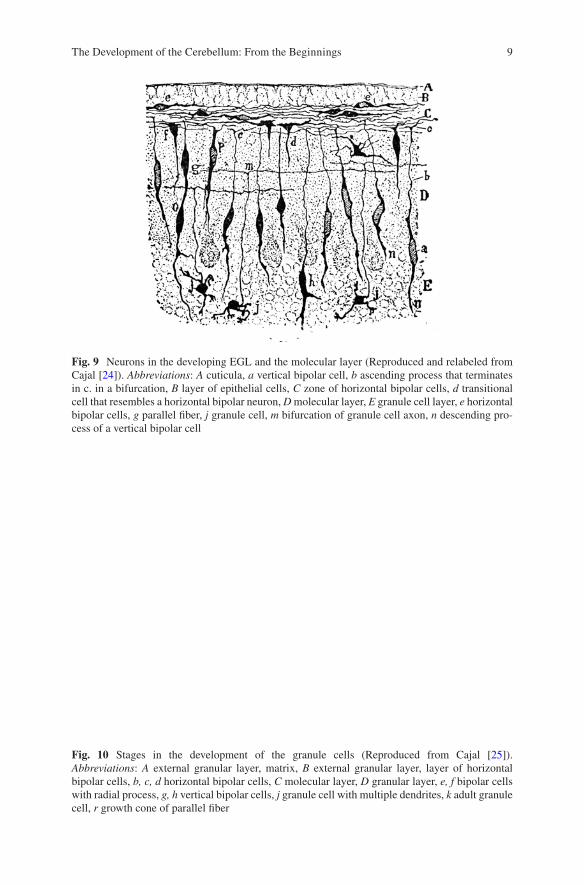
In his 1890a paper, Cajal described different cell types in the EGL and the molecular layer (Fig. 9). Horizontal, bipolar neurons, with horizontal axonal expan-sions extending in the length of the cerebellar folia, occur in the deep layer of the EGL. Bipolar neurons with radially oriented processes occur in the molecular layer (Fig. 9). Strange as it may seem to us now, Cajal did not recognize these neurons as stages in the migrating granule cells, at least, with his scientific rigor, he judged that he had too little material to draw this conclusion. Later he identified the origin of the parallel fibers from the horizontal bipolar neurons, the emergence of a third, proto-plasmic process, and the translocation of the nucleus in this process through the molecular layer into the internal granular layer (Fig. 10). Here its rounded cell body bears multiple dendrites most of which are resorbed when it settles deep in the granular layer in regions were the mossy fiber rosettes have attained their adult form (Fig. 10) [25]. The parallel fibers are stacked from the bottom of the molecular layer upward. A similar gradient as present for the differentiation of the Purkinje cells in different lobules of the cerebellum was found for the differentiation of the granule cells [3].
Granule cell precursors use Bergmann glial fibers for their migration [97]. These fibers, with their typical lateral processes and their attachment to the meningeal surface
Fig. 8 Section through the cerebellar cortex of a neonate. Carmine staining 1 upper layer of the EGL (Basalschichte), 2 second granular layer, 3 molecular layer (radiär gestreifte Schichte), 4 Purkinje cell layer (tangentielle Schichte), 5 permanent granular layer (Reproduced from Obersteiner [90])
J. Voogd
9
Fig. 9 Neurons in the developing EGL and the molecular layer (Reproduced and relabeled from Cajal [24]). Abbreviations: A cuticula, a vertical bipolar cell, b ascending process that terminates in c. in a bifurcation, B layer of epithelial cells, C zone of horizontal bipolar cells, d transitional cell that resembles a horizontal bipolar neuron, D molecular layer, E granule cell layer, e horizontal bipolar cells, g parallel fiber, j granule cell, m bifurcation of granule cell axon, n descending pro-cess of a vertical bipolar cell
Fig. 10 Stages in the development of the granule cells (Reproduced from Cajal [25]). Abbreviations: A external granular layer, matrix, B external granular layer, layer of horizontal bipolar cells, b, c, d horizontal bipolar cells, C molecular layer, D granular layer, e, f bipolar cells with radial process, g, h vertical bipolar cells, j granule cell with multiple dendrites, k adult granule cell, r growth cone of parallel fiber
The Development of the Cerebellum: From the Beginnings

10
of the cerebellum, were described by Bergmann [13]. Bergmann glia have been described as originating from the Golgi epithelial cells by translocation of their cell bodies to the Purkinje cell layer [125] but have also been traced from cells proliferat-ing in the cerebellar white matter [126]. The orientation of the parallel fibers clearly is established very early as processes of the horizontal bipolar cells in the EGL. Purkinje cell dendritic arbors derive their plane shape and their orientation perpendicular to the parallel fibers from the interaction with these fibers during their development [3, 84]. However, the orientation of the parallel fibers in the long axis of the folia can be uncoupled after perinatal administration of methylazoxymethanol in rats [51].
Development of the Cerebellar Nuclei
The first study of the development of the cerebellar nuclei in different classes of vertebrates is by Rüdeberg [100]. In the tradition of Bergqvist and Källén [14], he traced the origin of the cerebellum from two, subsequent migration areas, A and B, from the ventricular neuroepithelium of the dorsal column of the first rhombomere. The dorsal part of the first migration area A gives rise to the external granular layer; its middle portion A2 merges with part of the second migration area B into the cell group A2B; its dorsal part, A1, develops outside the cerebellum, into the isthmic nucleus. The dorsal part of migration B gives rise to the Purkinje cell layer (Fig. 11). In birds cell group, A2B develops into the cerebellar nuclei, and in mammals, it gives rise to the lateral (dentate) nucleus. The interposed and fastigial nucleus stem from ventral parts of migration B. The development of the cerebellar nuclei in Cetacea follows the same pattern [63]. According to Korneliusen [62], all nuclei in the rat develop from the deep layer of migration B. The nomenclature used by Feirabend [36] for the early development of the chicken cerebellum is different, but his account of the origin of the cerebellar nuclei from the ventricular zone is very similar to that of Rüdeberg. The two migration layers were also recognized by Altman and Bayer [4–6] in the rat. The first migration layer, with exception of its ventral portion (Rüdeberg’s A1), gives rise to all cerebellar nuclei and was indicated as the nuclear transitory zone (NTZ). A second migration layer (Rüdeberg’s B) gives rise to the Purkinje cells. As a consequence, the future Purkinje cells migrate through the NTZ to reach their superficial position. The NTZ splits in a dorsomedial group of longitudinally oriented cells and a superficially located lateral group with a transverse orientation (Fig. 12). The latter migrates medially and gives rise to axons that cross in the cerebellar commissure forming the uncinate tract that takes origin from the fastigial nucleus. The superficial location and the origin of the unci-nate tract from this nucleus and its migration to a more ventral position were experi-mentally verified by Bourrat and Sotelo [17] (Fig. 12, inset). The longitudinally oriented neurons will develop into the interposed and lateral (dentate) nuclei. With the demonstration by Machold and Fishell [77] and Wang and Zoghbi [119] that glutamatergic neurons of the nuclei are derived from the upper rhombic lip, Rüdeberg’s migration A, or Altman’s nuclear transitory zone, became a layer of tangentially migrating neurons destined for the cerebellar nuclei.
J. Voogd

11
Development of Longitudinal Purkinje Cell Zones
Longitudinal Purkinje cell zones are among the first features of the cerebellum to develop as discrete multicellular clusters that will extend rostrocaudally as adult, monolayered zones. Purkinje cell zones were first identified by their projections to cerebellar and vestibular target nuclei and their afferent olivocerebellar fibers occupy [113, 114], illustrated in Fig. 13a–c. It should be noticed that the B zone (green) and the C1, C3, and Y zones (red) are restricted to the anterior lobe and the simplex lobule, and to lobule VIII and its hemisphere, the copula. Other zones extend over most of the rostro-caudal length of the cerebellar surface. Their devel-opment has been studied in serial, Nissl-stained sections in different species and by using Purkinje cell-specific markers. Their development was first studied by Korneliusen [61] in Cetacea. In Balaenoptera musculus (blue whale) and Balaenoptera physalus (fin whale) embryos, he distinguished four Purkinje cell clusters in the cortical anlage, each cluster being topographically related to one of the incipient cerebellar nuclei (Fig. 14). Clusters are clearly demarcated and differ
Fig. 11 Transverse section of the cerebellar anlage from a 21 mm human embryo (Reproduced from Rüdeberg [100])
The Development of the Cerebellum: From the Beginnings

12
in the degree of differentiation of their cells. Raphe-like, cell-poor differentiations within the medullary substance demarcate the borders between the cluster/nuclear complexes. Clusters extend all over the length of the still smooth cerebellar sur-face. Three subdivisions are present in the medial cluster overlying similar differ-entiations within the medial nucleus. A narrow medial intermediate cluster is related to the small anterior interposed nucleus, the wide lateral intermediate clus-ter to the large posterior interposed nucleus, and the lateral cluster is topographi-cally related to the anlage of the lateral cerebellar nucleus. A very similar clustering in the incipient cortex was found in the rat [62] (Fig. 14). The same relations of the cerebellar nuclei were found as in whale embryos, but the lateral intermediate clus-ter, like its target nucleus, the posterior interposed, is smaller and of the same size as the medial intermediate zone and the anterior interposed nucleus. In the rat, a small, additional X zone was present between the lateral and lateral intermediate zone, related to the dorsolateral hump of the anterior interposed nucleus. The medial intermediate and the X clusters are partially covered by the adjoining clus-ters. Four Purkinje cell clusters were identified by Feirabend [36] in chick embryos. In later stages, migrating strands of granule cells (“granule cell raphes,” Fig. 15) are located between and within the clusters, subdividing them in smaller units. The existence of such a second generation of clusters has not been confirmed, but the Purkinje cell raphes have also been identified in mammals and have been used to delineate Purkinje cell clusters and zones in histochemical studies [59, 60, 75, 98]. Cerebellar zonation in early postnatal avian stages was documented by Braun et al. [20].
Fig. 12 Transverse section through the cerebellar anlage of a E17 rat embryo, showing the divi-sion of the nuclear transitory zone in a group of medially migrating, future fastigial nuclear neu-rons (fcf) that will give rise to the uncinate tract (hb), and a group of longitudinally oriented neurons (fci), the source of the interposed and lateral nuclei (Reproduced from Altman and Bayer [5]. Inset: Injection of horse radish peroxidase (stippled area) labels fibers of uncinate tract in the cerebellar commissure and cells of contralateral fastigial nucleus in a E16 rat embryo [17])
J. Voogd

13
Purkinje cell clusters have been identified during the development of the primate cerebellum. The first illustrations in human fetuses can be found in Langelaan [67] and Hochstetter [54]. They were studied in macaque monkey fetuses by Kappel [58]. She distinguished two sets of clusters. Those destined to develop in the adult A, C2, and D1 and D2 zones reach the still smooth surface of the cerebellum early (Fig. 16). The clusters that will give rise to the future B, C1, and C3 zones reach the surface later. For some time, they are still partially covered by the neighboring clus-ters, a phenomenon also noticed for the same clusters in Korneliussen’s [64] paper on the rat corticogenesis. Korneliusen’s medial and lateral intermediate and his X zone clearly correspond to the monkey C1, C2, and C3 zones, respectively. In the
Fig. 13 (a) Diagram of the Purkinje cell zones in a flattened map of the cortex of the cerebellum of the rat. The cerebellar and vestibular target nuclei of the zones are indicated in (b), the source of their climbing fiber afferents in a flattened map of the inferior olive in (c). A map of the distribution of zebrin-positive (black) and zebrin-negative Purkinje cells is illustrated in (d). Zebrin-positive zones are numbers 1–7. Note that the Purkinje cells of the B, C1, C3, and Y zones are zebrin negative
The Development of the Cerebellum: From the Beginnings

14
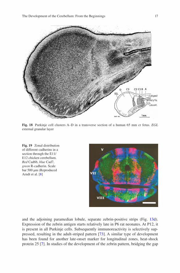
monkey fetus, cell strands connect the C1 and C3 clusters with the anterior inter-posed nucleus (Fig. 17). The same Purkinje cell clusters also can be recognized in human fetuses, where the large size of the lateral D cluster should be noticed (Fig. 18). The differentiation of the human dentate nucleus in a dorsomedial portion with an early differentiating coils, and a late developing ventrocaudal part, was first described by Weidenreich [124]. The general conclusion of these studies is that Purkinje cell clusters transform directly into the adult pattern of Purkinje cell zones.
Fig. 14 Transverse section and a diagram of the flattened cerebellar cortex showing Purkinje cell clusters in a 30 mm cr rat embryo and a 17 cm cr Balaenoptera physalus embryo (Modified from Korneliusen [61, 62])
J. Voogd

15
Fig. 15 Granule cell raphes in a 14-day chick embryo. (a) Loyez stain of anterior lobe, the EGl, and the granule cell raphes are stained. (b) Nissl stain. EGL external granular layer, P Purkinje cell clusters (Courtesy Dr. Hans Feirabend)
Nothing is known about the development of the detailed (somato) topical patterns [34] in the Purkinje cell zones.
The role of cadherins, adhesion molecules that play an important role in cerebel-lar development, was reviewed by Redies et al. [99]. Different cadherins are expressed by Purkinje cell clusters early in chick embryos and provide an adhesive code for parasagittal cell domains in avian and mammalian embryos (Fig. 19) and characterize interconnected grisea, such as Purkinje cell clusters and the cerebellar nuclei [8, 86]. In mice, these cadherin domains resemble the Purkinje cell zones as they are known in rats.
Wassef and Sotelo [120] and Wassef et al. [123] traced the development of Purkinje cell clusters in rats, using markers that are expressed by all adult Purkinje cells. Not all Purkinje cell clusters express these markers during development. Different patterns of labeling were observed for different markers. Whether this is caused by a different phenotype of the immature Purkinje cells or by a difference in time scale of the expression of the different markers is not clear. The number of clusters identified was greater than in previous studies, and, therefore, a comparison with them was not attempted.
Another set of Purkinje cell-specific antibodies was developed by Hawkes and Leclerc ([48]: mapQ-113, zebrin I) and Brochu et al. ([21], zebrin II). The epitope of zebrin II was found to be aldolase C [1]. These antibodies stain a subpopulation of Purkinje cells. Multiple longitudinal strips of zebrin-negative Purkinje cells in the anterior lobe and the simplex lobule, in the posterior cerebellum in the pyramis
The Development of the Cerebellum: From the Beginnings

16
Fig. 16 Photographs of the rostral aspect of reconstructions of the Purkinje cell layer of the cer-ebellum of four fetuses of the rhesus monkey. Clusters are indicated with different shadings. Note the superficial location of Purkinje cells of the early arriving clusters D, C2, and A in the youngest fetus and the gradual emergence at the surface of the later arriving clusters B, C1, and C3. Compare with sections of 55-, 65-, and 70-day-old fetuses in Fig. 16. Abbreviations: Fl flocculus, prf pri-mary fissure (Reproduced from Kappel [58])
Fig. 17 Coronal section through the cerebellum of a 55-day-old rhesus monkey fetus. Note super-ficial location of the Purkinje cells of the early arriving clusters A, C2, and D, which still partially cover the later arriving deep clusters B, C1, and C3. Abbreviations: cr restiform body, IntA anterior interposed nucleus, v4 fourth ventricle (Reproduced from Kappel [58])
J. Voogd
17
Fig. 18 Purkinje cell clusters A–D in a transverse section of a human 65 mm cr fetus. EGL external granular layer
Fig. 19 Zonal distribution of different cadherins in a section through the E11/E12 chicken cerebellum. Red Cad6b, blue Cad7, green R-cadherin. Scale bar 500 μm (Reproduced Arndt et al. [8]
and the adjoining paramedian lobule, separate zebrin-positive strips (Fig. 13d). Expression of the zebrin antigen starts relatively late in P6 rat neonates. At P12, it is present in all Purkinje cells. Subsequently immunoreactivity is selectively sup-pressed, resulting in the adult-striped pattern [73]. A similar type of development has been found for another late-onset marker for longitudinal zones, heat-shock protein 25 [7]. In studies of the development of the zebrin pattern, bridging the gap
The Development of the Cerebellum: From the Beginnings

18
between prenatal clusters and adult zebrin-negative and zebrin-positive strips proved to be difficult [68].
One of the problems is that zebrin-positive and zebrin-negative strips do not map one to one on the Purkinje cell zones defined by their corticonuclear and olivo-cerebellar identity. The zebrin immunoreactivity of these Purkinje cell zones was established by Voogd et al. [118], Voogd and Ruigrok [117], and Sugihara and Shinoda [107]. Their studies also revealed a number of additional, narrow zebrin-positive strips that were formally discarded as satellite bands. In the rat hemisphere, the B, C1, C3, and Y zones were found to be zebrin negative, the intercalated C2, D1, and D2 zones were zebrin positive (compare Fig. 13a, d). In the vermis, the A zone consists of multiple zebrin-positive and zebrin-negative subzones. Earlier publications on differences in birth date between the Purkinje cells of different clusters [37] were succeeded by the viral labeling studies of Hashimoto and Mikoshiba [47] that showed that Purkinje cells in mice born at E11.5 form clusters that will develop into the zebrin-positive (C2, D1, and D2) zones, whereas Purkinje cells born at E12.5 develop in the zebrin-negative (B, C1, C3, and Y) zones [85] (Fig. 20). Earlier Kappel [58] found these late-born Purkinje cell clusters to arrive later at the cerebellar surface than the early-born clusters. Just as the number of zebrin-positive and zebrin-negative stripes increased in recent studies, the number of Purkinje cell clusters identified in E17.5 mice embryos increased to 54 on each side [39]. These authors traced the development of these clusters into the adult zebrin pattern. More recent developments in this field were reviewed by Arancillo et al. and in Leto et al. [74].
Development of Connections
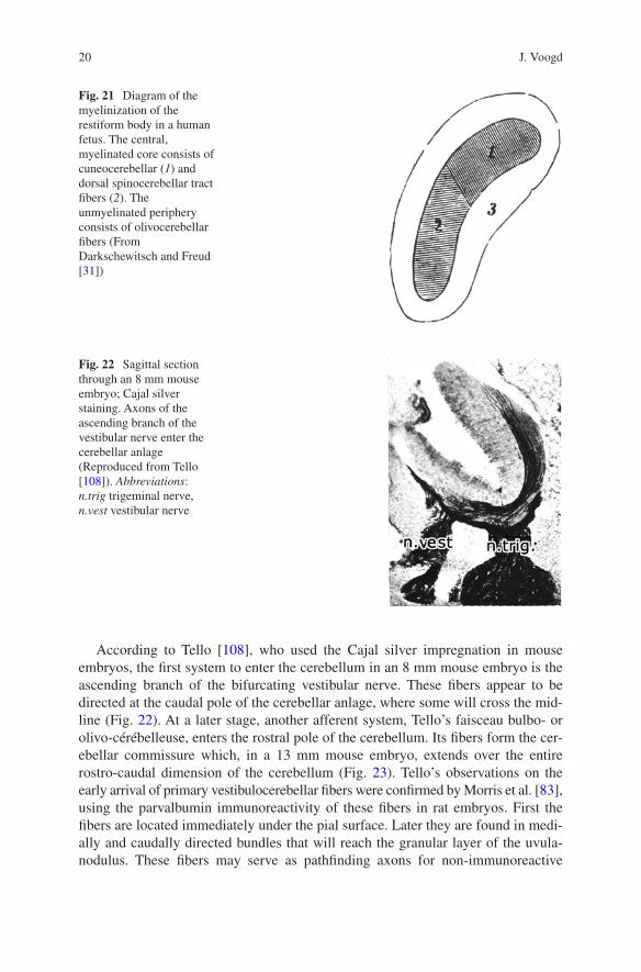
The development of the afferent climbing and mossy connections of the cerebellum has received more attention than the output systems of the cerebellar nuclei. A closed chapter in the study of the development of cerebellar connections is the study of their myelinization. Axonal systems acquire their myelin sheaths at different pre- and postnatal dates. Myelin-stained sections can provide information on their topography. The method was mostly used in human fetuses and neonates. Like modern MRI tractography. It does not provide information on the precise origin and termination of the tracts nor on the direction of impulse propagation. A good exam-ple is the dorsal spinocerebellar tract that bore the eponym Flechsig’s tract after its discovery in the myelogenetic studies of this author [38]. The localization of this tract in the restiform body was illustrated by Darkschewitsch and (Sigmund) Freud [31]. In a human fetus, it consists of a core of myelinated cuneocerebellar and dorsal spinocerebellar fibers and an unmyelinated periphery of olivocerebellar fibers (Fig. 21). Details on the intracerebellar topography were published by De Sanctis [32]. The state of the art at the end of the nineteenth century was reviewed by von Bechterew [12].
J. Voogd

19
Fig. 20 Distribution of Purkinje cells born on E10.5 and E12.5 superimposed on a map of the zebrin-positive (gray) and zebrin-negative strips of the cerebellum of the mouse. Early-born Purkinje cells constitute zebrin-positive bands; late-born Purkinje cells constitute zebrin-negative bands (Reproduced from Namba et al. [85])
The Development of the Cerebellum: From the Beginnings
20
According to Tello [108], who used the Cajal silver impregnation in mouse embryos, the first system to enter the cerebellum in an 8 mm mouse embryo is the ascending branch of the bifurcating vestibular nerve. These fibers appear to be directed at the caudal pole of the cerebellar anlage, where some will cross the mid-line (Fig. 22). At a later stage, another afferent system, Tello’s faisceau bulbo- or olivo-cérébelleuse, enters the rostral pole of the cerebellum. Its fibers form the cer-ebellar commissure which, in a 13 mm mouse embryo, extends over the entire rostro- caudal dimension of the cerebellum (Fig. 23). Tello’s observations on the early arrival of primary vestibulocerebellar fibers were confirmed by Morris et al. [83], using the parvalbumin immunoreactivity of these fibers in rat embryos. First the fibers are located immediately under the pial surface. Later they are found in medi-ally and caudally directed bundles that will reach the granular layer of the uvula-nodulus. These fibers may serve as pathfinding axons for non- immunoreactive
Fig. 21 Diagram of the myelinization of the restiform body in a human fetus. The central, myelinated core consists of cuneocerebellar (1) and dorsal spinocerebellar tract fibers (2). The unmyelinated periphery consists of olivocerebellar fibers (From Darkschewitsch and Freud [31])
Fig. 22 Sagittal section through an 8 mm mouse embryo; Cajal silver staining. Axons of the ascending branch of the vestibular nerve enter the cerebellar anlage (Reproduced from Tello [108]). Abbreviations: n.trig trigeminal nerve, n.vest vestibular nerve
J. Voogd

21
fibers, possibly belonging to secondary vestibulocerebellar fibers from the vestibular nuclei. The development of differential projections of cristae and maculae in mice to the uvula-nodulus was studied by Maklad and Fritsch [78].
Of the other mossy fiber afferent systems, the development of the spinocerebellar projection has received most attention. The bilateral, regular collateralization of spinocerebellar fibers that form multiple parasagittally oriented terminal fields in the granular layer was first described in our lab for mammals [114] and birds [112]. Lakke et al. [66] traced spinocerebellar axons with WGA HRP in chicken embryos. They enter the rostral cerebellum in Tello’s bulbocerebellar fascicle at the seventh incubation day. They course superficially, to enter the cerebellar commissure 2 days later. The bundle of spinocerebellar axons gives off collaterals which enter the Purkinje cell clusters, from where they extend into the molecular layer (Fig. 24). Spinocerebellar fibers disappear from the molecular layer, and terminal rosettes in the inner granular layer develop late before and after hatching [91]. In mammals, a similar sequence is present in the development of the spinocerebellar pathway. Their early entrance in the rostral cerebellar anlage in E13 mouse embryos, their superficial location, and their decussation in the cerebellar commissure are observed at E15. No parasagittal arrangement is visible at E19 [43]. According to Arsénio
Fig. 23 Sagittal section through a 13 mm mouse embryo; Cajal silver staining, showing the cer-ebellar commissure (Reproduced from Tello [108]). Abbreviations: comm cerebellar commissure, egl external granular layer, mes mesencephalon, plex. chor. choroid plexus, ventr. matrix ventricu-lar matrix
The Development of the Cerebellum: From the Beginnings

22
Nunes and Sotelo [9], the columnar distribution of spinocerebellar fibers in the rat develops postnatally from a more diffuse stage that was not observed in birds. A distinct topographical relationship of these columns to the zebrin pattern, i.e., to the Purkinje cell zones, was described by Ji and Hawkes [55]. According to these authors, despite the early zonal distribution of the mossy fibers being dependent on Purkinje cell clustering, granule cell-mossy fiber interactions if disturbed by chemi-cal ablation of the EGL result in blurring of this pattern [56].
Little is known about the development of other mossy fiber systems. For the development of the pontocerebellar projection, Bechterew [11] made an interesting observation. He found an early myelinating “spinal system” in the brachium pontis of human neonates that can be traced from the caudal pontine nuclei and the nucleus reticularis tegmenti pontis into the flocculus and the anterior cerebellum. The “cere-bral system” of the brachium pontis, which courses from the rostral pontine nuclei to the posterior cerebellum, is still unmyelinated at the time (Fig. 25). This is in accordance with more recent observations that the main projection of the caudal pontine nuclei, which receive their afferents from motor cortical areas, is to the anterior lobe, whereas rostral pontine nuclei that are innervated by cortical associa-tion areas mainly project to the caudal cerebellum [115]. Tolbert and Panneton [110] described transient extra-pontine cerebrocerebellar connections from the somatosensory cortex to the cerebellar cortex and nuclei in kittens using axonal transport of tritiated amino acids, horseradish peroxidase, or fluorescent dyes. These projections arise as collaterals from the pyramidal tract, passing through the supe-rior cerebellar peduncle to terminate in the nuclei, and caudal to the pons, bilaterally through the restiform body to be distributed as mossy-like fibers to the granular layer of the anterior lobe, the lobulus simplex and the paramedian lobule. The pro-jections to nuclei and cortex are somatotopically organized [94, 109]. Nuclear pro-jections are present at P6–P8, cortical projections between P8 and P10. After the seventh postnatal week, no cerebrocerebellar projections were present any more. Earlier, a similar transient pathway from the occipital region of the hemisphere to the paraflocculus was observed in neonatal rabbits [33].
Fig. 24 Bundles of spinocerebellar fibers in an 11-day incubation chick embryo are positionally related to the Purkinje cell clusters. Collaterals are seen to enter these clusters (Reproduced from Lakke et al. [66])
J. Voogd