Harrisons infectious disease 2 Ed Oxford handbook of ID and …Pathogenesis •Cellulitis caused by...
53
Musculoskeletal system Skin and soft tissue infections 4 Harrison’s infectious disease 2 nd Ed Oxford handbook of ID and MM
Transcript of Harrisons infectious disease 2 Ed Oxford handbook of ID and …Pathogenesis •Cellulitis caused by...

Musculoskeletal system
Skin and soft tissue infections 4
Harrison’s infectious disease 2nd Ed
Oxford handbook of ID and MM
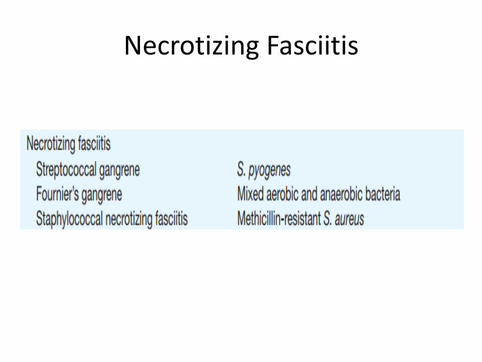
Necrotizing Fasciitis

Necrotizing fasciitis
• RAPIDLY progressing infection in the area between the fascia and deep subcutaneous tissue.
• Many risk factors increase the risk (see table next slide)
• Fibrous bands in this area prevents spread of infection– These bands are present in the head but not in the
extremities ( thus extremities are more susceptible)
– >50% in extermities
– 20% in perineum or buttocks ( esp in DM and alcoholics)
– 18% in trunk
– 9% head and neck

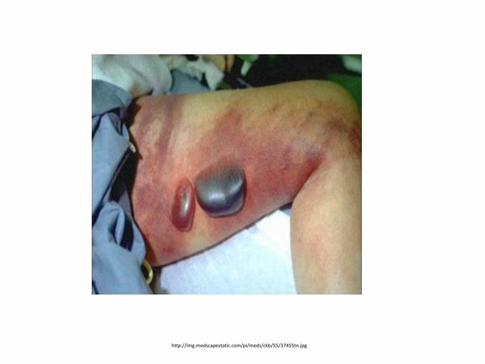
• Necrotizing fasciitis (GAS) and gas gangrene (anaerobic clostridia infection) also induce bulla formation.
• In the USA, the estimated incidence of invasive GAS infection is 3.5 cases per 100000 persons—necrotizing infections account for 6% of these.
Risk factors associated with necrotizing fasciitis
Malnutrition
Patient conditions
Immuno-compromised
Poor blood supply
Skin trauma in last 3 months
Breaks in mucosa of GI or GU tracts (anaerobes)
-Hypo-albuminemia
-Alcoholism
-Cirrhosis
- >50 Year olds
-Obesity
-Cancer
-Steroid therapy
-Heart disease
-PVD
-DM
-Burns-penetrating trauma-IV drugs-surgery
-colon cancer-diverticulae-hemorrhoids or fissues-urethral tears-cirrhosis
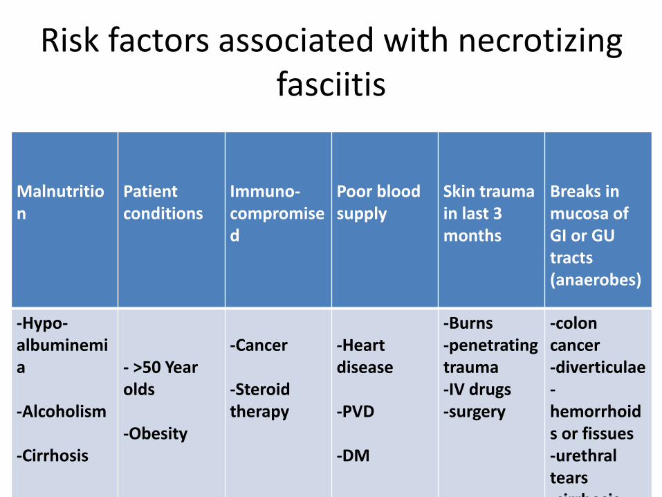
Signs and Symptoms – occur in order
• Pain/tenderness• Unexplained fever (Early diagnosis may be difficult when pain or
unexplained fever is the only presenting manifestation, remember infection is deep, might not present with pain yet )
• Swelling• Dark red induration• BULLAE, filled with blueish or purple fluid• Thrombosis of dermal blood vessels (The affected area becomes
anaesthetic as a result of small vessel thrombosis and destruction of superficial nerves)
• Extension to deep fascia with rapid spread• Most progressed symptoms : toxicity , shock and multi organ
failures
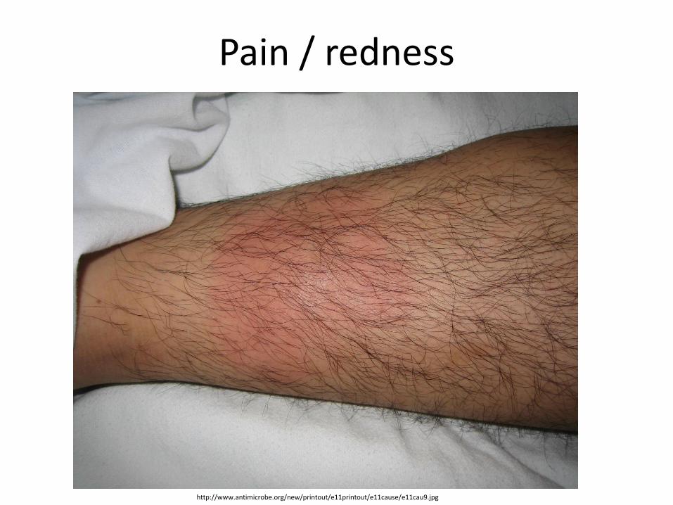
Pain / redness
http://www.antimicrobe.org/new/printout/e11printout/e11cause/e11cau9.jpg
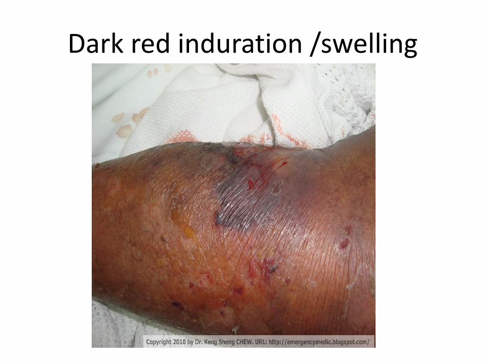
Dark red induration /swelling
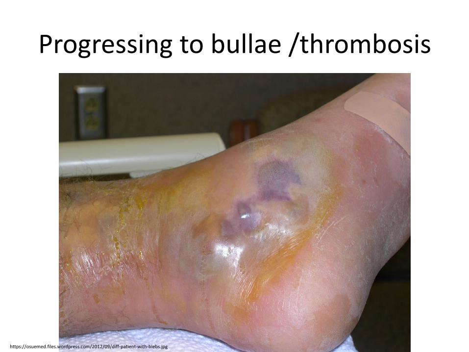
Progressing to bullae /thrombosis
https://osuemed.files.wordpress.com/2012/09/diff-patient-with-blebs.jpg
http://img.medscapestatic.com/pi/meds/ckb/55/37455tn.jpg

Microbiology causes:
• A) Polymicrobial (• Type I necrotizing fasciitis involves at least one anaerobic species ( Bacteroides or Peptostreptococcus spp.), as well as one or more facultative anaerobic species (e.g. non-GAS, E. coli, Enterobacter, Klebsiella, Proteus spp.).
• usually a mix of aerobes and anaerobic bacteria ( clostridium perfringens)
• 1 - Break in Gastrointestinal or Genitourinary mucosa, typically on trunk and extremities
• 2- Fournier's Gangrene (in genitalia/perineal area)• 3- mixed infection usually have comorbid states ( DM,
PVD, immunecompromised)

Microbiology causes…cont
• B) Type II necrotizing fasciitis is usually caused by GAS alone or in combination with other species (e.g. S. aureus). Group A , Beta hemolytic strep (GAS), S. pyogenes +- S. aureus
• Strains of MRSA that produce the PantonValentine leukocidin (PVL) toxin have been reported to cause necrotizing fasciitis.
• 1) usually following trauma in otherwise healthy individualor IV drug abusers (skin popping)
2) Fasciitis progresses to skin contusions due to seeding by transient bactermia3) Gas production if mixed infections occurs!!! (you need anaerobes)4) Severe toxicity and renal impairments shock5) Myositis ( destruction of muscle tissue markedly increases CPK)6) Mortality is high (upto 50%!)
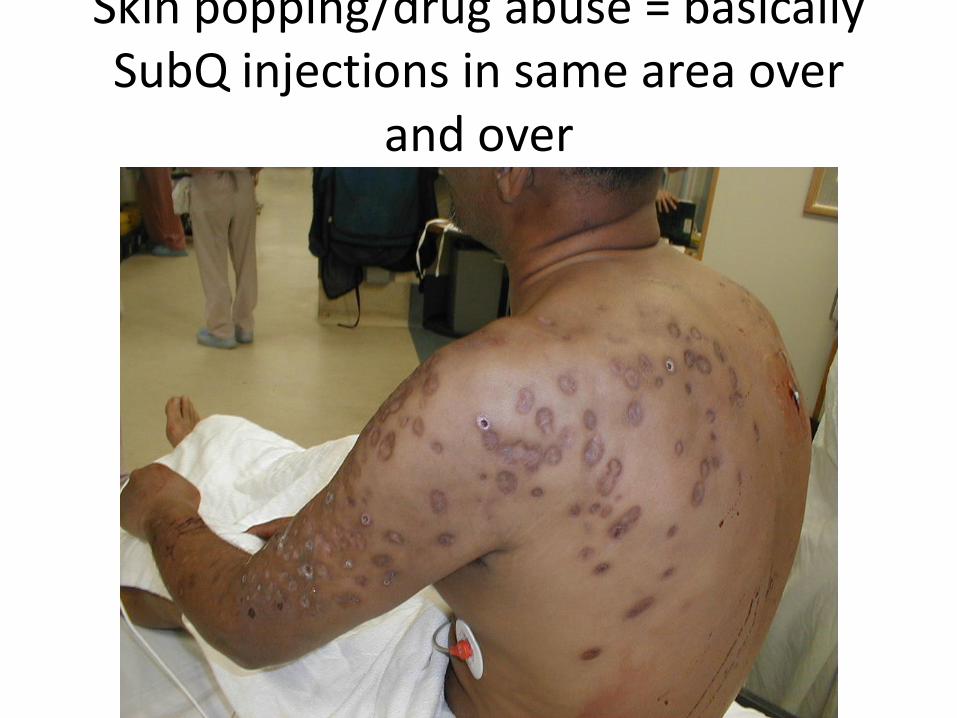
Skin popping/drug abuse = basically SubQ injections in same area over
and over

• Necrotizing fasciitis caused by mixed aerobic-anaerobic bacteria begins with a breach in the integrity of a mucous membrane barrier, such as the mucosa of the gastrointestinal or genitourinary tract.
• The portal can be a malignancy, a diverticulum, a hemorrhoid, an anal fissure, or a urethral tear.
• Other predisposing factors include peripheral vascular disease, diabetes mellitus, surgery, and penetrating injury to the abdomen.
• Leakage into the perineal area results in a syndrome called Fournier’s gangrene, characterized by massive swelling of the scrotum and penis with extension into the perineum or the abdominal wall and legs.
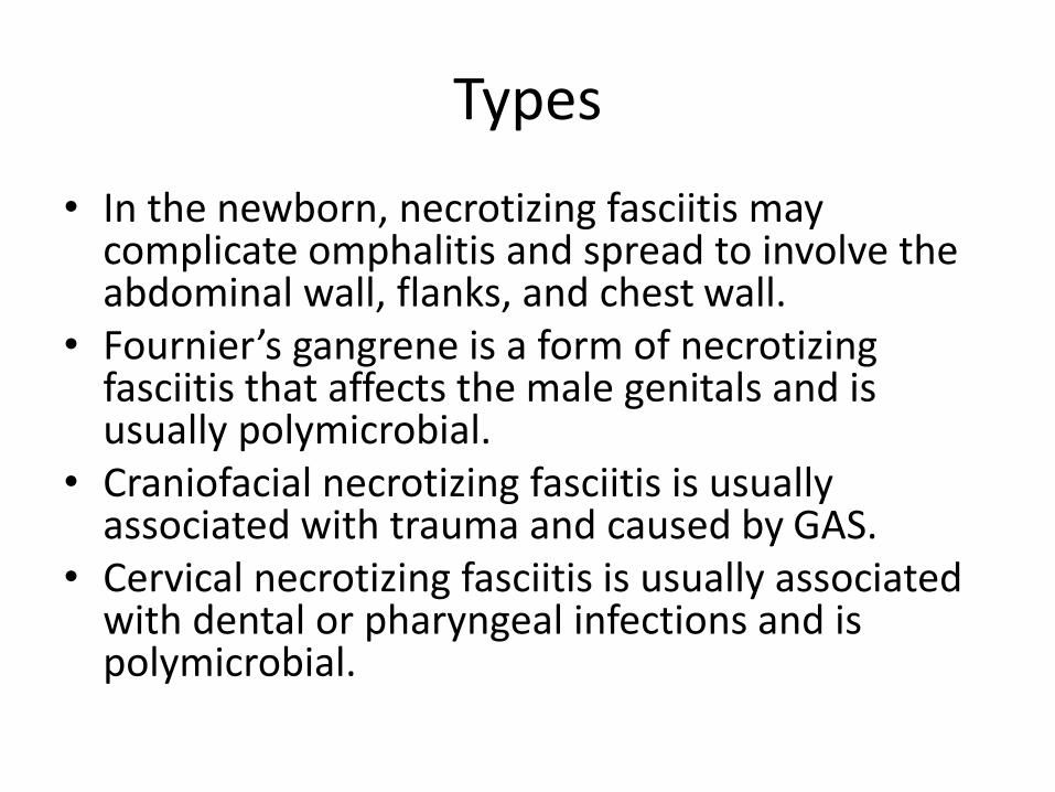
Types
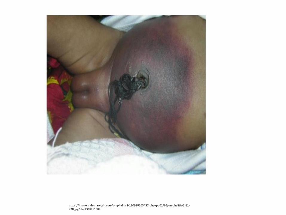
• In the newborn, necrotizing fasciitis may complicate omphalitis and spread to involve the abdominal wall, flanks, and chest wall.
• Fournier’s gangrene is a form of necrotizing fasciitis that affects the male genitals and is usually polymicrobial.
• Craniofacial necrotizing fasciitis is usually associated with trauma and caused by GAS.
• Cervical necrotizing fasciitis is usually associated with dental or pharyngeal infections and is polymicrobial.
https://image.slidesharecdn.com/omphalitis2-120928165437-phpapp01/95/omphalitis-2-11-728.jpg?cb=1348851384

Dx of necrotizing fasciitis
Clinical findings are suggestive + surgical exploration/sample:a) Altered mental statusb) Soft tissue infection signs (redness/swelling/pain) 70-80%
of casesBullae Pain is typically exaggerated out of examTenderness is outside the red erythematous borders (indicates further progress)
c) are only seen in ¼ of casesd) Fever in less than 50% of the cases!e) Low BP in 21% f) crepitation (feeling of air pockets under skin upon
examination) in 20%
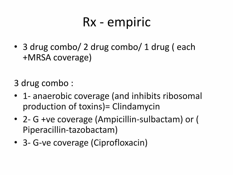
Rx - empiric
• 3 drug combo/ 2 drug combo/ 1 drug ( each +MRSA coverage)
3 drug combo :
• 1- anaerobic coverage (and inhibits ribosomal production of toxins)= Clindamycin
• 2- G +ve coverage (Ampicillin-sulbactam) or ( Piperacillin-tazobactam)
• 3- G-ve coverage (Ciprofloxacin)
• 2 drug combo (Cefotaxime covers G+ and G-bacteria) + (anaerobic coverage by metronidazole or clindamycin)
• 1 drug combo ( Carbapenem 0 Imipenem, meropenem, ertapenem)
• The MRSA coverage to be added to any chosen empiric regimen includes = Vancomycin or Linezolid
• Hemorrhagic bullae may indicate presence of vibrio vulnificus, in which case doxycycline is used

Rx.
• Surgical debridement, and treatment in hospital Emergency surgical exploration and debridement confirm the diagnosis and are the mainstay of therapy.
• Reducing compartment pressure in extremities
• Prophylaxis for exposed house hold members (penicillin, rifampin, clindamycin or azirthromycin)
Gas gangrene ( Clostridium infection)
Gas production due to anaerobic bacteria
• Typically due to contaminated DEEP wounds -no oxygen- (surgery, car crash..etc) to introduce spores of G+ve clostridia into the wound
• Also progresses similarly to other types: fasciitis toxemia organ failure
• Gangrene usually occurs following muscle injury and contamination of the wound by soil or foreign material containing clostridial spores.
• C. perfringens is the predominant cause (80–95%), and its pathological effects are mediated by α and λ toxins
Cont.. etiology and pathogenesis
• Spontaneous or non-traumatic gas gangrene may occur in the absence of an obvious wound.
• This form is usually caused by C. septicum and associated with intestinal abnormalities, e.g. colonic cancer, diverticulitis, bowel infarction, necrotizing enterocolitis. •
Clinical features
• • The incubation period is usually 2–3 days but may be shorter.
• • Patients present with acute onset of excruciating pain and signs of shock (fever, tachycardia, hypotension, jaundice, renal failure).
• • Local edema and tenderness may be the only early signs, or there may be an open wound, herniation of muscle, a serosanguinous and foul smelling discharge, crepitus, skin discoloration, and necrosis.
• • Progression is rapid, and death may occur within hours
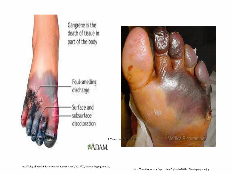
https://clinicalscienceblogsimone.files.wordpress.com/2013/02/gangrene-pictures-3.jpg
http://blog.almawiclinic.com/wp-content/uploads/2015/07/Foot-with-gangrene.jpghttp://healthooze.com/wp-content/uploads/2012/12/wet-gangrene.jpg

Diagnosis •
• The diagnosis is usually clinical but may be confirmed by Gram stain of the wound or aspirate.
• • Liquid anaerobic cultures may be positive within 6h.
• • Plain radiographs may show gas in the affected tissues
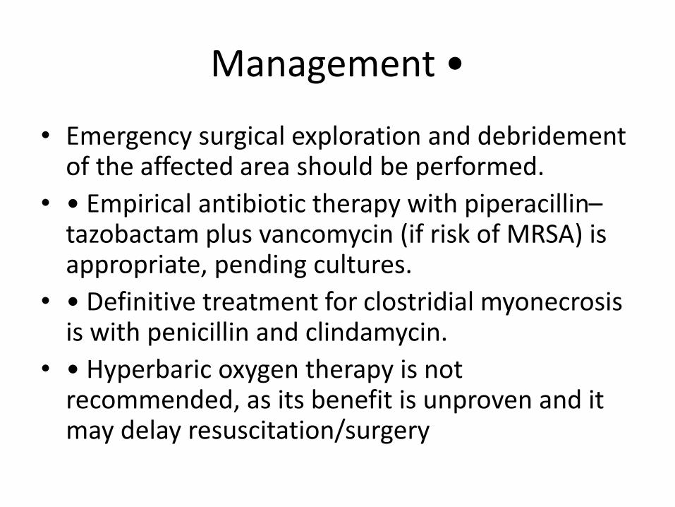
Management •
• Emergency surgical exploration and debridement of the affected area should be performed.
• • Empirical antibiotic therapy with piperacillin–tazobactam plus vancomycin (if risk of MRSA) is appropriate, pending cultures.
• • Definitive treatment for clostridial myonecrosis is with penicillin and clindamycin.
• • Hyperbaric oxygen therapy is not recommended, as its benefit is unproven and it may delay resuscitation/surgery

Cellulitis
• Cellulitis is an acute inflammatory condition of the skin that is characterized by:
localized pain erythema, swelling, and heat (inflammation signs).
Usually caused by indigenous flora colonizing the skin (S. aureus and S. pyogenes) or by a variety of non colonizing exogenous bacteria. To detect the source of the exogenous bacteria involved in cellulitis a thorough history (+ epidemiologic data) is needed, as these bacteria occupy small niches in nature.
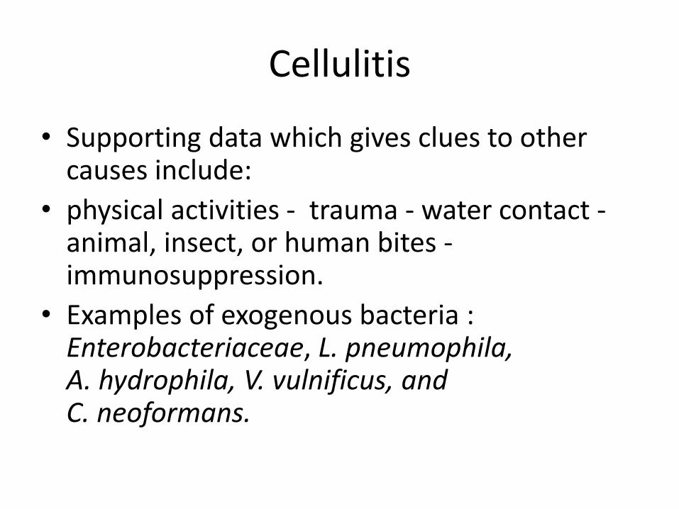
Cellulitis
• Supporting data which gives clues to other causes include:
• physical activities - trauma - water contact -animal, insect, or human bites -immunosuppression.
• Examples of exogenous bacteria :Enterobacteriaceae, L. pneumophila, A. hydrophila, V. vulnificus, and C. neoformans.
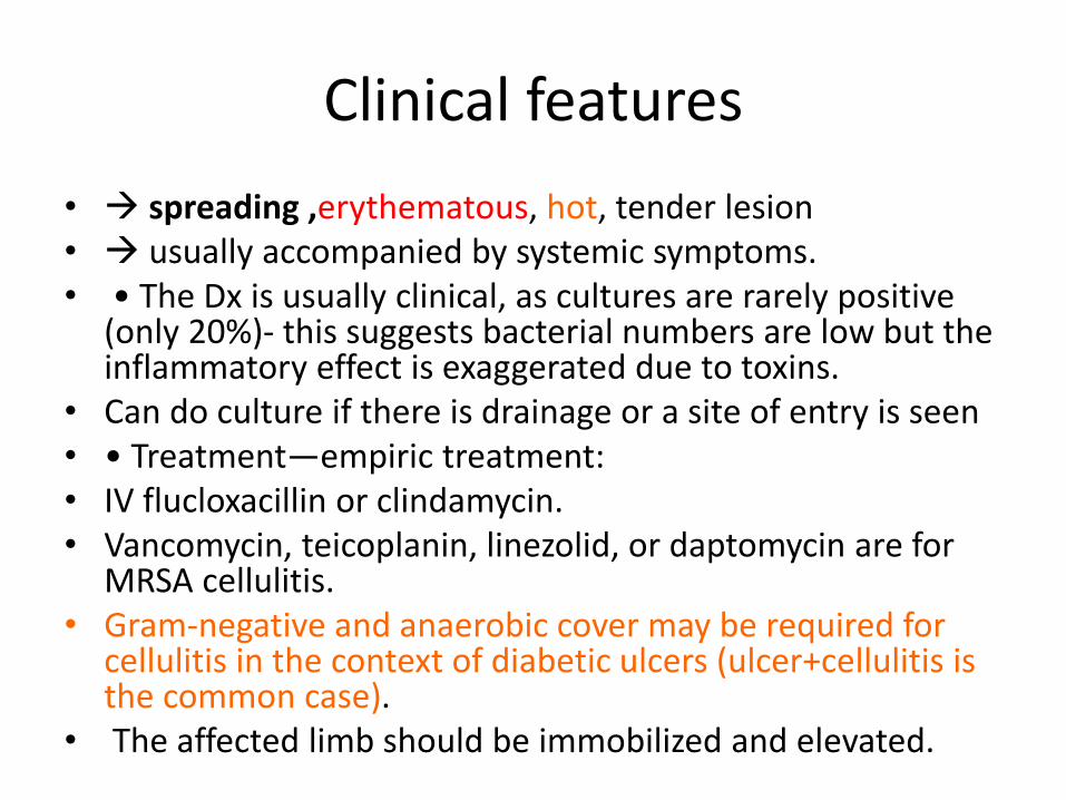
Clinical features
• spreading ,erythematous, hot, tender lesion• usually accompanied by systemic symptoms.• • The Dx is usually clinical, as cultures are rarely positive
(only 20%)- this suggests bacterial numbers are low but the inflammatory effect is exaggerated due to toxins.
• Can do culture if there is drainage or a site of entry is seen• • Treatment—empiric treatment:• IV flucloxacillin or clindamycin.• Vancomycin, teicoplanin, linezolid, or daptomycin are for
MRSA cellulitis. • Gram-negative and anaerobic cover may be required for
cellulitis in the context of diabetic ulcers (ulcer+cellulitis is the common case).
• The affected limb should be immobilized and elevated.

https://www.youtube.com/watch?v=4l8FxmMlgQA

Remember; acute and spreading
https://www.google.com/url?sa=i&rct=j&q=&esrc=s&source=images&cd=&ved=2ahUKEwjJ6vK_07HZAhUC16QKHZvjALEQjxx6BAgAEAI&url=https%3A%2F%2Famwell.com%2Fcm%2Fconditions%2Fcellulitis%2F&psig=AOvVaw0fo79yBSq0tU3pOduFHrom&ust=1519118392872439
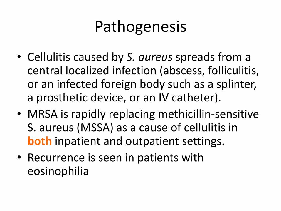
Pathogenesis
• Cellulitis caused by S. aureus spreads from a central localized infection (abscess, folliculitis, or an infected foreign body such as a splinter, a prosthetic device, or an IV catheter).
• MRSA is rapidly replacing methicillin-sensitive S. aureus (MSSA) as a cause of cellulitis in both inpatient and outpatient settings.
• Recurrence is seen in patients with eosinophilia

• Cellulitis due to S. pyogenes is more rapidly spreading, diffuse process that is frequently associated with lymphangitis and fever.
• Recurrent streptococcal cellulitis of the lower extremities may be caused by organisms of group A, C, or G in association with chronic venous stasis or with saphenous venectomy for coronary artery bypass surgery.
• Also recurrent streptococcal cellulitis is seen among patients with chronic lymphedema resulting from elephantiasis, lymph node dissection, or Milroy’s disease.
• This is all due to the fact that streptococci use the lymphatic system in their spread.

• Cellulitis caused by (group B Streptococcus) occurs mostly in elderly patients (usually patients with diabetes mellitus or peripheral vascular disease.
• H. influenzae typically causes periorbital cellulitis in children in association with sinusitis, otitis media, or epiglottitis.
• It is unclear if this form of cellulitis will become less common as a result of the efficacy of the H. influenzae type b vaccine.
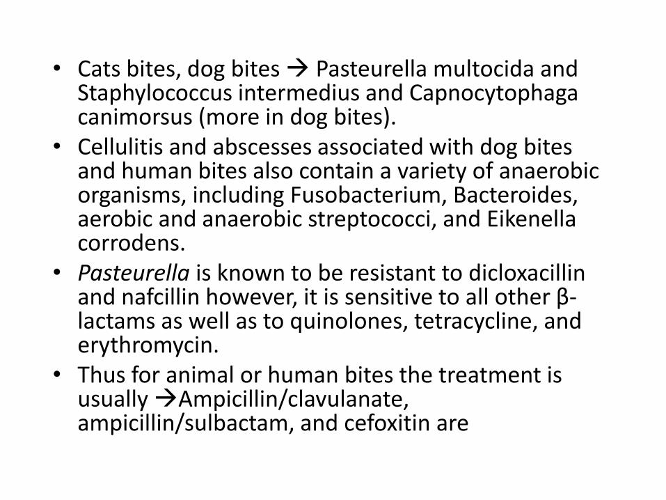
• Cats bites, dog bites Pasteurella multocida and Staphylococcus intermedius and Capnocytophagacanimorsus (more in dog bites).
• Cellulitis and abscesses associated with dog bites and human bites also contain a variety of anaerobic organisms, including Fusobacterium, Bacteroides, aerobic and anaerobic streptococci, and Eikenellacorrodens.
• Pasteurella is known to be resistant to dicloxacillin and nafcillin however, it is sensitive to all other β-lactams as well as to quinolones, tetracycline, and erythromycin.
• Thus for animal or human bites the treatment is usually Ampicillin/clavulanate, ampicillin/sulbactam, and cefoxitin are
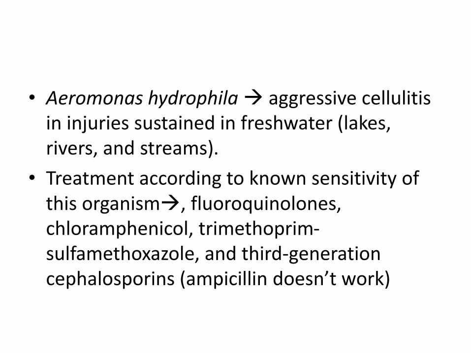
• Aeromonas hydrophila aggressive cellulitis in injuries sustained in freshwater (lakes, rivers, and streams).
• Treatment according to known sensitivity of this organism, fluoroquinolones, chloramphenicol, trimethoprim-sulfamethoxazole, and third-generation cephalosporins (ampicillin doesn’t work)

P. aeruginosa
• Causes 3 types of infections in MSS• 1 ecthyma gangrenosum in neutropenic patients• 2 hot-tub folliculitis• 3 cellulitis following penetrating injury (usually stepping
on a nail) • Commonly seen in hospital setting/immune compromised
patients.• Rx: surgical inspection and drainage/debridement (recall
biofilm of pseudomonas) • empirical treatment : • -aminoglycoside - a third-generation cephalosporin
(ceftazidime, cefoperazone, or cefotaxime) -semisynthetic penicillin (ticarcillin, mezlocillin, or piperacillin), or a fluoroquinolone(not in pediatric patient)
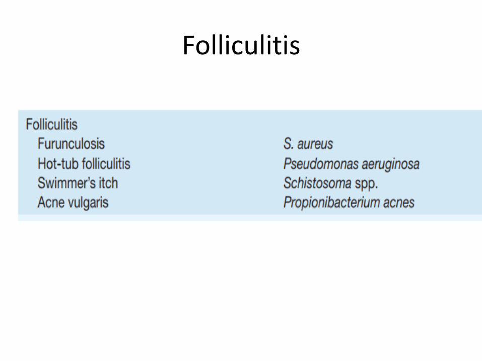
Folliculitis

https://library.med.utah.edu/kw/derm/pages/in04_10.htm

Folliculitis
• • A superficial infection of the hair follicles and apocrine structures.
• Causative organisms: S. aureus (commonest), P. aeruginosa (‘hot tub’ folliculitis), Enterobacteriaceae (complication of acne), Candida spp., and M. furfur (in patients taking corticosteroids).
• Eosinophilic pustular folliculitis occurs in AIDS patients. • Clinically: lesions consist of small, erythematous, pruritic
papules, often with a central pustule. • • Treatment—empiric treatment is with oral flucloxacillin.• If the clinical response is slowconsider other pathogens.

http://diseasespictures.com/folliculitis-symptoms-causes-pictures-treatment-home-remedies/
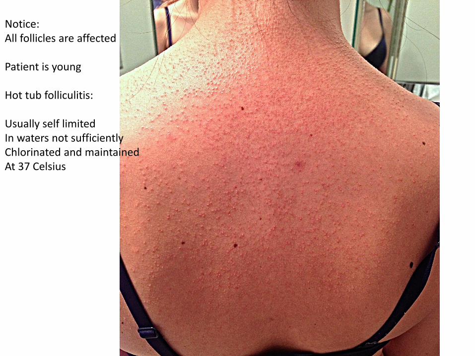
Notice:All follicles are affected
Patient is young
Hot tub folliculitis:
Usually self limitedIn waters not sufficientlyChlorinated and maintainedAt 37 Celsius
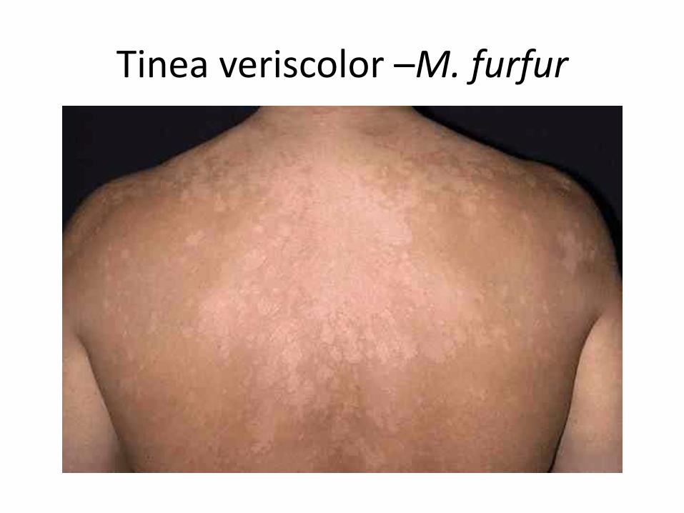
Tinea veriscolor –M. furfur
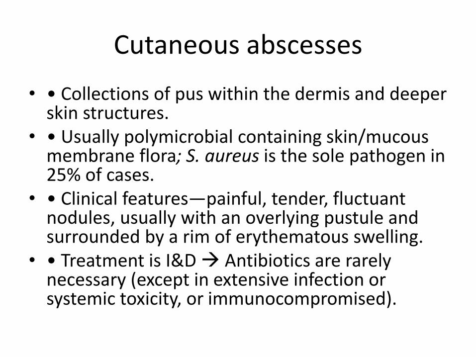
Cutaneous abscesses
• • Collections of pus within the dermis and deeper skin structures.
• • Usually polymicrobial containing skin/mucous membrane flora; S. aureus is the sole pathogen in 25% of cases.
• • Clinical features—painful, tender, fluctuant nodules, usually with an overlying pustule and surrounded by a rim of erythematous swelling.
• • Treatment is I&D Antibiotics are rarely necessary (except in extensive infection or systemic toxicity, or immunocompromised).
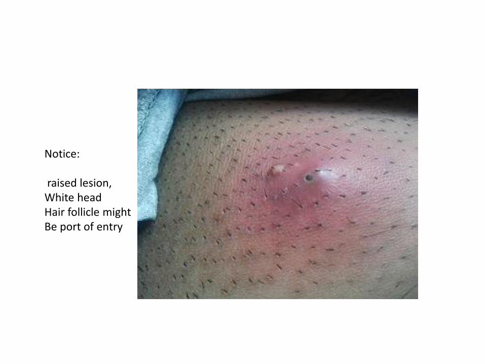
Notice:
raised lesion,White headHair follicle mightBe port of entry

Furuncles and carbuncles
• • A furuncle (boil) is a deep inflammatory nodule that usually develops from preceding folliculitis.
• Occur in areas of the hairy skin, e.g. face, neck, axillae, and buttocks.
• • A carbuncle is a larger, deeper lesion made of multiple abscesses extending into the subcutaneous fat.
• Usually occur at the nape of the neck, on the back, or on the thighs.
• Patients may be systemically unwell. • • Outbreaks of furunculosis caused by MSSA and MRSA
have been described in groups of individuals with close contact, e.g. families, prisons, and sports teams.

Rx for furnucles
• • application of moist heat promotes localization and spontaneous drainage.
• Large lesions require surgical drainage.
• Systemic antibiotics are indicated 1- fever, 2-cellulitis 3-lesions are located near the nose or lip.
• Outbreaks control with chlorhexidine soapsand stop sharing of clothing articles or towels, and decolonization of staph.

• Sebaceous glands that empty into the hair follicle maybe blocked and cause a swelling similar to an abscess (sebaceous cyst).
• Infection of sweat glands (hidradenitis suppurativa) can also mimic infection of hair follicles, particularly in the axillae.
• Chronic folliculitis is uncommon except in acne vulgaris, where constituents of the normal flora (e.g., Propionibacterium acnes) may play a role.
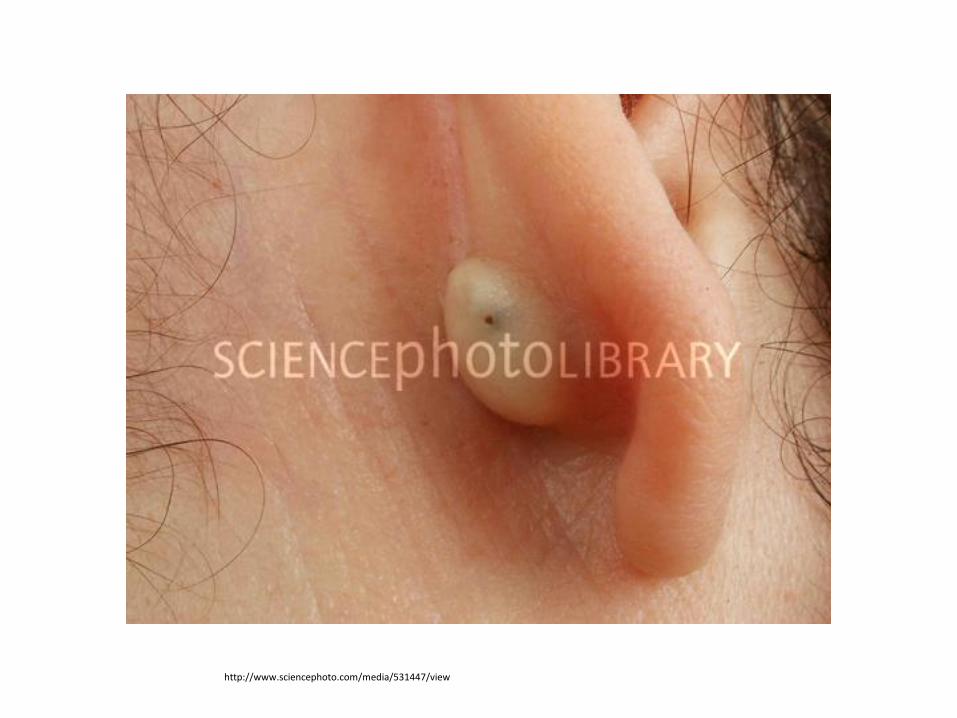
http://www.sciencephoto.com/media/531447/view

HidradenitisSuppurativa
Usually in sweaty areasWhere skin folds ( axilla,Buttocks, breasts, inner thighs)
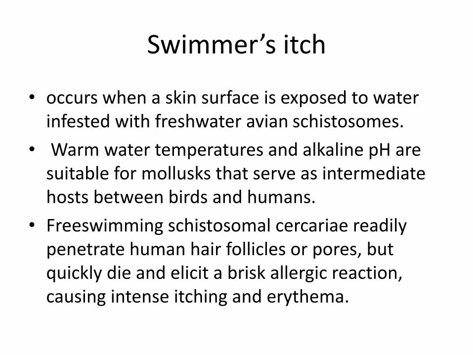
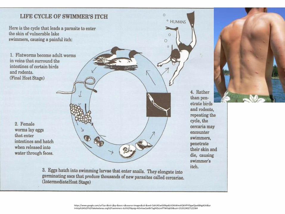
Swimmer’s itch
• occurs when a skin surface is exposed to water infested with freshwater avian schistosomes.
• Warm water temperatures and alkaline pH are suitable for mollusks that serve as intermediate hosts between birds and humans.
• Freeswimming schistosomal cercariae readily penetrate human hair follicles or pores, but quickly die and elicit a brisk allergic reaction, causing intense itching and erythema.
https://www.google.com/url?sa=i&rct=j&q=&esrc=s&source=images&cd=&ved=2ahUKEwiG09qa6LHZAhWmsKQKHfYYDgwQjxx6BAgAEAI&url=http%3A%2F%2Flakeleelanau.org%2Fswimmers-itch%2F&psig=AOvVaw2an8V7yglHZoLmPTWOqb5X&ust=1519124027122364
Erysipelas
• Erysipelas is due to S. pyogenes and is characterized by an abrupt onset of fiery-red swelling of the face or extremities.
• The distinctive features of erysipelas are well-defined indurated margins, particularly along the nasolabial fold; rapid progression; and intense pain.
• Flaccid bullae may develop during the second or third day of illness, but extension to deeper soft tissues is rare.
• Treatment : penicillin(flucloxacillin, clindamycin) or is effective• swelling may progress despite appropriate treatment, although
fever, pain, and the intense red color diminish.• Desquamation of the involved skin occurs 5–10 days into the
illness.• Infants and elderly adults are most commonly afflicted, and the
severity of systemic toxicity varies.