Harnessing fluorinesulfur contacts and multipolar ...sro.sussex.ac.uk/61588/1/main resubmission...
25
Harnessing fluorine-sulfur contacts and multipolar interactions for the design of p53 mutant Y220C rescue drugs Article (Accepted Version) http://sro.sussex.ac.uk Bauer, Matthias R, Jones, Rhiannon N, Baud, Matthias G J, Wilcken, Rainer, Boeckler, Frank M, Fersht, Alan, Joerger, Andreas C and Spencer, John (2016) Harnessing fluorine-sulfur contacts and multipolar interactions for the design of p53 mutant Y220C rescue drugs. ACS Chemical Biology, 11 (8). pp. 2265-2274. ISSN 1554-8929 This version is available from Sussex Research Online: http://sro.sussex.ac.uk/id/eprint/61588/ This document is made available in accordance with publisher policies and may differ from the published version or from the version of record. If you wish to cite this item you are advised to consult the publisher’s version. Please see the URL above for details on accessing the published version. Copyright and reuse: Sussex Research Online is a digital repository of the research output of the University. Copyright and all moral rights to the version of the paper presented here belong to the individual author(s) and/or other copyright owners. To the extent reasonable and practicable, the material made available in SRO has been checked for eligibility before being made available. Copies of full text items generally can be reproduced, displayed or performed and given to third parties in any format or medium for personal research or study, educational, or not-for-profit purposes without prior permission or charge, provided that the authors, title and full bibliographic details are credited, a hyperlink and/or URL is given for the original metadata page and the content is not changed in any way.
Transcript of Harnessing fluorinesulfur contacts and multipolar ...sro.sussex.ac.uk/61588/1/main resubmission...
Harnessing fluorinesulfur contacts and multipolar interactions for the design of p53 mutant Y220C rescue drugs
Article (Accepted Version)
http://sro.sussex.ac.uk
Bauer, Matthias R, Jones, Rhiannon N, Baud, Matthias G J, Wilcken, Rainer, Boeckler, Frank M, Fersht, Alan, Joerger, Andreas C and Spencer, John (2016) Harnessing fluorine-sulfur contacts and multipolar interactions for the design of p53 mutant Y220C rescue drugs. ACS Chemical Biology, 11 (8). pp. 2265-2274. ISSN 1554-8929
This version is available from Sussex Research Online: http://sro.sussex.ac.uk/id/eprint/61588/
This document is made available in accordance with publisher policies and may differ from the published version or from the version of record. If you wish to cite this item you are advised to consult the publisher’s version. Please see the URL above for details on accessing the published version.
Copyright and reuse: Sussex Research Online is a digital repository of the research output of the University.
Copyright and all moral rights to the version of the paper presented here belong to the individual author(s) and/or other copyright owners. To the extent reasonable and practicable, the material made available in SRO has been checked for eligibility before being made available.
Copies of full text items generally can be reproduced, displayed or performed and given to third parties in any format or medium for personal research or study, educational, or not-for-profit purposes without prior permission or charge, provided that the authors, title and full bibliographic details are credited, a hyperlink and/or URL is given for the original metadata page and the content is not changed in any way.

Harnessing Fluorine-Sulfur Contacts and Multipolar Interactions for the
Design of p53 Mutant Y220C Rescue Drugs
Matthias R. Bauer #1, Rhiannon N. Jones #2, Matthias G. J. Baud 1, Rainer Wilcken 1, Frank
M. Boeckler 3, Alan R. Fersht 1, Andreas C. Joerger *1,2,4,5 and John Spencer *2
1 MRC Laboratory of Molecular Biology, Francis Crick Avenue, Cambridge Biomedical
Campus, Cambridge CB2 0QH, United Kingdom.
2 Department of Chemistry, School of Life Sciences, University of Sussex, Falmer, Brighton,
East Sussex BN1 9QJ, UK.
3 Molecular Design and Pharmaceutical Biophysics, Institute of Pharmaceutical Sciences,
Eberhard Karls Universität Tübingen, Auf der Morgenstelle 8, 72076 Tübingen, Germany
4 German Cancer Consortium (DKTK), German Cancer Research Center (DKFZ), 69120
Heidelberg, Germany
5 Institute of Pharmaceutical Chemistry, Johann Wolfgang Goethe-University and Buchmann
Institute for Molecular Life Sciences, Max-von-Laue-Str. 9, 60438 Frankfurt am Main,
Germany
# M.R.B and R.N.J. contributed equally to this work
* Corresponding authors
ABSTRACT
Many oncogenic mutants of the tumor suppressor p53 are conformationally unstable,
including the frequently occurring Y220C mutant. We have previously developed
several small-molecule stabilizers of this mutant. One of these molecules, PhiKan083,
1-(9‐ethyl‐9H‐carbazole‐3‐yl)-N-methylmethanamine, binds to a mutation-induced
surface crevice with a KD =150 M, thereby increasing the melting temperature of the
protein and slowing its rate of aggregation. Incorporation of fluorine atoms into small
molecule ligands can substantially improve binding affinity to their protein targets.
We have, therefore, harnessed fluorine-protein interactions to improve the affinity of
this ligand. Step-wise introduction of fluorines at the carbazole ethyl anchor, which is
deeply buried within the binding site in the Y220C-PhiKan083 complex, led to a
five-fold increase in affinity for a 2,2,2-trifluoroethyl anchor (ligand efficiency of 0.3
kcal mol-1 atom-1). High-resolution crystal structures of the Y220C-ligand complexes
combined with quantum chemical calculations revealed favorable interactions of the
fluorines with protein backbone carbonyl groups (Leu145 and Trp146) and the sulfur
of Cys220 at the mutation site. Affinity gains were, however, only achieved upon
trifluorination, despite favorable interactions of the mono- and difluorinated anchors
with the binding pocket, indicating a trade-off between energetically favorable
protein-fluorine interactions and increased desolvation penalties. Taken together, the
optimized carbazole scaffold provides a promising starting point for the development
of high-affinity ligands to reactivate the tumor suppressor function of the p53 mutant
Y220C in cancer cells.
INTRODUCTION
The introduction of fluorine atoms into organic small molecules has become widespread in
drug discovery.1-5 Indeed, between 20-25% of drugs on the market are estimated to contain at
least one fluorine atom.6 Apart from modulating various important compound properties such
as logP, metabolic stability, basicity, and bioavailability, fluorine substituents have been also
successfully used for improving ligand binding affinities. 7-9 This can be achieved via
intramolecular stabilization of favorable ligand binding conformations, modulation of
polarity or basicity of neighbouring functional groups of the ligand, or direct fluorine-protein
interactions.1, 8 These interactions can be further classified into polar interactions with
hydrogen bond donors (e.g. backbone NH, polarized Cα-H, polar side chains, and protein
bound water), as observed in the binding of type II statins to the HMGCoA enzyme,10
hydrophobic interactions with lipophilic side chains, and orthogonal multipolar interactions,
which can be also described as n→π* interactions,11 with backbone carbonyl groups, amide
containing side chains (Asn and Gln), and guanidinium groups (Arg).
A number of case studies on the use of fluorine interactions in rational drug design have been
reported. Olsen et al. conducted a systematic fluorine scan with a thrombin inhibitor,
revealing favorable interactions with backbone amides and Cα protons that yielded
significantly improved binding affinity 12 The introduction of fluorine substituents also
significantly increased the potency of kinesin spindle protein inhibitors, abl kinase inhibitors,
and peptidic elastase inhibitors.13-16 Vulpetti et al. identified fluorinated fragments binding to
trypsin by 19F NMR screening and crystal structure determination and described a general
approach to identify fluorophilic hot-spots in proteins using crystal and computational
analysis.17 Recently, Pollock et al. investigated the impact of fluorine-protein interactions on

the binding affinity of a menin-MLL inhibitor and introduced their computational algorithm
FMAP which aims to facilitate the rational design of fluorine-protein interactions.18
Here, we have harnessed fluorine interactions for the development of mutant p53 rescue
drugs. The tumor suppressor p53 plays a key role in regulating cell-cycle arrest, DNA repair,
apoptosis or cellular senescence.19-21 In virtually all human cancers, p53 is inactivated either
by mutation or overexpression of negative regulators such as MDM2 or MDMX, which leads
to proteasomal degradation of p53.22 The cancer mutation Y220C, which accounts for an
estimated 100,000 new cancer cases per year worldwide, significantly destabilizes the p53
DNA-binding domain (DBD) and impairs its function via increased thermal denaturation.21, 23
We have previously developed small-molecule stabilizers of p53-Y220C, such as Phikan083,
PhiKan5196, and PhiKan7088 (Figure 1), which bind to a mutation-induced surface crevice
on the DBD, thereby stabilizing the protein, slowing its unfolding and aggregation, and in
some cases restoring tumor suppressor activity in cancer cells harboring the p53-Y220C
mutation.24-28 In this study we aimed at improving the potency of the carbazole-based
compound Phikan083 and employed ab inito quantum-chemical calculations to probe
potential interaction energy gains upon fluorination of the ethyl anchor. We have synthesized
mono-, di- and, tri- fluorinated 9H-fluoroethyl carbazoles, evaluated their binding affinities
via differential scanning fluorimetry (DSF) and isothermal titration calorimetry (ITC), and
determined their binding mode by X-ray crystallography. We found that trifluorination
significantly improved the binding affinity by approximately five-fold compared with
PhiKan083 (1), whereas both monofluoro and difluoro analogues were less potent than the
parent compound.
Insert Figure 1 here

RESULTS AND DISCUSSION
Quantum chemical calculations
In the crystal structure of p53-Y220C in complex with PhiKan083 (pdb: 2VUK), the ethyl
moiety of PhiKan083 is in close proximity to the carbonyl groups of Leu145 and Trp146, and
the thiol group of Cys220. Given the frequent and well-characterized interactions between
organofluorine groups and protein backbone amides, as well as the less frequently observed
interactions between fluorine and sulfur atoms,9 we investigated whether gains in binding
affinity could be achieved via fluorinated ethyl substituents using DFT-D calculations at
BLYP-D3/def2-SVP level with a truncated model of PhiKan083 bound to the p53-Y220C
binding pocket (Figure 2B). Except for the sulfur atom of Cys220, all heavy atoms of the
Y220C binding pocket as well as the nitrogen and C-3 atom of the pyrrole ligand model were
kept frozen during the calculations.
Distances between the backbone amides of Leu145 and Trp146 and the nearest C-F groups
were between 3.0 Å and 3.3 Å in the optimized structures, which is in good agreement with
typical CF…C=O distances (3.0 Å – 3.7 Å) for orthogonal multipolar interactions.16 C-F…S
and C-F…HS distances ranged from 2.8 Å to 3.6 Å, which is also in good agreement with
experimentally observed distances for fluorine-sulfur contacts (2.8 Å - 3.4 Å) in protein
structures.9 Our DFT-D3 calculations indicated that the relative interaction energies (ΔΔE)
for all fluorinated ethyl groups improved by at least -2 kcal/mol compared with the N-ethyl
reference energy ΔE (ΔE = Ecomplex - (Ereceptor + Eligand)) (Figure 2B). The energetically most
favorable conformation (ΔΔE = -6.5 kcal/mol) of the 2-fluoroethyl group was orientation 1
(Figure 2B), in which the C-F vector points towards the backbone amides of Leu145 and
Trp146, predicting two potential orthogonal multipolar interactions between the fluorine
atom and both carbonyl groups. Orientations 2 and 3 of the 2-fluoroethyl group, where the

fluorines were oriented towards the sulfhydryl group of Cys220, were energetically less
favorable, with respective ΔΔE values of -2.2 kcal/mol and -3.7 kcal/mol. The relative
interaction energy of conformation 1 of the difluoro ethyl moiety (ΔΔE = -6.9 kcal/mol) was
similar to the most favored 2-fluoroethyl conformation (see Figure S1 for difluoro ethyl
conformations 2 and 3 and their DFT-D energies), whereas the trifluoro-substituted ethyl
anchor was energetically less favorable with a ΔΔE value of -4.2 kcal/mol (Figure 2B).
However, the calculated DFT-D3 energies only yield an estimate of the ligand-protein
interaction at the chosen computational level in a model system of small size and neglect
other contributions to the free energy of binding such as entropic changes and desolvation
penalties.
Insert Fig 2 here
Compound synthesis
We devised PhiKan083 (1) analogues 2-4 (Figure 3) with a mono-, di- or trifluoroethyl
anchor, in addition to their counterparts 5-6 bearing a dimethylamine instead of a
monomethylamine group. We envisaged that having different substitution patterns (e.g.,
secondary vs tertiary amine) at the level of the pendant, solvent exposed amino group would
provide additional structure-activity relationship and inform on the influence of the amino
side chain on the overall potency and binding mode of 1 and fluorinated analogues.
Insert Fig 3 here
1 and 2 were obtained from commercial sources (see Experimental Section). The synthesis of
3, 4, 5, and 6 was straightforward, and is described in Scheme 1. Attempts at alkylation of
intermediate 9 were unsuccessful. Alkylation of 9H-carbazole-3-carbaldehyde (9)29 with the

appropriate alkyl halide or tosylate was achieved in the presence of Cs2CO3 in DMF, and
provided intermediates 7-8. Low yields were due to a competing fluorine elimination
pathway which has been previously reported by Suehiro et al..30 Further introduction of the
amine side chain by reductive amination afforded 3, 4, 5, and 6. Two reductive amination
procedures were attempted; titanium (IV) isopropoxide-sodium borohydride and sodium
triacetoxyborohydride and yields in both cases were comparable.31, 32 Indeed, the titanium-
based method was used for the reductive amination of 7 and 8 but we anticipate that sodium
triacetoxyborohydride would be equally effective.
Scheme 1. Synthesis and overall yield of compounds 3, 4, 5 and 6 using the two-step synthesis
described.
Synthesis of 3, 4, 5 and 6. (a) R1I (difluoroethyl) or R1OTs (trifluoroethyl), Cs2CO3, DMF, µwave, 150°C, 30 min; (b)
amine, Ti(OiPr)4, Et3N, EtOH/DCM, rt, 18 h then NaBH4, rt, 8 h; (c) NaBH(OAc)3, HNMe2, AcOH, THF, rt.
Compound R1 R2 Yield
(step a)
Yield
(step b)
3
44 58
4
50 40
5
NA a 82

6
50 56
a not applicable, precursor was bought from TCI UK.
Biophysical evaluation
We tested PhiKan083 (1) and analogues 2-6 for their stabilization of the p53-Y220C DNA-
binding domain (DBD) using differential scanning fluorimetry (DSF) (Table 1). The N-2,2,2-
trifluoroethyl substituted carbazoles 4 and 6 increased stability of the p53-Y220C DBD by
1.2 K at a compound concentration of 125 μM, showing a clear improvement over their N-
ethyl substituted counterparts PhiKan083 (1) and 5 (ΔTm = 0.8 K and ΔTm =0.6 K,
respectively). In contrast, the difluorinated (2) and monofluorinated (3) carbazoles were
significantly worse than the parent compound in terms of protein stabilization, with
respective thermal shifts of 0.3 K and 0.4 K. Dissociation constants (KD) were determined by
isothermal titration calorimetry (ITC) (Table 1). The substitution pattern of the solvent-
exposed amine had a minor effect on affinity, with the additional methyl group in 5 resulting
in a 30% drop in affinity. The trifluoro-substituted carbazoles 4 and 6 were the most potent
compounds and showed ITC KD values of 28 and 37 µM, respectively (Figure 4), which
corresponds to a 5-fold increase in affinity compared to the non-fluorinated parent
compounds (1) and (5). Despite prediction of improved interaction energies by DFT-D
calculations, the mono- and difluorinated analogues, 2 and 3, failed to display improved
affinities. They had KD values of 101 μM and 138 μM, respectively (Figure S2).
Table 1. Thermal shift data and KD values
compound DSF ΔTm at
125 µM
ITC KD
(µM)
1 0.8 124a
2 0.4 138
3 0.3 101
4 1.2 28
5 0.6 169
6 1.2 37 a data taken from Boeckler et al.24
Insert Figure 4 here
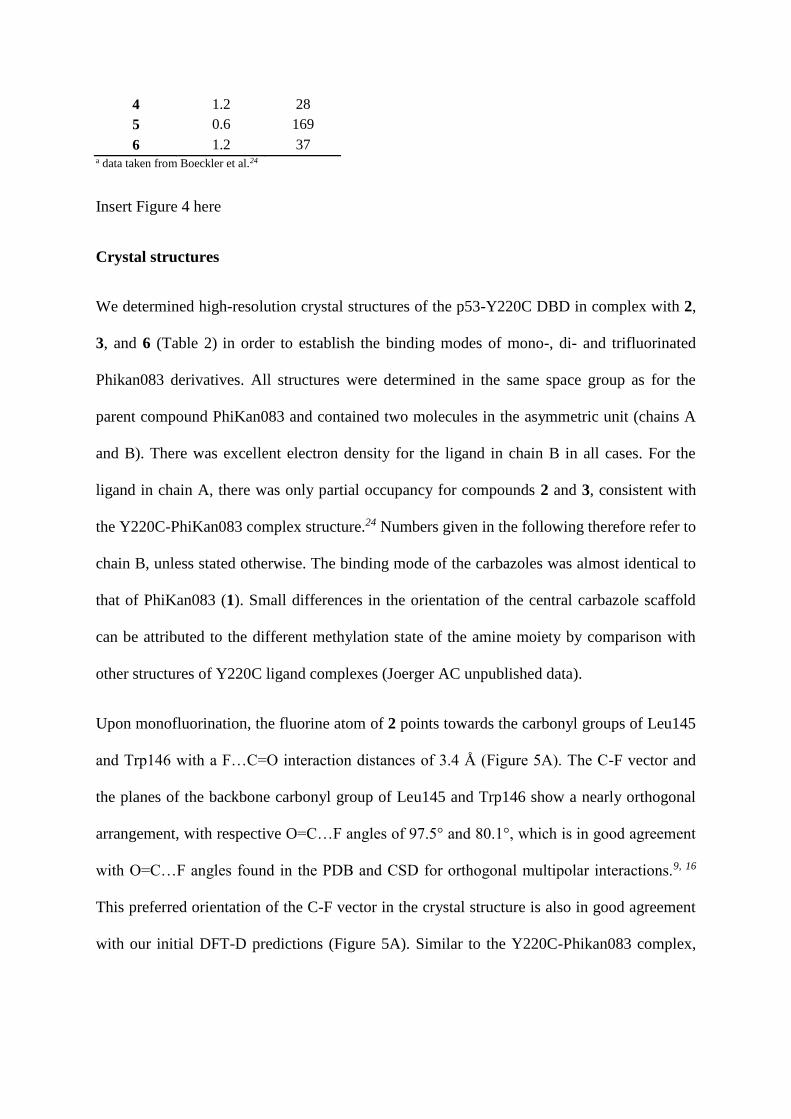
Crystal structures
We determined high-resolution crystal structures of the p53-Y220C DBD in complex with 2,
3, and 6 (Table 2) in order to establish the binding modes of mono-, di- and trifluorinated
Phikan083 derivatives. All structures were determined in the same space group as for the
parent compound PhiKan083 and contained two molecules in the asymmetric unit (chains A
and B). There was excellent electron density for the ligand in chain B in all cases. For the
ligand in chain A, there was only partial occupancy for compounds 2 and 3, consistent with
the Y220C-PhiKan083 complex structure.24 Numbers given in the following therefore refer to
chain B, unless stated otherwise. The binding mode of the carbazoles was almost identical to
that of PhiKan083 (1). Small differences in the orientation of the central carbazole scaffold
can be attributed to the different methylation state of the amine moiety by comparison with
other structures of Y220C ligand complexes (Joerger AC unpublished data).
Upon monofluorination, the fluorine atom of 2 points towards the carbonyl groups of Leu145
and Trp146 with a F…C=O interaction distances of 3.4 Å (Figure 5A). The C-F vector and
the planes of the backbone carbonyl group of Leu145 and Trp146 show a nearly orthogonal
arrangement, with respective O=C…F angles of 97.5° and 80.1°, which is in good agreement
with O=C…F angles found in the PDB and CSD for orthogonal multipolar interactions.9, 16
This preferred orientation of the C-F vector in the crystal structure is also in good agreement
with our initial DFT-D predictions (Figure 5A). Similar to the Y220C-Phikan083 complex,
the side chain of Cys220, in immediate vicinity to the ethyl anchor, adopts two alternative,
albeit very similar conformations.
For the N-2-difluoroethyl anchor of 3, we observed two alternative conformations in the p53-
Y220C binding pocket (Figure 5B and C). In both conformations, one fluorine atom interacts
with the carbonyl groups of Leu145 and Trp146 in an almost identical fashion as observed
for compound 2. The second fluorine interacts with the thiol group of Cys220, pointing either
towards Pro151 (Figure 5C) or towards Leu145 at the bottom of the binding pocket (Figure
5B), which is essentially the result of a 120° rotation around the ethyl anchor C-C bond.
Interestingly, only one of the two Cys conformations was observed in chain A, coinciding
with a preferential orientation of the difluoroethyl anchor in the orientation highlighted in
Figure 5B whereby the fluorine forms a weak hydrogen bond with the SH group of Cys220.
In both orientations, the fluorine atom interacts with the thiol group at a distance of about 3.2
Å, which is more or less the sum of the van der Waals radii of sulfur and fluorine (rF =
1.47Å; rS = 1.80 Å).1, 33
In the structure of the most potent compound 6, the CF3 group aligns well with the different
fluorine positions observed for monofluorinated 2 and difluorinated 3 (Figure 5D), i.e., it
interacts with the backbone carbonyl groups of Leu145 and Trp146 as well as with the thiol
group of Cys220. Analysis of C-S…F angles in the complexes with di and trifluorinated
compounds suggests that fluorine interacts with Cys220 via weak hydrogen bonding with the
polarized proton of the thiol function and via sulfur -hole bonding34-36 at an angle close to
180˚.
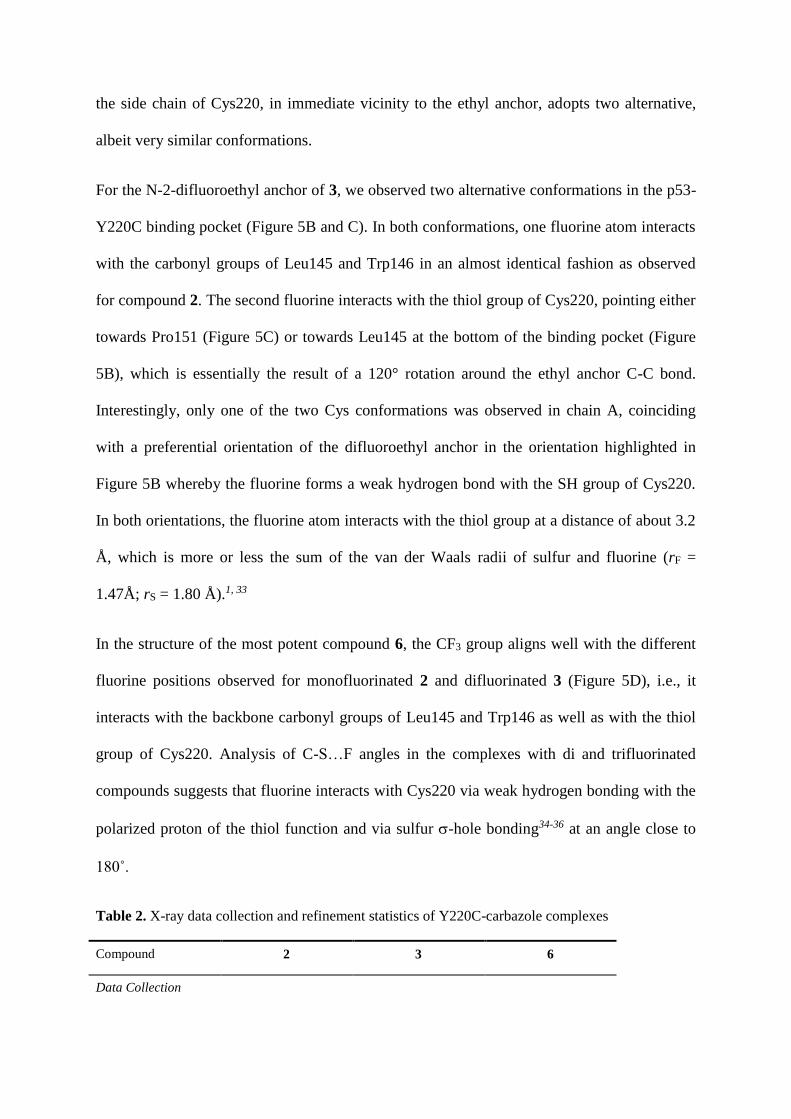
Table 2. X-ray data collection and refinement statistics of Y220C-carbazole complexes
Compound 2 3 6
Data Collection
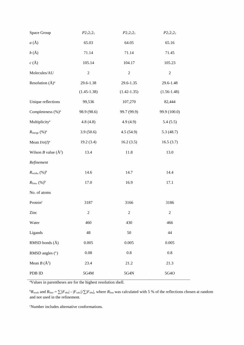
Space Group P212121 P212121 P212121
a (Å) 65.03 64.05 65.16
b (Å) 71.14 71.14 71.45
c (Å) 105.14 104.17 105.23
Molecules/AU 2 2 2
Resolution (Å)a 29.6-1.38
(1.45-1.38)
29.6-1.35
(1.42-1.35)
29.6-1.48
(1.56-1.48)
Unique reflections 99,536 107,270 82,444
Completeness (%)a 98.9 (98.6) 99.7 (99.9) 99.9 (100.0)
Multiplicitya 4.8 (4.8) 4.9 (4.9) 5.4 (5.5)
Rmerge (%)a 3.9 (50.6) 4.5 (54.9) 5.3 (48.7)
Mean I/(I)a 19.2 (3.4) 16.2 (3.5) 16.5 (3.7)
Wilson B value (Å2) 13.4 11.8 13.0
Refinement
Rwork, (%)b 14.6 14.7 14.4
Rfree, (%)b 17.0 16.9 17.1
No. of atoms
Proteinc 3187 3166 3186
Zinc 2 2 2
Water 460 430 466
Ligands 48 50 44
RMSD bonds (Å) 0.005 0.005 0.005
RMSD angles () 0.08 0.8 0.8
Mean B (Å2) 23.4 21.2 21.3
PDB ID 5G4M 5G4N 5G4O
aValues in parentheses are for the highest resolution shell.
bRwork and Rfree = ∑||Fobs| - |Fcalc||/∑|Fobs|, where Rfree was calculated with 5 % of the reflections chosen at random
and not used in the refinement.
cNumber includes alternative conformations.

Insert Fig 5 here please
Discussion
Incorporation of fluorine atoms into the N-ethyl anchor of the p53-Y220C stabilizer
PhiKan083 (1) yielded two compounds, 4 and 6, with substantially increased p53-Y220C
stabilization (1.2 K at 125 μM) and Y220C binding affinity. Compared with the parent
compounds PhiKan083 (1) and 5, the ITC KD values improved by a factor of 5 and the free
energy of binding by approximately -0.9 kcal/mol (at 293K). Typically, C-F…C=O
orthogonal multipolar interactions with optimal geometry raise the binding free enthalpy by
about -0.2 to -0.3 kcal/mol, 37 which suggests that the observed C-F…C=O interactions with
the carbonyl groups of Leu145 and Trp146 are the main factor for the potency increase of the
trifluorinated PhiKan083 derivatives. The DFT-D calculations are also consistent with the
binding modes 2 and 3 observed in our crystal structures, as the preferred orientation of
fluorine towards the backbone carbonyl groups of Leu145 and Trp146 in these structures
indicates that interactions of the protein carbonyl groups with fluorine atoms contribute more
strongly to the affinity increase than interactions with the thiol group of Cys220 or apolar
protons of hydrophobic side chains. The preferred interaction with the protein backbone then
seems to direct the orientation of additional fluorine atoms in the binding pocket, interacting
with the thiol group of Cys220 via either a hydrogen bond or a planar sulfur -hole
interaction.
Although DFT-D calculations predicted improved interaction energies for mono-fluorinated
and di-fluorinated PhiKan083 derivatives, this was not reflected in the experimentally
determined dissociation constants and stability data for compounds 2 and 3, which were close
to that of the parent compound PhiKan083. These discrepancies between theoretical and
experimental data are likely due to different desolvation penalties associated with each
fluorinated group. Fluorine can act as a weak hydrogen-bond acceptor in a 2,2-difluoromethyl
group and even more strongly in a 2-fluoromethyl group, although hydrogen-bond strength
was found to be significantly weaker than for the conventional hydrogen-bond acceptor
acetophenone.38 For example, difluoroalkyl groups have been used as thiol surrogates in drug
discovery because of their similar steric properties and the acidity of the terminal hydrogen
resulting from the high polarization of the C-H bond by the geminal fluorine atoms.39 To
estimate desolvation penalties of fluorinated N-ethyl anchors, we calculated solvation
energies for N-ethylcarbazole, N-2-fluoroethylcarbazole, N-2,2-difluoroethylcarbazole, and
N-2,2,2-trifluoroethylcarbazole with Jaguar using the SM8 water model at BLYP-D3/6-
31G** level of theory (Table 3 and Figure S3).40 Both mono and difluorinated carbazoles
showed solvation energies that were by -1.0 and -2.8 kcal mol-1 larger than the solvation
energy of N-ethylcarbazole. The solvation energy of the trifluorinated carbazole was identical
to N-ethylcarbazole (-3.1 kcal/mol). Hence, the higher polarity of the 2-fluoroethyl and 2,2-
difluoroethyl moieties in the PhiKan083 derivatives 2 and 3 and the associated desolvation
penalties are most likely the key factors counteracting and quenching favorable fluorine-
protein interactions in these compounds.
In conclusion, targeting Cys220 and the backbone carbonyl groups of Leu145 and Trp146 via
fluorine bonding helped to further optimize the carbazole scaffold. N-3,3,3-Trifluoroethyl
substituted carbazoles 4 and 6 exhibit a high ligand efficiency (LE = 0.3 kcal mol-1 atom-1),
although binding is still relatively weak for these fragment-like molecules. Nevertheless, they
represent a promising starting point for the development of high affinity small-molecule
stabilizers of the oncogenic Y220C mutant.

Table 3. Calculated solvation energies for DFT-D3 optimized carbazoles
carbazole anchor a solvation energy
(kcal/mol) b
N-ethyl -3.1
N-2-fluoroethyl -5.9
N-2,2-difluoroethyl -4.1
N-2,2,2-trifluoroethyl -3.1
aSubstitution of 9H-carbazole. Structures of DFT-D3 optimized compounds are shown in Figure S3.
bSolvation energies were calculated from single point calculations of the global minimum conformation of each compound
in Jaguar at BLYP-D3/6-31G** level using the SM8 water model.
MATERIALS & METHODS
Protein expression and purification
Stabilized p53-Y220C DBD (residues 94-312) was expressed and purified as previously
described.26
Differential Scanning Fluorimetry (DSF)
DSF was performed as described.26 Briefly, melting temperatures (Tm) were determined using
8 µM protein (stabilized p53-Y220C DBD) and 10x SYPRO orange (Life Technologies) in a
25 mM KPi pH 7.2, 150 mM NaCl, 1mM TCEP assay buffer at a final DMSO concentration
of 5% (v/v). ΔTm values were calculated as: ΔTm = Tm (protein+compound) - Tm (protein)).
All samples were measured in triplicate.
Isothermal Titration Calorimetry (ITC)
ITC experiments were conducted as described.26 Samples for the cell unit contained 50 μM
protein in a 25 mM KPi pH 7.2, 150 mM NaCl, 1mM TCEP, 5% (v/v) DMSO assay buffer.
The syringe contained 2 - 5 mM compound in the same assay buffer.
Quantum chemical calculations
QM calculations were conducted with TURBOMOLE 6.4, applying a BLYP(RI)-D3-def2-
SVP (DFT-D3) level of theory. A truncated model of the p53 Y220C binding pocket with an
N-ethyl-pyrrole ligand (truncated PhiKan083 model), comprising 140 atoms, was derived
from the crystal structure (pdb code: 2VUK). For geometry optimization all heavy atoms of
the binding pocket, except the sulfur atom of Cys220, and the nitrogen atom plus one carbon
atom of the ligand pyrrole ring were frozen (see supporting information). Interaction energies
were calculated as ΔE = Ecomplex - (Ereceptor + Eligand). Global minimum conformers, which
were used as input structures for solvation energy calculations, were determined by DFT-D3
calculations at BLYP(RI)-D3-def2-TZVP level of theory with TURBOMOLE 6.4. For N-2-
Fluoroethylcarbazole and N-2,2-Difluoroethylcarbazole, we rotated the C-C axis of the ethyl
moiety step-wise by 60˚ to obtain six different conformers that were used as input structures
for the DFT-D3 calculations. Solvation energies were calculated with Jaguar 8.9
(Schrödinger, USA, NY) as single point calculations using the SM8 water model at BLYP-
D3/6-31G** level of theory.
X-ray crystallography
Crystals of the p53-Y220C DBD were grown at 18 C as described.41 Crystals were soaked
for either 90 min (6) or 4 h (2 and 3) in a 20 mM solution of compound in cyro buffer (19%
polyethylene glycol 4000, 20% glycerol, 10 mM sodium phosphate, pH 7.2, 100 mM Hepes,
pH 7.2, 150 mM NaCl) and flash frozen in liquid nitrogen. X-ray data sets were collected at
100 K at beamlines I03 and I04 of the Diamond Light Source, Oxford, UK. Indexing of the
datasets was performed using XDS 42 and scaling using the program SCALA 43 within the
CCP4 software suite 44. The structures of the Y220C-carbazole complexes were then solved
using the program PHENIX: 45 initial rigid body refinement using PDB entry 2J1X as a
starting model. The models were further refined using iterative cycles of manual model
building in COOT 46 and refinement with PHENIX. While there was clear electron density
for the ligands in chain B, there was only weak ligand density in chain A of the Y220C
complex with 2 and 3, indicating partial occupancy. The same observation had been made for
the parent compound PhiKan083.24 Accordingly, alternative states of the binding pocket were
refined: bound ligand and water network of the ligand-free structure. Data collection and
refinement statistics are given in Table 2. Structural figures were prepared using PyMOL
(www.pymol.org). The coordinates and structure factors of the complexes are deposited in
the Protein Data Bank (PDB ID: 5G4M, 5G4N, 5G4O).
General Procedures
All reactions were carried out in air unless otherwise stated, using commercial grade starting
materials, solvents and reagents. The progress of all reactions was monitored by thin layer
chromatography (TLC) using commercially available glass silica gel plates (60 Å, F254). The
mobile phase was generally a solvent mixture and the visualisation was undertaken using UV
light. Microwave reactions were conducted in a CEM Discovery microwave reactor. All
NMR spectra were measured on a Varian NMR 500 spectrometer at either 500 MHz (1H) or
126 MHz (13C). Chemical shifts are quoted in parts per million (ppm) (δ relative to a residual
solvent peak for 1H and 13C). Chromatographic purifications were undertaken using an ISCO
purification unit, Combi Flash RF 75 PSI, using Biotage silica gel columns. LC-MS purity
analyses were undertaken using a 5 μm C18 110 Å column. Percentage purities were
performed using a 30 min method in water/acetonitrile with 0.1% formic acid (5 min at 5%,
5–95% over 20 min, 5 min at 95%) with the UV set to 254 nm. All high-resolution mass

spectrometry was carried out at the EPSRC UK National Mass Spectrometry Facility
(NMSF), Swansea University, using a Thermo Scientific LTQ Orbitrap XL spectrometer.
Materials: 1-(9-(2-Fluoroethyl)-9H-carbazol-3-yl)-N-methylmethanamine (2) was purchased
from Enamine; its purity was assessed by LC-MS and found to be higher than 95% (LC-MS
purity > 95% (UV), ret. time = 10.83 min). 9-Ethyl-9H-carbazole-3-carbaldehyde was
purchased from TCI UK (>98% purity by manufacturer). 1-Iodo-2,2-difluoroethane and
2,2,2-trifluoroethyl p-toluenesulfonate were purchased from fluorochem. 9H-Carbazole-3-
carbaldehyde was synthesized according to a known procedure.29 Final compounds that were
tested had an LC purity of >95%.
Representative procedure for the synthesis of intermediates 7-8
9-(2,2-Difluoroethyl)9H-carbazole-3-carbaldehyde (7). To a suspension of 9H-carbazole-3-
carbaldehyde (196 mg, 1 mmol) and Cs2CO3 (652 mg, 2 mmol) in anhydrous DMF (5 mL)
was added 1-iodo-2,2-difluoroethane (172 mg, 2 mmol). The resulting suspension was stirred
under microwave irradiation at 150 °C for 30 minutes. The suspension was cooled and
diluted with H2O (20 mL). Crude product was extracted with ethyl acetate (EtOAc) (3 X 20
mL). The organic extracts were washed with brine (3 X 20 mL) and dried over anhydrous
MgSO4. The suspension was filtered and was concentrated to dryness in vacuo. Crude
product was purified by chromatography on silica gel using dichloromethane (DCM) as an
eluent to yield product as a pale yellow solid, yield 44% (115 mg, 0.44 mmol). 1H NMR (500
MHz, DMSO-d6) δ 10.06 (s, 1H), 8.73 (s, 1H), 8.33 – 8.17 (m, 1H), 7.99 (d, J = 8.4 Hz, 1H),
7.81 (d, J = 8.4 Hz, 1H), 7.76 – 7.66 (m, 1H), 7.60 – 7.47 (m, 1H), 7.36 – 7.24 (m, 1H), 6.50
(tt, 2JFH = 54.5 Hz, 3JHH = 3.2 Hz, 1H), 4.97 (td, 3JFH = 16.1 Hz, 3JHH = 3.2 Hz, 2H). 13C NMR
(126 MHz, DMSO-d6) δ 192.3, 144.6, 141.7, 129.4, 127.3, 127.2, 124.0, 123.0, 122.8, 121.1,
121.0, 115.1 (t, 1JFC = 241.9 Hz), 110.9, 110.8, 45.0 (t, 2JFC = 24.5 Hz). HRMS-ESI (m/z)
found 260.0884, calcd 260.0881 for [C15H11F2NO+H]+.
9-(2,2,2-Trifluoroethyl)-9H-carbazole-3-carbaldehyde (8). Using 2,2,2-trifluoroethyl p-
toluenesulfonate as an electrophile. Reaction carried out on a 0.48 mmol scale. Pale yellow
solid, yield 50% (68 mg, 0.24 mmol). 1H NMR (500 MHz, DMSO-d6) δ 10.07 (s, 1H), 8.75
(s, 1H), 8.28 (d, J = 8.3 Hz, 1H), 8.02 (d, J = 8.5 Hz, 1H), 7.90 (d, J = 8.5 Hz, 1H), 7.80 (d, J
= 8.3 Hz, 1H), 7.61 – 7.49 (m, 1H), 7.40 – 7.25 (m, 1H), 5.51 (q, 3JFH = 9.3 Hz, 2H).13C
NMR (126 MHz, DMSO-d6) δ 192.3, 144.2, 141.3, 129.8, 127.5, 127.4, 125.4 (q, 1JFC =
281.8 Hz), 124.0 123.3, 122.9, 121.5, 121.2, 110.9, 110.8, 44.1 (q, 2JFC = 33.5 Hz). HRMS-
ESI (m/z) found 278.0789, calcd 278.0787 for [C15H10F3NO+H]+.
Representative procedure for the synthesis of compounds 3, 4, 5 and 6
1-(9-(2,2-Difluoroethyl)-9H-carbazol-3-yl)-N-methylmethanamine (3). To a solution of 7 (75
mg, 0.29 mmol) in anhydrous EtOH/DCM (10 mL) was added methylamine hydrochloride
(39 mg, 0.58 mmol), triethylamine (61 μL, 0.44 mmol) and titanium (IV) isopropoxide (172
μL, 0.58 mmol). The resulting solution was stirred at room temperature for 18 h before the
addition of sodium borohydride (22 mg, 0.58 mmol). The solution was stirred at room
temperature for 8 hours before pouring into 2M aqueous ammonia (25 mL). The suspension
was filtered through celite and to the filtrate was added H2O. Crude product was extracted
with DCM, dried over anhydrous K2CO3. The suspension was filtered and the filtrate was
concentrated in vacuo to yield crude product that was purified by chromatography on silica
gel using DCM/MeOH 9/1 as an eluent to yield product as a pale yellow oil, yield 58% (46
mg, 0.16 mmol). 1H NMR (500 MHz, Acetonitrile-d3) δ 8.19 – 8.13 (m, 1H), 8.10 (s, 1H),
7.62 – 7.56 (m, 1H), 7.56 – 7.45 (m, 3H), 7.32 – 7.25 (m, 1H), 6.31 (tt,2JFH = 55.0 Hz, 3JHH =
3.2 Hz, 1H), 4.76 (td, 3JFH = 15.6 Hz, 3JHH = 3.2 Hz, 2H), 3.89 (s, 2H), 2.43 (s, 3H) (NH
missing due to D-H exchange). 13C NMR (126 MHz, Acetonitrile-d3) δ 141.1, 139.9, 132.5,
126.6, 125.9, 122.9, 122.8, 120.1, 119.7, 119.6, 114.8 (t, 1JFC = 241.9 Hz), 109.3, 109.0, 55.7,
44.9 (t, 2JFC = 25.6 Hz), 35.1. LC-MS purity = 96% (UV), ret. time = 11.01 min. HRMS-ESI
(m/z) found 275.1353, calcd 275.1354 for [C16H16F2N2+H]+.
1-(9-(2,2,2-Trifluoroethyl)-9H-carbazol-3-yl)-N-methylmethanamine (4). Reaction carried
out on a 0.31 mmol scale. Product isolated as a pale yellow solid, yield 40% (37 mg, 0.12
mmol). 1H NMR (500 MHz, acetonitrile-d3) δ 8.20 – 8.14 (m, 1H), 8.12 (s, 1H), 7.64 – 7.58
(m, 1H), 7.58 – 7.47 (m, 3H), 7.36 – 7.28 (m, 1H), 5.07 (q, 3JFH = 9.1 Hz, 2H), 3.89 (s, 2H),
2.43 (s, 3H) (NH missing due to D-H exchange). 13C NMR (126 MHz, DMSO-d6) 140.9,
140.2, 128.3, 127.7, 126.6, 126.1 (q, 1JFC = 281.2 Hz), 122.9 122.8, 121.3, 120.6, 120.5,
110.4, 110.1, 53.9, 44.0 (q, 2JFC = 33.4 Hz), 34.2. LC-MS purity = 97% (UV), ret. time =
11.55 min. HRMS-ESI (m/z) found 293.1259, calculated 293.1260 for [C16H15F3N2+H]+.
1-(9-Ethyl-9H-carbazol-3-yl)-N,N-dimethylmethanamine (5). Reaction carried out on a 0.44
mmol scale. Product isolated as a white solid, yield 82% (91 mg, 0.36 mmol). 1H NMR (500
MHz, Chloroform-d) δ 8.14 – 8.10 (m, 1H), 8.06 (s, 1H), 7.51 – 7.34 (m, 4H), 7.26 – 7.20
(m, 1H), 4.37 (q, J = 7.2 Hz, 2H), 3.64 (s, 2H), 2.33 (s, 5H), 1.45 (t, J = 7.2 Hz, 3H). 13C
NMR (126 MHz, Chloroform-d) δ 140.28, 139.3, 129.3, 127.1, 125.5, 122.9 (2C –
quaternary), 121.0, 120.4, 118.7, 108.4, 108.1, 64.7, 45.3 (2C), 37.5, 13.8. LC-MS purity
97% (UV), ret. time = 11.36 min. HRMS-ESI (m/z) found 208.1120, calculated 208.1121 for
[C15H14N]+ (loss of NMe2).
1-(9-(2,2,2-Trifluoroethyl)-9H-carbazol-3-yl)-N,N-dimethylmethanamine (6). Reaction
carried out on a 0.10 mmol scale. Product isolated as a white solid, yield 56% (17 mg, 0.05
mmol). 1H NMR (500 MHz, chloroform-d) δ 8.04 – 8.00 (m, 1H), 7.99 (s, 1H), 7.45 – 7.39

(m, 2H), 7.37 – 7.29 (m, 2H), 7.27 – 7.20 (m, 1H), 4.73 (q, 3JFH = 8.7 Hz, 2H), 3.62 (s, 2H),
2.28 (s, 6H). 13C NMR (126 MHz, chloroform-d) δ 140.9, 140.1, 129.8, 127.8, 126.4, 124.3
(q, 1JFC = 281.5 Hz), 123.6, 123.4, 121.3, 120.6, 120.4, 108.7, 108.5, 45.3 (q, 2JFC = 35.7 Hz),
44.9 (2C). LC-MS purity > 99% (UV), ret. time = 11.68 min. HRMS-ESI (m/z) found
307.1419, calcd 307.1417 for [C16H17F3N2+H]+.
1-(9-Carbazol-3-yl)-N,N-dimethylmethanamine (9). To a solution of 9H-carbazol-3-
carbaldehyde (45 mg, 0.26 mmol) in anhydrous THF (5 mL) was added dimethylamine (130
µL, 2.0 M solution in THF, 0.26 mmol) and acetic acid (12 µL, 0.26 mmol). Sodium
triacetoxyborohydride (80 mg, 0.39 mmol) was added and the resulting solution was stirred
for 18 h. Solvent was removed under a reduced pressure and to the residue was added DCM
and H2O. The solution was filtered through a hydrophobic frit and the filtrate was
concentrated in vacuo to yield crude product which was purified by chromatography on silica
gel (using DCM/MeOH 9/1 as an eluent) to yield product as a pale yellow solid, yield 88%
(51 mg, 0.22 mmol). 1H NMR (500 MHz, chloroform-d) δ 8.59 (s, 1H), 8.09 – 8.03 (m, 1H),
8.02 (s, 1H), 7.46 – 7.38 (m, 3H), 7.38 – 7.33 (m, 1H), 7.25 – 7.18 (m, 1H), 3.77 (s, 2H), 2.40
(s, 6H).13C NMR (126 MHz, chloroform-d) δ 139.9, 139.2, 127.5, 126.8, 125.9, 123.3, 123.0,
121.4, 120.3, 119.4, 110.7, 110.6, 63.9, 44.2 (2C). HRMS-ESI (m/z) found 180.0805,
calculated 180.0808 for [C13H10N]+ (loss of NMe2).
ACKNOWLEDGEMENTS
This work was funded by Worldwide Cancer Research Grant 14-1002 ’Rescuing thermally
unstable p53 mutants with small molecule stabilisers; new targeted cancer therapies’ and
ERC Advanced Grant 268506. RJ is funded by the University of Sussex (PhD studentship).
We thank the staff at Diamond beamlines I03 and I04 for technical assistance during data

collection. Access was supported in part by the EU FP7 infrastructure grant BIOSTRUCT-X
(contract no. 283570).
Supporting Information
Additional DFT-D data and coordinates used for calculations; ITC data for 2, 3, and 5; global
minimum conformers of fluorinated carbazole analogues and their calculated solvation
energies; scans of NMRs; LC purity.
REFERENCES
1. Muller, K., Faeh, C., and Diederich, F. (2007) Fluorine in pharmaceuticals: looking beyond intuition, Science 317, 1881-1886.
2. Wang, J., Sanchez-Rosello, M., Acena, J. L., del Pozo, C., Sorochinsky, A. E., Fustero, S., Soloshonok, V. A., and Liu, H. (2014) Fluorine in pharmaceutical industry: fluorine-containing drugs introduced to the market in the last decade (2001-2011), Chem Rev 114, 2432-2506.
3. Zhou, Y., Wang, J., Gu, Z., Wang, S., Zhu, W., Acena, J. L., Soloshonok, V. A., Izawa, K., and Liu, H. (2016) Next Generation of Fluorine-Containing Pharmaceuticals, Compounds Currently in Phase II-III Clinical Trials of Major Pharmaceutical Companies: New Structural Trends and Therapeutic Areas, Chem Rev 116, 422-518.
4. Hagmann, W. K. (2008) The many roles for fluorine in medicinal chemistry, J Med Chem 51, 4359-4369.
5. Kirk, K. L. (2006) Fluorine in medicinal chemistry: Recent therapeutic applications of fluorinated small molecules, J Fluorine Chem 127, 1013-1029.
6. Vulpetti, A., and Dalvit, C. (2012) Fluorine local environment: from screening to drug design, Drug Discov Today 17, 890-897.
7. Gerebtzoff, G., Li-Blatter, X., Fischer, H., Frentzel, A., and Seelig, A. (2004) Halogenation of drugs enhances membrane binding and permeation, Chembiochem 5, 676-684.
8. Bohm, H. J., Banner, D., Bendels, S., Kansy, M., Kuhn, B., Muller, K., Obst-Sander, U., and Stahl, M. (2004) Fluorine in medicinal chemistry, Chembiochem 5, 637-643.
9. Zhou, P., Zou, J., Tian, F., and Shang, Z. (2009) Fluorine bonding--how does it work in protein-ligand interactions?, J Chem Inf Model 49, 2344-2355.
10. Istvan, E. S., and Deisenhofer, J. (2001) Structural mechanism for statin inhibition of HMG-CoA reductase, Science 292, 1160-1164.
11. Kamer, K. J., Choudhary, A., and Raines, R. T. (2013) Intimate interactions with carbonyl groups: dipole-dipole or n-->pi*?, The Journal of organic chemistry 78, 2099-2103.
12. Olsen, J. A., Banner, D. W., Seiler, P., Obst Sander, U., D'Arcy, A., Stihle, M., Muller, K., and Diederich, F. (2003) A fluorine scan of thrombin inhibitors to map the fluorophilicity/fluorophobicity of an enzyme active site: evidence for C-F...C=O interactions, Angew Chem Int Ed Engl 42, 2507-2511.
13. Cox, C. D., Breslin, M. J., Mariano, B. J., Coleman, P. J., Buser, C. A., Walsh, E. S., Hamilton, K., Huber, H. E., Kohl, N. E., Torrent, M., Yan, Y., Kuo, L. C., and Hartman, G. D. (2005) Kinesin spindle protein (KSP) inhibitors. Part 1: The discovery of 3,5-diaryl-4,5-dihydropyrazoles as potent and selective inhibitors of the mitotic kinesin KSP, Bioorg Med Chem Lett 15, 2041-2045.
14. Cowan-Jacob, S. W., Fendrich, G., Floersheimer, A., Furet, P., Liebetanz, J., Rummel, G., Rheinberger, P., Centeleghe, M., Fabbro, D., and Manley, P. W. (2007) Structural biology contributions to the discovery of drugs to treat chronic myelogenous leukaemia, Acta Crystallogr D Biol Crystallogr 63, 80-93.
15. Hughes, D. L., Sieker, L. C., Bieth, J., and Dimicoli, J. L. (1982) Crystallographic study of the binding of a trifluoroacetyl dipeptide anilide inhibitor with elastase, J Mol Biol 162, 645-658.
16. Bissantz, C., Kuhn, B., and Stahl, M. (2010) A medicinal chemist's guide to molecular interactions, J Med Chem 53, 5061-5084.
17. Vulpetti, A., Schiering, N., and Dalvit, C. (2010) Combined use of computational chemistry, NMR screening, and X-ray crystallography for identification and characterization of fluorophilic protein environments, Proteins 78, 3281-3291.
18. Pollock, J., Borkin, D., Lund, G., Purohit, T., Dyguda-Kazimierowicz, E., Grembecka, J., and Cierpicki, T. (2015) Rational Design of Orthogonal Multipolar Interactions with Fluorine in Protein-Ligand Complexes, J Med Chem 58, 7465-7474.
19. Lane, D. P. (1992) Cancer. p53, guardian of the genome, Nature 358, 15-16. 20. Vousden, K. H., and Prives, C. (2009) Blinded by the Light: The Growing Complexity of p53, Cell
137, 413-431. 21. Joerger, A. C., Ang, H. C., and Fersht, A. R. (2006) Structural basis for understanding oncogenic
p53 mutations and designing rescue drugs, Proc Natl Acad Sci U S A 103, 15056-15061. 22. Joerger, A. C., and Fersht, A. R. (2010) The tumor suppressor p53: from structures to drug
discovery, Cold Spring Harb Perspect Biol 2, a000919. 23. Joerger, A. C., Bauer, M. R., Wilcken, R., Baud, M. G., Harbrecht, H., Exner, T. E., Boeckler, F. M.,
Spencer, J., and Fersht, A. R. (2015) Exploiting Transient Protein States for the Design of Small-Molecule Stabilizers of Mutant p53, Structure 23, 2246-2255.
24. Boeckler, F. M., Joerger, A. C., Jaggi, G., Rutherford, T. J., Veprintsev, D. B., and Fersht, A. R. (2008) Targeted rescue of a destabilized mutant of p53 by an in silico screened drug, Proc Natl Acad Sci U S A 105, 10360-10365.
25. Liu, X., Wilcken, R., Joerger, A. C., Chuckowree, I. S., Amin, J., Spencer, J., and Fersht, A. R. (2013) Small molecule induced reactivation of mutant p53 in cancer cells, Nucleic Acids Res 41, 6034-6044.
26. Wilcken, R., Liu, X. R., Zimmermann, M. O., Rutherford, T. J., Fersht, A. R., Joerger, A. C., and Boeckler, F. M. (2012) Halogen-Enriched Fragment Libraries as Leads for Drug Rescue of Mutant p53, J Am Chem Soc 134, 6810-6818.
27. Wilcken, R., Wang, G., Boeckler, F. M., and Fersht, A. R. (2012) Kinetic mechanism of p53 oncogenic mutant aggregation and its inhibition, Proc Natl Acad Sci U S A 109, 13584-13589.
28. Wang, G., and Fersht, A. R. (2012) First-order rate-determining aggregation mechanism of p53 and its implications, Proc Natl Acad Sci U S A 109, 13590-13595.
29. Chen, X., Mihalic, J., Fan, P., Liang, L., Lindstrom, M., Wong, S., Ye, Q., Fu, Y., Jaen, J., Chen, J. L., Dai, K., and Li, L. (2012) Discovery and characterization of a potent and selective antagonist of melanin-concentrating hormone receptor 2, Bioorg Med Chem Lett 22, 363-366.
30. Suehiro, M., Yang, G., Torchon, G., Ackerstaff, E., Humm, J., Koutcher, J., and Ouerfelli, O. (2011) Radiosynthesis of the tumor hypoxia marker [18F]TFMISO via O-[18F]trifluoroethylation reveals a striking difference between trifluoroethyl tosylate and iodide in regiochemical reactivity toward oxygen nucleophiles, Bioorg Med Chem 19, 2287-2297.
31. Abdel-Magid, A. F., Carson, K. G., Harris, B. D., Maryanoff, C. A., and Shah, R. D. (1996) Reductive Amination of Aldehydes and Ketones with Sodium Triacetoxyborohydride. Studies on Direct and Indirect Reductive Amination Procedures(1), The Journal of organic chemistry 61, 3849-3862.
32. Bhattacharyya, S. (1995) Reductive Alkylations of Dimethylamine Using Titanium(Iv) Isopropoxide and Sodium-Borohydride - an Efficient, Safe, and Convenient Method for the Synthesis of N,N-Dimethylated Tertiary-Amines, J Org Chem 60, 4928-4929.

33. Beno, B. R., Yeung, K. S., Bartberger, M. D., Pennington, L. D., and Meanwell, N. A. (2015) A Survey of the Role of Noncovalent Sulfur Interactions in Drug Design, J Med Chem 58, 4383-4438.
34. Zhang, X., Gong, Z., Li, J., and Lu, T. (2015) Intermolecular Sulfur...Oxygen Interactions: Theoretical and Statistical Investigations, J Chem Inf Model 55, 2138-2153.
35. Murray, J. S., Lane, P., and Politzer, P. (2008) Simultaneous alpha-Hole and Hydrogen Bonding by Sulfur- and Selenium-Containing Heterocycles, Int J Quantum Chem 108, 2770-2781.
36. Lange, A., Gunther, M., Buttner, F. M., Zimmermann, M. O., Heidrich, J., Hennig, S., Zahn, S., Schall, C., Sievers-Engler, A., Ansideri, F., Koch, P., Laemmerhofer, M., Stehle, T., Laufer, S. A., and Boeckler, F. M. (2015) Targeting the Gatekeeper MET146 of C-Jun N-Terminal Kinase 3 Induces a Bivalent Halogen/Chalcogen Bond, J Am Chem Soc 137, 14640-14652.
37. Paulini, R., Muller, K., and Diederich, F. (2005) Orthogonal multipolar interactions in structural chemistry and biology, Angew Chem Int Ed Engl 44, 1788-1805.
38. Dalvit, C., Invernizzi, C., and Vulpetti, A. (2014) Fluorine as a Hydrogen-Bond Acceptor: Experimental Evidence and Computational Calculations, Chem-Eur J 20, 11058-11068.
39. Narjes, F., Koehler, K. F., Koch, U., Gerlach, B., Colarusso, S., Steinkuhler, C., Brunetti, M., Altamura, S., De Francesco, R., and Matassa, V. G. (2002) A designed P1 cysteine mimetic for covalent and non-covalent inhibitors of HCV NS3 protease, Bioorg Med Chem Lett 12, 701-704.
40. Chamberlin, A. C., Cramer, C. J., and Truhlar, D. G. (2008) Performance of SM8 on a test to predict small-molecule solvation free energies, J Phys Chem B 112, 8651-8655.
41. Joerger, A. C., Ang, H. C., and Fersht, A. R. (2006) Structural basis for understanding oncogenic p53 mutations and designing rescue drugs, Proc Natl Acad Sci U S A 103, 15056-15061.
42. Kabsch, W. (2010) Xds, Acta Crystallogr D Biol Crystallogr 66, 125-132. 43. Evans, P. (2006) Scaling and assessment of data quality, Acta Crystallogr D Biol Crystallogr 62, 72-
82. 44. Winn, M. D., Ballard, C. C., Cowtan, K. D., Dodson, E. J., Emsley, P., Evans, P. R., Keegan, R. M.,
Krissinel, E. B., Leslie, A. G., McCoy, A., McNicholas, S. J., Murshudov, G. N., Pannu, N. S., Potterton, E. A., Powell, H. R., Read, R. J., Vagin, A., and Wilson, K. S. (2011) Overview of the CCP4 suite and current developments, Acta Crystallogr D Biol Crystallogr 67, 235-242.
45. Adams, P. D., Afonine, P. V., Bunkoczi, G., Chen, V. B., Davis, I. W., Echols, N., Headd, J. J., Hung, L. W., Kapral, G. J., Grosse-Kunstleve, R. W., McCoy, A. J., Moriarty, N. W., Oeffner, R., Read, R. J., Richardson, D. C., Richardson, J. S., Terwilliger, T. C., and Zwart, P. H. (2010) PHENIX: a comprehensive Python-based system for macromolecular structure solution, Acta Crystallogr D Biol Crystallogr 66, 213-221.
46. Emsley, P., Lohkamp, B., Scott, W. G., and Cowtan, K. (2010) Features and development of Coot, Acta Crystallogr D Biol Crystallogr 66, 486-501.