Harlequin onset sweating - jnnp.bmj.com · Harlequin syndrome: the sudden onset...
8
Journal of Neurology, Neurosurgery, and Psychiatry 1988;51:635-642 Harlequin syndrome: the sudden onset of unilateral flushing and sweating JAMES W LANCE,* PETER D DRUMMOND,* SIMON C GANDEVIA,* JOHN G L MORRISt From the Departments of Neurology, Prince Henry Hospital,* and Westmead Hospital,t Sydney, Australia SUMMARY Facial flushing and sweating were investigated in five patients who complained of the sudden onset of unilateral facial flushing in hot weather or when exercising vigorously. One patient probably suffered a brainstem infarct at the time that the unilateral flush was first noticed, and was left with a subtle Homer's syndrome on the side opposite to the flush. The other four had no other neurological symptoms and no ocular signs of Homer's syndrome. Thermal and emotional flushing and sweating were found to be impaired on the non-flushing side of the forehead in all five patients whereas gustatory sweating and flushing were increased on that side in four of the five patients, a combination of signs indicating a deficit of the second sympathetic neuron at the level of the third thoracic segment. CT and MRI of this area failed to disclose a structural lesion but latency from stimulation of the motor cortex and thoracic spinal cord to the third intercostal muscle was delayed on the non-flushing side in one patient. The complaint of unilateral flushing and sweating was abolished in one patient by ipsilateral stellate ganglionectomy. The unilateral facial flushing and sweating induced by heat in all five patients was thus a normal or excessive response by an intact sympathetic pathway, the other side failing to respond because of a sympathetic deficit. The onset in the four cases of peripheral origin followed strenuous exertion, which suggested that an anterior radicular artery may have become occluded at the third thoracic segment during torsion of the thoracic spine. One of the most mysterious, dramatic and colourful of autonomic syndromes is the sudden appearance of flushing limited to one side of the face, a symptom that does not appear to have received attention in the past. The descriptive title of "harlequin syndrome" comes to mind although the analogy has been drawn previously by paediatricians who apply the term "harlequin colour change" to vasomotor instability in the newborn causing flushing of the dependent half of the body.1 Unilateral sweating in response to spicy foods, heat and exercise may develop spontaneously as "idio- pathic hyperhidrosis" which is sometimes accom- panied by flushing2 3 and sometimes not.4 5 Unilateral flushing as a primary complaint does not seem to have been previously recorded. The stimulus for the present study was provided by Address for reprint requests: Professor J W Lance, Department of Neurology, The Prince Henry Hospital, Little Bay 2036, NSW, Australia. Received 19 June 1987 and in revised form 29 October 1987. Accepted 2 November 1987 five patients who observed asymmetry of facial flushing in hot weather or after exertion. Our in- vestigations were planned to determine which side was at fault and to elucidate the mechanism of the syndrome. Case reports The main clinical characteristics are summarised in table 1. Case 1 A woman aged 73 years first noticed asymmetry of flushing at the age of 64 after working hard in the garden on a hot day. When she looked in the mirror while washing her hands, she observed a distinct line of demarcation between the left half of her face which was red and the right half which retained its nornal colour. There were no accom- panying symptoms. Over the following 10 years she has often noticed the same phenomenon after exertion although the colour difference has become less marked over this time. She had not been aware of any change in sweating. There was no antecedent injury or illness of relevance. A fluctuating elevation of systolic blood pressure was first noted at the age of 68 years but no measurements had been recorded before then. On examination, the left pupil was slightly smaller than the right but there was no ptosis and the pupils reacted normally. The nervous and other systems 635 Protected by copyright. on August 24, 2019 by guest. http://jnnp.bmj.com/ J Neurol Neurosurg Psychiatry: first published as 10.1136/jnnp.51.5.635 on 1 May 1988. Downloaded from
Transcript of Harlequin onset sweating - jnnp.bmj.com · Harlequin syndrome: the sudden onset...

Journal of Neurology, Neurosurgery, and Psychiatry 1988;51:635-642
Harlequin syndrome: the sudden onset of unilateralflushing and sweatingJAMES W LANCE,* PETER D DRUMMOND,* SIMON C GANDEVIA,*JOHN G L MORRISt
From the Departments ofNeurology, Prince Henry Hospital,* and Westmead Hospital,t Sydney, Australia
SUMMARY Facial flushing and sweating were investigated in five patients who complained of thesudden onset of unilateral facial flushing in hot weather or when exercising vigorously. One patientprobably suffered a brainstem infarct at the time that the unilateral flush was first noticed, and was
left with a subtle Homer's syndrome on the side opposite to the flush. The other four had no otherneurological symptoms and no ocular signs of Homer's syndrome. Thermal and emotional flushingand sweating were found to be impaired on the non-flushing side of the forehead in all five patientswhereas gustatory sweating and flushing were increased on that side in four of the five patients, a
combination of signs indicating a deficit of the second sympathetic neuron at the level of the thirdthoracic segment. CT and MRI of this area failed to disclose a structural lesion but latency fromstimulation of the motor cortex and thoracic spinal cord to the third intercostal muscle was delayedon the non-flushing side in one patient. The complaint of unilateral flushing and sweating wasabolished in one patient by ipsilateral stellate ganglionectomy. The unilateral facial flushing andsweating induced by heat in all five patients was thus a normal or excessive response by an intactsympathetic pathway, the other side failing to respond because of a sympathetic deficit. The onsetin the four cases of peripheral origin followed strenuous exertion, which suggested that an anteriorradicular artery may have become occluded at the third thoracic segment during torsion of thethoracic spine.
One of the most mysterious, dramatic and colourfulof autonomic syndromes is the sudden appearance offlushing limited to one side of the face, a symptomthat does not appear to have received attention in thepast. The descriptive title of "harlequin syndrome"comes to mind although the analogy has been drawnpreviously by paediatricians who apply the term"harlequin colour change" to vasomotor instabilityin the newborn causing flushing of the dependent halfof the body.1
Unilateral sweating in response to spicy foods, heatand exercise may develop spontaneously as "idio-pathic hyperhidrosis" which is sometimes accom-panied by flushing2 3 and sometimes not.4 5Unilateral flushing as a primary complaint does notseem to have been previously recorded.The stimulus for the present study was provided by
Address for reprint requests: Professor J W Lance, Department ofNeurology, The Prince Henry Hospital, Little Bay 2036, NSW,Australia.
Received 19 June 1987 and in revised form 29 October 1987.Accepted 2 November 1987
five patients who observed asymmetry of facialflushing in hot weather or after exertion. Our in-vestigations were planned to determine which sidewas at fault and to elucidate the mechanism of thesyndrome.
Case reports
The main clinical characteristics are summarised in table 1.Case 1 A woman aged 73 years first noticed asymmetry offlushing at the age of 64 after working hard in the garden ona hot day. When she looked in the mirror while washing herhands, she observed a distinct line of demarcation betweenthe left half of her face which was red and the right halfwhich retained its nornal colour. There were no accom-panying symptoms. Over the following 10 years she hasoften noticed the same phenomenon after exertion althoughthe colour difference has become less marked over this time.She had not been aware of any change in sweating. Therewas no antecedent injury or illness of relevance. Afluctuating elevation of systolic blood pressure was firstnoted at the age of 68 years but no measurements had beenrecorded before then. On examination, the left pupil wasslightly smaller than the right but there was no ptosis and thepupils reacted normally. The nervous and other systems
635
Protected by copyright.
on August 24, 2019 by guest.
http://jnnp.bmj.com
/J N
eurol Neurosurg P
sychiatry: first published as 10.1136/jnnp.51.5.635 on 1 May 1988. D
ownloaded from

Table 1 Summary of clinical characteristics, ocular signs and investigations. See also tables 2 and 3
Case number
1 2 3 4 5
Age of onset (years) 64 54 37 32 27Present age (years) 73 58 42 39 29Sex F F M F MHypertension + + - - -Flushing, sweating L L L R R
Ptosis - R -Miosis - R - R RPupillary dilatation lag - R -- -Asymetry after
cocaine 5% - - lesstyramine 2%
Conclusion: ocular Horner's syndrome - R - - -
CT brain infarcts n n n dil. L. lat. V.thoracic spine n n
CT myelography nMRI brain n n
thoracic spine n n nDSA nL stellate abolished
ganglionectomy L. flushing
were otherwise normal apart from a blood pressure of200/80 mm Hg.Case 2 A woman aged 58 was walking in the sunlight withher daughter and grandchildren 4 years previously when shesuddenly felt giddy "as though everything were turningaround." She felt off balance and clutched a baby's pram forsupport. At the same time her daughter noticed that the leftside, of her face had become red and was sweating. Facialsweating subsided after 1 hour but her left facial flush persis-ted for 2 hours. Since that time any exertion such as house-work or walking in hot weather has made the left side of herface and neck flush and sweat, a source of embarrassmentthat has caused her to stay at home most of the summer. Shehad not noticed redness or watering of her left eye or block-age of her left nostril. The rest ofher body sweated normally.For the past year she had noticed transient weakness or"pins and needles" in her left arm and a burning sensationover the left side of her face and arm on occasions. Apartfrom a dull right frontal headache recurring intermittentlysince a car accident 22 years previously, she had experiencedno other symptoms. There was a 30 year history of hyper-tension which had proved difficult to control. On the firstexamination, there was a slight right ptosis but herpupils were symmetrical and reacted normally to light andaccommodation. Sensation to pinprick and temperature wasdiminished over the left half of her face and pharynx, withthe deficit extending over the left arm and left upper quad-rant of the trunk to below the nipple at the level of the sixththoracic dermatome. There was slight weakness of fingerflexors and small muscles of the hand on the left side and theleft upper limb reflexes appeared to be less brisk than theright. Blood pressure was 160/1 10 mm Hg. On repeated ex-aminations over the following year, her right pupil was no-ticed to be smaller than the left on occasions and the rightptosis was confirmed, the right palpebral fissure being 16mm less than the left. The upper limb reflexes were thenconsidered to be within the normal range of symmetry. Thediagnosis was thought to be a right lateral medullary infarct
but the possibility of syringomyelia could not be excluded onclinical grounds.Case 3 A 42 year old man had been working hard layingcables in his job as an electrician at the age of 37 when hewent to the changing room to wash and noticed that he wasflushed and sweating only on the left side of his forehead.One week later after playing squash he saw that only the leftside of his face was flushed and sweating. This asymmetryhad persisted for the past 5 years but sweating over the trunkand limbs remained symmetrical. He flushes and sweats withembarrassment over the left side of the forehead only. Forthe past 5 years he had been subject to right retro-orbitalheadaches of mild to moderate severity, recurring up to fourtimes each month, associated with nausea but these havenow subsided. His mother developed late onset diabetes. No
0:
Fig I Flushing on the right side of the face after exertion(case 4).
636 Lance, Drummond, Gandevia, Morris
Protected by copyright.
on August 24, 2019 by guest.
http://jnnp.bmj.com
/J N
eurol Neurosurg P
sychiatry: first published as 10.1136/jnnp.51.5.635 on 1 May 1988. D
ownloaded from

Harlequin syndrome: the sudden onset of unilateralflushing and sweating
abnormality was found on full examination of the nervousand other systems. Blood pressure was 130/80 mm Hg.Case 4 A woman aged 39 years, had finished playing ahard game of squash 7 years previously when a friend com-mented that the left side of her face remained pale and drywhile the right side was flushing and sweating normally.Ever since, a sharp line of demarcation has appeared be-tween the colour of each side of her face after 10 minutes ofstrenuous exertion (fig 1). She was not aware of any asym-metry of body sweating. At the time of onset of the symp-tom, she had just recovered from an infection of her left legthat had required treatment with antibiotics. Her motherhad developed late-onset diabetes. On examination her rightpupil was slightly smaller than the left but reacted normally.The nervous and other systems were otherwise normal.Blood pressure was 110/70 mm Hg.Case 5 A 29 year old male landscape gardener had alwayssweated and flushed on both sides of his face until 2 yearsago when his wife noticed that the right side of his foreheadand upper face was a bright red colour after a game ofsquash, while the left side of his face appeared pale. Thisasymmetry persisted for about 20 minutes. Since that timehe had flushed easily on exertion, or sometimes at rest on ahot day, with the flush involving chiefly the right side of theforehead, fading in intensity over the right side of the noseand the right cheek, sparing the chin and neck. At the sametime, the right side of his face, particularly the forehead,sweated more than the left whereas sweating remained sym-metrical over the remainder of his body. There were no othersymptoms and no history of birth trauma or anything else ofrelevance in his past health or family history. On exam-ination, his right pupil was smaller than the left but re-sponded normally to light and accommodation. There wasno ptosis. Subsequent testing revealed no evidence of aHorner's syndrome. No other abnormality was found on fullexamination of the nervous and other systems. His bloodpressure was 120/80 mm Hg.
Methods of investigation
The experiments were carried out in an air-conditioned lab-oratory maintained at 22 + I°C. The subjects' informedconsent to the procedures was obtained.
Facial flushing was measured thermographically and cap-illary pulsation was recorded by pulse transducers (photo-plethysmographs, Narco Bio-Systems) attached withadhesive washers to the forehead and cheeks.6 The range ofthe AGA 680 Thermovision camera was 7°C, and a heatsource, set at 35°C, was used for calibration. Change in am-plitude of capillary pulsations was expressed as the per-centage change from pretest level.6 Sweating on the upperand lower forehead was measured with an EvaporimeterEPI. Rate of evaporation was recorded in arbitrary unitsbecause calibration of the Evaporimeter was altered by plac-ing a protective stainless steel grill over the measuring head.6
Pupil diameter was recorded on infrared film in a smalllight-tight photographic chamber. Photographs were takenat an illumination of 153.5 lux, and after 5 and 30 seconds ofdarkness (0-04 lux) to investigate pupillary dilatation lag.Photographs were enlarged eight times so that diameter ofthe pupils and width of the palpebral fissures could be mea-sured with calipers. A symmetrical response to the installa-
tion in each eye of two drops of 5% cocaine (which blocksthe reuptake of noradrenaline) was used to confirm the pres-ence of normal sympathetic innervation of the pupil. Onanother occasion, eye drops of tyramine 2% (which releasesnoradrenaline from nerve terminals) were instilled to assessthe presence or absence of neurotransmitter depletion.
Facial flushing and sweating was induced by body heat-ing, embarrassment and the taste of chillies. The procedureshave been described in detail elsewhere.6 Thermoregulationwas tested by wrapping the patient in blankets and circu-lating hot air under the blankets until the patient sweated.Facial flushing after vigorous exercise (jogging around thehospital grounds and running up and down stairs) was mea-sured thermographically in three patients. The other twopatients, who were unable to exercise vigorously, wereheated until they flushed and sweated. To induce embarrass-ment, a tape recording was made of the patient singing chil-dren's songs, and this was played back while measurementswere carried out. Gustatory sweating and flushing was ob-served after tasting one-half teaspoon of Tabasco sauce.6The integrity of the first, second and third thoracic motor
roots was assessed in three patients by measurement of thelatencies of contraction of the thenar and appropriate inter-costal muscles respectively to stimulation of the motor cor-
Fig 2 Thermogram after exertion in case 5. The right sideof the forehead and right cheek increased in temperature by2°C, with a sharp line of demarcation from the left sidebecoming apparent (arrow). Temperature calibration(colour-coded in the original photograph) is at JOOCintervals. Temperature reference disc maintained at 35°C.
637
Protected by copyright.
on August 24, 2019 by guest.
http://jnnp.bmj.com
/J N
eurol Neurosurg P
sychiatry: first published as 10.1136/jnnp.51.5.635 on 1 May 1988. D
ownloaded from
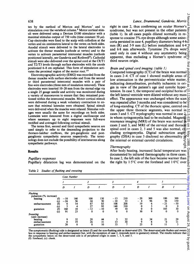
638
tex by the method of Merton and Morton7 and tostimulation over the vertebral column.8 Motor cortical stim-uli were delivered using a Devices Dl 80 stimulator with a
maximal stimulus output of 750 volts (time constant 50 ps).Cup electrodes were fixed to the scalp with collodion at thevertex and six centimetres lateral to the vertex on both sides.Anodal stimuli were delivered to the lateral electrodes toactivate the thenar muscles (cathode at vertex) and to thevertex to activate parasternal intercostal muscles (cathodepositioned laterally, see Gandevia and Rothwell9). Cathodalstimuli were also delivered over the spinal cord at the C8/T I
and T2/T3 levels through surface electrodes with the anodepositioned 6-8 cm cephalad. This form of stimulation acti-vates the proximal region of the motor root.10
Electromyographic activity (EMG) was recorded from thethenar muscles with surface electrodes and from the secondor third parastemal intercostal muscles with a pair offine-wire electrodes (three mm of insulation removed). Theseelectrodes were inserted 10-20 mm from the sternal edge viaa single 25 gauge needle and activity was monitored duringa variety of manoeuvres to ensure that they remained posi-tioned within the intercostal muscles. Motor cortical stimuliwere delivered during a weak voluntary contraction to en-sure that minimal latencies were obtained. Spinal stimuliwere delivered when the muscles were relaxed. Stimulus volt-ages were usually the same for recordings on both sides.Latencies were measured from a digital oscilloscope andwhere necessary up to eight responses were full-waverectified and averaged following cortical stimuli.The terms first, second and third sympathetic neuron are
used simply to refer to the descending projection to thethoraco-lumbar outflows, the pre-ganglionic and post-ganglionic sympathetic neurons respectively. The termi-nology does not exclude the possibility of interneurons alongsympathetic pathways.
Results
Pupillary responsesPupillary dilatation lag was demonstrated on the
Lance, Drummond, Gandevia, Morrisright in case 2, thus confirming an ocular Horner'ssyndrome, but was not present in the other patients(table 1). In all cases pupils dilated normally in re-
sponse to cocaine 5% eye drops although some aniso-coria persisted in case 4, pupillary diameters being 4 6mm (R) and 5 9 mm (L) before installation and 6 4and 6-8 mm afterwards. Tyramine 2% drops were
used only in case 4 without any asymmetry beingapparent, thus excluding a Horner's syndrome ofthird neuron origin.
Brain and spinal cord imaging (table 1)Computed tomography (CT) of the brain was normalin cases 2-4. CT of case 1 showed multiple areas oflow attenuation in the periventricular white matter,indicating demyelination, probably ischaemic in ori-gin in view of the patient's age and systolic hyper-tension. In case 5, the temporal and occipital horns ofthe left lateral ventricle were dilated without any masseffect. The appearance was unchanged when the scanwas repeated after 3 months and was considered to beof long-standing. CT of the thoracic spine, centred onthe upper three thoracic segments, was normal incases 3 and 5. CT myelography was normal in case 2,in whom syringomyelia had to be excluded. Magneticresonance imaging (MRI) of the brain was normal incases 2 and 3, and MRI of the cervical and thoracicspinal cord in cases 2, 3 and 5 was also normal, ex-
cluding syringomyelia. Digital subtraction angio-graphy (DSA) in case 5 disclosed no abnormality ofthe internal or external carotid circulations.
ThermographyAfter body heating, increased facial temperature was
documented by infrared thermography in three cases.In case 2, the left side of the face became warmer thanthe right by 1 5°C over the forehead and 1 0°C over
Table 2 Studies offlushing and sweating
Case Number
1 2 3 4 5
D I D I D I D I D I
Flushingpulsation (% increase)
heating (f) -10 160 100 150 -13 210 -20 300 100 300(c) -24 56 24 50 -10 33 - 4 96 56 150
embarrassment (f) 0 14 67 85 108 208 19 6 -21 -50(c) 5 4 27 95 140 310 0 5 0 0
gustation (f) 160 18 -12 -10 59 10 0 0 105 30(c) 91 23 0 0 0 0 30 9 0 0
Sweating(unit increase)
heating 9 32 20 44 3 32 0 34 21 29embarrassment 0 19 26 31 13 30 9 8 0 11gustation 20 0 8 10 33 7 25 9 11 3
The symptomatic (flushing) side is designated as intact (1) and the non-flushing side as denervated (D). The denervated side flushes and sweatsless in response to heating and embarrassment but, with the exception of case 2, responds more to gustatory stimuli. The results indicate thatthe sympathetic lesion on the denervated side is of peripheral origin in cases 1, 3, 4, and 5.(f): forehead; (c): cheek.
Protected by copyright.
on August 24, 2019 by guest.
http://jnnp.bmj.com
/J N
eurol Neurosurg P
sychiatry: first published as 10.1136/jnnp.51.5.635 on 1 May 1988. D
ownloaded from
Harlequin syndrome: the sudden onset of unilateralflushing and sweating
the cheek. In case 3, the left forehead and cheek be-came warmer than the right by 100C. In case 5, theright forehead and cheek temperature increased by2'C more than the left but heat loss from the chinremained symmetrical (fig 2).
Capillary pulsationsDuring the unilateral flush produced by body heating,the amplitude of capillary pulsations on theresponsive side increased by 150-300% over the fore-head and by 33-100% over the cheek while there waslittle or no increase on the opposite side (table 2). Thisindicated that sympathetic innervation was intact onthe side that flushed in response to heating (desig-nated I in table 2) and impaired on the non-responsivedenervated side (designated D in table 2) as describedby Drummond and Lance.6After embarrassment, asymmetry in pulsations was
detected in only three of the patients. Pulsation wasincreased on the affected side of the forehead in case1, the cheek in case 2 and both areas in case 3.The change in capillary pulsation reversed sides
after a gustatory stimulus in cases 1, 3, 4 and 5 but nochange was recorded in case 2 (table 2).
SweatingSweating over the face was assessed qualitatively bythe application of alizarin powder which changed col-our when moist (fig 3) and quantitatively by mea-surement of evaporation from the forehead (table 2).Sweating was greater on the flushing side of the facein all cases in response to body heating. In fourpatients the asymmetry was apparent over forehead,cheek and, to a lesser extent in some, the chin (fig 3).In case 5, an area of anhidrosis on the non-flushingside was noted over the medial aspect of the foreheadwhile sweating was only slightly diminished in otherareas. Embarrassment caused increased sweating onthe flushing side in cases 1, 3 and 5. The response to agustatory stimulus was reversed in cases 1, 3, 4 and 5with excessive sweating on the non-flushing side thathad sweated less during heating and embarrassment(table 2). These changes confirmed the presence of a
Right
Left
Fig 3 Sweating after exertion in case 4. The colour change
of alizarin powder applied to the face demonstrates that the
right side of the face sweats while the left remains dry.
IIIII
I
lorn
Fig 4 Typical electromyographic responses of the thirdintercostal muscles evoked by anodal stimulation of themotor cortex in case S. The latency was increased by 3-4ms on the left (non-flushing, non-sweating) side. Responsesto spinal stimulation were similarly prolonged (table 3),indicating impaired conduction in the third thoracic motor
root on the affected side. Vertical calibration: 200 ju V.
630
Protected by copyright.
on August 24, 2019 by guest.
http://jnnp.bmj.com
/J N
eurol Neurosurg P
sychiatry: first published as 10.1136/jnnp.51.5.635 on 1 May 1988. D
ownloaded from

640sympathetic lesion on the non-flushing side (D intable 2).
Clinical neurophysiologyBecause the sympathetic outflow accompanying thesecond and third thoracic roots was of particular in-terest the latency of the second and third intercostalmuscles to indirect stimulation of the motor cortexand to stimulation over the spinal cord was measured.In addition, the responses of the thenar muscles tospinal and cortical stimulation were assessed. Esti-mates of peripheral and central conduction times tothenar muscles were normal in the three patients stud-ied (cases 3, 4, 5; see table 3). In case 5, the latenciesto both cortical and spinal cord stimulation were pro-longed to the third intercostal muscle on the non-flushing side, exceeding those on the other side by 3-4and 4 2 ms respectively (fig 4). This patient showed adiminished interference pattern in the third interspaceon the non-flushing side. Case 4 showed normalperipheral and central motor conduction to the thirdintercostal muscles and this was demonstrated forboth second and third intercostal muscles in case 3.
Stellate ganglion block and ganglionectomyBecause case 2 was of fair complexion and was trou-bled by the unsightly flushing of the left side of herface, her left stellate ganglion was blocked by lig-nocaine 2% with abolition of flushing and sweatingon that side after body heating. The left stellate gan-glion was therefore removed and she has not noticedany flushing or sweating on that side in the 18 monthssince operation. As she already had a partial ocularHorner's syndrome on the right side the left Horner'ssyndrome resulting from operation restored a pleas-ing symmetry.
Blood glucose levelsFasting blood glucose levels were normal in all cases.
Table 3 Latency from stimulation of motor cortex andspinal cord to contraction of the thenar muscles andintercostal muscles in Cases 3, 4 and 5
Site of stimulation
Motor cortex Spinal cord
latency (ms) right left right leftcase 3 C8/Tl 22-6 20 9 16 9 14-8
T2 9-4 8-7 4-6 4-6T3 115 113 58 53
case 4 C8/TI 19 0 19-1 14 2 14.0T3 115 113 44 50
case 5 C8/T1 199 193 150 150T3 115 14.9* 51 9.3*
Significant asymmetry was demonstrated in case 5 for stimulation atboth motor cortical and spinal levels with the prolonged latencies (*)occurring on the non-flushing side.
Lance, Drummond, Gandevia, MorrisDiscussion
All five patients complained of the sudden onset offlushing and sweating on one side of the face, thusdirecting the attention of the clinician to this as thepresumably abnormal side. On the initial exam-ination of all five patients there was no ocular sign ofHorner's syndrome. Anisocoria was observed in cases4 and 5 but there was no pupillary dilatation lag onexposure to darkness, indicating that sympathetic in-nervation of the iris was intact.
In contrast, repeated observation of case 2 dis-closed an inconstant miosis and ptosis on the right(non-flushing) side with pupillary dilatation lag onthat side, thus demonstrating a mild right ocularHomer's syndrome. The normal response of the mi-otic pupil to cocaine eye drops in this instance indi-cates that the lesion was central.'I This, together withthe abrupt onset of unilateral flushing accompaniedby vertigo in a hypertensive patient, suggested abrainstem infarction. The residual upper quadranticimpairment of sensation to pinprick and temperaturewould be consistent with this diagnosis although itwas necessary to rule out the possibility of a syrinx byCT myelography and MRI. The fact that this patientfailed to sweat and flush on the right side in responseto heating but showed little asymmetry to embarrass-ment or gustatory stimuli indicates a lesion of the firstsympathetic neuron, a right central Homer's syn-drome.6 Because she had a fair skin that displayed herleft sided flush readily, she confined herself to thehouse on hot summer days. It was therefore decidedto remove her left stellate ganglion, a procedure thatabolished her untoward reaction to body heating. It isof interest that Wilson2 described a patient with syrin-gomyelia who did not show the ocular signs ofHomer's syndrome but was troubled by unilateralflushing and sweating which was relieved by ablationof the ipsilateral superior cervical ganglion. Gusta-tory sweating that develops after preganglionic cervi-cal sympathectomy is also abolished by stellateganglion block."2 1'The localisation of the site of the lesion in the other
four cases presented greater difficulty. The failure toflush and sweat in response to heat or embarrassmenton the non-symptomatic (non-flushing) side of theface was quantitatively similar to that found byDrummond and Lance6 in 21 patients with Horner'ssyndrome and in two patients with lesions of the sec-ond and third thoracic roots who had no ocular signsof Horner's syndrome. The unilateral flushing ofwhich the patients complained was therefore consid-ered to be a normal or excessive reaction to heatwhich was conspicuous in relation to the lack of re-sponse on the contralateral side caused by sym-pathetic deficit.
Protected by copyright.
on August 24, 2019 by guest.
http://jnnp.bmj.com
/J N
eurol Neurosurg P
sychiatry: first published as 10.1136/jnnp.51.5.635 on 1 May 1988. D
ownloaded from

Harlequin syndrome: the sudden onset of unilateralflushing and sweating
The enhancement of gustatory sweating andflushing on the side with reduced sympathetic activityin these four cases suggests that the lesion involves thesecond sympathetic neuron since this is a well recog-nised phenomenon developing after surgical sym-pathectomy, 12 - 15 possibly caused by the sprouting ofpreganglionic sympathetic fibres originally destinedfor salivary glands. In contrast to the patients de-scribed here, cases of idiopathic hyperhidrosis sweatand flush on the affected side in response to both gus-tatory and thermoregulatory stimuli.Impairment of sympathetic activity in one half of
the face without ipsilateral ptosis and miosis implies alesion at the level of the second or third thoracic seg-ments of the spinal cord, in the intermediolateral cellcolumn, root or white ramus. 6 The third thoracicroot probably carries the bulk of vasomotor and su-domotor fibres supplying the head and neck since sec-tioning the sympathetic trunk below the secondthoracic sympathetic ganglion produces anhidrosis ofthe ipsilateral face and neck.'7 Ocular sympatheticfibres most commonly leave the cord in the first tho-racic root. Reactions of cases 1, 3, 4 and 5 were simi-lar to those of two patients after surgical resection ofthe second and third thoracic roots reported byDrummond and Lance.6 The greater impairment ofsweating and flushing in the medial aspect of the fore-head in case 5 resembled the deficit after a post-ganglionic lesion'8 but the cheeks and chin were alsoinvolved in this patient to some extent. In spite ofthese localising signs, no lesion could be detected inthe upper thoracic segment of the spinal cord or para-vertebral area by CT or MRI. Latency of the secondand third intercostal muscles to stimulation of motorcortex and spinal cord showed no asymmetry in cases3 and 4, but latencies to the third intercostal muscleindicated that motor fibres had been compromised incase 5 on the non-flushing side and thus supports theconcept of an associated white ramus sympatheticlesion at that level.The first and second pairs of intercostal arteries
arise from the deep cervical arteries which arebranches of the costocervical trunks. The third tho-racic segment is supplied by an intercostal artery fromthe aorta in an area of the cord with poor collateralcirculation. The segmental artery divides into an ante-rior and posterior ramus. A spinal artery leaves eachposterior ramus and divides into a posterior and ananterior radicular artery which supply only the ap-propriate roots and do not contribute significantly tothe spinal cord circulation.'9 It is conceivable that ananterior radicular artery at the third thoracic segmentcould be occluded by torsion of the thoracic spine andthus give rise to the harlequin syndrome. It may berelevant that, of the four cases of peripheral origindescribed here, the onset was noted after a hard game
of squash in two (cases 4 and 5), strenuous exertionlaying cables (case 3) and working in the garden (case1). Fasting blood sugar was normal in all cases so thatdiabetes was not a contributing factor.
It is not clear whether flushing and sweating wasnormal or excessive on the side with intact sym-pathetic innervation. In this context, it is of interestthat Guttman20 reported hyperactivity of the sym-pathetic nervous system on the side of the face con-tralateral to cervical sympathectomy. The syndromeof unilateral flushing and sweating could therefore beexaggerated by a release phenomenon on the side op-posite to that of the sympathetic deficit, which wouldcompensate for loss of sweating and flushing on thedenervated side and serve to maintain normal heatregulation.
In conclusion, five patients are presented with thesudden onset of unilateral flushing and sweating re-sulting from a contralateral sympathetic deficit. Thesite of the lesion in one instance (case 2) was almostcertainly central but the evidence supports a periph-eral lesion, affecting the sympathetic outflow throughthe third thoracic root, in the other four cases. Thecause of such a selective radiculopathy remains un-known but a major structural lesion has been ex-cluded with reasonable certainty. The most likelyexplanation is occlusion of an anterior radicularartery during strenuous exertion.
The authors thank those who referred patients forthis investigation: Dr G Phipps (case 1); Dr GT Bassil(case 2); Dr R Joffe (case 3); Dr FS Lance (case 4) andDr JG Polgar (case 5), and to the patients for theircheerful submission to these investigations. Thefigures were prepared by the Department of MedicalIllustration, University of New South Wales. We aregrateful to the National Health and Medical Re-search Council of Australia, the Adolph Basser Trust,the JA Perini Family Trust and Mr and Mrs WarrenAnderson for their support. Richardson-Vicks PtyLtd kindly made the Evaporimeter available to us forthese investigations.
References
1 Esterly NB, Spraker MK. Neonatal skin problems. In:Dermatology. Vol. 2. Ed. Moschella SL, Hurley HJ.Philadelphia, Saunders, 1985:1882.
2 Wilson WC. Observations relating to the innervation ofthe sweat glands of the face. Clin Sci 1936;2;273-286.
3 Pearce JMS. Abnormal facial sweating. Br J Clin Pract1964;18:409-12.
4 Tankel HI. A case of gustatory sweating. J NeurolNeurosurg Psychiatry 195 1;14: 129-33.
5 Shafar J. The syndromes of the third neurone of the cer-vical sympathetic system. Am J Med 1966;40:97-109.
6 Drummond PD, Lance JW. Facial flushing and sweating
641
Protected by copyright.
on August 24, 2019 by guest.
http://jnnp.bmj.com
/J N
eurol Neurosurg P
sychiatry: first published as 10.1136/jnnp.51.5.635 on 1 May 1988. D
ownloaded from
642
mediated by the sympathetic nervous system. Brain1987;110:793-803.
7 Merton PA, Morton HB. Stimulation of the cerebralcortex in the intact human subject. Nature 1980;285:227.
8 Mills KR, Murray NMF. Corticospinal tract conductiontime in multiple sclerosis. Ann Neurol 1985;18:601-5.
9 Gandevia SC, Rothwell JC. Activation of the humandiaphragm from the motor cortex. J Physiol (Lond)1987;384: 109-18.
10 Mills KR, Murray NMF. Electrical stimulation over thehuman vertebral column. Which neural elements areexcited? Electroencephalogr Clin Neurophysiol 1986;63:582-9.
11 Jaffe NS. Localization of lesions causing Horner's syn-drome. Arch Ophthalmol 1950;44:710-28.
12 Haxton HA. Gustatory sweating. Brain 1948;71: 16-25.13 Ashby WB. Gustatory sweating and pilomotor changes.
Br J Surg 1960;47:406-10.14 Herxheimer A. Gustatory sweating and pilomotion. Br
Lance, Drummond, Gandevia, MorrisMed J 1958;1:688-9.
15 Bloor K. Gustatory sweating and other responses aftercervico-thoracic sympathectomy. Brain 1969;92:137-46.
16 Goetz RH. The surgical physiology of the sympatheticnervous system with special reference to cardio-vascular disorders. Int Abstr Surg 1948;87:417-39.
17 Schliack H. Segmental innervation and the clinicalaspects of spinal nerve root syndromes. In: Handbookof Clinical Neurology, Vol 2., Vinken PJ, Bruyn GW,eds. Amsterdam: North-Holland, 1969:157-77.
18 Morris JGL, Lee J, Lim CL. Facial sweating in Horner'ssyndrome. Brain 1984;107:751-8.
19 Herrick MK, Mills PE Jnr. Infarction of spinal cord.Two cases of selective gray matter involvement sec-ondary to asymptomatic aortic disease. Arch Neurol1971;24:228-41.
20 Guttmann L. The distribution of disturbances of sweatsecretion after extirpation of certain sympathetic cer-vical ganglia in man. J Anat 1940;74:537-49.
Protected by copyright.
on August 24, 2019 by guest.
http://jnnp.bmj.com
/J N
eurol Neurosurg P
sychiatry: first published as 10.1136/jnnp.51.5.635 on 1 May 1988. D
ownloaded from
![Harlequin investor slides[1]](https://static.fdocuments.us/doc/165x107/5483efa8b4af9faa0d8b4a1c/harlequin-investor-slides1.jpg)