HAND ANATOMY. Some Lovers Try Positions That They Can’t Handle Scaphoid, Lunate, Triquetrum,...
32
HAND ANATOMY
-
Upload
amice-paul -
Category
Documents
-
view
242 -
download
5
Transcript of HAND ANATOMY. Some Lovers Try Positions That They Can’t Handle Scaphoid, Lunate, Triquetrum,...
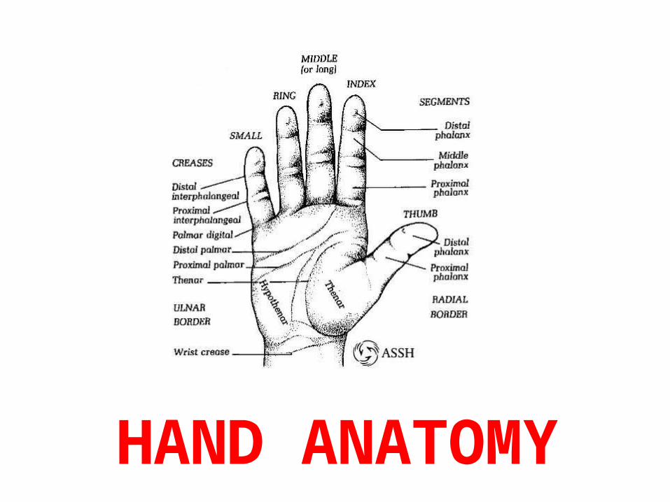
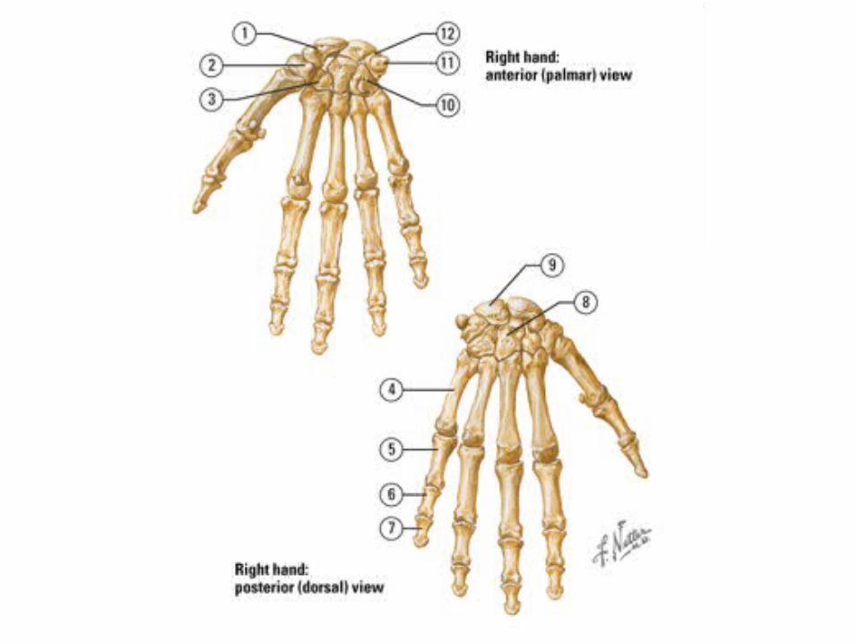
HAND ANATOMY
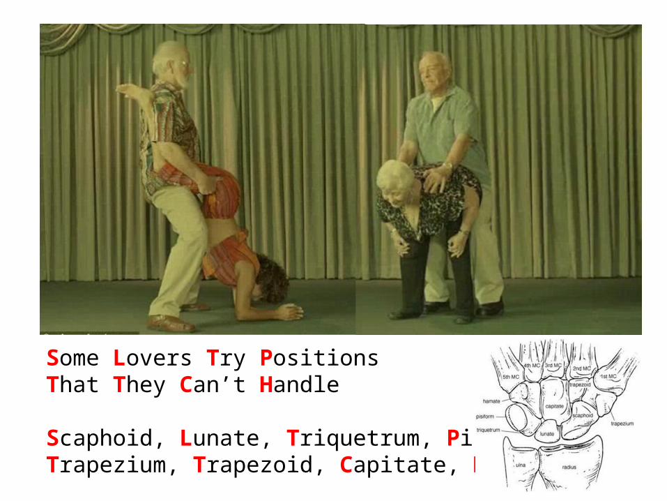
Some Lovers Try PositionsThat They Can’t Handle
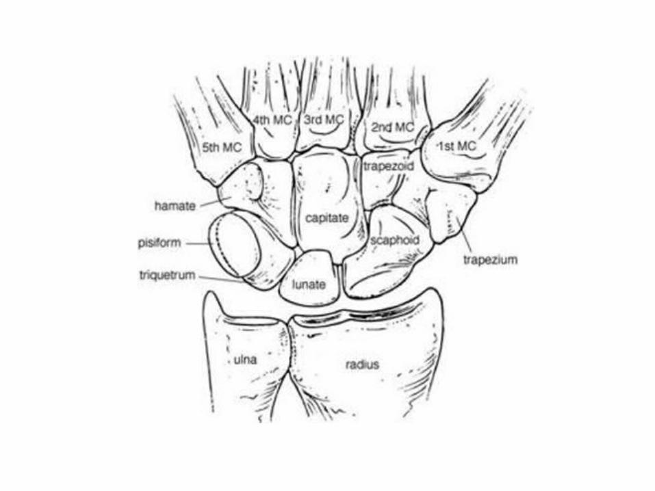
Scaphoid, Lunate, Triquetrum, PisiformTrapezium, Trapezoid, Capitate, Hamate
“Trapezium is under the thumb!”
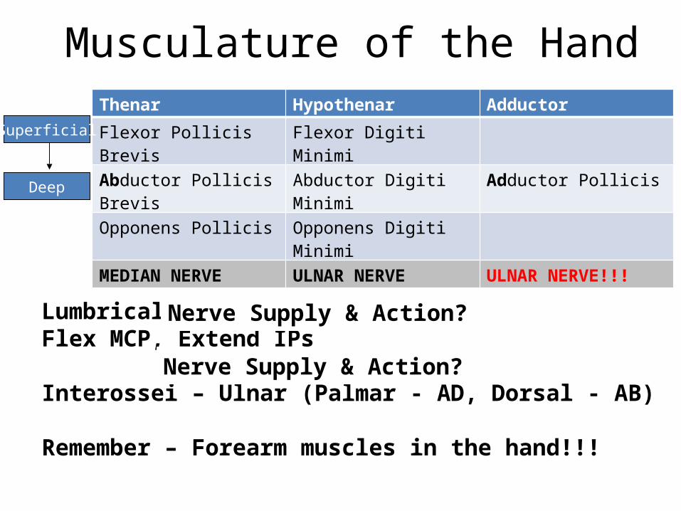
Lumbricals – Median (1 & 2), Ulnar (3 & 4) – Flex MCP, Extend IPs
Interossei – Ulnar (Palmar - AD, Dorsal - AB)
Remember – Forearm muscles in the hand!!!
Nerve Supply & Action?
Musculature of the HandThenar Hypothenar Adductor
Flexor Pollicis Brevis Flexor Digiti Minimi
Abductor Pollicis Brevis Abductor Digiti Minimi Adductor Pollicis
Opponens Pollicis Opponens Digiti Minimi
MEDIAN NERVE ULNAR NERVE ULNAR NERVE!!!
Superficial
Deep
Nerve Supply & Action?
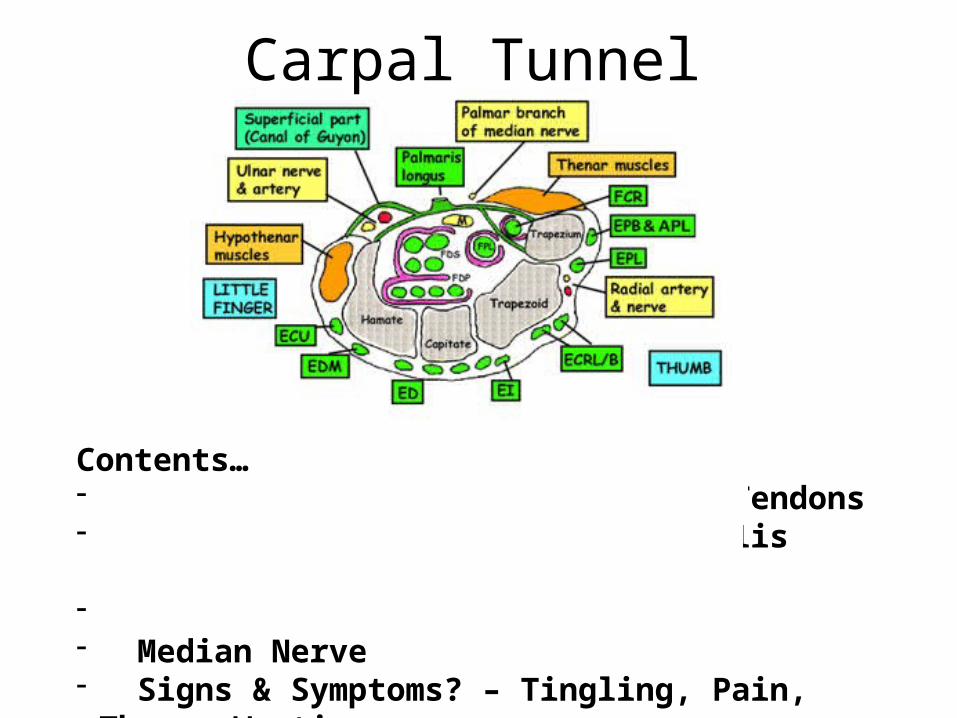
Carpal Tunnel
Contents…- 4 x Flexor Digitorum Profundus Tendons- 4 x Flexor Digitorum Superficialis Tendons- Flexor Pollicis Longus Tendon- Median Nerve- Signs & Symptoms? – Tingling, Pain, Thenar Wasting
Clinical Case 1• A 30-year-old man presents to A&E with incised wounds
to the anterior aspect of his wrist and forearm following a suicide attempt. Preliminary inspection reveals that the patient has four deep, linear incised wounds involving the palmar region of the wrist. The thumb and medial three fingers are noted to be gently flexed while the index finger is held in extension.
• Q - What specific anatomical structures do you think are injured?
• A - FDS and FDP of the index finger
Clinical Case 2
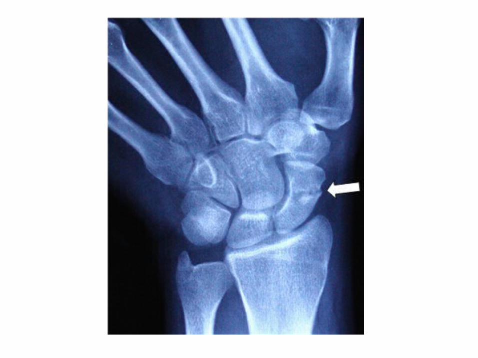
• A 16-year old male presents to A&E after slipping on ice. He states that he attempted to break his fall and fell on an outstretched hand. He complains of pain and swelling in the area indicated by the arrow in the photograph below.
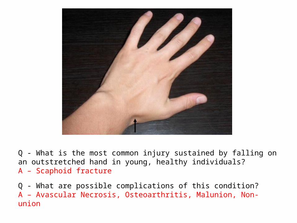
Q - What is the most common injury sustained by falling on an outstretched hand in young, healthy individuals?A – Scaphoid fracture
Q - What are possible complications of this condition?A – Avascular Necrosis, Osteoarthritis, Malunion, Non-union
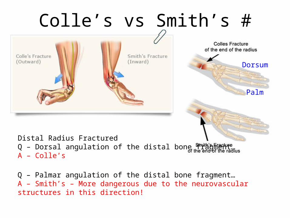
Colle’s vs Smith’s #
Distal Radius FracturedQ – Dorsal angulation of the distal bone fragment…A – Colle’s
Q – Palmar angulation of the distal bone fragment…A – Smith’s – More dangerous due to the neurovascular structures in this direction!
Dorsum
Palm

Boutonniere’s vs Swan Necking
Boutonniere’s – PIP & DIP…PIP flexed, DIP Hyper-extended
Swan Necking – PIP & DIP…PIP Hyper-extended, DIP Flexed
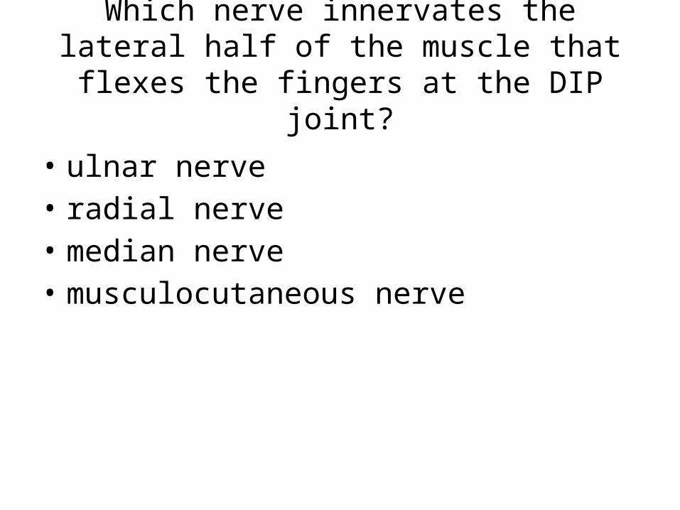
Which nerve innervates the lateral half of the muscle that flexes the fingers at the DIP joint?
• ulnar nerve• radial nerve• median nerve• musculocutaneous nerve

Which of the following structures does not pass through the carpal tunnel?
• Median nerve• Flexor carpi ulnaris• Flexor digitorum superficialis• Flexor digitorum profundus• Flexor pollicis longus
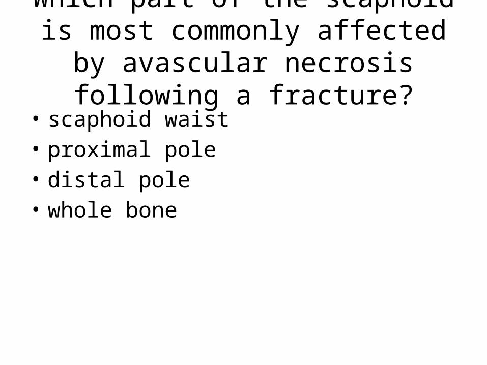
Which part of the scaphoid is most commonly affected by avascular
necrosis following a fracture?• scaphoid waist• proximal pole• distal pole• whole bone
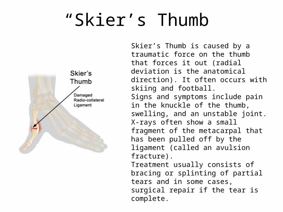
Skier’s Thumb is caused by a traumatic force on the thumb that forces it out (radial deviation is the anatomical direction). It often occurs with skiing and football.Signs and symptoms include pain in the knuckle of the thumb, swelling, and an unstable joint. X-rays often show a small fragment of the metacarpal that has been pulled off by the ligament (called an avulsion fracture).Treatment usually consists of bracing or splinting of partial tears and in some cases, surgical repair if the tear is complete.
“Skier’s Thumb”
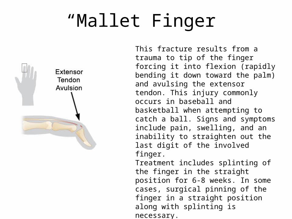
This fracture results from a trauma to tip of the finger forcing it into flexion (rapidly bending it down toward the palm) and avulsing the extensor tendon. This injury commonly occurs in baseball and basketball when attempting to catch a ball. Signs and symptoms include pain, swelling, and an inability to straighten out the last digit of the involved finger.Treatment includes splinting of the finger in the straight position for 6-8 weeks. In some cases, surgical pinning of the finger in a straight position along with splinting is necessary.
“Mallet Finger”
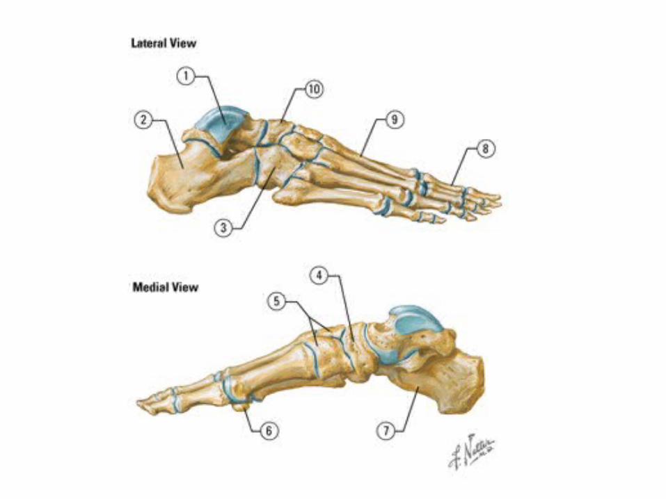
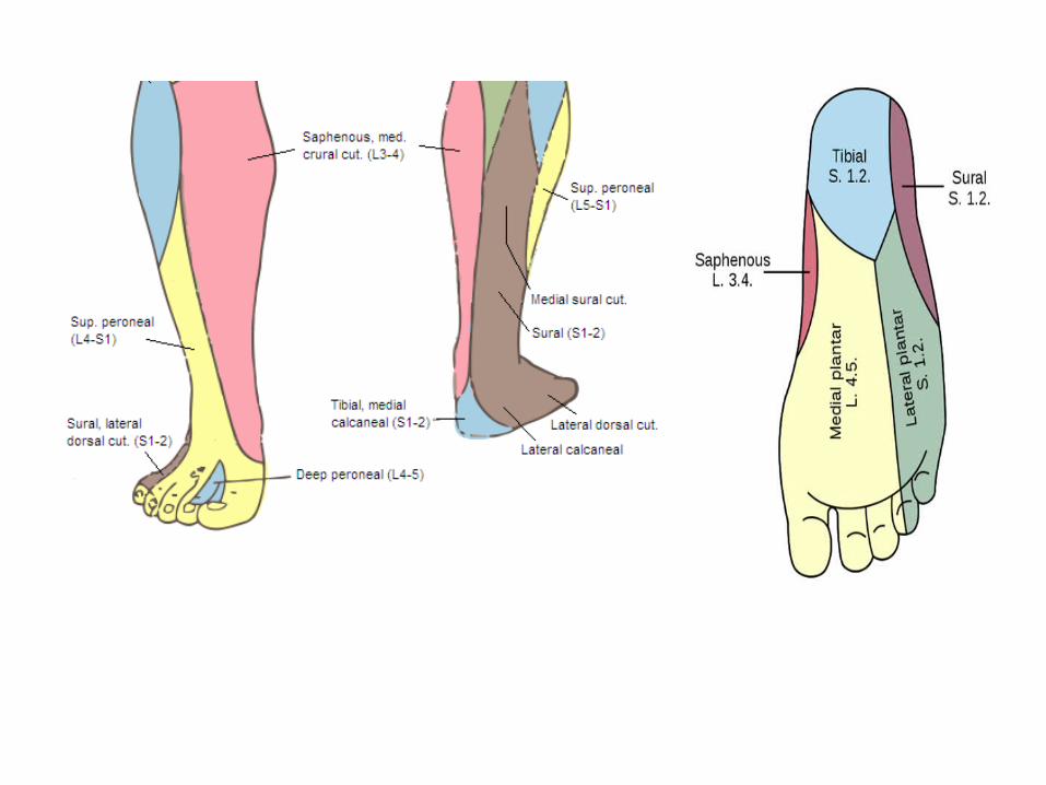
FOOT ANATOMY

Remember – Leg muscles in the foot!!!1st Layer of Sole:
Musculature of the Foot
Medial Plantar Nerve Lateral Plantar Nerve
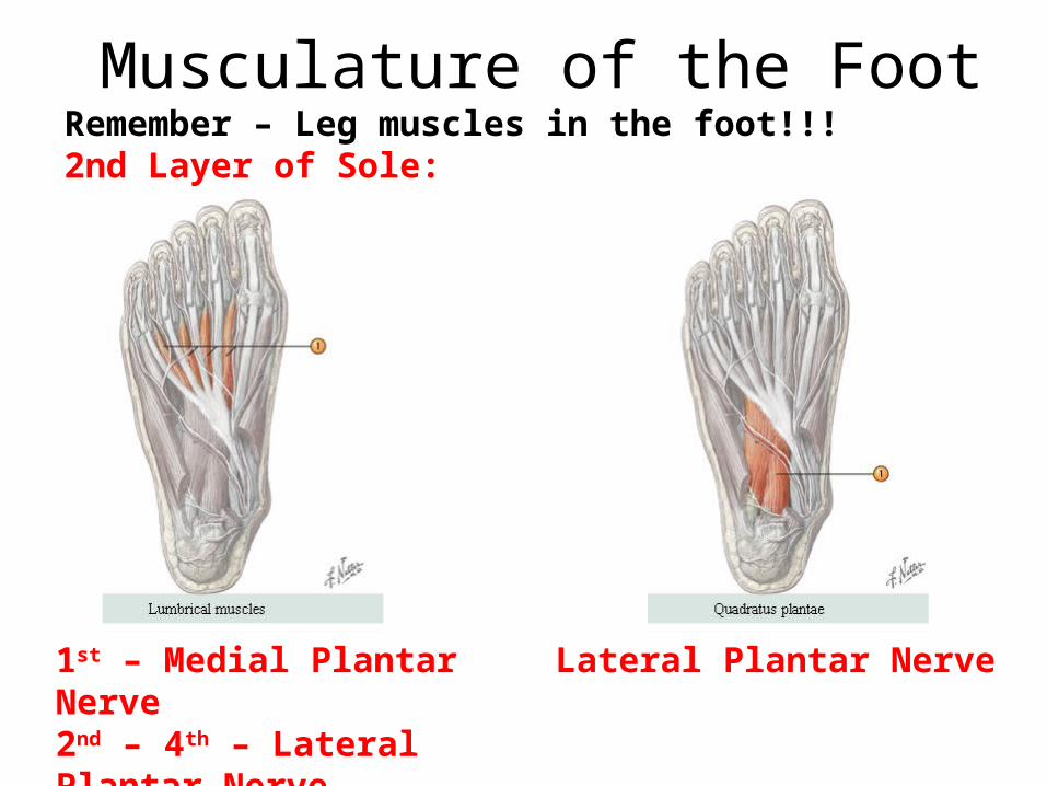
Remember – Leg muscles in the foot!!!2nd Layer of Sole:
Musculature of the Foot
1st – Medial Plantar Nerve2nd – 4th – Lateral Plantar Nerve
Lateral Plantar Nerve
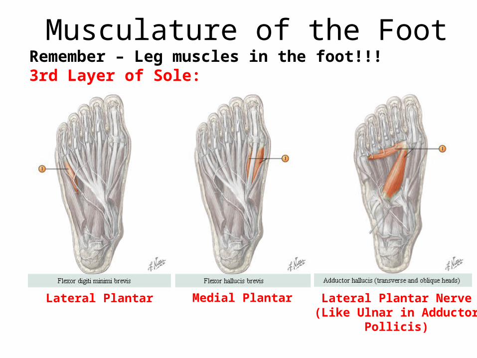
Remember – Leg muscles in the foot!!!3rd Layer of Sole:
Musculature of the Foot
Lateral Plantar Nerve(Like Ulnar in Adductor Pollicis)
Medial PlantarLateral Plantar
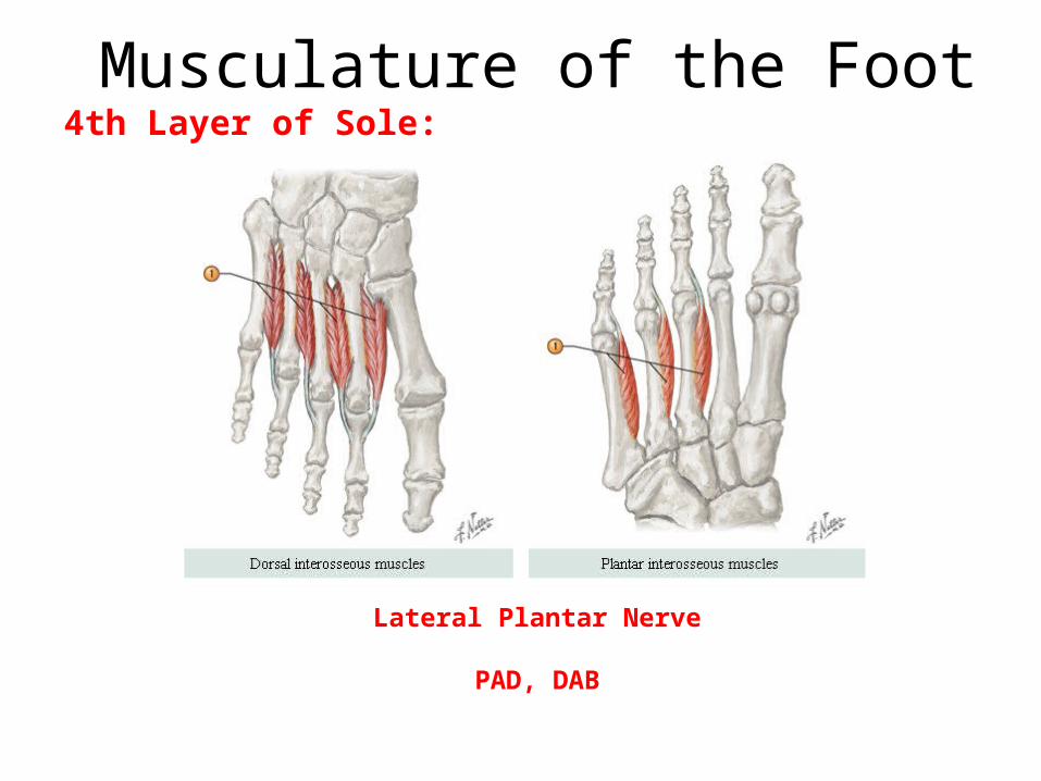
4th Layer of Sole:Musculature of the Foot
Lateral Plantar Nerve
PAD, DAB
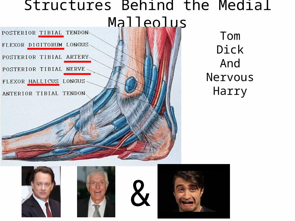
Structures Behind the Medial MalleolusTomDickAnd
NervousHarry
&
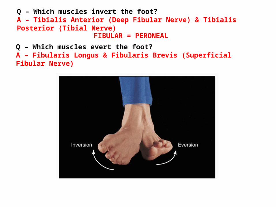
Q – Which muscles invert the foot?A – Tibialis Anterior (Deep Fibular Nerve) & Tibialis Posterior (Tibial Nerve)
FIBULAR = PERONEAL
Q – Which muscles evert the foot?A – Fibularis Longus & Fibularis Brevis (Superficial Fibular Nerve)
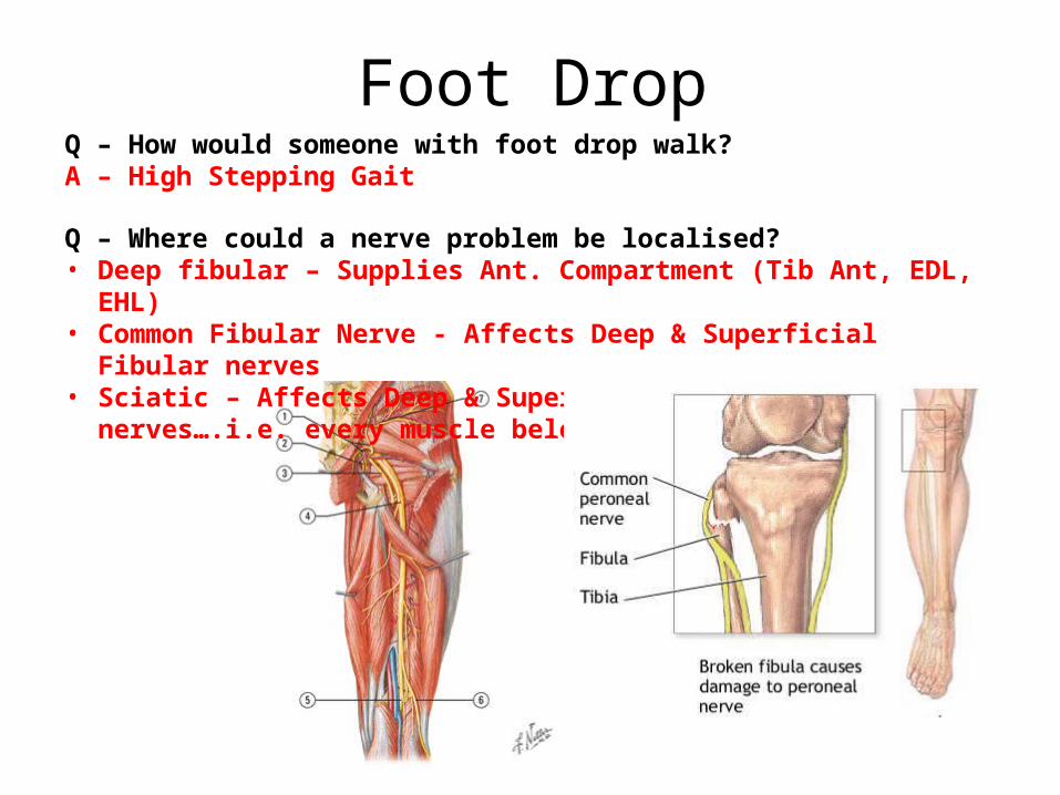
Foot DropQ – How would someone with foot drop walk?A – High Stepping Gait
Q – Where could a nerve problem be localised?• Deep fibular – Supplies Ant. Compartment (Tib Ant, EDL, EHL)• Common Fibular Nerve - Affects Deep & Superficial Fibular nerves• Sciatic – Affects Deep & Superficial Fibular & Tibial nerves….i.e. every muscle
below the knee
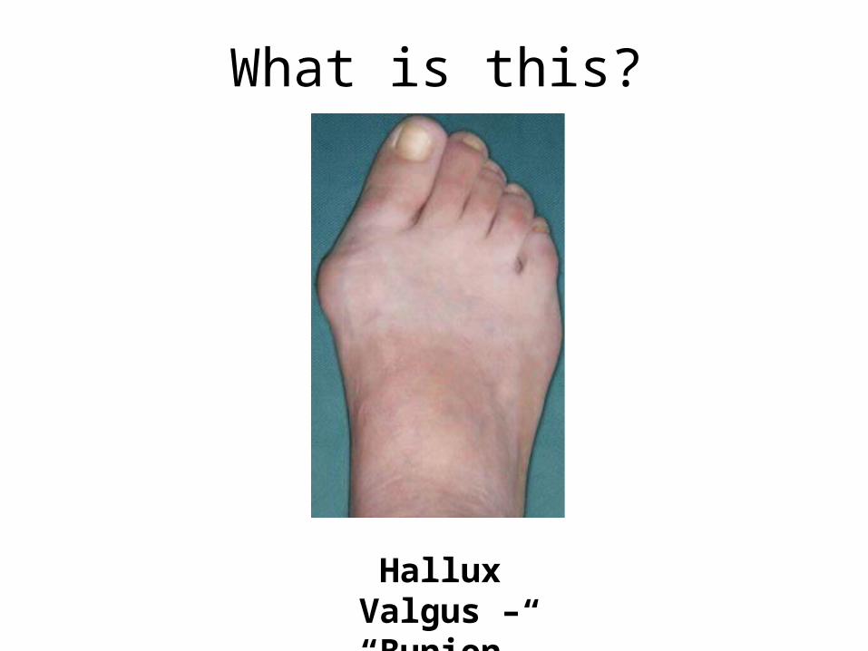
What is this?
Hallux Valgus – “Bunion”