Gyrodactylus salinae n. sp. (Platyhelminthes: Monogenea) infecting
12
RESEARCH Open Access Gyrodactylus salinae n. sp. (Platyhelminthes: Monogenea) infecting the south European toothcarp Aphanius fasciatus (Valenciennes) (Teleostei, Cyprinodontidae) from a hypersaline environment in Italy Giuseppe Paladini 1* , Tine Huyse 2 and Andrew P Shinn 1 Abstract Background: Historically, non-native species of Gambusia (Poeciliidae) have been used to control larval stages of the Asian tiger mosquito, Stegomyia albopicta Reinert, Harbach et Kitching, 2004 throughout Italy. The potential utility of indigenous populations of Aphanius fasciatus (Valenciennes) (Teleostei: Cyprinodontidae) as an appropriate alternative biological control is currently being explored. A sub-sample of ten fish collected from Cervia Saline, Italy (salinity 65 ppt; 30°C) to assess their reproductive capability in captivity, harboured a moderate infection of Gyrodactylus von Nordmann, 1832 (Platyhelminthes, Monogenea). A subsequent morphological and molecular study identified this as being a new species. Results: Gyrodactylus salinae n. sp. is described from the skin, fins and gills of A. fasciatus. Light and scanning electron microscopical (SEM) examination of the opisthaptoral armature and their comparison with all other recorded species suggested morphological similarities to Gyrodactylus rugiensoides Huyse et Volckaert, 2002 from Pomatoschistus minutus (Pallas). Features of the ventral bar, however, permit its discrimination from G. rugiensoides. Sequencing of the nuclear ribosomal DNA internal transcribed spacers 1 and 2 and the 5.8S rRNA gene and a comparison with all species listed in GenBank confirmed they are unique and represent a new species (most similar to Gyrodactylus anguillae Ergens, 1960, 8.3% pair-wise distance based on 5.8S+ITS2). This represents the first species of Gyrodactylus to be described from Aphanius and, to date, has the longest ITS1 (774 bp) sequenced from any Gyrodactylus. Additional sampling of Cervia Saline throughout the year, found G. salinae n. sp. to persist in conditions ranging from 35 ppt and 5°C in December to 65 ppt and 30°C in July, while in captivity a low level of infection was present, even in freshwater conditions (0 ppt). Conclusions: The ability of G. salinae n. sp. to tolerate a wide range of salinities and temperatures shows its potential to readily adapt to several environmental conditions. These findings, together with the fact that A. fasciatus is a protected species and is considered as a biological control organism, necessitate further studies on the ecology and virulence of G. salinae n. sp. * Correspondence: [email protected] 1 Institute of Aquaculture, University of Stirling, Stirling, FK9 4LA, Scotland, UK Full list of author information is available at the end of the article Paladini et al. Parasites & Vectors 2011, 4:100 http://www.parasitesandvectors.com/content/4/1/100 © 2011 Paladini et al; licensee BioMed Central Ltd. This is an Open Access article distributed under the terms of the Creative Commons Attribution License (http://creativecommons.org/licenses/by/2.0), which permits unrestricted use, distribution, and reproduction in any medium, provided the original work is properly cited.
Transcript of Gyrodactylus salinae n. sp. (Platyhelminthes: Monogenea) infecting
RESEARCH Open Access
Gyrodactylus salinae n. sp. (Platyhelminthes:Monogenea) infecting the south Europeantoothcarp Aphanius fasciatus (Valenciennes)(Teleostei, Cyprinodontidae) froma hypersaline environment in ItalyGiuseppe Paladini1*, Tine Huyse2 and Andrew P Shinn1
Abstract
Background: Historically, non-native species of Gambusia (Poeciliidae) have been used to control larval stages ofthe Asian tiger mosquito, Stegomyia albopicta Reinert, Harbach et Kitching, 2004 throughout Italy. The potentialutility of indigenous populations of Aphanius fasciatus (Valenciennes) (Teleostei: Cyprinodontidae) as an appropriatealternative biological control is currently being explored. A sub-sample of ten fish collected from Cervia Saline, Italy(salinity 65 ppt; 30°C) to assess their reproductive capability in captivity, harboured a moderate infection ofGyrodactylus von Nordmann, 1832 (Platyhelminthes, Monogenea). A subsequent morphological and molecularstudy identified this as being a new species.
Results: Gyrodactylus salinae n. sp. is described from the skin, fins and gills of A. fasciatus. Light and scanningelectron microscopical (SEM) examination of the opisthaptoral armature and their comparison with all otherrecorded species suggested morphological similarities to Gyrodactylus rugiensoides Huyse et Volckaert, 2002 fromPomatoschistus minutus (Pallas). Features of the ventral bar, however, permit its discrimination from G. rugiensoides.Sequencing of the nuclear ribosomal DNA internal transcribed spacers 1 and 2 and the 5.8S rRNA gene and acomparison with all species listed in GenBank confirmed they are unique and represent a new species (mostsimilar to Gyrodactylus anguillae Ergens, 1960, 8.3% pair-wise distance based on 5.8S+ITS2). This represents the firstspecies of Gyrodactylus to be described from Aphanius and, to date, has the longest ITS1 (774 bp) sequenced fromany Gyrodactylus. Additional sampling of Cervia Saline throughout the year, found G. salinae n. sp. to persist inconditions ranging from 35 ppt and 5°C in December to 65 ppt and 30°C in July, while in captivity a low level ofinfection was present, even in freshwater conditions (0 ppt).
Conclusions: The ability of G. salinae n. sp. to tolerate a wide range of salinities and temperatures shows itspotential to readily adapt to several environmental conditions. These findings, together with the fact that A.fasciatus is a protected species and is considered as a biological control organism, necessitate further studies onthe ecology and virulence of G. salinae n. sp.
* Correspondence: [email protected] of Aquaculture, University of Stirling, Stirling, FK9 4LA, Scotland, UKFull list of author information is available at the end of the article
Paladini et al. Parasites & Vectors 2011, 4:100http://www.parasitesandvectors.com/content/4/1/100
© 2011 Paladini et al; licensee BioMed Central Ltd. This is an Open Access article distributed under the terms of the Creative CommonsAttribution License (http://creativecommons.org/licenses/by/2.0), which permits unrestricted use, distribution, and reproduction inany medium, provided the original work is properly cited.
BackgroundToothcarp is a colloquial term used to describemembers of the order Cyprinodontiformes, which com-prises ten fish families, namely Anablepidae, Aplocheilidae,Cyprinodontidae, Fundulidae, Goodeidae, Nothobranchii-dae, Poeciliidae, Profundulidae, Rivulidae and Valenciidae[1]. Within the family Cyprinodontidae, the south Eur-opean toothcarp Aphanius fasciatus (Valenciennes) is oneof the more commonly occurring neritic species withinthe Mediterranean and is characteristically known to bean eurythermic and euryhaline fish species, for its abilityto tolerate a wide range of temperatures (5-39°C) andsalinities (0-180 ppt), respectively [2]. Aphanius fasciatushas a widespread distribution along the Italian coastline,principally in brackish waters [3], although increasinganthropogenic activity has caused a general decline innumbers. For this reason, A. fasciatus is considered to be aspecies that is “dying out” and as such, is listed underAppendix III “Protected Fauna Species” after the BernConvention on the conservation of European wildlife andnatural habitats, and under Annex II of the Council Direc-tive 92/43/EEC on the conservation of natural habitatsand of wild fauna and flora in the European Community[4]. It has recently been used as a biological control againstthe larvae of the Asian tiger mosquito Stegomyia albopicta(= Aedes albopictus) Reinert, Harbach et Kitching, 2004which are vectors for a range of human infectious diseases,including Chikungunya fever, dengue fever, West Nilefever and yellow fever [5-7].During a recent research study investigating the artifi-
cial reproduction of A. fasciatus in captivity as part of alarge scale restocking and mosquito control initiative inItaly [8,9], several specimens collected, under licence, ona number of occasions were found to harbour an infec-tion of Gyrodactylus von Nordmann, 1832, principallyon the skin and fins and, to a lesser degree, on the gills.Infected tissues were removed from the moribund fishand subsequently sent to the Institute of Aquaculture,University of Stirling (Scotland, UK) for identification.Given the importance of A. fasciatus as a protectedspecies and its utility as an alternative indigeneous bio-logical control agent to the introduced Gambusia spp.(Poeciliidae), more information on its Gyrodactylusfauna was needed.
MethodsSpecimens collection and preparationA total of ten adult A. fasciatus (total length 4-7 cm;weight ~3 g) was collected during July 2008 from iso-lated pools in Cervia Saline, located in the EmiliaRomagna region in northern Italy, and fixed in 70%ethanol. All ten specimens collected from the drying,landlocked pools were moribund individuals, a
consequence of reduced water availability and increasedalgal growth. The skin, fins and gills of each fish weresubsequently screened for metazoan parasites using anOlympus SZ40 stereomicroscope at ×4 magnification.Specimens of Gyrodactylus were removed usingmounted triangular surgical needles. All ten fish wereinfected but given the condition of the fish on collec-tion, the intensity of infection can only be estimated atbetween 10-30 parasites per fish.The alcohol-fixed parasites were subsequently rinsed
in distilled water and representatives prepared as wholemounts using ammonium picrate glycerine following theprocedure detailed by Malmberg [10]. Additional speci-mens had their opisthaptors removed using a scalpel,which were then individually subjected to proteolyticdigestion on glass slides, as described in Paladini et al.[11]. The largely tissue-free opisthaptoral hook prepara-tions were then mounted in ammonium picrate glycer-ine using an 18 × 18 mm coverslip, the edges of whichwere sealed with a commercial brand of nail varnish.The corresponding body of each specimen of Gyrodacty-lus was fixed in 90% ethanol for subsequent molecularcharacterisation.For scanning electron microscopy (SEM), single speci-
mens of Gyrodactylus were subjected to full proteolyticdigestion on 11 mm round glass coverslips to obtaintissue-free attachment hooks. Each digestion tookapproximately 60 min, with 3 μl of digestion solutionbeing added every 10 min, punctuated by the additionof 5 μl distilled water for five times to remove digestedtissue residues and debris. For each step, the digestedhook preparations were placed in a Petri dish to protectthem from extraneous dust and placed in an incubatorat 55°C to help digestion, followed by a final incubationat 40°C overnight to dry. The position of hooks on eachcoverslip was subsequently marked using tiny, adhesive,triangular labels positioned using forceps under anOlympus BX51 compound microscope at ×10 magnifi-cation. The coverslips were then attached onto alumi-nium stubs with bi-adhesive round labels, sputter-coatedwith gold using an Edwards S150B sputter coater andthen examined using a JEOL JSM5200 SEM operating atan accelerating voltage of 10 kv.
Morphological analysisFor the morphological study, images of the opisthaptoralhard parts and the male copulatory organ (MCO) werecaptured at magnifications of ×40 and ×100 oil immer-sion using MRGrab 1.0.0.4 (Carl Zeiss Vision GmbH,2001) software and a Zeiss AxioCam MRc digitalcamera mounted on an Olympus BX51 compoundmicroscope, using a ×0.75 interfacing lens. Drawings ofthe taxonomic features were made from the captured
Paladini et al. Parasites & Vectors 2011, 4:100http://www.parasitesandvectors.com/content/4/1/100
Page 2 of 12
images. Each Gyrodactylus specimen was subjected tomorphometric analysis taking a total of 27 point-to-point measurements on the opisthaptoral hooks using aJVC KY-F30B 3CCD video camera mounted on anOlympus BH2 microscope using a ×2.5 interfacing lensat ×100 oil immersion and the KS300 (ver.3.0) (CarlZeiss Vision GmbH, 1997) image analysis software,combined with the specific macro for gyrodactylids,Point-R (Bron & Shinn, University of Stirling). Thepoint-to-point hook measurements for the specimensare given in micrometres as the mean ± 1 standarddeviation followed by the range in parentheses, andfollow those described in Shinn et al. [12], plus threeadditional measurements of the dorsal bar (total length,width and attachment point length).The gyrodactylid material prepared from A. fasciatus
was compared to type material of Gyrodactylus rugien-soides Huyse et Volckaert, 2002 (paratypes acc. nos.BMNH 2002.2.14.2-3), a species with morphologicallysimilar marginal hooks, held in the Parasitic Worms col-lection at The Natural History Museum, London, UK. Inaddition, type material of the six Gyrodactylus speciesknown to parasitise cyprinodontid hosts, held in theU.S. National Parasite Collection, Beltsville, Maryland,USA, was examined, the marginal hooks re-drawn andcompared to the new specimens collected fromA. fasciatus. These are Gyrodactylus cyprinodontisMizelle et Kritsky, 1967 (holotype and paratype acc. no.USNPC 62951), Gyrodactylus hargisi Williams et Rogers,1971 (paratypes acc. no. USNPC 71760), Gyrodactylusmobilensis Williams et Rogers, 1971 (paratypes acc. no.USNPC 71762), Gyrodactylus nevadensis Mizelle etKritsky, 1967 (holotype and paratype acc. no. USNPC62954), Gyrodactylus saratogensis Mizelle et Kritsky,1967 (paratype acc. no. USNPC 62956) and Gyrodacty-lus tularosae Kritsky et Stockwell, 2005 (paratype acc.no. USNPC 94780).
Molecular analysisThe bodies of 2 specimens were individually transferredto a 0.2 ml microcentrifuge tube containing 5 μl ofmilli-Q water and digested by the addition of 5 μl oflysis solution consisting of 1×PCR buffer (Eurogentec,Seraing, Belgium), 0.45% (v/v) Tween 20, 0.45% (v/v)NP 40 and 60 μg/ml of proteinase K (Sigma, Poole,UK). The samples were incubated at 65°C for 25 min,followed by 10 min at 95°C to inactivate the proteinase.The primer pairs ITS1A (5’-GTAACAAGGTTTCCGTAGGTG-3’) and ITS2 (5’-TCCTCCGCTTAGT-GATA- 3’) [13] were used to amplify a fragment span-ning the 3’ end of the 18S rRNA gene, the internaltranscribed spacer 1 (ITS1), the 5.8S rRNA gene, ITS2,and the 5’ end of the 28S rRNA gene. The amplificationreactions (20 μl) consisted of 1×PCR buffer, 1.5 mM
MgCl2 (Eurogentec, Seraing, Belgium), 200 μM of eachdNTP (Amersham Pharmacia Biotech, Uppsala,Sweden), 1 μM of each primer (Eurogentec, Seraing,Belgium), 2 μl lysate, 1 unit Taq polymerase (Eurogen-tec, Seraing, Belgium) and milli-Q water. The mixtureswere heated for 4 min at 96°C and subjected to 35cycles of 1 min at 95°C, 1 min at 50°C and 2 min at 72°C, followed by a final extension at 72°C for 7 min. ThePCR products were visualised using ethidium bromideon a 1.2% agarose gel. The products were then purifiedby means of GFX columns according to the manufac-turer’s instructions (Amersham Pharmacia Biotech,Uppsala, Sweden). Both DNA strands were sequencedusing a Big Dye Chemistry Cycle Sequencing Kit (ver-sion 1.1) in a 3130 DNA Analyzer (Applied Biosystems,Belgium). The PCR primers and 2 internal primers,ITS1R (5’-ATTT GCGTTCGAGAGACCG-3’) andITS2F (5’-TGGTGGATCA CTCGGCTCA-3’) [14], wereused for sequencing.The obtained sequences were subjected to a BLAST
search (available at http://www.ncbi.nih.gov/BLAST/)to identify similar sequences among other species ofGyrodactylus in GenBank [15]. The sequences with thehighest similarity to our sequences were downloaded andthe 5.8S and ITS2 fragments were aligned in Clustal Wimplemented in MEGA 4 [16]. Pair-wise genetic distanceswere computed in MEGA 4 according to the evolutionarymodel that was selected by jModelTest 0.1.1 [17]. Thesequences were also scanned for repeat elements using theprogram Tandem Repeats Finder [18].
ResultsGyrodactylus salinae n. spType hostAphanius fasciatus (Valenciennes), Cyprinodontidae("South European toothcarp”, “nono”).Site of infectionSkin, fins and occasionally gills.Type localityCervia Saline, Emilia Romagna region, Italy (44°14’N, 12°20’E).Environmental conditions under which specimenswere collectedSalinity and temperature in July 2008, 65 ppt and 30°C,respectively.Type materialFifteen specimens were studied for light microscopy.Holotype (acc. no. BMNH 2011.5.19.1) and four para-types (acc. nos. BMNH 2011.5.19.2-5) are deposited inthe parasitic worm collection at The Natural HistoryMuseum, London. Additionally, three paratypes (acc. no.M-521) are deposited in the gyrodactylid collection heldat the Institute of Parasitology, Academy of Sciences ofthe Czech Republic, České Budĕjovice; four paratypes
Paladini et al. Parasites & Vectors 2011, 4:100http://www.parasitesandvectors.com/content/4/1/100
Page 3 of 12
(acc. nos. AHC 35118-35121) are deposited in the Aus-tralian Helminthological Collection (AHC) of The SouthAustralian Museum (SAMA), North Terrace, Adelaide;and three paratypes (acc. nos. USNPC 104748-104750)are deposited in the United States National Parasite Col-lection, Beltsville, Maryland, USA.Molecular sequence dataThe sequence fragment of approximately 1379 bp encod-ing partial 18S (32 bp), ITS1 (774 bp), 5.8S (158 bp),ITS2 (402 bp) and partial 28S (13 bp) is deposited inGenBank under accession no. JF950559.GeneralA species profile including host and taxonomic details isprovided on the on-line databases http://www.gyrodb.net[19] and http://www.monodb.org[20].EtymologyNamed after the Italian generic name for a hypersalinewater body i.e. “salina” (= saline in English) and thebroad salinity tolerance exhibited by this species ofGyrodactylus.
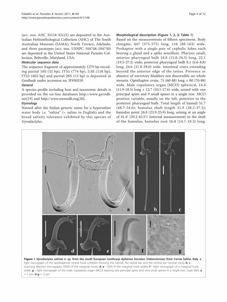
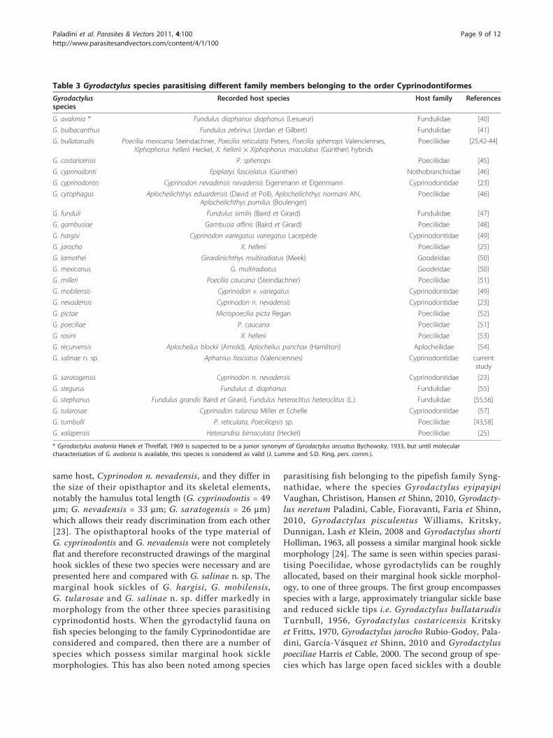
Morphological description (Figure 1, 2, 3; Table 1)Based on the measurements of fifteen specimens. Bodyelongate, 447 (375-575) long, 116 (88-163) wide.Prohaptor with a single pair of cephalic lobes eachbearing a gland and a spike sensillum. Pharynx small,anterior pharyngeal bulb 18.8 (15.0-24.5) long, 22.1(19.5-27.2) wide; posterior pharyngeal bulb 8.1 (6.6-8.8)long, 24.6 (21.8-29.0) wide. Intestinal crura extendingbeyond the anterior edge of the testes. Presence orabsence of excretory bladders not discernible on wholemounts. Opisthaptor ovate, 75 (60-88) long × 80 (70-88)wide. Male copulatory organ (MCO) spherical, 14.4(11.9-18.5) long × 12.7 (10.1-17.6) wide, armed with oneprincipal spine and 9 small spines in a single row. MCOposition variable, usually on the left, posterior to theposterior pharyngeal bulb. Total length of hamuli 51.7(48.7-54.6); hamulus shaft length 31.9 (28.2-37.3);hamulus point 24.8 (23.9-25.9) long, arising at an angleof 41.4° (39.2-43.5°) (internal measurement) to the shaftof the hamulus; hamulus root 16.8 (14.7-18.3) long.
Figure 1 Gyrodactylus salinae n. sp. from the south European toothcarp Aphanius fasciatus (Valenciennes) from Cervia Saline, Italy. a -light micrograph of the opisthaptoral central hook complex showing the hamuli, the dorsal bar and the ventral bar (ventral view); b, c -scanning electron micrographs (SEM) of the marginal hooks; d, e - SEM of the marginal hook sickles; f - light micrograph of a marginal hooksickle; g - light micrograph of the male copulatory organ (MCO) bearing one principal spine and nine small spines in a single row. Scale bars: a= 5 μm; b-g = 3 μm.
Paladini et al. Parasites & Vectors 2011, 4:100http://www.parasitesandvectors.com/content/4/1/100
Page 4 of 12
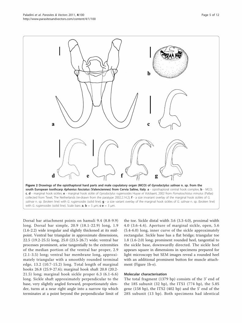
Dorsal bar attachment points on hamuli 9.4 (8.8-9.9)long. Dorsal bar simple, 20.9 (18.1-22.9) long, 1.9(1.6-2.2) wide irregular and slightly thickened at its mid-point. Ventral bar triangular in approximate dimensions,22.5 (19.2-25.5) long, 25.0 (23.5-26.7) wide; ventral barprocesses prominent, arise tangentially to the extremitiesof the median portion of the ventral bar proper, 2.9(2.1-3.5) long; ventral bar membrane long, approxi-mately triangular with a smoothly rounded terminaledge, 13.2 (10.7-15.2) long. Total length of marginalhooks 26.8 (25.9-27.6); marginal hook shaft 20.8 (20.2-21.5) long; marginal hook sickle proper 6.3 (6.1-6.6)long. Sickle shaft approximately perpendicular to thebase, very slightly angled forward, proportionately slen-der, turns at a near right angle into a narrow tip whichterminates at a point beyond the perpendicular limit of
the toe. Sickle distal width 3.6 (3.3-4.0), proximal width4.0 (3.6-4.4). Aperture of marginal sickle, open, 5.6(5.4-6.0) long, inner curve of the sickle approximatelyrectangular. Sickle base has a flat bridge; triangular toe1.8 (1.6-2.0) long; prominent rounded heel, tangential tothe sickle base, downwardly directed. The sickle heelappears square in dimensions in specimens prepared forlight microscopy but SEM images reveal a rounded heelwith an additional prominent button for muscle attach-ment (Figure 1b-e).
Molecular characterisationThe total fragment (1379 bp) consists of the 3’ end ofthe 18S subunit (32 bp), the ITS1 (774 bp), the 5.8Sgene (158 bp), the ITS2 (402 bp) and the 5’ end of the28S subunit (13 bp). Both specimens had identical
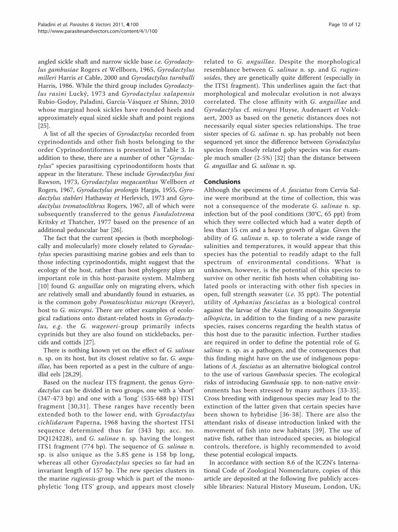
Figure 2 Drawings of the opisthaptoral hard parts and male copulatory organ (MCO) of Gyrodactylus salinae n. sp. from thesouth European toothcarp Aphanius fasciatus (Valenciennes) from Cervia Saline, Italy. a - opisthaptoral central hook complex; b - MCO;c, d - marginal hook sickles; e - marginal hook sickle of Gyrodactylus rugiensoides Huyse et Volckaert, 2002 from Pomatoschistus minutus (Pallas)collected from Texel, The Netherlands (re-drawn from the paratype 2002.2.14.2); f - a size invariant overlay of the marginal hook sickles of G.salinae n. sp. (broken line) with G. rugiensoides (solid line); g - a size variant overlay of the marginal hook sickles of G. salinae n. sp. (broken line)with G. rugiensoides (solid line). Scale bars: a, b = 5 μm; c-e = 3 μm.
Paladini et al. Parasites & Vectors 2011, 4:100http://www.parasitesandvectors.com/content/4/1/100
Page 5 of 12
sequences. Gyrodactylus salinae n. sp. appeared mostclosely related to Gyrodactylus species belonging to theG. (Paranephrotus) and G. (Neonephrotus) sub-genera(sub-genera according to Malmberg [10]) based on thepair-wise distances (Tamura-Nei gamma corrected dis-tances [21] using the 5.8S-ITS2 fragment, Table 2); it ismost closely related to Gyrodactylus anguillae Ergens,
1960 (8.3%; acc. no. AB063294) and Gyrodactylusmicropsi Gläser, 1974 (8.6%; acc. no. AF328868). Thepair-wise distance with G. rugiensoides, the specieswhose attachment hooks are morphologically similar tothose of G. salinae n. sp., amounted to 14%. Of allavailable ITS rDNA sequences of Gyrodactylus (146 onGenBank), G. salinae n. sp. has the longest ITS1
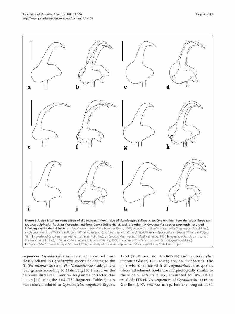
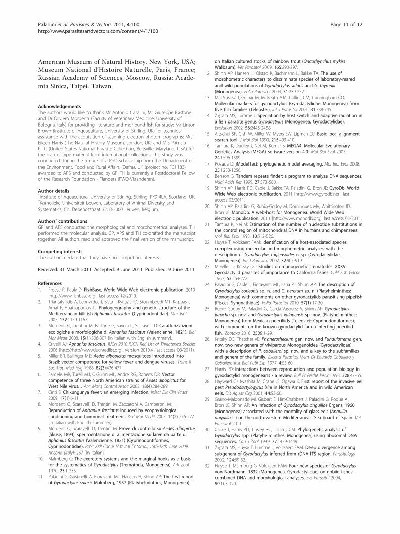
Figure 3 A size invariant comparison of the marginal hook sickle of Gyrodactylus salinae n. sp. (broken line) from the south Europeantoothcarp Aphanius fasciatus (Valenciennes) from Cervia Saline (Italy), with the other six Gyrodactylus species previously recordedinfecting cyprinodontid hosts. a - Gyrodactylus cyprinodontis Mizelle et Kritsky, 1967; b - overlay of G. salinae n. sp. with G. cyprinodontis (solid line);c - Gyrodactylus hargisi Williams et Rogers, 1971; d - overlay of G. salinae n. sp. with G. hargisi (solid line); e - Gyrodactylus mobilensis Williams et Rogers,1971; f - overlay of G. salinae n. sp. with G. mobilensis (solid line); g - Gyrodactylus nevadensis Mizelle et Kritsky, 1967; h - overlay of G. salinae n. sp. withG. nevadensis (solid line); i - Gyrodactylus saratogensis Mizelle et Kritsky, 1967; j - overlay of G. salinae n. sp. with G. saratogensis (solid line);k - Gyrodactylus tularosae Kritsky et Stockwell, 2005; l - overlay of G. salinae n. sp. with G. tularosae (solid line). Scale bars = 3 μm.
Paladini et al. Parasites & Vectors 2011, 4:100http://www.parasitesandvectors.com/content/4/1/100
Page 6 of 12
fragment (774 bp), followed by Gyrodactylus teuchisLautraite, Blanc, Thiery, Daniel et Vigneulle, 1999 (720bp; acc. no. AJ249350). The ITS1 has a GC content of42.1% and an imperfect repeat of an 8 bp motif(GAGAGAGT), starting at position 101 (copynumber
4.4). The ITS1 of G. anguillae and G. micropsi did nothave any repeat element. The 5.8S rRNA gene is 158bp long, which is 1 bp longer than all other Gyrodacty-lus species sequenced so far; the ITS2 (402 bp) has amedian size.
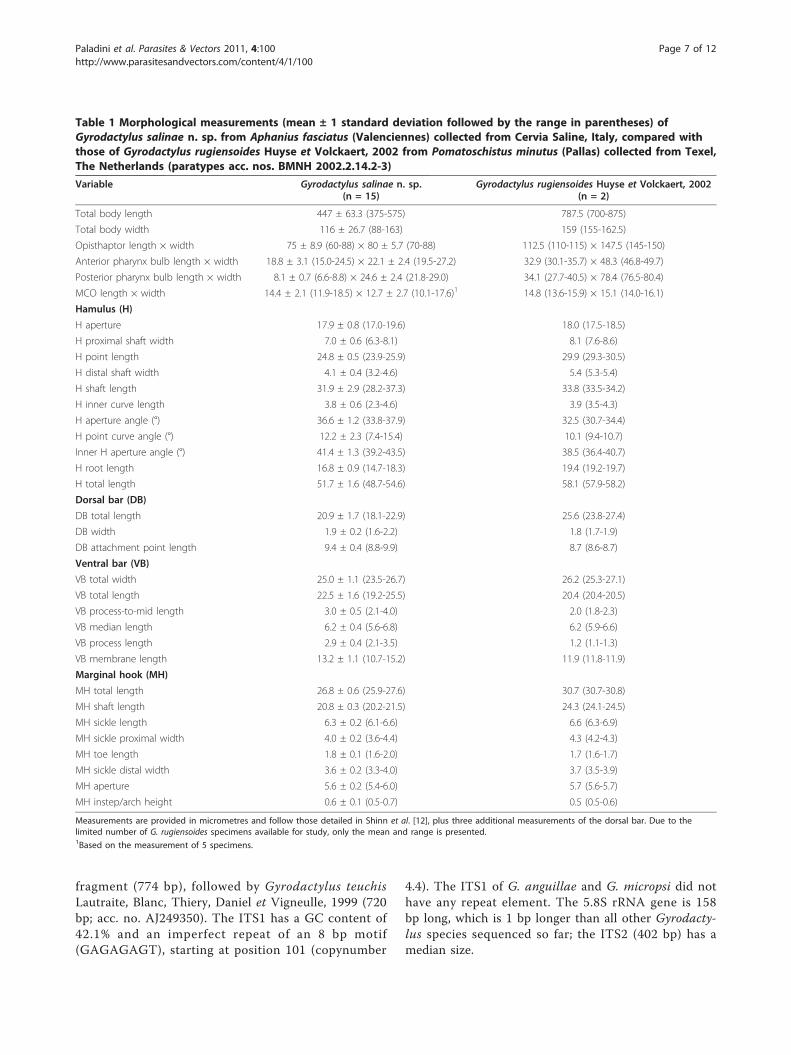
Table 1 Morphological measurements (mean ± 1 standard deviation followed by the range in parentheses) ofGyrodactylus salinae n. sp. from Aphanius fasciatus (Valenciennes) collected from Cervia Saline, Italy, compared withthose of Gyrodactylus rugiensoides Huyse et Volckaert, 2002 from Pomatoschistus minutus (Pallas) collected from Texel,The Netherlands (paratypes acc. nos. BMNH 2002.2.14.2-3)
Variable Gyrodactylus salinae n. sp.(n = 15)
Gyrodactylus rugiensoides Huyse et Volckaert, 2002(n = 2)
Total body length 447 ± 63.3 (375-575) 787.5 (700-875)
Total body width 116 ± 26.7 (88-163) 159 (155-162.5)
Opisthaptor length × width 75 ± 8.9 (60-88) × 80 ± 5.7 (70-88) 112.5 (110-115) × 147.5 (145-150)
Anterior pharynx bulb length × width 18.8 ± 3.1 (15.0-24.5) × 22.1 ± 2.4 (19.5-27.2) 32.9 (30.1-35.7) × 48.3 (46.8-49.7)
Posterior pharynx bulb length × width 8.1 ± 0.7 (6.6-8.8) × 24.6 ± 2.4 (21.8-29.0) 34.1 (27.7-40.5) × 78.4 (76.5-80.4)
MCO length × width 14.4 ± 2.1 (11.9-18.5) × 12.7 ± 2.7 (10.1-17.6)1 14.8 (13.6-15.9) × 15.1 (14.0-16.1)
Hamulus (H)
H aperture 17.9 ± 0.8 (17.0-19.6) 18.0 (17.5-18.5)
H proximal shaft width 7.0 ± 0.6 (6.3-8.1) 8.1 (7.6-8.6)
H point length 24.8 ± 0.5 (23.9-25.9) 29.9 (29.3-30.5)
H distal shaft width 4.1 ± 0.4 (3.2-4.6) 5.4 (5.3-5.4)
H shaft length 31.9 ± 2.9 (28.2-37.3) 33.8 (33.5-34.2)
H inner curve length 3.8 ± 0.6 (2.3-4.6) 3.9 (3.5-4.3)
H aperture angle (°) 36.6 ± 1.2 (33.8-37.9) 32.5 (30.7-34.4)
H point curve angle (°) 12.2 ± 2.3 (7.4-15.4) 10.1 (9.4-10.7)
Inner H aperture angle (°) 41.4 ± 1.3 (39.2-43.5) 38.5 (36.4-40.7)
H root length 16.8 ± 0.9 (14.7-18.3) 19.4 (19.2-19.7)
H total length 51.7 ± 1.6 (48.7-54.6) 58.1 (57.9-58.2)
Dorsal bar (DB)
DB total length 20.9 ± 1.7 (18.1-22.9) 25.6 (23.8-27.4)
DB width 1.9 ± 0.2 (1.6-2.2) 1.8 (1.7-1.9)
DB attachment point length 9.4 ± 0.4 (8.8-9.9) 8.7 (8.6-8.7)
Ventral bar (VB)
VB total width 25.0 ± 1.1 (23.5-26.7) 26.2 (25.3-27.1)
VB total length 22.5 ± 1.6 (19.2-25.5) 20.4 (20.4-20.5)
VB process-to-mid length 3.0 ± 0.5 (2.1-4.0) 2.0 (1.8-2.3)
VB median length 6.2 ± 0.4 (5.6-6.8) 6.2 (5.9-6.6)
VB process length 2.9 ± 0.4 (2.1-3.5) 1.2 (1.1-1.3)
VB membrane length 13.2 ± 1.1 (10.7-15.2) 11.9 (11.8-11.9)
Marginal hook (MH)
MH total length 26.8 ± 0.6 (25.9-27.6) 30.7 (30.7-30.8)
MH shaft length 20.8 ± 0.3 (20.2-21.5) 24.3 (24.1-24.5)
MH sickle length 6.3 ± 0.2 (6.1-6.6) 6.6 (6.3-6.9)
MH sickle proximal width 4.0 ± 0.2 (3.6-4.4) 4.3 (4.2-4.3)
MH toe length 1.8 ± 0.1 (1.6-2.0) 1.7 (1.6-1.7)
MH sickle distal width 3.6 ± 0.2 (3.3-4.0) 3.7 (3.5-3.9)
MH aperture 5.6 ± 0.2 (5.4-6.0) 5.7 (5.6-5.7)
MH instep/arch height 0.6 ± 0.1 (0.5-0.7) 0.5 (0.5-0.6)
Measurements are provided in micrometres and follow those detailed in Shinn et al. [12], plus three additional measurements of the dorsal bar. Due to thelimited number of G. rugiensoides specimens available for study, only the mean and range is presented.1Based on the measurement of 5 specimens.
Paladini et al. Parasites & Vectors 2011, 4:100http://www.parasitesandvectors.com/content/4/1/100
Page 7 of 12
CommentsMorphologically, the opisthaptoral hooks, notably themarginal hook sickles, of G. salinae n. sp. are similar tothose of G. rugiensoides described from the sand gobyPomatoschistus minutus (Pallas) collected from Texel,The Netherlands [22] (Figure 2d-g). Given the potentialoverlapping distribution of these two fish hosts withinthe Mediterranean Sea [1], it is important to detail thesubtle features in hook morphology that permit theirdiscrimination from one another. Two paratypes ofG. rugiensoides were examined for morphologicalcomparison with G. salinae n. sp. The paratypes werere-measured and the marginal hook sickle re-drawn andoverlaid with G. salinae n. sp. for a direct comparison(Table 1; Figure 2e-g). There was a good agreementbetween the measurements obtained in the currentstudy and those presented in Huyse & Volckaert [22].The present study, however, provides an additional setof measurements taken from the type material ofG. rugiensoides for direct comparison with the opisthap-toral features of G. salinae n. sp. Although the hamuliroots in both species narrow after their union with theshaft, giving the anterior edge of the dorsal bar attach-ment point a small but distinct edge, this appears to bemore prominent in G. rugiensoides than on the hamuliof G. salinae n. sp. The ventral bar attachment pointsalso differ; those of G. rugiensoides appear flat and rec-tangular, while those of G. salinae n. sp. are indented.The prominent ventral bar processes and the longer,slender ventral bar membrane of G. salinae n. sp. con-tributes to the discrimination of the two species. Thesimilar morphology of the marginal hook sickles of bothspecies though, requires careful examination. The unionof the marginal hook shaft with the sickle divides thewidth of the sickle base into 3:2 (heel:toe) in bothspecies, however, the sickle base is deeper in G. rugien-soides than in G. salinae n. sp. with a more angular, rec-tangular heel and a steeper faced, more robust toe
(Figure 2f-g). The sickle shaft and sickle tip of G. rugien-soides is broader, proportionately so, than that ofG. salinae n. sp. giving the latter the appearance of hav-ing a more open deeper sickle aperture. A comparisonof soft body features suggests that the posterior pharynxbulb of G. rugiensoides (78.4 μm in diameter) is consid-erably larger than that of G. salinae n. sp. whichmeasures in 24.6 μm in diameter. Although Huyse &Volckaert [22] described the MCO of G. rugiensoides asarmed with one principal spine and five small spines, acloser examination of the paratypes shows thatG. rugiensoides possesses nine small spines, as that ofG. salinae n. sp. The MCOs of the two species are simi-lar in length (G. salinae n. sp. 14.4 μm vs G. rugien-soides 14.8 μm) but that of G. rugiensoides is slightlywider (12.7 vs 15.1).A molecular comparison of G. rugiensoides and
G. salinae n. sp. showed that they were quite distinct.The pair-wise distance amounted to 14% (based on the5.8S + ITS2 fragment). The ITS1 sequences were moredifficult to align, due to length differences (up to 132bp). Both species belong to the so-called marine rugien-sis-group that also includes G. anguillae.Additional sampling A. fasciatus from the Cervia
Saline at several time points throughout the year, foundthat G. salinae n. sp. was present on their hosts inwaters ranging from 35 ppt and 5°C during Decemberto 65 ppt and 30°C during July in the wild, while undercaptive conditions, fish even maintained a low level ofinfection in freshwater (0 ppt) (pers. obs.).
DiscussionGyrodactylus salinae n. sp. is the first species to beformally described from Aphanius fasciatus and, alsothe first from the genus Aphanius Nardo, 1827. Over420 species of Gyrodactylus have been described [19,20]and only six are known to parasitise cyprinodontids, allof them are recorded from Cyprinodon spp. (see Table 3).These are G. cyprinodontis, G. nevadensis andG. saratogensis, all from Cyprinodon nevadensis nevaden-sis Eigenmann et Eigenmann; G. hargisi and G. mobilen-sis from Cyprinodon variegatus variegatus Lacepède, andG. tularosae from Cyprinodon tularosa Miller et Echelle.No supporting molecular data, however, is available forany of these species. Morphologically, the marginal hooksickles of the Gyrodactylus species described from cypri-nodontid hosts are markedly different (Figure 3). Themarginal hook sickles of these species were re-drawnfrom the paratypes and holotypes, where available, and acomparison of their morphology with G. salinae n. sp. isgiven in Figure 3. The marginal hook sickles ofG. cyprinodontis, G. nevadensis and G. saratogensis aremorphologically very similar to one another (seeFigure 3a, g, i). These three species were found on the
Table 2 Pair-wise genetic distances based on the 5.8Sand ITS2 rDNA fragment of Gyrodactylus salinae n. sp.and the Gyrodactylus species showing highest similarityin the BLAST search on GenBank (Tamura-Nei + gammamodel)
Gyrodactylus species 1 2 3 4 5 6 7
1. G. salinae n. sp.
2. G. anguillae 0.083
3. G. rugiensis 0.135 0.139
4. G. micropsi 0.086 0.088 0.113
5. G. cf. micropsi 0.097 0.076 0.121 0.048
6. G. rugiensoides 0.140 0.151 0.023 0.124 0.132
7. G. eyipayipi 0.179 0.189 0.225 0.193 0.188 0.232
8. G. longidactylus 0.112 0.098 0.128 0.075 0.091 0.137 0.191
Paladini et al. Parasites & Vectors 2011, 4:100http://www.parasitesandvectors.com/content/4/1/100
Page 8 of 12
same host, Cyprinodon n. nevadensis, and they differ inthe size of their opisthaptor and its skeletal elements,notably the hamulus total length (G. cyprinodontis = 49μm; G. nevadensis = 33 μm; G. saratogensis = 26 μm)which allows their ready discrimination from each other[23]. The opisthaptoral hooks of the type material ofG. cyprinodontis and G. nevadensis were not completelyflat and therefore reconstructed drawings of the marginalhook sickles of these two species were necessary and arepresented here and compared with G. salinae n. sp. Themarginal hook sickles of G. hargisi, G. mobilensis,G. tularosae and G. salinae n. sp. differ markedly inmorphology from the other three species parasitisingcyprinodontid hosts. When the gyrodactylid fauna onfish species belonging to the family Cyprinodontidae areconsidered and compared, then there are a number ofspecies which possess similar marginal hook sicklemorphologies. This has also been noted among species
parasitising fish belonging to the pipefish family Syng-nathidae, where the species Gyrodactylus eyipayipiVaughan, Christison, Hansen et Shinn, 2010, Gyrodacty-lus neretum Paladini, Cable, Fioravanti, Faria et Shinn,2010, Gyrodactylus pisculentus Williams, Kritsky,Dunnigan, Lash et Klein, 2008 and Gyrodactylus shortiHolliman, 1963, all possess a similar marginal hook sicklemorphology [24]. The same is seen within species parasi-tising Poecilidae, whose gyrodactylids can be roughlyallocated, based on their marginal hook sickle morphol-ogy, to one of three groups. The first group encompassesspecies with a large, approximately triangular sickle baseand reduced sickle tips i.e. Gyrodactylus bullatarudisTurnbull, 1956, Gyrodactylus costaricensis Kritskyet Fritts, 1970, Gyrodactylus jarocho Rubio-Godoy, Pala-dini, García-Vásquez et Shinn, 2010 and Gyrodactyluspoeciliae Harris et Cable, 2000. The second group of spe-cies which has large open faced sickles with a double
Table 3 Gyrodactylus species parasitising different family members belonging to the order Cyprinodontiformes
Gyrodactylusspecies
Recorded host species Host family References
G. avalonia * Fundulus diaphanus diaphanus (Lesueur) Fundulidae [40]
G. bulbacanthus Fundulus zebrinus (Jordan et Gilbert) Fundulidae [41]
G. bullatarudis Poecilia mexicana Steindachner, Poecilia reticulata Peters, Poecilia sphenops Valenciennes,Xiphophorus hellerii Heckel, X. hellerii × Xiphophorus maculatus (Günther) hybrids
Poeciliidae [25,42-44]
G. costaricensis P. sphenops Poeciliidae [45]
G. cyprinodonti Epiplatys fasciolatus (Günther) Nothobranchiidae [46]
G. cyprinodontis Cyprinodon nevadensis nevadensis Eigenmann et Eigenmann Cyprinodontidae [23]
G. cytophagus Aplocheilichthys eduardensis (David et Poll), Aplocheilichthys normani Ahl,Aplocheilichthys pumilus (Boulenger)
Poeciliidae [46]
G. funduli Fundulus similis (Baird et Girard) Fundulidae [47]
G. gambusiae Gambusia affinis (Baird et Girard) Poeciliidae [48]
G. hargisi Cyprinodon variegatus variegatus Lacepède Cyprinodontidae [49]
G. jarocho X. hellerii Poeciliidae [25]
G. lamothei Girardinichthys multiradiatus (Meek) Goodeidae [50]
G. mexicanus G. multiradiatus Goodeidae [50]
G. milleri Poecilia caucana (Steindachner) Poeciliidae [51]
G. mobilensis Cyprinodon v. variegatus Cyprinodontidae [49]
G. nevadensis Cyprinodon n. nevadensis Cyprinodontidae [23]
G. pictae Micropoecilia picta Regan Poeciliidae [52]
G. poeciliae P. caucana Poeciliidae [51]
G. rasini X. hellerii Poeciliidae [53]
G. recurvensis Aplocheilus blockii (Arnold), Aplocheilus panchax (Hamilton) Aplocheilidae [54]
G. salinae n. sp. Aphanius fasciatus (Valenciennes) Cyprinodontidae currentstudy
G. saratogensis Cyprinodon n. nevadensis Cyprinodontidae [23]
G. stegurus Fundulus d. diaphanus Fundulidae [55]
G. stephanus Fundulus grandis Baird et Girard, Fundulus heteroclitus heteroclitus (L.) Fundulidae [55,56]
G. tularosae Cyprinodon tularosa Miller et Echelle Cyprinodontidae [57]
G. turnbulli P. reticulata, Poeciliopsis sp. Poeciliidae [43,58]
G. xalapensis Heterandria bimaculata (Heckel) Poeciliidae [25]
* Gyrodactylus avalonia Hanek et Threlfall, 1969 is suspected to be a junior synonym of Gyrodactylus arcuatus Bychowsky, 1933, but until molecularcharacterisation of G. avalonia is available, this species is considered as valid (J. Lumme and S.D. King, pers. comm.).
Paladini et al. Parasites & Vectors 2011, 4:100http://www.parasitesandvectors.com/content/4/1/100
Page 9 of 12
angled sickle shaft and narrow sickle base i.e. Gyrodacty-lus gambusiae Rogers et Wellborn, 1965, Gyrodactylusmilleri Harris et Cable, 2000 and Gyrodactylus turnbulliHarris, 1986. While the third group includes Gyrodacty-lus rasini Lucký, 1973 and Gyrodactylus xalapensisRubio-Godoy, Paladini, García-Vásquez et Shinn, 2010whose marginal hook sickles have rounded heels andapproximately equal sized sickle shaft and point regions[25].A list of all the species of Gyrodactylus recorded from
cyprinodontids and other fish hosts belonging to theorder Cyprinodontiformes is presented in Table 3. Inaddition to these, there are a number of other “Gyrodac-tylus“ species parasitising cyprinodontiform hosts thatappear in the literature. These include Gyrodactylus foxiRawson, 1973, Gyrodactylus megacanthus Wellborn etRogers, 1967, Gyrodactylus prolongis Hargis, 1955, Gyro-dactylus stableri Hathaway et Herlevich, 1973 and Gyro-dactylus trematoclithrus Rogers, 1967, all of which weresubsequently transferred to the genus FundulotremaKritsky et Thatcher, 1977 based on the presence of anadditional peduncular bar [26].The fact that the current species is (both morphologi-
cally and molecularly) more closely related to Gyrodac-tylus species parasitising marine gobies and eels than tothose infecting cyprinodontids, might suggest that theecology of the host, rather than host phylogeny plays animportant role in this host-parasite system. Malmberg[10] found G. anguillae only on migrating elvers, whichare relatively small and abundantly found in estuaries, asis the common goby Pomatoschistus microps (Krøyer),host to G. micropsi. There are other examples of ecolo-gical radiations onto distant-related hosts in Gyrodacty-lus, e.g. the G. wageneri-group primarily infectscyprinids but they are also found on sticklebacks, per-cids and cottids [27].There is nothing known yet on the effect of G. salinae
n. sp. on its host, but its closest relative so far, G. angu-illae, has been reported as a pest in the culture of angu-illid eels [28,29].Based on the nuclear ITS fragment, the genus Gyro-
dactylus can be divided in two groups, one with a ‘short’(347-473 bp) and one with a ‘long’ (535-688 bp) ITS1fragment [30,31]. These ranges have recently beenextended both to the lower end, with Gyrodactyluscichlidarum Paperna, 1968 having the shortest ITS1sequence determined thus far (343 bp; acc. no.DQ124228), and G. salinae n. sp. having the longestITS1 fragment (774 bp). The sequence of G. salinae n.sp. is also unique as the 5.8S gene is 158 bp long,whereas all other Gyrodactylus species so far had aninvariant length of 157 bp. The new species clusters inthe marine rugiensis-group which is part of the mono-phyletic ‘long ITS’ group, and appears most closely
related to G. anguillae. Despite the morphologicalresemblance between G. salinae n. sp. and G. rugien-soides, they are genetically quite different (especially inthe ITS1 fragment). This underlines again the fact thatmorphological and molecular evolution is not alwayscorrelated. The close affinity with G. anguillae andGyrodactylus cf. micropsi Huyse, Audenaert et Volck-aert, 2003 as based on the genetic distances does notnecessarily equal sister species relationships. The truesister species of G. salinae n. sp. has probably not beensequenced yet since the difference between Gyrodactylusspecies from closely related goby species was for exam-ple much smaller (2-5%) [32] than the distance betweenG. anguillae and G. salinae n. sp.
ConclusionsAlthough the specimens of A. fasciatus from Cervia Sal-ine were moribund at the time of collection, this wasnot a consequence of the moderate G. salinae n. sp.infection but of the pool conditions (30°C, 65 ppt) fromwhich they were collected which had a water depth ofless than 15 cm and a heavy growth of algae. Given theability of G. salinae n. sp. to tolerate a wide range ofsalinities and temperatures, it would appear that thisspecies has the potential to readily adapt to the fullspectrum of environmental conditions. What isunknown, however, is the potential of this species tosurvive on other neritic fish hosts when cohabiting iso-lated pools or interacting with other fish species inopen, full strength seawater (i.e. 35 ppt). The potentialutility of Aphanius fasciatus as a biological controlagainst the larvae of the Asian tiger mosquito Stegomyiaalbopicta, in addition to the finding of a new parasitespecies, raises concerns regarding the health status ofthis host due to the parasitic infection. Further studiesare required in order to define the potential role of G.salinae n. sp. as a pathogen, and the consequences thatthis finding might have on the use of indigenous popu-lations of A. fasciatus as an alternative biological controlto the use of various Gambusia species. The ecologicalrisks of introducing Gambusia spp. to non-native envir-onments has been stressed by many authors [33-35].Cross breeding with indigenous species may lead to theextinction of the latter given that certain species havebeen shown to hybridise [36-38]. There are also theattendant risks of disease introduction linked with themovement of fish into new habitats [39]. The use ofnative fish, rather than introduced species, as biologicalcontrols, therefore, is highly recommended to avoidthese potential ecological impacts.In accordance with section 8.6 of the ICZN’s Interna-
tional Code of Zoological Nomenclature, copies of thisarticle are deposited at the following five publicly acces-sible libraries: Natural History Museum, London, UK;
Paladini et al. Parasites & Vectors 2011, 4:100http://www.parasitesandvectors.com/content/4/1/100
Page 10 of 12
American Museum of Natural History, New York, USA;Museum National d’Histoire Naturelle, Paris, France;Russian Academy of Sciences, Moscow, Russia; Acade-mia Sinica, Taipei, Taiwan.
AcknowledgementsThe authors would like to thank Mr Antonio Casalini, Mr Giuseppe Bastoneand Dr Oliviero Mordenti (Faculty of Veterinary Medicine, University ofBologna, Italy) for providing literature and moribund fish for study; Mr LintonBrown (Institute of Aquaculture, University of Stirling, UK) for technicalassistance with the acquisition of scanning electron photomicrographs; MrsEileen Harris (The Natural History Museum, London, UK) and Mrs PatriciaPilitt (United States National Parasite Collection, Beltsville, Maryland, USA) forthe loan of type material from international collections. This study wasconducted during the tenure of a PhD scholarship from the Department ofthe Environment, Food and Rural Affairs (Defra), UK (project no. FC1183)awarded to APS and conducted by GP. TH is currently a Postdoctoral Fellowof the Research Foundation - Flanders (FWO-Vlaanderen).
Author details1Institute of Aquaculture, University of Stirling, Stirling, FK9 4LA, Scotland, UK.2Katholieke Universiteit Leuven, Laboratory of Animal Diversity andSystematics, Ch. Deberiotstraat 32, B-3000 Leuven, Belgium.
Authors’ contributionsGP and APS conducted the morphological and morphometrical analyses, THperformed the molecular analysis. GP, APS and TH co-drafted the manuscripttogether. All authors read and approved the final version of the manuscript.
Competing interestsThe authors declare that they have no competing interests.
Received: 31 March 2011 Accepted: 9 June 2011 Published: 9 June 2011
References1. Froese R, Pauly D: FishBase, World Wide Web electronic publication. 2010
[http://www.fishbase.org], last access 12/2010.2. Triantafyllidis A, Leonardos I, Bista I, Kyriazis ID, Stoumboudi MT, Kappas I,
Amat F, Abatzopoulos TJ: Phylogeography and genetic structure of theMediterranean killifish Aphanius fasciatus (Cyprinodontidae). Mar Biol2007, 152:1159-1167.
3. Mordenti O, Trentini M, Bastone G, Savoia L, Scaravelli D: Caratterizzazioniecologiche e morfologiche di Aphanius fasciatus (Valencienne, 1821). BiolMar Medit 2008, 15(1):306-307 [In Italian with English summary].
4. Crivelli AJ: Aphanius fasciatus. IUCN 2010 IUCN Red List of Threatened Species2006 [http://http//www.iucnredlist.org], Version 2010.4 (last access 03/2011).
5. Miller BR, Ballinger ME: Aedes albopictus mosquitoes introduced intoBrazil: vector competence for yellow fever and dengue viruses. Trans RSoc Trop Med Hyg 1988, 82(3):476-477.
6. Sardelis MR, Turell MJ, O’Guinn ML, Andre RG, Roberts DR: Vectorcompetence of three North American strains of Aedes albopictus forWest Nile virus. J Am Mosq Control Assoc 2002, 18(4):284-289.
7. Cinti S: Chikungunya fever: an emerging infection. Infect Dis Clin Pract2009, 17(1):6-11.
8. Mordenti O, Scaravelli D, Trentini M, Zaccaroni A, Gamberoni M:Reproduction of Aphanius fasciatus induced by ecophysiologicalconditioning and hormonal treatment. Biol Mar Medit 2007, 14(2):276-277[In Italian with English summary].
9. Mordenti O, Scaravelli D, Trentini M: Prove di controllo su Aedes albopictus(Skuse, 1894): sperimentazione di alimentazione su larve da parte diAphanius fasciatus (Valencienne, 1821) (Cyprinodontiformes,Cyprinodontidae). Proc XXII Congr Naz Ital Entomol, 15th-18th June 2009,Ancona (Italy): 267 [In Italian].
10. Malmberg G: The excretory systems and the marginal hooks as a basisfor the systematics of Gyrodactylus (Trematoda, Monogenea). Ark Zool1970, 23:1-235.
11. Paladini G, Gustinelli A, Fioravanti ML, Hansen H, Shinn AP: The first reportof Gyrodactylus salaris Malmberg, 1957 (Platyhelminthes, Monogenea)
on Italian cultured stocks of rainbow trout (Oncorhynchus mykissWalbaum). Vet Parasitol 2009, 165:290-297.
12. Shinn AP, Hansen H, Olstad K, Bachmann L, Bakke TA: The use ofmorphometric characters to discriminate species of laboratory-rearedand wild populations of Gyrodactylus salaris and G. thymalli(Monogenea). Folia Parasitol 2004, 51:239-252.
13. Matĕjusová I, Gelnar M, McBeath AJA, Collins CM, Cunningham CO:Molecular markers for gyrodactylids (Gyrodactylidae: Monogenea) fromfive fish families (Teleostei). Int J Parasitol 2001, 31:738-745.
14. Ziętara MS, Lumme J: Speciation by host switch and adaptive radiation ina fish parasite genus Gyrodactylus (Monogenea, Gyrodactylidae).Evolution 2002, 56:2445-2458.
15. Altschul SF, Gish W, Miller W, Myers EW, Lipman DJ: Basic local alignmentsearch tool. J Mol Biol 1990, 215:403-410.
16. Tamura K, Dudley J, Nei M, Kumar S: MEGA4: Molecular EvolutionaryGenetics Analysis (MEGA) software version 4.0. Mol Biol Evol 2007,24:1596-1599.
17. Posada D: jModelTest: phylogenetic model averaging. Mol Biol Evol 2008,25:1253-1256.
18. Benson G: Tandem repeats finder: a program to analyze DNA sequences.Nucl Acids Res 1999, 27:573-580.
19. Shinn AP, Harris PD, Cable J, Bakke TA, Paladini G, Bron JE: GyroDb. WorldWide Web electronic publication. 2011 [http://www.gyrodb.net], lastaccess 03/2011.
20. Shinn AP, Paladini G, Rubio-Godoy M, Domingues MV, Whittington ID,Bron JE: MonoDb. A web-host for Monogenea. World Wide Webelectronic publication. 2011 [http://www.monodb.org], last access 03/2011.
21. Tamura K, Nei M: Estimation of the number of nucleotide substitutions inthe control region of mitochondrial DNA in humans and chimpanzees.Mol Biol Evol 1993, 10:512-526.
22. Huyse T, Volckaert FAM: Identification of a host-associated speciescomplex using molecular and morphometric analyses, with thedescription of Gyrodactylus rugiensoides n. sp. (Gyrodactylidae,Monogenea). Int J Parasitol 2002, 32:907-919.
23. Mizelle JD, Kritsky DC: Studies on monogenetic trematodes. XXXVI.Gyrodactylid parasites of importance to California fishes. Calif Fish Game1967, 53:264-272.
24. Paladini G, Cable J, Fioravanti ML, Faria PJ, Shinn AP: The description ofGyrodactylus corleonis sp. n. and G. neretum sp. n. (Platyhelminthes:Monogenea) with comments on other gyrodactylids parasitising pipefish(Pisces: Syngnathidae). Folia Parasitol 2010, 57(1):17-30.
25. Rubio-Godoy M, Paladini G, García-Vásquez A, Shinn AP: Gyrodactylusjarocho sp. nov. and Gyrodactylus xalapensis sp. nov. (Platyhelminthes:Monogenea) from Mexican poeciliids (Teleostei: Cyprinodontiformes),with comments on the known gyrodactylid fauna infecting poeciliidfish. Zootaxa 2010, 2509:1-29.
26. Kritsky DC, Thatcher VE: Phanerothecium gen. nov. and Fundulotrema gen.nov. two new genera of viviparous Monogenoidea (Gyrodactylidae),with a description of P. caballeroi sp. nov, and a key to the subfamiliesand genera of the family. Excerta Parasitol Mem Dr Eduardo Caballero yCaballero Inst Biol Publ Esp 1977, 4:53-60.
27. Harris PD: Interactions between reproduction and population biology ingyrodactylid monogeneans - a review. Bull Fr Pêche Piscic 1993, 328:47-65.
28. Hayward CJ, Iwashita M, Crane JS, Ogawa K: First report of the invasive eelpest Pseudodactylogyrus bini in North America and in wild Americaneels. Dis Aquat Org 2001, 44:53-60.
29. Grano-Maldonado MI, Gisbert E, Hirt-Chabbert J, Paladini G, Roque A,Bron JE, Shinn AP: An infection of Gyrodactylus anguillae Ergens, 1960(Monogenea) associated with the mortality of glass eels (Anguillaanguilla L.) on the north-western Mediterranean Sea board of Spain. VetParasitol 2011.
30. Cable J, Harris PD, Tinsley RC, Lazarus CM: Phylogenetic analysis ofGyrodactylus spp. (Platyhelminthes: Monogenea) using ribosomal DNAsequences. Can J Zool 1999, 77:1439-1449.
31. Ziętara MS, Huyse T, Lumme J, Volckaert FAM: Deep divergence amongsubgenera of Gyrodactylus inferred from rDNA ITS region. Parasitology2002, 124:39-52.
32. Huyse T, Malmberg G, Volckaert FAM: Four new species of Gyrodactylusvon Nordmann, 1832 (Monogenea, Gyrodactylidae) on gobiid fishes:combined DNA and morphological analyses. Sys Parasitol 2004,59:103-120.
Paladini et al. Parasites & Vectors 2011, 4:100http://www.parasitesandvectors.com/content/4/1/100
Page 11 of 12
33. Leyse KE, Lawler SP, Strange T: Effects of an alien fish, Gambusia affinis,on an endemic California fairy shrimp, Linderiella occidentalis:implications for conservation of diversity in fishless waters. Biol Conserv2004, 118:57-65.
34. García-Berthou E, Alcaraz C, Pou-Rovira Q, Zamora L, Coenders G, Feo C:Introduction pathways and establishment rates of invasive aquaticspecies in Europe. Can J Fish Aquat Sci 2005, 62:453-463.
35. Reichard M, Watters BR, Wildekamp RH, Sonnenberg R, Nagy B, Polačik M,Valdesalici S, Cellerino A, Cooper BJ, Hengstler H, Rosenstock J, Sainthouse I:Potential negative impacts and low effectiveness in the use of Africanannual killifish in the biocontrol of aquatic mosquito larvae intemporary water bodies. Parasit Vectors 2010, 3:89.
36. Ryan MJ, Wagner WE Jr: Asymmetries in mating preferences betweenspecies: female swordtails prefer heterospecific males. Science 1987,236:595-597.
37. Rosenfield J, Kodric-Brown A: Sexual selection promotes hybridizationbetween Pecos pupfish, Cyprinodon pecosensis-C. variegatus andsheepshead minnow. J Evol Biol 2003, 16:595-606.
38. Wong BBM, Fisher HS, Rosenthal GG: Species recognition by maleswordtails via chemical cues. Behav Ecol 2005, 16:818-822.
39. Daszak P, Cunningham AA, Hyatt AD: Emerging infectious diseases ofwildlife - threats to biodiversity and human health. Science 2000,287:443-449.
40. Dechtiar A, Christie WJ: Survey of the parasite fauna of Lake Ontariofishes, 1961-1971. In Parasites of Fishes in the Canadian Waters of the GreatLakes Great Lakes Fishery Commission Technical Report no 51. Edited by:Nepszy SJ. Ann Arbor, Michigan; 1988:66-95.
41. Mayes MA: New species of Gyrodactylus and Dactylogyrus (Trematoda:Monogenea) from fishes of Nebraska. J Parasitol 1977, 63:805-809.
42. Turnbull ER: Gyrodactylus bullatarudis n. sp. from Lebistes reticulatusPeters with a study of its life-cycle. Can J Zool 1956, 34:583-594.
43. Harris PD: Species of Gyrodactylus von Nordmann, 1832 (Monogenea:Gyrodactylidae) from poeciliid fishes, with a description of G. turnbullisp. nov. from the guppy, Poecilia reticulata Peters. J Nat Hist 1986,20:183-191.
44. Dove ADM, Ernst I: Concurrent invaders - four exotic species ofMonogenea now established on exotic freshwater fishes in Australia. IntJ Parasitol 1998, 28:1755-1764.
45. Kritsky DC, Fritts TH: Monogenetic trematodes from Costa Rica with theproposal of Anacanthocotyle gen. n. (Gyrodactylidae: Isancistrinae). ProcHelminthol Soc Wash 1970, 37:63-68.
46. Paperna I: Monogenetic trematodes collected from freshwater fishes inGhana. Second report. Bamidgeh 1968, 20:88-90.
47. Hargis WJ: Monogenetic trematodes of Gulf of Mexico fishes. Part I. Thesuperfamily Gyrodactyloidea. Biol Bull 1955, 108:125-137.
48. Rogers WA, Wellborn TL Jr: Studies on Gyrodactylus (Trematoda:Monogenea) with descriptions of five new species from thesoutheastern U.S. J Parasitol 1965, 51:977-982.
49. Williams EH Jr, Rogers WA: Two new species of Gyrodactylus (Trematoda:Monogenea) and a redescription and new host record for G. prolongisHargis, 1955. J Parasitol 1971, 57:845-847.
50. Mendoza-Palmero CA, Sereno-Uribe AL, Salgado-Maldonado G: Two newspecies of Gyrodactylus von Nordmann, 1832 (Monogenea:Gyrodactylidae) parasitizing Girardinichthys multiradiatus(Cyprinodontiformes: Goodeidae), an endemic freshwater fish fromcentral Mexico. J Parasitol 2009, 95:315-318.
51. Harris PD, Cable J: Gyrodactylus poeciliae n. sp. and G. milleri n. sp.(Monogenea: Gyrodactylidae) from Poecilia caucana (Steindachner) inVenezuela. Sys Parasitol 2000, 47:79-85.
52. Cable J, van Oosterhout C, Barson N, Harris PD: Gyrodactylus pictae n. sp.(Monogenea: Gyrodactylidae) from the Trinidadian swamp guppyPoecilia picta Regan, with a discussion on species of Gyrodactylus vonNordmann, 1832 and their poeciliid hosts. Sys Parasitol 2005, 60:159-164.
53. Lucký Z: Gyrodactylus rasini n. sp. (Monogenoidea: Gyrodactylidae) aparasite on the gills of Xiphophorus hellerii bred as an aquarium fish inCzechoslovakia. Vet Med 1973, 18:647-652.
54. Rukmini C, Madhavi R: Gyrodactylus recurvensis n. sp. (Monogenea,Gyrodactylidae) from larvivorous fishes Aplocheilus panchax and A.blocki. Indian J Helminthol 1989, 6:17-20.
55. Mueller JF: Further studies on North American Gyrodactyloidea. Am MidlNat 1937, 18:207-219.
56. King SD, Cone DK: Morphological and molecular taxonomy of a newspecies of fundulotrema and comments on Gyrodactylus stephanus(Monogenea: Gyrodactylidae) from Fundulus heteroclitus (Actinopterygii:Cyprinodontiformes) in Nova Scotia, Canada. J Parasitol 2009,95(4):846-849.
57. Kritsky DC, Stockwell CA: New species of Gyrodactylus (Monogenoidea,Gyrodactylidae) from the white sands pupfish, Cyprinodon tularosa, inNew Mexico. Southwest Nat 2005, 50(3):312-317.
58. An L, Jara CA, Cone DK: Five species of Gyrodactylus Nordmann, 1832(Monogenea) from fresh-water fishes of Peru. Can J Zool 1991,69:1199-1202.
doi:10.1186/1756-3305-4-100Cite this article as: Paladini et al.: Gyrodactylus salinae n. sp.(Platyhelminthes: Monogenea) infecting the south European toothcarpAphanius fasciatus (Valenciennes) (Teleostei, Cyprinodontidae) from ahypersaline environment in Italy. Parasites & Vectors 2011 4:100.
Submit your next manuscript to BioMed Centraland take full advantage of:
• Convenient online submission
• Thorough peer review
• No space constraints or color figure charges
• Immediate publication on acceptance
• Inclusion in PubMed, CAS, Scopus and Google Scholar
• Research which is freely available for redistribution
Submit your manuscript at www.biomedcentral.com/submit
Paladini et al. Parasites & Vectors 2011, 4:100http://www.parasitesandvectors.com/content/4/1/100
Page 12 of 12