Gut Microbiota Promote Hematopoiesis to Control Bacterial ... thesis.pdf · Gut Microbiota Promote...
62
Gut Microbiota Promote Hematopoiesis to Control Bacterial Infection Thesis by Arya Khosravi In Partial Fulfillment of the Requirements for the degree of Doctor of Philosophy CALIFORNIA INSTITUTE OF TECHNOLOGY Pasadena, California 2014 (Defended 3/24/14)
Transcript of Gut Microbiota Promote Hematopoiesis to Control Bacterial ... thesis.pdf · Gut Microbiota Promote...

Gut Microbiota Promote Hematopoiesis to
Control Bacterial Infection
Thesis by
Arya Khosravi
In Partial Fulfillment of the Requirements for the degree
of
Doctor of Philosophy
CALIFORNIA INSTITUTE OF TECHNOLOGY
Pasadena, California
2014
(Defended 3/24/14)

iii
2014
Arya Khosravi
All Rights Reserved

iii
ACKNOWLEDGEMENTS
The work presented here could not be accomplished without the support and guidance of Sarkis
Mazmanian, the members of the Mazmanian lab, and my committee members. Further, I am
indebted to my collaborators for their contributions. I would like to thank the USC-Caltech
MD/PhD program for providing me the opportunity to pursue training as a physician scientist.
Finally, none of this could have been accomplished without the love and support of my better half.
Marburg, you are my light. Thank you for being such a rad cat.
iv
ABSTRACT
The commensal microbiota impacts specific immune cell populations and their functions at
peripheral sites, such as gut mucosal tissues. However, it remains unknown whether gut microbiota
control immunity through regulation of hematopoiesis at primary immune sites. We reveal that
germ-free mice display reduced proportions and differentiation potential of specific myeloid cell
progenitors of both yolk sac and bone marrow origin. Homeostatic innate immune defects may lead
to impaired early responses to pathogens. Indeed, following systemic infection with Listeria
monocytogenes, germ-free and oral antibiotic-treated mice display increased pathogen burden and
acute death. Recolonization of germ-free mice with a complex microbiota restores defects in
myelopoiesis and resistance to Listeria. These findings reveal that gut bacteria direct innate immune
cell development via promoting hematopoiesis, contributing to our appreciation of the deep
evolutionary connection between mammals and their microbiota.
v
TABLE OF CONTENTS
Acknowledgements ........................................................................................................ iii
Abstract ........................................................................................................................... iv
Table of Contents ............................................................................................................ v
Chapter I: Introduction .................................................................................................... 1
Chapter II: The microbiota protects against infection .................................................... 4
Commensal microbes directly resist enteric pathogens ........................................... 4
Gut microbes promote barrier immunity ................................................................. 6
The microbiota primes mucosal immune resistance to pathogen invasion ............. 6
Commensal microbes prevent infection at colonization sites beyond the gut ........ 7
Commensal microbes promote host resistance to systemic infection ..................... 9
Defects in host-microbial symbiosis may predicate susceptibility to infection .... 10
Conclusions ............................................................................................................. 13
Figures ..................................................................................................................... 14
Chapter III: Gut microbes drive steady-state hematopoiesis ....................................... 17
Germ-free animals display global defects in innate immune cells ........................ 18
Commensal microbes enhance myelopoiesis ........................................................ 19
Figures ..................................................................................................................... 22
Chapter IV: Microbiota-driven hematopoiesis protects against systemic infection .... 29
Tissue-resident phagocytes mediate protection by commensal microbes ............ 32
Commensal bacterial signals mediate maintenance of myelopoiesis ................... 32
Figures ..................................................................................................................... 33
Chapter V: Findings and Discussion ............................................................................ 41
Figures ..................................................................................................................... 44
Material and methods .................................................................................................... 45
Bibliography .................................................................................................................. 49

1
C h a p t e r 1
INTRODUCTION
The discovery of antibiotics in the last century is one of the most significant achievements of
modern medicine. Pathogens that once devastated entire civilizations, such as Mycobacterium
tuberculosis, could finally be controlled, suggesting a triumph over infectious disease. However, the
rampant rise of antibiotic resistance among pathogens, compounded by a drying pipeline of novel
antibiotic development by pharmaceutical companies, has rendered current therapeutic strategies
ineffective. As such, it is speculated that we are entering a post-antibiotic era where pathogens once
again reign with limited opposition, and a minor scrape may pose the risk of a fatal infection
(Alanis, 2005; Kahrstrom, 2013). To combat the renewed threat of pathogenic microorganisms,
clinical approaches toward eradicating infectious disease must evolve.
The recent increase in the severity and incidence of Clostridium difficile-associated diarrhea
(CDAD) is emblematic of medicine’s current failings as well as its possible future. The disruption
of intestinal microbiota, most commonly by antibiotics, prompts infection by C. difficile, resulting
in disease that ranges from mild diarrhea to fulminant colitis (Bartlett, 2002). Once fatal, the advent
of antibiotics consigned infection to a manageable disease. However, the spread of antibiotic-
resistant, hypervirulent strains in recent years has created an epidemic that is exceedingly difficult
to manage (Loo et al., 2005). Currently, 20–25% of patients experience relapsing disease, further
reflecting the reduced efficacy of antibiotic therapy (Bartlett, 2002). Besieged by an unrelenting
pathogen, clinicians began to supplement patients with the fecal contents of healthy donors in an

2
attempt to reestablish the natural resistance afforded by the microbiota against C. difficile. Fecal
transplantation embraces the hygiene hypothesis, which argues that microbial exposure, particularly
commensal microbes, is beneficial to host health. This approach of administering microbes to
combat disease is in shocking contrast to standard medical practices of the last century that, abiding
by the principles of germ theory, indiscriminately target microbes as a means of promoting
individual health. Yet, as fecal transplantation achieves a 91% primary cure rate (Brandt et al.,
2012), it insists upon a reassessment of our clinical strategy toward preventing and treating
infectious disease and suggests a possible role of commensal microbes in mediating protection
against pathogenic microorganisms.
The commensal microbiota is primarily comprised of indigenous bacteria that colonize the external
interfaces of its host. Co-evolution has resulted in microbes with extensive and diverse impacts on
multiple aspects of host biology, including nutrient acquisition and immune development (Kau et
al., 2011; Round and Mazmanian, 2009). Appropriately, conditions that disrupt the symbiotic host-
microbial coexistence significantly alter predisposition to a wide spectrum of metabolic and
inflammatory disorders, which include diabetes, metabolic syndrome, inflammatory bowel disease,
asthma and multiple sclerosis (Hill et al., 2012; Lee et al., 2011; Mazmanian et al., 2008; Vijay-
Kumar et al., 2010). We further advance these studies by revealing the microbiota is essential in
maintaining immune integrity against pathogenic microbes by driving immune development within
primary and secondary lymphoid tissues. To provide background for these studies, Chapter 2 will
review current literature regarding the influence of commensal microbiota on host immune
responses, particularly as it relates to promoting resistance against infectious disease. Chapter 3 will
present new data revealing the contribution of the microbiota in maintaining systemic populations

3
of myeloid cells by driving steady-state hematopoiesis. Chapter 4 will show this influence is
essential for promoting resistance against systemic bacterial infection. Finally, Chapter 5 will
summarize and contextualize these new finding, as well as discuss future directions regarding how
this work may contribute to the prevention and treatment of infectious disease.

4
C h a p t e r 2
THE MICROBIOTA PROTECTS AGAINST INFECTION
The development of enteric infection, following antibiotic use, has long been observed in both
clinical practice and in animal models of disease (Bartlett, 2002). This observation suggests that the
commensal microbiota is essential in protecting against infection by pathogenic microorganisms.
The utilization of animal models to study the microbiota, including germ-free (GF) mice that lack
microbial exposure, has revealed significant insight into the diverse and intricate contribution of the
commensal microbes towards mediating resistance against infectious disease.
Commensal Microbes Directly Resist Enteric Pathogens
The commensal microbiota achieves resistance against opportunistic infection, in part by competing
for resources required by pathogens to establish infection. GF mice are highly susceptible to enteric
infection with Salmonella enterica serovar Typhimurium (STm), a human-specific enteric
pathogen, and Citrobacter rodentium, a murine pathogen used to model infection with
enterohemorragic and enteropathogenic Escherichia coli (Kamada et al., 2012; Ng et al., 2013;
Sekirov et al., 2008). However, colonization of GF mice with isolated commensal microbes protects
against infection by STm or C. rodentium, in part due to the enhanced glycan acquisition
capabilities of the transferred bacteria which outcompete and eventually displace pathogenic
microbes (Kamada et al., 2012). Alternatively, antibiotic-depletion of the microbiota in colonized
mice (Specific pathogen free; SPF) results in a spike in free glycans, which promotes pathogen

5
expansion and increases the risk for enteric infection (Ng et al., 2013). These findings reveal direct
competition by commensal microbes for nutrients as a means of limiting infection by pathogens at
sites of colonization.
Recent studies show that certain enteric pathogens actively trigger host inflammation which favors
pathogen invasion and dissemination (Lupp et al., 2007; Sekirov et al., 2008; Stecher et al., 2007).
Further, these reports surprisingly demonstrate that pathogen-induced inflammation adversely
affects the microbiota, depleting populations of beneficial bacteria. By reducing the numbers of
commensal microbes via triggering inflammatory responses, pathogens have unimpeded nutrient
access. Collectively, there is growing evidence for the notion that pathogens and symbiotic bacteria
are engaged in an ‘evolutionary combat’, with the host serving as the battlefield. However,
instances in which commensal microbes are unable to directly prevent invasion by pathogenic
microbes, protection is indirectly achieved by commensal microbes through the modulation of host
immune responses.
Gut Microbes Promote Barrier Immunity
In addition to competing with invading pathogens, the commensal microbiota directly promote host
immune responses which are protective against infection. Immune modulation by commensal
microbes is essential for establishing host-microbial coexistence (Round and Mazmanian, 2009).
We now appreciate that this influence extends into supporting protection against infectious disease
6
both by promoting barrier immunity as well as priming immune defenses against pathogen insult
(Figure 1-1).
Immune modulation by the microbiota occurs through commensal-derived signals, such as
microbial associated molecular patterns (MAMPs). Host recognition of MAMPs is achieved by
pathogen recognition receptors (PRRs), such as Toll-like receptors (TLRs). At mucosal surfaces,
these commensal-derived signals drive epithelial production of mucin, secretion of immunoglobulin
A (IgA), and the expression of antimicrobial peptides (AMPs), which collectively limit microbial
(beneficial and pathogenic) contact with mucosal tissue (Hooper and Macpherson, 2010; Moreau et
al., 1978; Petersson et al., 2011). One such example is commensal-driven expression of RegIIIγ by
intestinal epithelial cells (Figure 1-2). RegIIIγ is a C-type lectin that possesses antimicrobial activity
against Gram-positive microbes (Cash et al., 2006). Expression of RegIIIγ requires TLR recognition
of commensal MAMPs (Vaishnava et al., 2011). As such, disruption of the microbiota, as through
antibiotic treatment, reduces production of RegIIIγ, resulting in a breakdown of barrier immunity.
As a consequence, antibiotic-treated mice are highly susceptible to opportunistic infection with
enteric pathogens such as vancomycin-resistant enterococcus (VRE) (Brandl et al., 2008). VRE is a
common cause of antibiotic-associated diarrhea and, similar to C. difficile, exceedingly difficult to
treat. However, supplementation of antibiotic-treated mice with purified MAMPs is sufficient to
prime RegIIIγ expression and achieve resistance against infection. Herein is another example of
how current treatment strategies predispose the host to secondary infections, and how efforts to
maintain the integrity of the microbiota or supplement it during antibiotic treatment may be
effective in limiting susceptibly to opportunistic pathogens.
7
The Microbiota Primes Mucosal Immune Resistance to Pathogen Invasion
Under conditions in which barrier resistance fails, commensal microbes continue to limit pathogen
infection and dissemination by enhancing immune clearance responses. One such means by which
the microbiota promotes host resistance is through priming expression of interleukin-1 β (IL-1β).
IL-1β is a proinflammatory cytokine that is expressed in an inactive form (pro-IL-1β), which is
subsequently cleaved by caspases following inflammasome activation (Rathinam et al., 2012).
Intestinal mononuclear phagocytes isolated from GF mice express reduced levels of pro-IL-1β,
compared to cells isolated from mice with an intact microbiota (specific pathogen-free; SPF)
(Franchi et al., 2012). Cleavage of pro-IL-1β into its active form occurs after challenge with
pathogenic microorganisms, such as STm, but not following exposure to commensal microbes. This
would suggest that commensal microbes promote pro-IL-1β expression among intestinal
mononuclear cells, which is specifically activated following pathogen insult. This selective
activation of proinflammatory cytokines is one possible means by which the host is able to
distinguish between beneficial and harmful microbes. Appropriately, commensal-driven pro-IL-1β
expression enhances resistance to enteric infection with STm.
Additional mucosal immune responses are influenced by the microbiota include driving
differentiation of T-helper 17 (Th17) cells and expression of IL-22 by intestinal NKp46+ cells
(Ivanov et al., 2009; Satoh-Takayama et al., 2008). While the role of these cells in mitigating host-
commensal co-existence remains unknown, both cell types are critical in combating mucosal
infection with C. rodentium. It appears that the microbiota may drive certain immune responses,

8
including the production of pro-IL-1β, with the primary purpose of promoting resistance to
pathogenic infection.
Commensal Microbes Prevent Infection at Colonization Sites Beyond the Gut
While many studies investigating the contribution of the microbiota in resisting infections have
focused within the gut, colonization by commensal microbes at other epithelial surfaces also affords
protection against pathogenic microorganisms. Skin microbes prime local development of Th1,
Th17 and IL-17+ gamma-delta T cells (Naik et al., 2012). Cutaneous T cell differentiation by
commensal microbes is achieved through MAMP-driven IL-1β signaling. This response is
independent of the intestinal microbiota as oral antibiotic treatment, which reduces intestinal Th1
and Th17 cells, has no effect on the immune profile within the skin. Furthermore, colonization of
GF mice with the prominent skin commensal Staphylococcus epidermidis is sufficient to rescue the
defective immune response in GF mice. Priming of these immune responses by skin microbes is
instrumental in promoting resistance against cutaneous infection with Leishmania major. Here we
see compartmentalized immune modulation by commensal microbes leads to site-specific
protection against infectious disease.
Immune protection is also achieved by commensal microbes residing within the respiratory mucosa.
Antibiotic-treated mice display reduced resistance to influenza infection (Ichinohe et al., 2011).
Disease susceptibility is characterized by defective IL-1β production as well as reduced dendritic
cell recruitment and T cell priming. As a consequence, antibiotic-treated animals display attenuated

9
T cell and B cell responses following viral infection. Interestingly, depletion of the microbiota did
not enhance susceptibility to infection with herpes simplex virus type 2 or Legionella pneumophila,
indicating specificity for pathogens to which the microbiota promotes resistance. Intranasal
inoculation with purified MAMPs, such as LPS, is sufficient to restore protective immunity to
infection, as is, surprisingly, intrarectal MAMP administration. These findings suggest that the
imunoprotective properties of commensal microbes are not limited to the sites of colonization, but
rather may extend to distal compartments and may even support host resistance against systemic
infection.
Commensal Microbes Promote Host Resistance to Systemic Infection
While commensal microbes are physically restricted to external sites of colonization, their influence
on host immune responses extends into systemic compartments. This concept was revealed with the
finding that GF mice display a diminished splenic CD4+ T cell profile (Mazmanian et al., 2005).
Monocolonization of GF mice with Bacteroides fragilis, a prominent intestinal microbe, is
sufficient to promote CD4+ T cell development within the spleen. The role of commensal microbes
in driving systemic immune maturation suggests that disruption of the microbiota may compromise
host resistance to systemic infection.
Deliberate depletion of the microbiota reduces resistance to systemic infection with Lymphocytic
Choriomeningitis Virus (LCMV) (Abt et al., 2012). Antibiotic-treated mice display increased viral
burden as a consequence of attenuated anti-viral immune responses following infection.

10
Macrophages isolated from antibiotic-treated mice are deficient in type I and II interferon (IFN)
signaling, as well as in controlling viral replication ex vivo. This defect in innate immune resistance
contributes to an impaired adaptive immune response, which includes deficient expansion and
cytolytic activity of LCMV-specific CD8+ T cells, as well as reduced serum titers of anti-LCMV
IgG. Furthermore, anti-viral immunity among microbiota-depleted mice may be further
compromised by altered transcriptional regulation proinflammatory genes following infection.
Splenic mononuclear cells, isolated from GF mice, demonstrate reduced production of
proinflammatory cytokines following stimulated with purified MAMPs (Ganal et al., 2012). This
defective response is associated with reduced transcription of various inflammatory response genes
due to chromatin modification of the promoter region. These studies reveal a remarkable role for
commensal microbes in programing host systemic defense responses during steady-state conditions.
Furthermore, as this influence is reversible, temporary depletion of the microbiota is sufficient to
compromise systemic immune resistance to pathogen invasion.
In addition to priming anti-viral immune responses during steady-state conditions, commensal
microbes may also protect against systemic bacteremia. Neutrophils isolated from the bone marrow
of antibiotic-treated or GF mice are attenuated in killing extracellular pathogens Staphylococcus
aureus and Streptococcus pneumonia, ex vivo (Clarke et al., 2010). This defect was reproduced in
mice deficient in Nod1, a PRR which recognizes peptidoglycan derived meso-diaminopimelic acid
(mesoDAP), but not in mice deficient in other PRRs. As mesoDAP is expressed by commensal
microbes, it was speculated that the microbiota may directly prime neutrophil killing activity.
Appropriately, peptidoglycan from intestinal microbes is detected within the bone marrow,
indicating that commensal microbes are able to directly stimulate neutrophils within systemic

11
tissues. Furthermore, neutrophil antimicrobial activity among antibiotic-treated mice is rescued
following stimulation with Nod1 ligand. While it remains to be shown that the absence or disruption
of the microbiota actually reduces resistance to bacterial infection, these collective findings suggest
that immune priming by commensal microbes is critical in promoting host resistance against
systemic infections.
Defects in Host-Microbial Symbiosis May Predicate Susceptibility to Infection
Factors that determine an individual’s susceptibility to infectious disease remain largely unknown.
Here we suggest that environmental and genetic influences that disrupt the microbiota or impede
host sensing of commensal-derived signals may confer vulnerability to pathogen infection (Figure
1-3). As discussed earlier, depletion of the microbiota through antibiotics is sufficient to
compromise host immune function and increase the risk of opportunistic infection. Other
environmental factors that disrupt the composition of the microbiota, including gastrointestinal
infection or diet, additionally may serve as a risk factor for disease (Bäckhed et al., 2007; Lupp et
al., 2007). Susceptibility to infection may even persist long after exposure to the microbiota-
disrupting agent. Tracking the intestinal commensal profile among patients taking oral antibiotics
show a recovery in the composition of the microbiota following cessation of therapy (Dethlefsen
and Relman, 2011). However, there is a delay of several weeks to months between the final
antibiotic administration and recovery of the microbiota to the pre-treatment composition. In animal
models this delay was associated with increased susceptibility to infection, reflecting the persistent
consequences of antibiotic therapy (Ubeda et al., 2010). Additionally, certain individuals display
alterations for up to four years after antibiotic treatment, indicating a defect in microbiota resilience

12
(Jakobsson et al., 2010). We speculate that such a defect, while asymptomatic, may compromise the
protective contribution of the commensal microbiota to host immunity and weaken resistance
against pathogenic insult.
Defects in host sensing of the beneficial influence of commensal microbes may also serve as a risk
factor for disease. Nod2 is an intracellular PRR that recognizes muramyl dipeptide, a conserved
structural moiety of bacterial peptidoglycan (Maeda et al., 2005). Nod2 signaling promotes
expression of Paneth cell α-defensin, a class of antimicrobial peptides that, similar to RegIIIγ, limits
microbial contact with host tissue (Kobayashi et al., 2005). As a consequence of the diminished α-
defensin production, Nod2-deficient mice display heightened susceptibility to gastroenteritis by
Listeria monocytogenes. Furthermore, as homozygous mutations in this receptor are associated with
increased incidence of Crohn’s disease, defects in host sensing of commensal signals may be a risk
factor for inflammatory bowel disease (IBD) by reducing clearance of pathogenic bacteria (Maeda
et al., 2005). Indeed, the finding that adhesive and invasive E. coli (AIEC) are tightly associated
with the intestinal epithelium among patients with Crohn’s disease may support this notion (Eaves-
Pyles et al., 2008).
Finally, the genetic selection of one’s microbiota composition may reflect individual susceptibility
to infection. NIH Swiss (NIH) mice are naturally resistant to gastrointestinal infection with C.
rodentium compared to C3H/HeJ (HeJ) mice, which develop lethal disease (Willing et al., 2011).
Resistance among NIH mice is associated with increased expression of IL-22 and RegIIIβ, relative
to HeJ mice. As the microbiota drives the expression of both antimicrobial mediators, susceptibility

13
to infection may be a function of gut bacterial community composition. To test this hypothesis, HeJ
mice were depleted of microbiota through antibiotic treatment, and colonized with intestinal
microbes from NIH mice. The bacterial community profile of transplanted mice was shown to
resemble that of the NIH donor. Remarkably, transfer of commensal microbes from NIH to HeJ
mice is sufficient to promote resistance to infection. Protection is associated with increased
expression of IL-22 and RegIIIβ, and protection is lost following neutralization of IL-22.
Reciprocally, transplantation of HeJ microbiota to NIH mice increased disease burden to C.
rodentium. Finally, pups in the subsequent generation inherit the microbiomes transferred to their
parents. Offspring display resistance patterns to C. rodentium infection relative to their microbiota
composition, rather than their genetics. These data suggest that familial history of infectious disease
may not only reflect the inheritance of susceptibility genes, but possibly the vertical transmission of
a microbiota that is less protective against pathogen challenge.
Conclusion
The evidence summarized in this review suggests that disruption of the microbiota through
environmental influences may compromise immune function, leading to increased susceptibility to
infectious disease. These studies emphasize the importance of commensal microbes in mediating
host immune integrity during infection with pathogenic microorganisms. While these studies have
focused on the contribution of the microbiota in modulating functional responses by mucosal and
systemic immune cell populations (including cytokine processing, production of antimicrobial
peptides and phagocytic activity), little is known regarding the role of commensal microbes in
mediating immune cell development. In the next three chapters, we present new data that

14
demonstrates that commensal microbes promote hematopoiesis to expand myeloid cells that
populate systemic sites, and serve as a first line defense against infectious disease.

15
Figure 1-1. The Intestinal Microbiota Promotes Three Levels of Protection Against Enteric
Infection (I) Saturation of colonization sites and competition for nutrients by the microbiota limit
pathogen association with host tissue. (II) Commensal microbes prime barrier immunity by driving
expression of mucin, immunoglobulin A (IgA) and antimicrobial peptides (AMPs) that further
prevent pathogen contact with host mucosa. (III) Finally, the microbiota enhances immune
responses to invading pathogens. This is achieved by promoting IL-22 expression by T cells and
NKp46+ cells, which increases epithelial resistance against infection, as well as priming secretion of
IL-1β by intestinal monocytes (MФ) and dendritic cells (DCs), which promotes recruitment of
inflammatory cells into the site of infection. Under conditions in which the microbiota is absent,
such as following antibiotic treatment, there is reduced competition, barrier resistance and immune
defense against pathogen invasion.
16
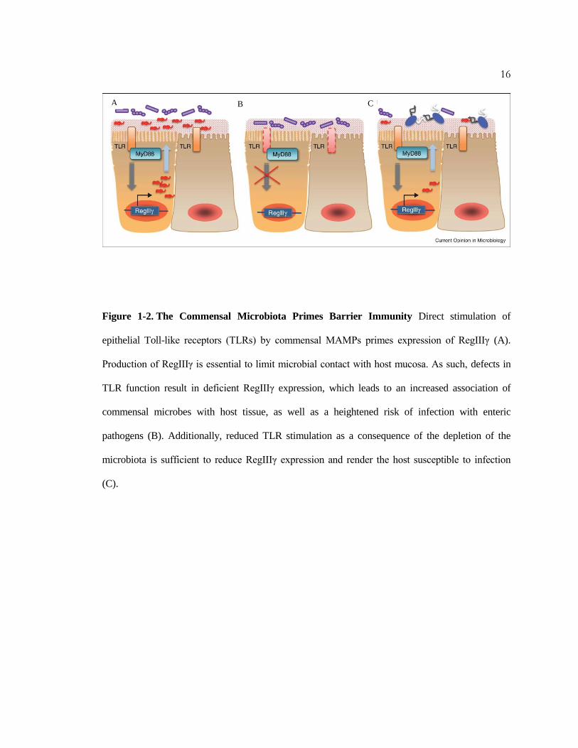
Figure 1-2. The Commensal Microbiota Primes Barrier Immunity Direct stimulation of
epithelial Toll-like receptors (TLRs) by commensal MAMPs primes expression of RegIIIγ (A).
Production of RegIIIγ is essential to limit microbial contact with host mucosa. As such, defects in
TLR function result in deficient RegIIIγ expression, which leads to an increased association of
commensal microbes with host tissue, as well as a heightened risk of infection with enteric
pathogens (B). Additionally, reduced TLR stimulation as a consequence of the depletion of the
microbiota is sufficient to reduce RegIIIγ expression and render the host susceptible to infection
(C).
A B C
17
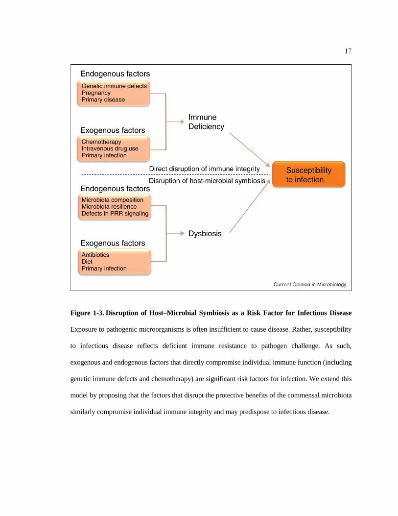
Figure 1-3. Disruption of Host–Microbial Symbiosis as a Risk Factor for Infectious Disease
Exposure to pathogenic microorganisms is often insufficient to cause disease. Rather, susceptibility
to infectious disease reflects deficient immune resistance to pathogen challenge. As such,
exogenous and endogenous factors that directly compromise individual immune function (including
genetic immune defects and chemotherapy) are significant risk factors for infection. We extend this
model by proposing that the factors that disrupt the protective benefits of the commensal microbiota
similarly compromise individual immune integrity and may predispose to infectious disease.
18
C h a p t e r 3
GUT MICROBES DRIVE STEADY-STATE HEMATOPOIESIS
The immune system begins to develop in utero, but full maturation requires both genetic and
environmental signals that further shape immunity after birth. Lymphoid and myeloid cells develop
largely from hematopoietic stem cells (HSCs) within primary tissues, where molecular cues
orchestrate immune cell differentiation from uncommitted HSCs and progenitor cells via regulation
of transcription factors and epigenetic modifications (Weissman, 1994). Additionally, certain
phagocyte populations (including Langerhans cells and microglia), derived from embryonic
precursors, are maintained independently of HSCs (Sieweke and Allen, 2013). Genetic
contributions (i.e., molecular cues encoded by the host genome) to lineage commitment pathways
that control the myeloid repertoire are well studied (Georgopoulos, 2002). However, environmental
factors that influence hematopoiesis have not been extensively defined. Based on emerging data that
the microbiota represents an integral environmental factor in shaping numerous features of the
immune system, we reasoned that gut bacteria may be controlling central immunity. We report
herein that commensal microbes promote the maintenance of both HSC and embryonic-derived
myeloid cells during steady-state conditions. The absence of commensal microbes leads to defects
in several innate immune cell populations (including neutrophils, monocytes and macrophages)
within systemic sites. By controlling the differentiation of innate immune cells, the gut microbiota
prepares the host to rapidly mount immune responses upon pathogen encounter, as germ-free and
antibiotic treated mice are impaired in clearance of systemic bacterial infection. Our study reveals
that gut microbes evolved to actively shape immunity at its core, via regulation of hematopoiesis.

19
Germ-free Animals Display Global Defects in Innate Immune Cells
The commensal gut microbiota profoundly influences cellular proportions, migration and functions
of various immune cell subsets. Recent studies have provided numerous examples illustrating how
gut bacteria modulate innate and adaptive immune responses at mucosal surfaces during infection,
inflammation and autoimmunity (Kamada et al., 2013; Round and Mazmanian, 2009). With such
pervasive effects, we reasoned that the microbiota may regulate hematopoiesis—the developmental
programming of the immune system. Initially, to determine if the microbiota has global effects on
systemic immune cell populations, we profiled myeloid cells in the spleen of colonized (SPF;
specific pathogen free) and germ-free (GF) mice. Indeed, GF animals display reduced proportions
and total numbers of F4/80hi and F4/80
lo cells compared to SPF mice (Figures 3-1A-C). F4/80
hi cells
are mainly macrophages, while F4/80lo splenocytes are a heterogeneous population of macrophages,
monocytes and neutrophils (Schulz et al., 2012). Intriguingly, all three cell subsets are reduced in
GF mice (Figure 3-2A). Furthermore, treatment of SPF mice with antibiotics also results in
diminished myeloid cell populations in the spleen (Figure 3-2B). Thus, gut bacteria dynamically
influence innate immune cell proportions at secondary immune sites in the periphery.
Myeloid cell precursors differentiate into various phagocyte lineages that are stored in the bone
marrow, and are a major source of cells that populate peripheral tissues (Geissmann et al., 2010).
The reduction of splenic macrophages, monocytes and neutrophils in GF mice suggests that defects
in host immunity may include compromised development in primary immune sites. Accordingly,
we observed a reduction of myeloid cells within the bone marrow of GF mice (Figures 3-3A-C). A

20
similar decrease was observed in the liver, a site of alternative immune cell development (Figure 3-
2C). A global defect in myeloid cell populations in primary immune sites of GF mice demonstrates
that gut bacteria shape the architecture of the immune system early in cellular development.
Commensal Microbes Enhance Myelopoiesis
We reasoned that reductions in several phagocytic cell subsets in GF mice may reflect a primary
defect in the maintenance of myeloid cell populations. To test if commensal microbes promote
myelopoiesis, we pulsed SPF and GF mice with 5-Ethynyl-2´-deoxyuridine (EdU), a thymidine
analog, to compare the percentage of dividing leukocytes. Both F4/80hi and F4/80
lo phagocytes from
GF mice showed reduced EdU incorporation compared to SPF animals (Figure 3-4A, B). F4/80hi
macrophages are largely derived from embryonic yolk sac progenitors and are maintained
independently of HSCs (Schulz et al., 2012; Sieweke and Allen, 2013). F4/80lo leukocytes,
however, are of hematopoietic origin, and reduced EdU incorporation by these cells in GF mice
indicates defects in the expansion and/or differentiation of bone marrow progenitor cells (Schulz et
al., 2012). These studies uncover a role for commensal microbes in promoting the maintenance of
both splenic yolk sac-derived and HSC-derived myeloid cells.
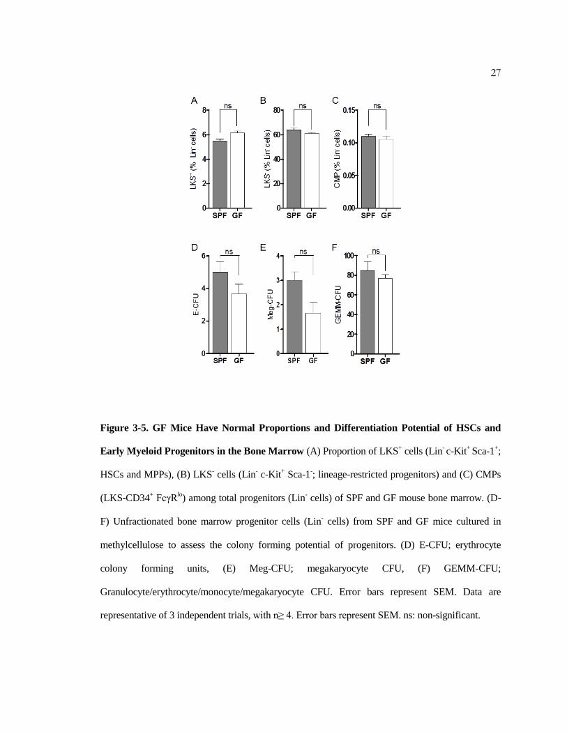
The reduction of F4/80lo cells in GF mice led us to further investigate the contribution of
commensal microbes on HSCs and myeloid progenitor cells in the bone marrow. No differences
were detected in the proportion or differentiation potential of LKS+ cells (HSCs and multipotent
progenitors; MPPs), LKS- cells (total lineage-restricted progenitors), or common myeloid

21
progenitor cells (CMPs) between SPF and GF mice (Figure 3-5A-F). Remarkably, GF mice are
significantly reduced in the proportion of bone marrow granulocyte and/or monocyte progenitors
(GMPs), identified as LKS- CD34
+ FcγR
hi cells (Figure 3-6A). GMPs consist of progenitor cells,
downstream of HSCs and CMPs during hematopoiesis, with restricted myeloid differentiation
potential (Akashi et al., 2000). To further examine the effects of gut microbiota on innate immune
cells, we tested if commensal microbes affect the differentiation potential and self-maintenance
capacity of GMPs. Methylcellulose culture of LKS- CD34
+ FcγR
hi cells from GF mice displayed
reduced granulocyte (G-CFU) and monocyte (M-CFU) colony formation compared to cells from
SPF mice (Figure 3-6B). Furthermore, LKS- CD34
+ FcγR
hi cells isolated from GF mice in primary
methylcellulose culture yielded fewer of c-Kit+ CD11b
- progenitor cells compared to SPF GMPs
(Figure 3-6C). This suggests that the ability of GMPs to maintain cells with progenitor potential is
defective in the absence of commensal microbes (Rodrigues et al., 2008). Consistent with this
notion, secondary cultures of unfractionated cells derived from GF GMPs generated fewer colonies
compared to cells isolated from SPF mice (Figure 3-6D). The commensal microbiota therefore
promotes steady-state myelopoiesis by specifically maintaining GMP proportions and enhancing
their differentiation into mature myeloid cells in the bone marrow.
Extramedullary hematopoiesis (outside the bone marrow) further contributes to the maintenance
and inflammatory responses of tissue-resident phagocytic cells (Jenkins et al., 2011; Massberg et
al., 2007; Robbins et al., 2012; Swirski et al., 2009). We therefore investigated whether commensal
microbes influence the hematopoietic potential of progenitors located in the spleen. Similar to
GMPs from the bone marrow, splenocytes isolated from GF mice displayed reduced colony
formation in methylcellulose compared to SPF mice, with significant reductions in both neutrophil
22
and monocyte production (Figures 3-7A-B). Overall, we conclude that the microbiota shapes innate
immune profiles by promoting myeloid progenitor development and differentiation in the bone
marrow and extramedullary sites, revealing that gut bacteria control immunity at its core—during
hematopoiesis.

23
Figure 3-1. GF Mice Are Deficient in Resident Myeloid Cell Populations in the Spleen (A-C)
Splenic phagocyte profile among SPF and GF mice. Representative flow cytometry plots (A), cell
proportions (B), and total cell number (C) of CD11blo
F4/80hi
and CD11bhi F4/80
lo splenic cells in
SPF and GF mice. For all panels, data are representative of at least 3 independent trials, with n≥ 4
mice / group. Each symbol represents data from a single animal. Error bars represent standard error
of mean (SEM). *p<0.05, **p<0.01.

24
Figure 3-2. GF and Antibiotic-Treated Mice Have Reduced Populations of Myeloid Cells in
Systemic Sites (A) Frequency of splenic neutrophils (CD11b+ GR1
hi Ly6c
lo), monocytes (CD11b
+
Ly6chi
GR1hi) and macrophages (CD11b
+ GR1
- F4/80
lo) among SPF and GF mice. (B) Frequency of
splenic CD11b+ F4/80
hi and CD11b
+ F4/80
lo phagocytes among untreated mice (Ctl) and SPF mice
treated with oral antibiotics (Abx). (C) Frequency of liver CD11b+ F4/80
hi macrophages recovered
from SPF or GF mice. Error bars represent SEM. Data are representative of 2-3 independent trials,
with n≥ 4. *p<0.05, **p<0.01. PMN: polymorphonuclear cells; Mono: monocytes; MФ :
macrophages.

25
Figure 3-3. GF Mice Are Deficient in Bone Marrow Myeloid Cell Populations (A-C) Bone
marrow populations of neutrophils (Gr1hi CD115
neg) and monocytes (Gr1
hi CD115
hi) among SPF
and GF mice. Representative flow cytometry plots (A), cell proportions (B) and total cell number
(C) within the bone marrow of SPF and GF mice. For all panels, data are representative of at least 3
independent trials, with n≥ 4 mice / group. Each symbol represents data from a single animal. Error
bars represent standard error of mean (SEM). *p<0.05, **p<0.01. PMN: polymorphonuclear cells;
Mono: monocytes.
26
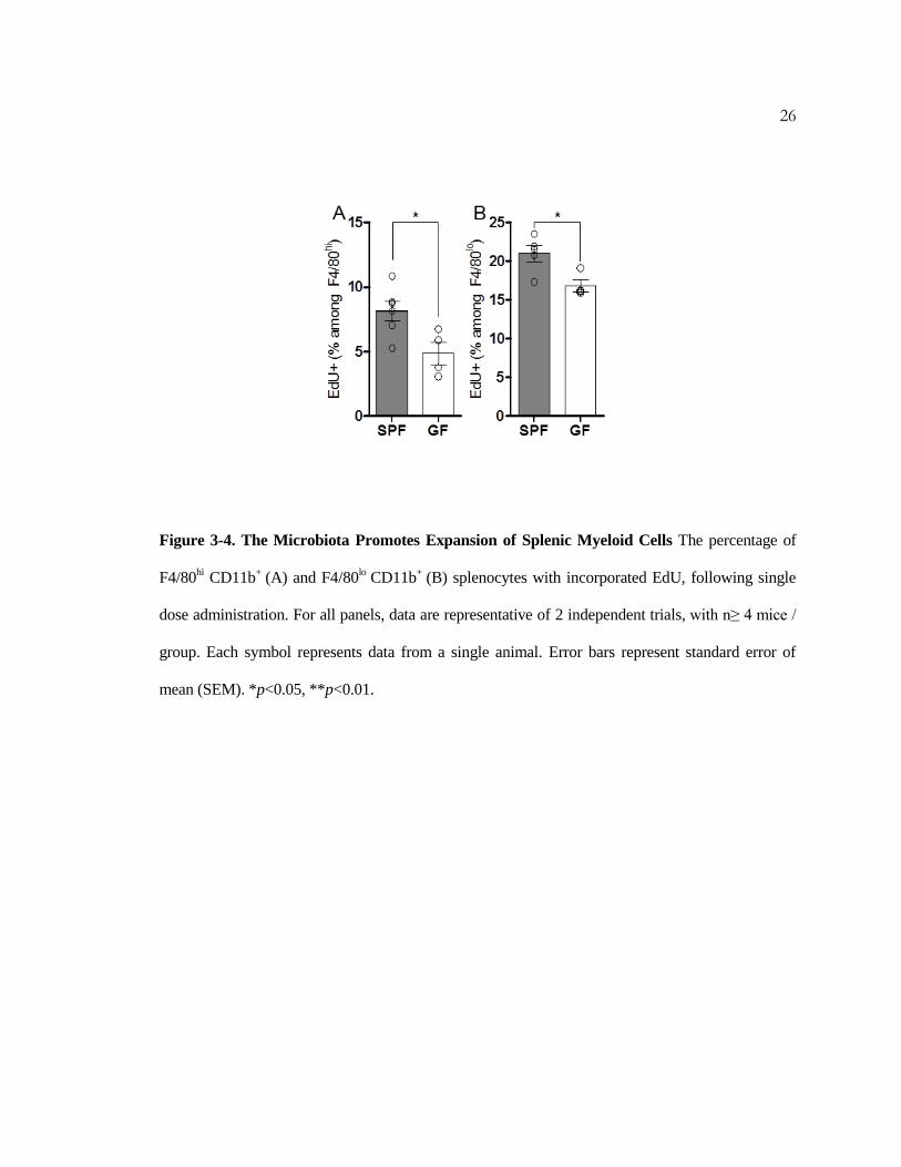
Figure 3-4. The Microbiota Promotes Expansion of Splenic Myeloid Cells The percentage of
F4/80hi CD11b
+ (A) and F4/80
lo CD11b
+ (B) splenocytes with incorporated EdU, following single
dose administration. For all panels, data are representative of 2 independent trials, with n≥ 4 mice /
group. Each symbol represents data from a single animal. Error bars represent standard error of
mean (SEM). *p<0.05, **p<0.01.
27
Figure 3-5. GF Mice Have Normal Proportions and Differentiation Potential of HSCs and
Early Myeloid Progenitors in the Bone Marrow (A) Proportion of LKS+ cells (Lin
- c-Kit
+ Sca-1
+;
HSCs and MPPs), (B) LKS- cells (Lin
- c-Kit
+ Sca-1
-; lineage-restricted progenitors) and (C) CMPs
(LKS-CD34+ FcγR
lo) among total progenitors (Lin
- cells) of SPF and GF mouse bone marrow. (D-
F) Unfractionated bone marrow progenitor cells (Lin- cells) from SPF and GF mice cultured in
methylcellulose to assess the colony forming potential of progenitors. (D) E-CFU; erythrocyte
colony forming units, (E) Meg-CFU; megakaryocyte CFU, (F) GEMM-CFU;
Granulocyte/erythrocyte/monocyte/megakaryocyte CFU. Error bars represent SEM. Data are
representative of 3 independent trials, with n≥ 4. Error bars represent SEM. ns: non-significant.

28
Figure 3-6. The Microbiota Directs Myelopoiesis (A) The frequency of LKS- CD34
+ FcγR
hi
granulocyte and/or monocyte progenitors (GMPs) among lineage negative (Lin-) progenitors from
bone marrow of SPF and GF mice, as assessed by flow cytometry. (B) Distribution of cell types
following purified LKS- CD34
+ FcγR
hi cell culture in methylcellulose medium. Colonies were
identified and counted to assess the proportion of granulocyte-monocytes (GM-CFU; black),
granulocytes (G-CFU; blue) and monocytes (M-CFU; green). (C) Total numbers of c-Kit+
CD11b-
progenitors from methylcellulose cultures of LKS- CD34
+ FcγR
hi progenitors, as assessed by flow
cytometry. (D) Cells harvested from methylcellulose cultures of LKS- CD34
+ FcγR
hi progenitors
were re-plated at equal numbers in fresh methylcellulose, and cultured to assess their colony
forming capacity. For each panel, data are representative of at least 2-3 independent trials, with n≥ 4
/ group. Each symbol represents data from a single animal. Error bars represent SEM. *p<0.05 for
all panels. **p<0.05 (comparing total CFU between SPF and GF for (B)), ***p<0.05 (comparing
G-CFU between SPF and GF for (B)), ****p<0.05 (comparing M-CFU between SPF and GF for
(B)). CFU: colony forming units.

29
Figure 3-7. Commensal Microbes Promote Extramedullary Hematopoiesis (A and B) Splenic
cells isolated from SPF and GF mice were cultured in methylcellulose to assess the colony forming
capacity of progenitors from SPF and GF mice. Total CFUs (A), and GM-CFUs, G-CFUs and M-
CFUs (B) are shown. For each panel, data are representative of at least 2-3 independent trials, with
n≥ 4 / group. Each symbol represents data from a single animal. Error bars represent SEM. *p<0.05
for all panels. **p<0.05 (comparing total CFU between SPF and GF for (B)), ***p<0.05
(comparing G-CFU between SPF and GF for (B)), ****p<0.05 (comparing M-CFU between SPF
and GF for (B)). CFU: colony forming units.

30
C h a p t e r 4
MICROBIOTA-DRIVEN HEMATOPOIESIS PROTECTS AGAINST SYSTEMIC
INFECTION
Commensal microbes have previously been shown to influence functional responses by various
phagocytic cells during bacterial and viral infection (Clarke et al., 2010; Franchi et al., 2012;
Ganal et al., 2012; Ichinohe et al., 2011). However, the role of the microbiota in promoting
hematopoiesis, and its contribution towards host health, has not been previously studied. As
revealed in Chapter 3, naïve GF animals display reductions in both proportions and total cell
numbers of tissue-resident F4/80hi and F4/80
lo phagocytes compared to SPF mice (Figures 3-1A-
C). Furthermore, treatment of SPF mice with antibiotics also results in diminished resident
phagocytic cells (Figure 3-2B). Tissue-resident cells are essential in mediating acute resistance
against pathogenic microorganisms by restricting bacterial dissemination, as well as coordinating
the recruitment of additional immune cells to the site of infection (Pamer, 2004; Sieweke and
Allen, 2013). Therefore, we investigated whether the reduced populations of these phagocytic
cells, as a consequence of absent or diminished colonization by commensal microbes, increases
susceptibility to infectious disease.
Tissue-Resident Phagocytes Mediate Protection by Commensal Microbes
We sought to test the impact of commensal microbes on myeloid cell differentiation by employing
infection models where innate immunity is vital for an effective immune response. SPF and GF
31
mice were infected intravenously (i.v.) with the model pathogen, Listeria monocytogenes. SPF mice
challenged systemically with L. monocytogenes effectively control infection, as previously
described (Figure 4-1A) (Serbina et al., 2012; Shi et al., 2011). However, GF mice rapidly succumb
at the same inoculum (Figure 4-1A). Heightened susceptibility to infection among GF mice was
associated with a significant increase in splenic and liver bacterial burden 24 and 72 hours post-
infection (hpi), demonstrating a defect in early resistance to Listeria infection (Figures 4-1B-D).
Susceptibility to infection is not restricted to L. monocytogenes, as GF mice also displayed
increased disease burden following systemic challenge with Staphylococcus aureus (Figure 4-1E).
Interestingly, SPF mice treated orally with broad-spectrum antibiotics are also impaired in
controlling Listeria, indicating that protection by commensal microbes is an active process and is
subject to loss following depletion of gut microbiota (Figure 4-1F). Collectively, these data reveal
that commensal microbes are critical for rapid and potent systemic immune responses to acute
bacterial infection.
To confirm that defects in myelopoiesis contribute to increased disease burden in GF mice,
phagocytic cells were depleted with clodronate-loaded liposomes (CL) prior to infection with L.
monocytogenes (van Rooijen et al., 1996). CL pre-treatment increased susceptibility to Listeria
infection (Figure 4-2A,B), confirming the importance of resident cells in pathogen resistance
(Aichele et al., 2003; Kastenmuller et al., 2012). Importantly, depletion of resident phagocytes
rendered both SPF and GF mice equally susceptible to infection, resulting in similar splenic disease
burden 24 hpi (Figure 4-2A), and rapid death within 48 hpi (Figure 4-2B). While functional defects
in myeloid cells may potentially contribute to increased disease in GF mice, we did not detect
differences during in vitro Listeria killing by macrophages from SPF or GF mice (Figure 4-2C).

32
Furthermore, CD11b+ myeloid cells isolated from either SPF or GF donors were equally sufficient
in providing protection when transferred into GF mice prior to infection (Figure 4-2D), suggesting
that reduced cell proportions are likely the primary defect in GF mice. These studies confirm the
importance of microbiota-driven myelopoiesis in promoting host resistance during systemic
infection.
Effective responses to L. monocytogenes requires coordination between innate and adaptive
immune cells, resulting in pathogen clearance and protective immunity (Pamer, 2004). Thus, we
investigated whether additional immune cells beyond tissue-resident phagocytes may mediate
commensal-derived protection to Listeria infection. We show that adaptive immunity is not
required for protection by the microbiota during acute infection (Figure 4-3A), nor are GF mice
deficient in developing long-term protective immunity against subsequent infection (Figure 4-3B).
Furthermore, the selective expansion of myeloid cells during acute infection (called emergency
hematopoiesis) which is necessary for mediating delayed resistance to L. monocytogenes (following
48 hpi), was maintained in GF mice (Figure 4-3C) (Serbina et al., 2009; Serbina et al., 2003).
Finally, while there are fewer inflammatory neutrophils and monocytes recruited to the spleen
following infection (Figure 4-3D), a possible consequence of increased apoptosis (Figure 4-3E),
these cells were not required for commensal-mediated protection against L. monocytogenes (Figure
4-3F, G). Together, these findings demonstrate that hematopoietic defects in tissue-resident myeloid
cells prior to infection of GF mice (i.e., during steady-state hematopoiesis) is the primary cause of
impaired control of Listeria.

33
Commensal Bacterial Signals Mediate Maintenance of Myelopoiesis
The molecular mechanism(s) by which commensal microbes promote steady-state expansion of
bone marrow- and yolk sac-derived myeloid cells remains unknown. Microbial associated
molecular patterns (MAMPs) and microbial metabolites, such as short chain fatty acids (SCFAs),
have been shown to modulate various aspects of the host immune response (Chu and Mazmanian,
2013; Clarke et al., 2010; Smith et al., 2013). Furthermore, MyD88 (an adaptor for recognition of
many MAMPs) was recently shown to promote GMP expansion and differentiation (Fiedler et al.,
2013). Accordingly, we sought to address whether commensal-derived factors are involved in the
maintenance of myeloid cells under naïve conditions. Recolonization of GF mice with a complex
microbiota and oral treatment with MAMPs, but not SCFAs, was sufficient to promote recovery of
GMP-derived myeloid cells (neutrophils and monocytes) within the bone marrow (Figure 4-4A, B).
Importantly, only recolonization of GF mice with an SPF microbiota was sufficient to restore
splenic populations of F4/80hi macrophages and F4/80
lo splenocytes (i.e., neutrophils, monocytes
and macrophages) (Figure 4-4C and data not shown). Therefore, while MAMP treatment is
necessary for the maintenance of bone marrow-derived myeloid cells, colonization with a live and
complex microbiota is required to promote complete myelopoiesis (including yolk sac-derived
macrophages). Finally, only recolonization of GF animals, and not oral MAMP treatment, was
sufficient to restore the defect in GF mice to systemic challenge with L. monocytogenes (Figure 4-
4D and data not shown). Collectively, these studies reveal that the microbiota provides complex
molecular signals that actively promote the hematopoietic differentiation of myeloid cells, resulting
in peripheral phagocyte populations that function as sentinels for the early detection and control of
systemic bacterial infection.

34
Figure 4-1. The Microbiota Promotes Early Resistance to Systemic Infection (A-C) SPF and
GF mice were infected with L. monocytogenes and assessed for survival (A) and splenic bacterial
burden at 24 (B) and 72 (C) hours post- infection (hpi). (D) Liver bacterial burden among SPF and
GF mice, 72 hpi. (E) SPF and GF mice infected with S. aureus. Kidney bacterial burden assessed 5
days post-infection . (F) SPF mice treated with antibiotics (Abx) and untreated controls (Ctl) were
infected with L. monocytogenes and splenic bacterial burden was measured 72 hpi. For all panels,
data are representative of at least 2-3 independent trials, with n≥ 4 / group. Each symbol represents
data from a single animal. Error bars represent SEM. *p<0.05, **p<0.01, *** p<0.05 log-rank test
used for survival curves in (A).

35
Figure 4-2. The Microbiota Promotes Resistance to Infection via Tissue-Resident Cells (A-C)
SPF and GF mice depleted of tissue-resident cells prior to infection with L. monocytogenes and
assessed for splenic bacterial burden 24 hpi (A) and survival (B). (C) Peritoneal macrophages
isolated from SPF or GF mice, untreated or stimulated with interferon-γ (IFNγ), infected with L.
monocytogenes. Recovery of intracellular bacteria measured over time. Data is non-significant for
all time points measured, except where indicated (untreated SPF vs. GF, 4 hpi). (D) Splenic
bacterial burden, 24 hpi, following transfer of splenic CD11b+ cells from SPF or GF donors . For all
panels, data are representative of at least 2-3 independent trials, with n≥ 4 / group. Each symbol
represents data from a single animal. Error bars represent SEM. *p<0.05, **p<0.01. CL:
clodronate-loaded liposomes.

36
Figure 4-3. Resident Phagocytes Mediated Commensal-Enhanced Protection Against
Infectious Disease (A) SPF and GF Rag-/-
mice infected with L. monocytogenes, splenic bacterial
burden assessed 72 hpi. (B) SPF and GF mice were immunized with L. monocytogenes ΔactA. 45
days after immunization, SPF and GF mice, as well as naïve, non-immunized SPF controls, were
infected with wild-type (WT) L. monocytogenes. Splenic bacteria burden of the WT strain was
measured at 72 hpi. Note: two of the four naïve, non-immunized SPF mice died following infection,
prior to the 72 hour time point (data not shown). (C) BrdU incorporation among bone marrow
neutrophils (CD11b+
GR1hi
) and monocytes (CD11b+ CD115
+), 72 hpi. (D) Percentage of splenic
neutrophils (Gr1hi Ly6C
lo) and monocytes (Gr1
hi Ly6C
hi) among SPF and GF mice, 72 hpi. (E)
Annexin V+ bone marrow monocytes, 72 hpi. (F) SPF and GF mice infected with L.

37
monocytogenes, following neutrophil depletion. Splenic bacterial burden assessed at 72 hpi. (G)
Splenic bacterial burden of SPF and GF mice, reconstituted with bone marrow from WT or CCR2-/-
mice, 72 hpi. SPF mice reconstituted with CCR2-/-
bone marrow display a two-fold reduction in
splenic CFUs compared to GF CCR2-/-
mice. For all panels, data are representative of 2-3
independent trials, with n≥ 4/ group. Each symbol represents data from a single animal. Error bars
represent SEM. *p<0.05, **p<0.01. PMN: polymorphonuclear cells; Mono: monocytes.

38
Figure 4-4. Recolonization of GF Mice Restores Immune Integrity Against Systemic
Listeriosis (A) Neutrophil (GR1hi
CD115-) and (B) monocyte (GR1
hi CD115
+) bone marrow
profiles from SPF, GF, recolonized GF mice and MAMP or SCFA-treated GF mice. (C) F4/80hi
splenic macrophage profile among SPF, GF, recolonized GF mice and GF mice treated with
MAMPs or SCFAs. (D) Splenic bacterial burden 72 hpi among SPF, GF and recolonized GF mice
infected with L. monocytogenes. For all panels, data are representative of at least 2 independent
trials, with n≥4 / group. Each symbol represents data from a single animal. Error bars represent
standard error of mean (SEM). *p<0.05, **p<0.01. Recol: recolonized; MAMPs: molecular
associated molecular patterns; SCFAs: short chain fatty acids.

39
C h a p t e r 5
FINDINGS AND DISCUSSION
Advances in understanding host-microbial symbiosis have revealed that the gut microbiota control
the phenotype, migration and activity of multiple innate and adaptive immune cells (Belkaid and
Naik, 2013; Chu and Mazmanian, 2013). Disruption or alteration of commensal communities
impacts host susceptibility to various disorders, particularly at sites of microbial colonization such
as the intestines, respiratory mucosa and skin epithelium (Kamada et al., 2013). In addition to
modulating functional immune outcomes, the microbiota is necessary for maintaining circulating
populations of neutrophils and CD4+ T cells in the spleen (Bugl et al., 2013; Mazmanian et al.,
2005), suggesting a possible contribution by gut microbiota to the development of the immune
system. Herein, we reveal that gut bacteria regulate hematopoiesis within primary immune sites,
providing a unifying explanation for previous observations of the widespread effects by the
microbiota on the immune system.
Our study uncovers that the microbiota promotes steady-state myeloid cell development by driving
the expansion of yolk sac-derived macrophages, as well as enhancing the numbers and
differentiation potential of GMPs in the bone marrow. Furthermore, as a consequence of the
reduced populations of resident phagocytes, which serve as a first line defense against invading
pathogens, GF mice are more susceptible to systemic infection with L. monocytogenes. Interesting,
despite multiple immune abnormalities having been previously described in GF mice, the increased
susceptibility to systemic infection with L. monocytogenes appears to be specific to the reduced
40
proportions of resident myeloid cells. Previous studies have shown that commensal microbes prime
neutrophil killing of Streptococcus pneumoniae and Staphylococcus aureus (Clarke et al., 2010).
Further, the microbiota enhances host resistance to viral infection by promoting expression of type-
1 interferon by splenic phagocytes (Abt et al., 2012). However, we were not able to detect defects in
the functional activity of phagocytes isolated from GF mice, as related to protection against L.
monocytogenes. Peritoneal macrophages isolated from naïve SPF and GF mice displayed equivalent
killing of Listeria, ex vivo. Additionally, SPF and GF phagocytes expressed similar levels of TNFα
and NO following infection, which is essential for limiting Listeria dissemination (data not shown).
Finally, splenic phagocytes isolated from SPF and GF donors were equally sufficient to provide
protection against infection when transferred into GF recipients. These data suggests that a primary
defect in the maintenance of resident phagocytes in GF animals is responsible for the increased
susceptibility to systemic infection. However, it remains possible that the microbiota primes
additional immune responses, not described here, that further contributes to mediating host
protection against infectious disease.
While our studies reveal steady-state hematopoiesis is compromised in GF mice, emergency
hematopoiesis, or the selective expansion of myeloid cell following infection, is maintained
independent of commensal microbes. One possible explanation for this contrast is that the
expression of cytokines and growth factors following infection, as well as direct stimulation by
microbial ligands, may rescue hematopoietic defects in GF mice otherwise present under steady-
state/non-inflammatory conditions. We propose a model whereby a primary defect in hematopoiesis
in GF or antibiotic-treated mice compromises multiple tissue-resident innate immune cell
populations prior to infection, leading to blunted early responses upon subsequent pathogen
encounter (see diagram in Figure 5-1). While tissue-resident phagocytes directly mediate early

41
resistance to infection, these cells are also essential for recruiting additional phagocytes
(monocytes) which is essential for maintaining resistance (Coombes et al., 2012; Kastenmuller et
al., 2012). GF mice therefor display exacerbated disease severity as a consequence of diminished
phagocyte recruitment into infected tissues. While our studies focus on innate immunity due to its
role in rapid control of early Listeria infection, impaired microbiota-mediated hematopoiesis may
also extend to the adaptive immune system, providing an explanation for observations that
peripheral T, B and iNKT cell populations are altered in GF mice (Ivanov et al., 2008; Macpherson
and Uhr, 2004; Mazmanian et al., 2005; Olszak et al., 2012).
How commensal microbes (presumably in the gut) are able to control immune responses in distant
sites such as the bone marrow remains incompletely understood. It has recently been shown that
mice deficient in MyD88 signaling display reductions in systemic myeloid cell populations and
GMP numbers (Fiedler et al., 2013; Yanez et al., 2013), similar to our findings in GF mice. Further,
as microbial ligands are detected in systemic sites, including the bone marrow (Clarke et al., 2010),
commensal-derived MAMPs that originate in the gut may mediate steady-state myelopoiesis in
primary immune sites. Accordingly, we show that oral treatment with MAMPs is sufficient to
rescue GMP-mediated expansion of neutrophils and monocytes in GF mice. However, MAMP
treatment alone is inadequate to expand splenic F4/80hi and F4/80
lo cells, indicating that additional
commensal-derived signals are necessary to influence site-specific HSC- and yolk sac-derived
myeloid cells. Interestingly, recolonization of adult GF mice with SPF microbiota is insufficient to
restore splenic F4/80hi macrophages to the levels found in SPF mice. This may suggest that
complete rescue requires either colonization from birth or colonization with specific microbes that
were not transferred into GF mice. In addition to microbial ligands or metabolites translocating
42
from the gut into the circulation to directly stimulate progenitor cells, other explanations for how the
microbiota affects hematopoiesis may include a role for myeloid cell growth factors. In support of
this notion, preliminary data suggest that GF mice are reduced in M-CSF transcript levels in the gut
(data not shown), though further work is need to uncover the complex molecular mechanism(s) by
which commensal bacteria signal from the gut to distant primary immune organs.
Finally, we speculate that these findings may be relevant to human infections. Evidence that
depletion of the microbiota leads to transient immune suppression suggests factors that disrupt
commensal microbes, including that clinical antibiotic use may, paradoxically, be a risk factor for
susceptibility to opportunistic pathogens. Furthermore, the spread of antibiotic-resistance among
pathogens, paired with a dwindling supply of effective antibiotics, has necessitated alternative
strategies to combat infections (Khosravi and Mazmanian, 2013). As certain commensal microbes
have been previously shown to express molecules with unique immunomodulatory properties, it is
possible such microbial products may be developed into therapeutics to treat infectious diseases.
Whereas traditional antibiotics work by through direct microbicidal activity, indiscriminately killing
both pathogenic and commensal microbes, immunomodulatory therapeutics would enhance host
immune responses to promote pathogen clearance. Such a strategy may specifically target
pathogenic microbes and thereby reduce the risk of secondary inflammatory disease caused by
depleting commensal microbial communities, as currently occurs with antibiotic use. The concepts
proposed herein, if validated in humans, may herald future medical approaches that combine
antibiotics with immunomodulatory microbial molecules as revolutionary combination treatments
to address the reemerging crisis of infectious diseases.

43
Figure 5-1. A Proposed Model For How the Microbiota Mediates Host Resistance to Systemic
Infection Commensal microbes stimulate bone marrow and splenic myelopoiesis during naïve
conditions (in the absence of infection), expanding systemic pools of mature myeloid cells in SPF
mice that are essential for restricting pathogen dissemination upon acute infection. GF mice have
reduced proportions and differentiation potential by GMPs during the steady-state, as well as
diminished expansion of yolk sac-derived macrophages, impairing the immune response to
infection with L. monocytogenes. This model suggests that conditions under which the microbiota is
disrupted may result in deficient expansion of myeloid cells, compromising host resistance to
infectious disease.
44
M a t e r i a l a n d M e t h o d s
Animal Studies
Specific pathogen-free (SPF) C57BL/6 mice were purchased from Taconic Farms. Germ-free (GF)
C57BL/6 and C57BL/6 Rag-/-
mice were bred and raised in sterile gnotobiotic flexible film isolators
at the California Institute of Technology. Mice at 8-12 weeks of age were infected via retro-orbital
injection with 3x10⁴ colony forming units (CFU) of Listeria monocytogenes 10403S. Splenic and
liver bacterial CFU were assessed 24-72 hpi by microbiological plating. In some experiments, SPF
and GF mice were immunized with 3x10⁴ CFU L. monocytogenes ΔactA (Lara-Tejero and Pamer,
2004), and immunized mice and non-immunized controls were infected with 2x10⁵ CFU of wild-
type (WT) L. monocytogenes 45-day post immunization, with splenic bacterial burden measured 72
hpi. Alternatively, SPF and GF mice were infected with 1x10⁷ CFU of S. aureus (strain Newman)
via tail vein injection and kidney bacterial burden assessed 5 days post-infection. For microbiota
depletion studies, SPF mice were treated with 1 mg/ml of ampicillin (Auromedics), neomycin
sulfate (Fisher), streptomycin (Sigma) and 0.5 mg/ml of vancomycin (Sagent) in the drinking water
for 4-5 weeks. Mice were taken off antibiotics 4 days prior to infection. Antibiotic-treated and
untreated SPF mice were infected with 3x10⁴ CFU of L. monocytogenes, and splenic bacterial
burden was assessed 72 hpi. GF mice were recolonized by gavage with cecal contents of SPF mice.
Alternatively, GF mice were treated with MAMPs through the addition of heat killed Escherichia
coli strain Nissle (Lodinova-Zadnikova and Sonnenborn, 1997) or autoclaved cecal contents from
SPF mice in water (~1x10⁹ CFU/ml in drinking water). For treatment with short chain fatty acids,
sodium proprionate (Sigma), sodium butyrate (Sigma), and sodium acetate (Sigma) was added to
drinking water at previously described concentrations (25mM, 40mM and 67.5mM, respectively)

45
(Smith et al., 2013). Mice were recolonized or treated with microbial ligands or metabolites for 4
weeks prior to cellular analysis and infectious studies. Animals were cared for under established
protocols and IACUC guidelines from the California Institute of Technology.
Cellular Analysis
Spleens were either mechanically disrupted via passage through 100 µm mesh filters (BD
Biosciences) or digested in 0.5 mg/ml of Collagenase D (Roche) and 0.5 mg/ml of DNase I
(Worthington). Bone marrow was collected by flushing femurs with PBS containing 0.5% BSA and
5mM EDTA. Single cell suspensions were removed of red blood cells (RBC lysis buffer, Sigma).
Mature myeloid cells were evaluated by staining with antibodies to GR1 (RB6-8C5), Ly6C (HK
1.4), CD11b (M1/70), CD115 (AFS98) and F4/80 (BM8). Mouse hematopoietic stem and
progenitor cells (HSPCs) were isolated from bone marrow by a combination of MACS magnetic
bead purification (Miltenyi) and fluorescence activated cell sorting (FACS). Lineage marker-
negative cells (Lin-) were first separated using a MACS lineage cell depletion kit (containing
antibodies against CD5 (53-7.3), CD45R (B220; RA3-6B2), CD11b, Gr-1, 7-4 (15BS) and Ter-119
(Ter-119)) and an autoMACS Separator (Miltentyi). Lin- cells were then further stained with c-Kit
(CD117; 3C1), Sca-1 (D7), CD16/CD32 (93), CD34 (RAM34). Populations of LKS+ cells (Lin
- c-
Kit+ Sca-1
+; HSCs and MPPs), Lin
- c-Kit
+ Sca-1
- (LKS
-) CD34
+ FcγR
lo cells (CMPs) and LKS
-
CD34+ FcγR
hi cells (GMPs) were analyzed by flow cytometry. LKS
- CD34
+ FcγR
hi cells were
FACS sorted using an Aria cell sorter (BD Biosciences). Steady-state cell proliferation was
measured by intraperitoneal (i.p.) injection of 500 µg EdU (Life Technologies) and EdU
incorporation among splenic myeloid cells was measured 24 hours later via Click-it EdU assay kit
46
(Life Technologies). To measure cell proliferation during Listeria infection, mice were injected i.p.
with 100 µg BrdU (Sigma), and BrdU incorporation among progenitor and mature myeloid cells
was determined 3 hours later via a BrdU detection kit (eBioscience). Apoptosis and cell viability
was assessed by staining with Annexin V (eBioscience) and 7-Aminoactinomycin-D (Invitrogen).
Listeria-killing assays were conducted as previously described (Portnoy et al., 1989). Briefly,
peritoneal macrophages were collected from naïve SPF and GF mice. Adherent cells were
stimulated with 100 U/ml of interferon gamma (IFNγ) (PeproTech) or left untreated for 24 hours.
Macrophages were washed and infected with L. monocytogenes at a multiplicity of infection (MOI)
of 10. Cells were washed 30 minutes later and fresh media with 5 μg/ml of Gentamycin (Phoenix)
was added. Cells were washed and lysed at various time points to quantitate intracellular Listeria
via microbiological plating. Antibodies were purchased from eBioscience, BD Bioscience, Miltenyi
or Biolegend. Data were collected on a FACSCalibur or LSR Fortessa (BD Bioscience) and
analyzed with FlowJo software (TreeStar).
Cell Depletion and Adoptive Transfer
Resident phagocytes were depleted by intravenous (i.v.) treatment with 100 μl of clodronate-loaded
liposomes (CL; FormuMax) 48 hours prior to infection. CD11b+ splenocytes were isolated from
naïve SPF and GF mice using CD11b microbeads (Miltenyi). 2x10⁶ CD11b
+ cells (>90% purity)
were transferred into GF recipients 24 hours prior to infection with L. monocytogenes. CFU burden
were assessed 24 hpi. CCR2-/-
chimeras were generated by transferring bone marrow from WT or
CCR2-/-
donors into SPF or GF recipients that had been lethally irradiated (1000 rads) 48 hours
prior. Mice were infected with 3x10⁴ CFU of L. monocytogenes 8 weeks post reconstitution, and

47
splenic bacterial burden was assessed 72 hpi. For neutrophil depletion, SPF and GF mice were
injected i.p. with 0.5 mg of anti-Ly6G antibody (Bioxpress), or saline control, 24 hours prior to
infection with L. monocytogenes.
CFU Assays
To evaluate hematopoietic potential, 1×10³ Lin
- or 1x10
3 LKS
- CD34
+ FcγR
hi cells or 2x10
5
splenocytes were plated in triplicate in MethoCult GF M3434 (StemCell Technologies)
methylcellulose-based medium and incubated for 7 days in 37oC with 5% CO2, after which the
colonies were counted on the basis of their morphological characteristics in accordance with the
manufacturer’s instructions. On the same day, cells were harvested, counted and stained for c-Kit
and CD11b expression for progenitor quantification by flow cytometry. For re-plating assays, 5x10⁴
cells from the first culture were plated in triplicate in a secondary culture of fresh MethoCult GF
M3434, and colonies were counted after 7 days of incubation.

48
B i b l i o g r a p h y
The work presented in Chapters 1-5 is collected from published articles (Khosravi and Mazmanian,
2013) and (Khosravi et al., 2014).
Abt, Michael C., Osborne, Lisa C., Monticelli, Laurel A., Doering, Travis A., Alenghat, T.,
Sonnenberg, Gregory F., Paley, Michael A., Antenus, M., Williams, Katie L., Erikson, J., et al.
(2012). Commensal Bacteria Calibrate the Activation Threshold of Innate Antiviral Immunity.
Immunity 37, 158-170.
Aichele, P., Zinke, J., Grode, L., Schwendener, R.A., Kaufmann, S.H., and Seiler, P. (2003).
Macrophages of the splenic marginal zone are essential for trapping of blood-borne particulate
antigen but dispensable for induction of specific T cell responses. J Immunol 171, 1148-1155.
Akashi, K., Traver, D., Miyamoto, T., and Weissman, I.L. (2000). A clonogenic common
myeloid progenitor that gives rise to all myeloid lineages. Nature 404, 193-197.
Alanis, A.J. (2005). Resistance to Antibiotics: Are We in the Post-Antibiotic Era? Archives of
Medical Research 36, 697-705.
Bäckhed, F., Manchester, J.K., Semenkovich, C.F., and Gordon, J.I. (2007). Mechanisms
underlying the resistance to diet-induced obesity in germ-free mice. Proceedings of the
National Academy of Sciences 104, 979-984.
Bartlett, J.G. (2002). Clinical practice. Antibiotic-associated diarrhea. The New England journal
of medicine 346, 334-339.

49
Belkaid, Y., and Naik, S. (2013). Compartmentalized and systemic control of tissue immunity
by commensals. Nat Immunol 14, 646-653.
Brandl, K., Plitas, G., Mihu, C.N., Ubeda, C., Jia, T., Fleisher, M., Schnabl, B., DeMatteo, R.P.,
and Pamer, E.G. (2008). Vancomycin-resistant enterococci exploit antibiotic-induced innate
immune deficits. Nature 455, 804-807.
Brandt, L.J., Aroniadis, O.C., Mellow, M., Kanatzar, A., Kelly, C., Park, T., Stollman, N.,
Rohlke, F., and Surawicz, C. (2012). Long-term follow-up of colonoscopic fecal microbiota
transplant for recurrent Clostridium difficile infection. The American journal of
gastroenterology 107, 1079-1087.
Bugl, S., Wirths, S., Radsak, M.P., Schild, H., Stein, P., André, M.C., Müller, M.R., Malenke, E.,
Wiesner, T., Märklin, M., et al. (2013). Steady-state neutrophil homeostasis is dependent on
TLR4/TRIF signaling. Blood 121, 723-733.
Cash, H.L., Whitham, C.V., Behrendt, C.L., and Hooper, L.V. (2006). Symbiotic Bacteria
Direct Expression of an Intestinal Bactericidal Lectin. Science 313, 1126-1130.
Chu, H., and Mazmanian, S.K. (2013). Innate immune recognition of the microbiota promotes
host-microbial symbiosis. Nat Immunol 14, 668-675.
Clarke, T.B., Davis, K.M., Lysenko, E.S., Zhou, A.Y., Yu, Y., and Weiser, J.N. (2010).
Recognition of peptidoglycan from the microbiota by Nod1 enhances systemic innate
immunity. Nat Med 16, 228-231.
Coombes, Janine L., Han, S.-J., van Rooijen, N., Raulet, David H., and Robey, Ellen A. (2012).
Infection-Induced Regulation of Natural Killer Cells by Macrophages and Collagen at the
Lymph Node Subcapsular Sinus. Cell Reports 2, 124-135.
50
Dethlefsen, L., and Relman, D.A. (2011). Incomplete recovery and individualized responses of
the human distal gut microbiota to repeated antibiotic perturbation. Proc Natl Acad Sci U S A
108 Suppl 1, 4554-4561.
Eaves-Pyles, T., Allen, C.A., Taormina, J., Swidsinski, A., Tutt, C.B., Jezek, G.E., Islas-Islas,
M., and Torres, A.G. (2008). Escherichia coli isolated from a Crohn's disease patient adheres,
invades, and induces inflammatory responses in polarized intestinal epithelial cells.
International journal of medical microbiology : IJMM 298, 397-409.
Fiedler, K., Kokai, E., Bresch, S., and Brunner, C. (2013). MyD88 is involved in myeloid as
well as lymphoid hematopoiesis independent of the presence of a pathogen. American journal
of blood research 3, 124-140.
Franchi, L., Kamada, N., Nakamura, Y., Burberry, A., Kuffa, P., Suzuki, S., Shaw, M.H., Kim,
Y.G., and Nunez, G. (2012). NLRC4-driven production of IL-1beta discriminates between
pathogenic and commensal bacteria and promotes host intestinal defense. Nat Immunol 13,
449-456.
Ganal, Stephanie C., Sanos, Stephanie L., Kallfass, C., Oberle, K., Johner, C., Kirschning, C.,
Lienenklaus, S., Weiss, S., Staeheli, P., Aichele, P., et al. (2012). Priming of Natural Killer Cells
by Nonmucosal Mononuclear Phagocytes Requires Instructive Signals from Commensal
Microbiota. Immunity 37, 171-186.
Geissmann, F., Manz, M.G., Jung, S., Sieweke, M.H., Merad, M., and Ley, K. (2010).
Development of Monocytes, Macrophages, and Dendritic Cells. Science 327, 656-661.
Georgopoulos, K. (2002). Haematopoietic cell-fate decisions, chromatin regulation and ikaros.
Nat Rev Immunol 2, 162-174.
51
Hill, D.A., Siracusa, M.C., Abt, M.C., Kim, B.S., Kobuley, D., Kubo, M., Kambayashi, T.,
LaRosa, D.F., Renner, E.D., Orange, J.S., et al. (2012). Commensal bacteria-derived signals
regulate basophil hematopoiesis and allergic inflammation. Nat Med 18, 538-546.
Hooper, L.V., and Macpherson, A.J. (2010). Immune adaptations that maintain homeostasis
with the intestinal microbiota. Nat Rev Immunol 10, 159-169.
Ichinohe, T., Pang, I.K., Kumamoto, Y., Peaper, D.R., Ho, J.H., Murray, T.S., and Iwasaki, A.
(2011). Microbiota regulates immune defense against respiratory tract influenza A virus
infection. Proc Natl Acad Sci U S A 108, 5354-5359.
Ivanov, I.I., Atarashi, K., Manel, N., Brodie, E.L., Shima, T., Karaoz, U., Wei, D., Goldfarb,
K.C., Santee, C.A., Lynch, S.V., et al. (2009). Induction of Intestinal Th17 Cells by Segmented
Filamentous Bacteria. Cell 139, 485-498.
Ivanov, I.I., Frutos, R.d.L., Manel, N., Yoshinaga, K., Rifkin, D.B., Sartor, R.B., Finlay, B.B.,
and Littman, D.R. (2008). Specific Microbiota Direct the Differentiation of IL-17-Producing
T-Helper Cells in the Mucosa of the Small Intestine. Cell Host & Microbe 4, 337-349.
Jakobsson, H.E., Jernberg, C., Andersson, A.F., Sjolund-Karlsson, M., Jansson, J.K., and
Engstrand, L. (2010). Short-term antibiotic treatment has differing long-term impacts on the
human throat and gut microbiome. PLoS One 5, e9836.
Jenkins, S.J., Ruckerl, D., Cook, P.C., Jones, L.H., Finkelman, F.D., van Rooijen, N.,
MacDonald, A.S., and Allen, J.E. (2011). Local macrophage proliferation, rather than
recruitment from the blood, is a signature of TH2 inflammation. Science 332, 1284-1288.
Kahrstrom, C.T. (2013). Entering a post-antibiotic era? Nat Rev Micro 11, 146-146.

52
Kamada, N., Kim, Y.G., Sham, H.P., Vallance, B.A., Puente, J.L., Martens, E.C., and Nunez,
G. (2012). Regulated virulence controls the ability of a pathogen to compete with the gut
microbiota. Science 336, 1325-1329.
Kamada, N., Seo, S.-U., Chen, G.Y., and Nunez, G. (2013). Role of the gut microbiota in
immunity and inflammatory disease. Nat Rev Immunol 13, 321-335.
Kastenmuller, W., Torabi-Parizi, P., Subramanian, N., Lammermann, T., and Germain, R.N.
(2012). A spatially-organized multicellular innate immune response in lymph nodes limits
systemic pathogen spread. Cell 150, 1235-1248.
Kau, A.L., Ahern, P.P., Griffin, N.W., Goodman, A.L., and Gordon, J.I. (2011). Human
nutrition, the gut microbiome and the immune system. Nature 474, 327-336.
Khosravi, A., and Mazmanian, S.K. (2013). Disruption of the gut microbiome as a risk factor
for microbial infections. Current Opinion in Microbiology 16, 221-227.
Khosravi, A., Yanez, A., Price, J.G., Chow, A., Merad, M., Goodridge, H.S., and Mazmanian,
S.K. (2014). Gut microbiota promote hematopoiesis to control bacterial infection. Cell Host
Microbe 15, 374-381.
Kobayashi, K.S., Chamaillard, M., Ogura, Y., Henegariu, O., Inohara, N., Nuñez, G., and
Flavell, R.A. (2005). Nod2-Dependent Regulation of Innate and Adaptive Immunity in the
Intestinal Tract. Science 307, 731-734.
Lee, Y.K., Menezes, J.S., Umesaki, Y., and Mazmanian, S.K. (2011). Proinflammatory T-cell
responses to gut microbiota promote experimental autoimmune encephalomyelitis. Proc Natl
Acad Sci U S A 108 Suppl 1, 4615-4622.

53
Lodinova-Zadnikova, R., and Sonnenborn, U. (1997). Effect of preventive administration of a
nonpathogenic Escherichia coli strain on the colonization of the intestine with microbial
pathogens in newborn infants. Biology of the neonate 71, 224-232.
Loo, V.G., Poirier, L., Miller, M.A., Oughton, M., Libman, M.D., Michaud, S., Bourgault,
A.M., Nguyen, T., Frenette, C., Kelly, M., et al. (2005). A predominantly clonal multi-
institutional outbreak of Clostridium difficile-associated diarrhea with high morbidity and
mortality. The New England journal of medicine 353, 2442-2449.
Lupp, C., Robertson, M.L., Wickham, M.E., Sekirov, I., Champion, O.L., Gaynor, E.C., and
Finlay, B.B. (2007). Host-Mediated Inflammation Disrupts the Intestinal Microbiota and
Promotes the Overgrowth of Enterobacteriaceae. Cell Host & Microbe 2, 119-129.
Macpherson, A.J., and Uhr, T. (2004). Induction of Protective IgA by Intestinal Dendritic
Cells Carrying Commensal Bacteria. Science 303, 1662-1665.
Maeda, S., Hsu, L.-C., Liu, H., Bankston, L.A., Iimura, M., Kagnoff, M.F., Eckmann, L., and
Karin, M. (2005). Nod2 Mutation in Crohn's Disease Potentiates NF-κB Activity and IL-1ß
Processing. Science 307, 734-738.
Massberg, S., Schaerli, P., Knezevic-Maramica, I., Kollnberger, M., Tubo, N., Moseman, E.A.,
Huff, I.V., Junt, T., Wagers, A.J., Mazo, I.B., et al. (2007). Immunosurveillance by
hematopoietic progenitor cells trafficking through blood, lymph, and peripheral tissues. Cell
131, 994-1008.
Mazmanian, S.K., Liu, C.H., Tzianabos, A.O., and Kasper, D.L. (2005). An
immunomodulatory molecule of symbiotic bacteria directs maturation of the host immune
system. Cell 122, 107-118.

54
Mazmanian, S.K., Round, J.L., and Kasper, D.L. (2008). A microbial symbiosis factor prevents
intestinal inflammatory disease. Nature 453, 620-625.
Moreau, M.C., Ducluzeau, R., Guy-Grand, D., and Muller, M.C. (1978). Increase in the
population of duodenal immunoglobulin A plasmocytes in axenic mice associated with
different living or dead bacterial strains of intestinal origin. Infect Immun 21, 532-539.
Naik, S., Bouladoux, N., Wilhelm, C., Molloy, M.J., Salcedo, R., Kastenmuller, W., Deming, C.,
Quinones, M., Koo, L., Conlan, S., et al. (2012). Compartmentalized control of skin immunity
by resident commensals. Science 337, 1115-1119.
Ng, K.M., Ferreyra, J.A., Higginbottom, S.K., Lynch, J.B., Kashyap, P.C., Gopinath, S., Naidu,
N., Choudhury, B., Weimer, B.C., Monack, D.M., et al. (2013). Microbiota-liberated host sugars
facilitate post-antibiotic expansion of enteric pathogens. Nature advance online publication.
Olszak, T., An, D., Zeissig, S., Vera, M.P., Richter, J., Franke, A., Glickman, J.N., Siebert, R.,
Baron, R.M., Kasper, D.L., et al. (2012). Microbial Exposure During Early Life Has Persistent
Effects on Natural Killer T Cell Function. Science 336, 489-493.
Pamer, E.G. (2004). Immune responses to Listeria monocytogenes. Nat Rev Immunol 4, 812-
823.
Petersson, J., Schreiber, O., Hansson, G.C., Gendler, S.J., Velcich, A., Lundberg, J.O., Roos, S.,
Holm, L., and Phillipson, M. (2011). Importance and regulation of the colonic mucus barrier in
a mouse model of colitis. American journal of physiology Gastrointestinal and liver physiology
300, G327-333.
Portnoy, D.A., Schreiber, R.D., Connelly, P., and Tilney, L.G. (1989). Gamma interferon limits
access of Listeria monocytogenes to the macrophage cytoplasm. The Journal of Experimental
Medicine 170, 2141-2146.
55
Rathinam, V.A.K., Vanaja, S.K., and Fitzgerald, K.A. (2012). Regulation of inflammasome
signaling. Nat Immunol 13, 333-332.
Robbins, C.S., Chudnovskiy, A., Rauch, P.J., Figueiredo, J.-L., Iwamoto, Y., Gorbatov, R.,
Etzrodt, M., Weber, G.F., Ueno, T., van Rooijen, N., et al. (2012). Extramedullary
Hematopoiesis Generates Ly-6Chigh Monocytes That Infiltrate Atherosclerotic Lesions.
Circulation 125, 364-374.
Rodrigues, N.P., Boyd, A.S., Fugazza, C., May, G.E., Guo, Y., Tipping, A.J., Scadden, D.T.,
Vyas, P., and Enver, T. (2008). GATA-2 regulates granulocyte-macrophage progenitor cell
function. Blood 112, 4862-4873.
Round, J.L., and Mazmanian, S.K. (2009). The gut microbiota shapes intestinal immune
responses during health and disease. Nat Rev Immunol 9, 313-323.
Satoh-Takayama, N., Vosshenrich, C.A., Lesjean-Pottier, S., Sawa, S., Lochner, M., Rattis, F.,
Mention, J.J., Thiam, K., Cerf-Bensussan, N., Mandelboim, O., et al. (2008). Microbial flora
drives interleukin 22 production in intestinal NKp46+ cells that provide innate mucosal
immune defense. Immunity 29, 958-970.
Schulz, C., Gomez Perdiguero, E., Chorro, L., Szabo-Rogers, H., Cagnard, N., Kierdorf, K.,
Prinz, M., Wu, B., Jacobsen, S.E., Pollard, J.W., et al. (2012). A lineage of myeloid cells
independent of Myb and hematopoietic stem cells. Science 336, 86-90.
Sekirov, I., Tam, N.M., Jogova, M., Robertson, M.L., Li, Y., Lupp, C., and Finlay, B.B. (2008).
Antibiotic-induced perturbations of the intestinal microbiota alter host susceptibility to enteric
infection. Infect Immun 76, 4726-4736.
Serbina, N.V., Hohl, T.M., Cherny, M., and Pamer, E.G. (2009). Selective expansion of the
monocytic lineage directed by bacterial infection. J Immunol 183, 1900-1910.
56
Serbina, N.V., Salazar-Mather, T.P., Biron, C.A., Kuziel, W.A., and Pamer, E.G. (2003).
TNF/iNOS-producing dendritic cells mediate innate immune defense against bacterial
infection. Immunity 19, 59-70.
Serbina, N.V., Shi, C., and Pamer, E.G. (2012). Monocyte-mediated immune defense against
murine Listeria monocytogenes infection. Adv Immunol 113, 119-134.
Shi, C., Hohl, T.M., Leiner, I., Equinda, M.J., Fan, X., and Pamer, E.G. (2011). Ly6G+
neutrophils are dispensable for defense against systemic Listeria monocytogenes infection. J
Immunol 187, 5293-5298.
Sieweke, M.H., and Allen, J.E. (2013). Beyond Stem Cells: Self-Renewal of Differentiated
Macrophages. Science 342.
Smith, P.M., Howitt, M.R., Panikov, N., Michaud, M., Gallini, C.A., Bohlooly, Y.M.,
Glickman, J.N., and Garrett, W.S. (2013). The microbial metabolites, short-chain fatty acids,
regulate colonic Treg cell homeostasis. Science 341, 569-573.
Stecher, B., Robbiani, R., Walker, A.W., Westendorf, A.M., Barthel, M., Kremer, M., Chaffron,
S., Macpherson, A.J., Buer, J., Parkhill, J., et al. (2007). Salmonella enterica Serovar
Typhimurium Exploits Inflammation to Compete with the Intestinal Microbiota. PLoS Biol 5,
e244.
Swirski, F.K., Nahrendorf, M., Etzrodt, M., Wildgruber, M., Cortez-Retamozo, V., Panizzi, P.,
Figueiredo, J.L., Kohler, R.H., Chudnovskiy, A., Waterman, P., et al. (2009). Identification of
splenic reservoir monocytes and their deployment to inflammatory sites. Science 325, 612-616.
Ubeda, C., Taur, Y., Jenq, R.R., Equinda, M.J., Son, T., Samstein, M., Viale, A., Socci, N.D.,
van den Brink, M.R., Kamboj, M., et al. (2010). Vancomycin-resistant Enterococcus
57
domination of intestinal microbiota is enabled by antibiotic treatment in mice and precedes
bloodstream invasion in humans. J Clin Invest 120, 4332-4341.
Vaishnava, S., Yamamoto, M., Severson, K.M., Ruhn, K.A., Yu, X., Koren, O., Ley, R.,
Wakeland, E.K., and Hooper, L.V. (2011). The Antibacterial Lectin RegIIIγ Promotes the
Spatial Segregation of Microbiota and Host in the Intestine. Science 334, 255-258.
van Rooijen, N., Sanders, A., and van den Berg, T.K. (1996). Apoptosis of macrophages
induced by liposome-mediated intracellular delivery of clodronate and propamidine. J
Immunol Methods 193, 93-99.
Vijay-Kumar, M., Aitken, J.D., Carvalho, F.A., Cullender, T.C., Mwangi, S., Srinivasan, S.,
Sitaraman, S.V., Knight, R., Ley, R.E., and Gewirtz, A.T. (2010). Metabolic Syndrome and
Altered Gut Microbiota in Mice Lacking Toll-Like Receptor 5. Science 328, 228-231.
Weissman, I.L. (1994). Developmental switches in the immune system. Cell 76, 207-218.
Willing, B.P., Vacharaksa, A., Croxen, M., Thanachayanont, T., and Finlay, B.B. (2011).
Altering host resistance to infections through microbial transplantation. PLoS One 6, e26988.
Yanez, A., Goodridge, H.S., Gozalbo, D., and Gil, M.L. (2013). TLRs control hematopoiesis
during infection. European journal of immunology 43, 2526-2533.


![Gut microbiota and metabolite alterations …...the existence of a gut microbiota-bone axis [14–18], and the gut microbiota is a major regulator of bone mineral density (BMD) via](https://static.fdocuments.us/doc/165x107/5f0ecd4a7e708231d441023f/gut-microbiota-and-metabolite-alterations-the-existence-of-a-gut-microbiota-bone.jpg)