Gunnar Heuser* and Sylvia A. Heuser Functional brain MRI ...
9
Rev Environ Health 2017; 32(3): 291–299 Gunnar Heuser* and Sylvia A. Heuser Functional brain MRI in patients complaining of electrohypersensitivity after long term exposure to electromagnetic fields DOI 10.1515/reveh-2017-0014 Received April 3, 2017; accepted May 25, 2017; previously published online July 5, 2017 Abstract Introduction: Ten adult patients with electromagnetic hypersensitivity underwent functional magnetic resonance imaging (fMRI) brain scans. All scans were abnormal with abnormalities which were consistent and similar. It is pro- posed that fMRI brain scans be used as a diagnostic aid for determining whether or not a patient has electromagnetic hypersensitivity. Over the years we have seen an increasing number of patients who had developed multi system com- plaints after long term repeated exposure to electromag- netic fields (EMFs). These complaints included headaches, intermittent cognitive and memory problems, intermit- tent disorientation, and also sensitivity to EMF exposure. Regular laboratory tests were within normal limits in these patients. The patients refused to be exposed to radioactiv- ity. This of course ruled out positron emission tomography (PET) and single-photon emission computed tomography (SPECT) brain scanning. This is why we ordered fMRI brain scans on these patients. We hoped that we could document objective abnormalities in these patients who had often been labeled as psychiatric cases. Materials and methods: Ten patients first underwent a regular magnetic resonance imaging (MRI) brain scan, using a 3 Tesla Siemens Verio MRI open system. A func- tional MRI study was then performed in the resting state using the following sequences: 1. A three-dimensional, T1-weighted, gradient-echo (MPRAGE) 2. Resting state network. The echo-planar imaging (EPI) sequences for this resting state blood oxygenation level dependent (BOLD) scan were then post processed on a 3D workstation and the independent component analy- sis was performed separating out the various networks. 3. Arterial spin labeling. 4. Tractography and fractional anisotropy. Results: All ten patients had abnormal functional MRI brain scans. The abnormality was often described as hyper connectivity of the anterior component of the default mode in the medial orbitofrontal area. Other abnormali- ties were usually found. Regular MRI studies of the brain were mostly unremarkable in these patients. Conclusion: We propose that functional MRI studies should become a diagnostic aid when evaluating a patient who claims electrohypersensitivity (EHS) and has otherwise normal studies. Interestingly, the differential diagnosis for the abnormalities seen on the fMRI includes head injury. It turns out that many of our patients indeed had a history of head injury which was then followed sometime later by the development of EHS. Many of our patients also had a his- tory of exposure to potentially neurotoxic chemicals, espe- cially mold. Head injury and neurotoxic chemical exposure may make a patient more vulnerable to develop EHS. Keywords: electrohypersensitivity (EHS); electromagnetic field (EMF); fMRI; multiple chemical sensitivity (MCS). Introduction In the past the senior author (G.H.) practiced clinical toxi- cology and in that capacity saw more than 1000 patients who had suffered exposure to toxic chemicals. Their impairment was often neurologic with loss of memory function, headaches, intermittent confusion, problems with balance, and other symptoms. This impairment had persisted, at times for years after exposure to these toxic chemicals had ceased. Some of these patients had devel- oped sensitivity to even small amounts of chemicals result- ing in multiple chemical sensitivity (MCS). More than 60 patients were eventually studied and the results were pub- lished in a peer reviewed journal [1]. All of these patients had single-photon emission computed tomography *Corresponding author: Gunnar Heuser, MD, PhD, Formerly UCLA Medical Center, Department of Medicine, PO Box 5066, El Dorado Hills, Los Angeles, CA 95762, USA, Phone: +(310) 500-0041, E-mail: [email protected], Website: emfdoc.com; Emeritus Cedars Sinai Medical Center, Department of Medicine, Los Angeles, CA, USA; Former Member of Brain Research Institute, UCLA Medical Center, Los Angeles, CA, USA Sylvia A. Heuser: Environmental, Medical, Research and Information Center (EMRIC), Santa Barbara, CA, USA Unauthenticated Download Date | 12/7/17 11:31 PM
Transcript of Gunnar Heuser* and Sylvia A. Heuser Functional brain MRI ...
Rev Environ Health 2017; 32(3): 291–299
Gunnar Heuser* and Sylvia A. Heuser
Functional brain MRI in patients complaining of electrohypersensitivity after long term exposure to electromagnetic fieldsDOI 10.1515/reveh-2017-0014Received April 3, 2017; accepted May 25, 2017; previously published online July 5, 2017
Abstract
Introduction: Ten adult patients with electromagnetic hypersensitivity underwent functional magnetic resonance imaging (fMRI) brain scans. All scans were abnormal with abnormalities which were consistent and similar. It is pro-posed that fMRI brain scans be used as a diagnostic aid for determining whether or not a patient has electromagnetic hypersensitivity. Over the years we have seen an increasing number of patients who had developed multi system com-plaints after long term repeated exposure to electromag-netic fields (EMFs). These complaints included headaches, intermittent cognitive and memory problems, intermit-tent disorientation, and also sensitivity to EMF exposure. Regular laboratory tests were within normal limits in these patients. The patients refused to be exposed to radioactiv-ity. This of course ruled out positron emission tomography (PET) and single-photon emission computed tomography (SPECT) brain scanning. This is why we ordered fMRI brain scans on these patients. We hoped that we could document objective abnormalities in these patients who had often been labeled as psychiatric cases.Materials and methods: Ten patients first underwent a regular magnetic resonance imaging (MRI) brain scan, using a 3 Tesla Siemens Verio MRI open system. A func-tional MRI study was then performed in the resting state using the following sequences:1. A three-dimensional, T1-weighted, gradient-echo
(MPRAGE)2. Resting state network. The echo-planar imaging (EPI)
sequences for this resting state blood oxygenation level
dependent (BOLD) scan were then post processed on a 3D workstation and the independent component analy-sis was performed separating out the various networks.
3. Arterial spin labeling.4. Tractography and fractional anisotropy.
Results: All ten patients had abnormal functional MRI brain scans. The abnormality was often described as hyper connectivity of the anterior component of the default mode in the medial orbitofrontal area. Other abnormali-ties were usually found. Regular MRI studies of the brain were mostly unremarkable in these patients.Conclusion: We propose that functional MRI studies should become a diagnostic aid when evaluating a patient who claims electrohypersensitivity (EHS) and has otherwise normal studies. Interestingly, the differential diagnosis for the abnormalities seen on the fMRI includes head injury. It turns out that many of our patients indeed had a history of head injury which was then followed sometime later by the development of EHS. Many of our patients also had a his-tory of exposure to potentially neurotoxic chemicals, espe-cially mold. Head injury and neurotoxic chemical exposure may make a patient more vulnerable to develop EHS.
Keywords: electrohypersensitivity (EHS); electromagnetic field (EMF); fMRI; multiple chemical sensitivity (MCS).
IntroductionIn the past the senior author (G.H.) practiced clinical toxi-cology and in that capacity saw more than 1000 patients who had suffered exposure to toxic chemicals. Their impairment was often neurologic with loss of memory function, headaches, intermittent confusion, problems with balance, and other symptoms. This impairment had persisted, at times for years after exposure to these toxic chemicals had ceased. Some of these patients had devel-oped sensitivity to even small amounts of chemicals result-ing in multiple chemical sensitivity (MCS). More than 60 patients were eventually studied and the results were pub-lished in a peer reviewed journal [1]. All of these patients had single-photon emission computed tomography
*Corresponding author: Gunnar Heuser, MD, PhD, Formerly UCLA Medical Center, Department of Medicine, PO Box 5066, El Dorado Hills, Los Angeles, CA 95762, USA, Phone: +(310) 500-0041, E-mail: [email protected], Website: emfdoc.com; Emeritus Cedars Sinai Medical Center, Department of Medicine, Los Angeles, CA, USA; Former Member of Brain Research Institute, UCLA Medical Center, Los Angeles, CA, USASylvia A. Heuser: Environmental, Medical, Research and Information Center (EMRIC), Santa Barbara, CA, USA
UnauthenticatedDownload Date | 12/7/17 11:31 PM
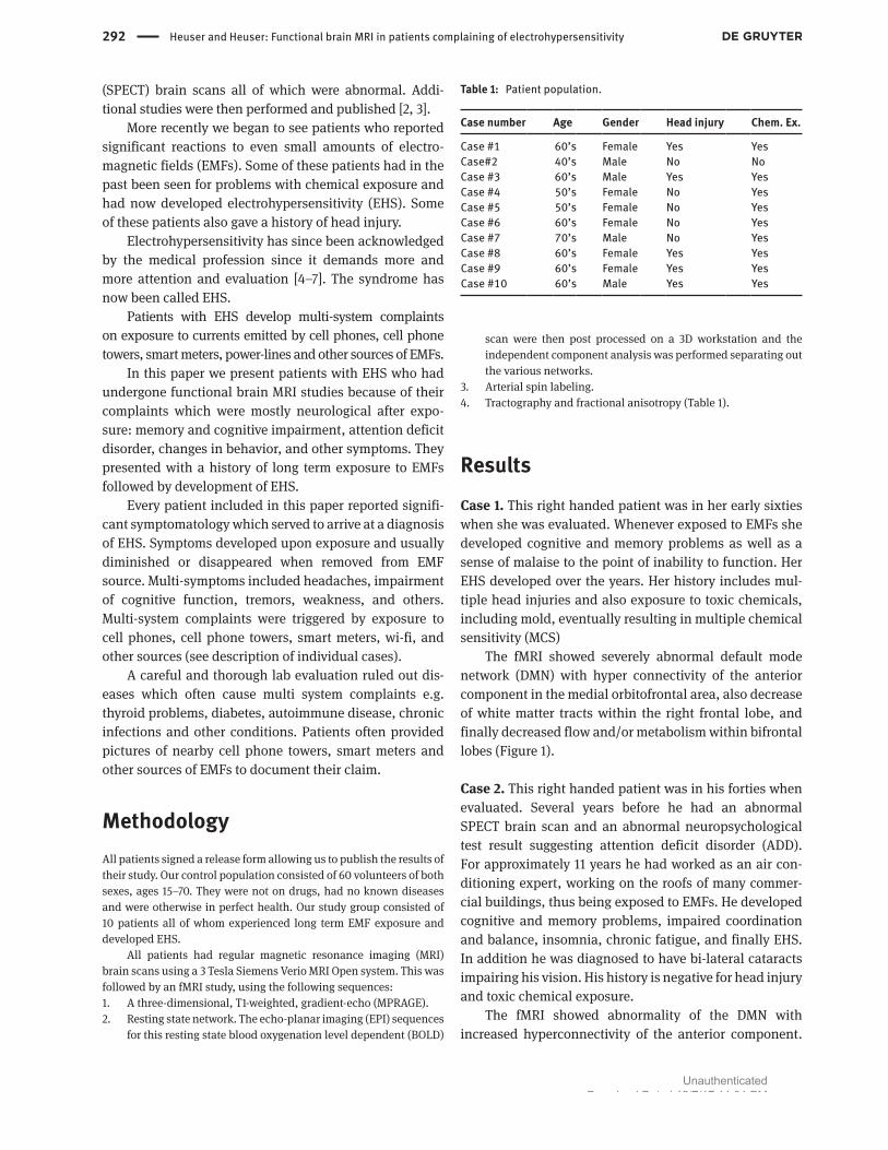
292 Heuser and Heuser: Functional brain MRI in patients complaining of electrohypersensitivity
(SPECT) brain scans all of which were abnormal. Addi-tional studies were then performed and published [2, 3].
More recently we began to see patients who reported significant reactions to even small amounts of electro-magnetic fields (EMFs). Some of these patients had in the past been seen for problems with chemical exposure and had now developed electrohypersensitivity (EHS). Some of these patients also gave a history of head injury.
Electrohypersensitivity has since been acknowledged by the medical profession since it demands more and more attention and evaluation [4–7]. The syndrome has now been called EHS.
Patients with EHS develop multi-system complaints on exposure to currents emitted by cell phones, cell phone towers, smart meters, power-lines and other sources of EMFs.
In this paper we present patients with EHS who had undergone functional brain MRI studies because of their complaints which were mostly neurological after expo-sure: memory and cognitive impairment, attention deficit disorder, changes in behavior, and other symptoms. They presented with a history of long term exposure to EMFs followed by development of EHS.
Every patient included in this paper reported signifi-cant symptomatology which served to arrive at a diagnosis of EHS. Symptoms developed upon exposure and usually diminished or disappeared when removed from EMF source. Multi-symptoms included headaches, impairment of cognitive function, tremors, weakness, and others. Multi-system complaints were triggered by exposure to cell phones, cell phone towers, smart meters, wi-fi, and other sources (see description of individual cases).
A careful and thorough lab evaluation ruled out dis-eases which often cause multi system complaints e.g. thyroid problems, diabetes, autoimmune disease, chronic infections and other conditions. Patients often provided pictures of nearby cell phone towers, smart meters and other sources of EMFs to document their claim.
MethodologyAll patients signed a release form allowing us to publish the results of their study. Our control population consisted of 60 volunteers of both sexes, ages 15–70. They were not on drugs, had no known diseases and were otherwise in perfect health. Our study group consisted of 10 patients all of whom experienced long term EMF exposure and developed EHS.
All patients had regular magnetic resonance imaging (MRI) brain scans using a 3 Tesla Siemens Verio MRI Open system. This was followed by an fMRI study, using the following sequences:1. A three-dimensional, T1-weighted, gradient-echo (MPRAGE).2. Resting state network. The echo-planar imaging (EPI) sequences
for this resting state blood oxygenation level dependent (BOLD)
scan were then post processed on a 3D workstation and the independent component analysis was performed separating out the various networks.
3. Arterial spin labeling.4. Tractography and fractional anisotropy (Table 1).
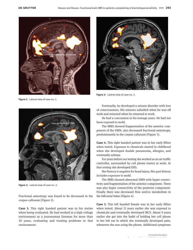
ResultsCase 1. This right handed patient was in her early sixties when she was evaluated. Whenever exposed to EMFs she developed cognitive and memory problems as well as a sense of malaise to the point of inability to function. Her EHS developed over the years. Her history includes mul-tiple head injuries and also exposure to toxic chemicals, including mold, eventually resulting in multiple chemical sensitivity (MCS)
The fMRI showed severely abnormal default mode network (DMN) with hyper connectivity of the anterior component in the medial orbitofrontal area, also decrease of white matter tracts within the right frontal lobe, and finally decreased flow and/or metabolism within bifrontal lobes (Figure 1).
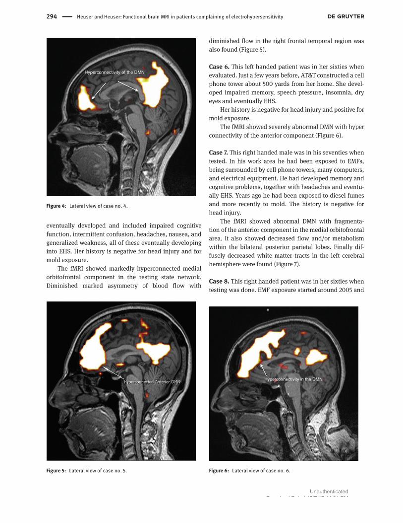
Case 2. This right handed patient was in his forties when evaluated. Several years before he had an abnormal SPECT brain scan and an abnormal neuropsychological test result suggesting attention deficit disorder (ADD). For approximately 11 years he had worked as an air con-ditioning expert, working on the roofs of many commer-cial buildings, thus being exposed to EMFs. He developed cognitive and memory problems, impaired coordination and balance, insomnia, chronic fatigue, and finally EHS. In addition he was diagnosed to have bi-lateral cataracts impairing his vision. His history is negative for head injury and toxic chemical exposure.
The fMRI showed abnormality of the DMN with increased hyperconnectivity of the anterior component.
Table 1: Patient population.
Case number Age Gender Head injury Chem. Ex.
Case #1 60 ’s Female Yes YesCase#2 40 ’s Male No NoCase #3 60 ’s Male Yes YesCase #4 50 ’s Female No YesCase #5 50 ’s Female No YesCase #6 60 ’s Female No YesCase #7 70 ’s Male No YesCase #8 60 ’s Female Yes YesCase #9 60 ’s Female Yes YesCase #10 60 ’s Male Yes Yes
UnauthenticatedDownload Date | 12/7/17 11:31 PM
Heuser and Heuser: Functional brain MRI in patients complaining of electrohypersensitivity 293
Fractional anisotropy was found to be decreased in the corpus callosum (Figure 2).
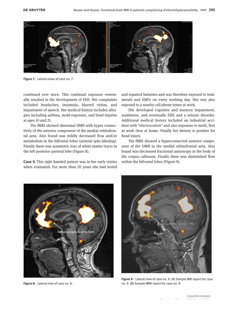
Case 3. This right handed patient was in his sixties when being evaluated. He had worked in a high voltage environment as a journeyman lineman for more than 30 years, evaluating and treating problems in that environment.
Eventually, he developed a seizure disorder with loss of consciousness. His seizures subsided when he was off work and returned when he returned to work.
He had a concussion in his teenage years. He had not been exposed to mold.
The fMRI showed fragmentation of the anterior com-ponent of the DMN, also decreased fractional anisotropy, predominantly in the corpus callosum (Figure 3).
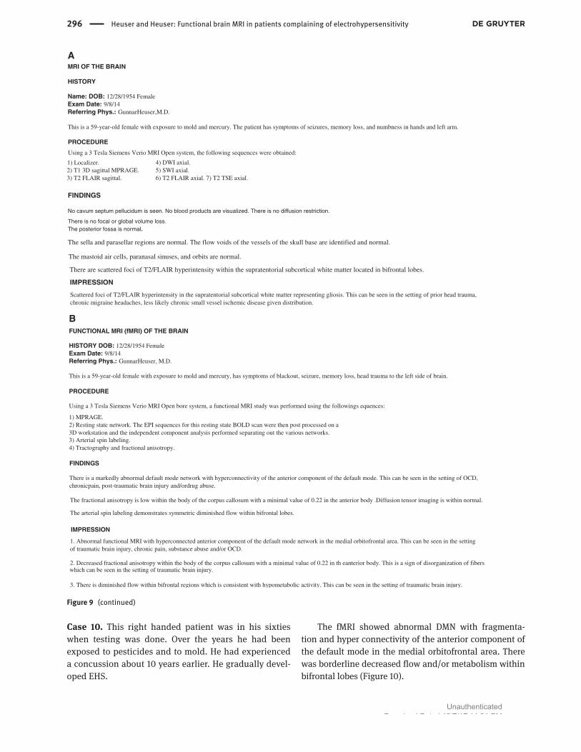
Case 4. This right handed patient was in her early fifties when tested. Exposure to chemicals started in childhood when she developed double pneumonia, allergies, and eventually asthma
For years before our testing she worked as an air traffic controller, surrounded by cell phone towers at work. In that setting she developed EHS.
Her history is negative for head injury. Her past history includes exposure to mold.
The fMRI showed abnormal DMN with hyper connec-tivity and fragmentation of the anterior component. There was also hyper connectivity of the posterior component. Finally there was decreased flow and/or metabolism in the bifrontal lobes (Figure 4).
Case 5. This left handed female was in her early fifties when tested. About 15 years earlier she was exposed to chemicals and eventually developed MCS. About 9 years earlier she got into the habit of holding her cell phone to her left ear in which she eventually developed pain, whenever she was using the phone. Additional symptoms
Figure 1: Lateral view of case no. 1.
Figure 2: Lateral view of case no. 2.
Figure 3: Lateral view of case no. 3.
UnauthenticatedDownload Date | 12/7/17 11:31 PM
294 Heuser and Heuser: Functional brain MRI in patients complaining of electrohypersensitivity
eventually developed and included impaired cognitive function, intermittent confusion, headaches, nausea, and generalized weakness, all of these eventually developing into EHS. Her history is negative for head injury and for mold exposure.
The fMRI showed markedly hyperconnected medial orbitofrontal component in the resting state network. Diminished marked asymmetry of blood flow with
diminished flow in the right frontal temporal region was also found (Figure 5).
Case 6. This left handed patient was in her sixties when evaluated. Just a few years before, AT&T constructed a cell phone tower about 500 yards from her home. She devel-oped impaired memory, speech pressure, insomnia, dry eyes and eventually EHS.
Her history is negative for head injury and positive for mold exposure.
The fMRI showed severely abnormal DMN with hyper connectivity of the anterior component (Figure 6).
Case 7. This right handed male was in his seventies when tested. In his work area he had been exposed to EMFs, being surrounded by cell phone towers, many computers, and electrical equipment. He had developed memory and cognitive problems, together with headaches and eventu-ally EHS. Years ago he had been exposed to diesel fumes and more recently to mold. The history is negative for head injury.
The fMRI showed abnormal DMN with fragmenta-tion of the anterior component in the medial orbitofrontal area. It also showed decreased flow and/or metabolism within the bilateral posterior parietal lobes. Finally dif-fusely decreased white matter tracts in the left cerebral hemisphere were found (Figure 7).
Case 8. This right handed patient was in her sixties when testing was done. EMF exposure started around 2005 and
Figure 5: Lateral view of case no. 5. Figure 6: Lateral view of case no. 6.
Figure 4: Lateral view of case no. 4.
UnauthenticatedDownload Date | 12/7/17 11:31 PM
Heuser and Heuser: Functional brain MRI in patients complaining of electrohypersensitivity 295
continued ever since. This continual exposure eventu-ally resulted in the development of EHS. Her complaints included headaches, insomnia, blurred vision, and impairment of speech. Her medical history includes aller-gies including asthma, mold exposure, and head injuries at ages 10 and 21.
The fMRI showed abnormal DMN with hyper connec-tivity of the anterior component of the medial orbitofron-tal area. Also found was mildly decreased flow and/or metabolism in the bifrontal lobes (arterial spin labeling). Finally there was symmetric loss of white matter tracts in the left posterior parietal lobe (Figure 8).
Case 9. This right handed patient was in her early sixties when evaluated. For more than 20 years she had tested
and repaired batteries and was therefore exposed to toxic metals and EMFs on every working day. She was also exposed to a nearby cell phone tower at work.
She developed cognitive and memory impairment, numbness, and eventually EHS and a seizure disorder. Additional medical history included an industrial acci-dent with “electrocution” and also exposure to mold, first at work then at home. Finally her history is positive for head injury.
The fMRI showed a hyperconnected anterior compo-nent of the DMN in the medial orbitofrontal area. Also found was decreased fractional anisotropy in the body of the corpus callosum. Finally there was diminished flow within the bifrontal lobes (Figure 9).
Figure 7: Lateral views of case no. 7.
Figure 8: Lateral view of case no. 8.Figure 9: Lateral view of case no. 9. (A) Sample MRI report for case no. 9. (B) Sample fMRI report for case no. 9.
UnauthenticatedDownload Date | 12/7/17 11:31 PM
296 Heuser and Heuser: Functional brain MRI in patients complaining of electrohypersensitivity
MRI OF THE BRAIN
HISTORY
Name: DOB: 12/28/1954 FemaleExam Date: 9/8/14Referring Phys.: GunnarHeuser,M.D.
This is a 59-year-old female with exposure to mold and mercury. The patient has symptoms of seizures, memory loss, and numbness in hands and left arm.
PROCEDURE
Using a 3 Tesla Siemens Verio MRI Open system, the following sequences were obtained:
1) Localizer.2) T1 3D sagittal MPRAGE.3) T2 FLAIR sagittal.
4) DWI axial.5) SWI axial.6) T2 FLAIR axial. 7) T2 TSE axial.
FINDINGS
No cavum septum pellucidum is seen. No blood products are visualized. There is no diffusion restriction.
There is no focal or global volume loss.The posterior fossa is normal.
The sella and parasellar regions are normal. The flow voids of the vessels of the skull base are identified and normal.
The mastoid air cells, paranasal sinuses, and orbits are normal.
There are scattered foci of T2/FLAIR hyperintensity within the supratentorial subcortical white matter located in bifrontal lobes.
Scattered foci of T2/FLAIR hyperintensity in the supratentorial subcortical white matter representing gliosis. This can be seen in the setting of prior head trauma, chronic migraine headaches, less likely chronic small vessel ischemic disease given distribution.
A
FUNCTIONAL MRI (fMRI) OF THE BRAIN
B
HISTORY DOB: 12/28/1954 FemaleExam Date: 9/8/14Referring Phys.: GunnarHeuser, M.D.
This is a 59-year-old female with exposure to mold and mercury, has symptoms of blackout, seizure, memory loss, head trauma to the left side of brain.
PROCEDURE
Using a 3 Tesla Siemens Verio MRI Open bore system, a functional MRI study was performed using the followings equences:
1) MPRAGE.2) Resting state network. The EPI sequences for this resting state BOLD scan were then post processed on a3D workstation and the independent component analysis performed separating out the various networks.3) Arterial spin labeling.4) Tractography and fractional anisotropy.
FINDINGS
There is a markedly abnormal default mode network with hyperconnectivity of the anterior component of the default mode. This can be seen in the setting of OCD,chronicpain, post-traumatic brain injury and/ordrug abuse.
The fractional anisotropy is low within the body of the corpus callosum with a minimal value of 0.22 in the anterior body .Diffusion tensor imaging is within normal.
The arterial spin labeling demonstrates symmetric diminished flow within bifrontal lobes.
IMPRESSION
1. Abnormal functional MRI with hyperconnected anterior component of the default mode network in the medial orbitofrontal area. This can be seen in the setting of traumatic brain injury, chronic pain, substance abuse and/or OCD.
2. Decreased fractional anisotropy within the body of the corpus callosum with a minimal value of 0.22 in th eanterior body. This is a sign of disorganization of fibers
3. There is diminished flow within bifrontal regions which is consistent with hypometabolic activity. This can be seen in the setting of traumatic brain injury.
which can be seen in the setting of traumatic brain injury.
IMPRESSION
Figure 9 (continued)
Case 10. This right handed patient was in his sixties when testing was done. Over the years he had been exposed to pesticides and to mold. He had experienced a concussion about 10 years earlier. He gradually devel-oped EHS.
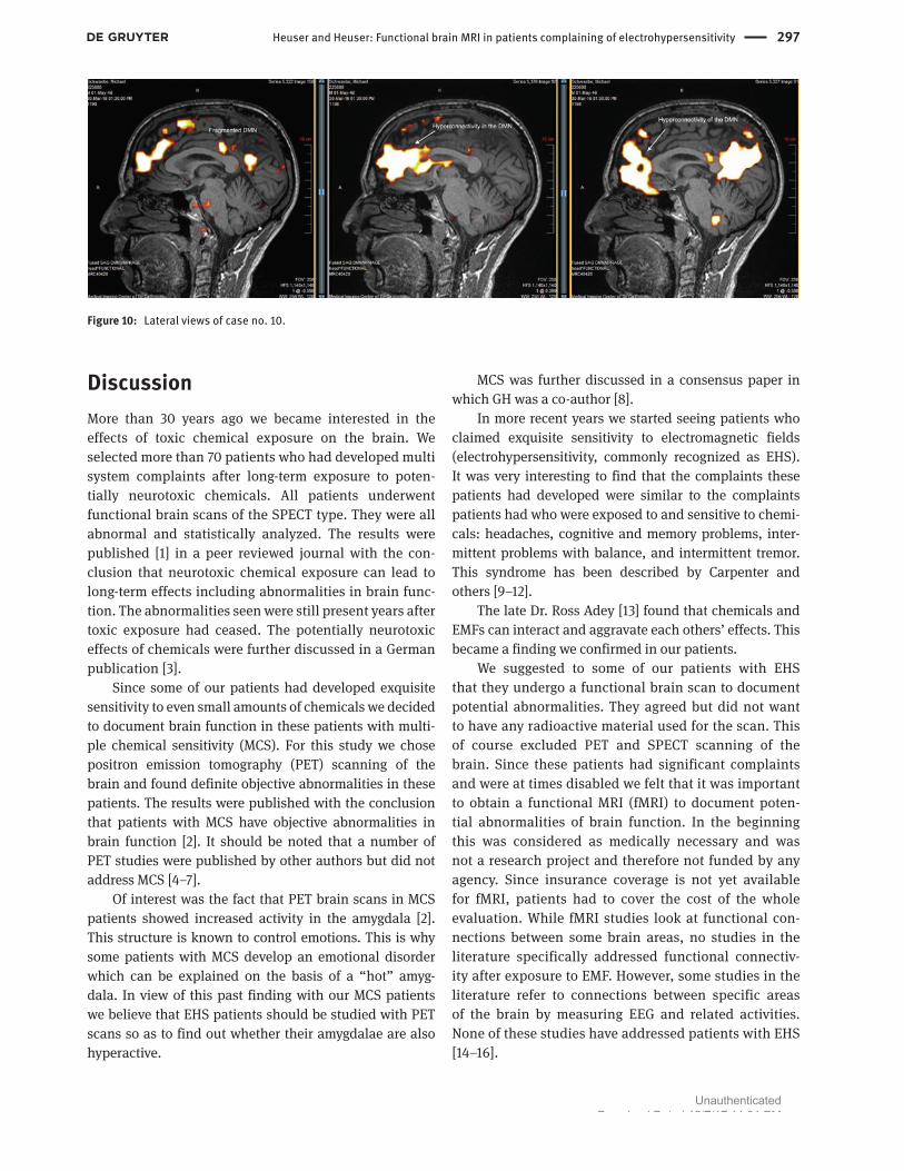
The fMRI showed abnormal DMN with fragmenta-tion and hyper connectivity of the anterior component of the default mode in the medial orbitofrontal area. There was borderline decreased flow and/or metabolism within bifrontal lobes (Figure 10).
UnauthenticatedDownload Date | 12/7/17 11:31 PM
Heuser and Heuser: Functional brain MRI in patients complaining of electrohypersensitivity 297
DiscussionMore than 30 years ago we became interested in the effects of toxic chemical exposure on the brain. We selected more than 70 patients who had developed multi system complaints after long-term exposure to poten-tially neurotoxic chemicals. All patients underwent functional brain scans of the SPECT type. They were all abnormal and statistically analyzed. The results were published [1] in a peer reviewed journal with the con-clusion that neurotoxic chemical exposure can lead to long-term effects including abnormalities in brain func-tion. The abnormalities seen were still present years after toxic exposure had ceased. The potentially neurotoxic effects of chemicals were further discussed in a German publication [3].
Since some of our patients had developed exquisite sensitivity to even small amounts of chemicals we decided to document brain function in these patients with multi-ple chemical sensitivity (MCS). For this study we chose positron emission tomography (PET) scanning of the brain and found definite objective abnormalities in these patients. The results were published with the conclusion that patients with MCS have objective abnormalities in brain function [2]. It should be noted that a number of PET studies were published by other authors but did not address MCS [4–7].
Of interest was the fact that PET brain scans in MCS patients showed increased activity in the amygdala [2]. This structure is known to control emotions. This is why some patients with MCS develop an emotional disorder which can be explained on the basis of a “hot” amyg-dala. In view of this past finding with our MCS patients we believe that EHS patients should be studied with PET scans so as to find out whether their amygdalae are also hyperactive.
MCS was further discussed in a consensus paper in which GH was a co-author [8].
In more recent years we started seeing patients who claimed exquisite sensitivity to electromagnetic fields (electrohypersensitivity, commonly recognized as EHS). It was very interesting to find that the complaints these patients had developed were similar to the complaints patients had who were exposed to and sensitive to chemi-cals: headaches, cognitive and memory problems, inter-mittent problems with balance, and intermittent tremor. This syndrome has been described by Carpenter and others [9–12].
The late Dr. Ross Adey [13] found that chemicals and EMFs can interact and aggravate each others’ effects. This became a finding we confirmed in our patients.
We suggested to some of our patients with EHS that they undergo a functional brain scan to document potential abnormalities. They agreed but did not want to have any radioactive material used for the scan. This of course excluded PET and SPECT scanning of the brain. Since these patients had significant complaints and were at times disabled we felt that it was important to obtain a functional MRI (fMRI) to document poten-tial abnormalities of brain function. In the beginning this was considered as medically necessary and was not a research project and therefore not funded by any agency. Since insurance coverage is not yet available for fMRI, patients had to cover the cost of the whole evaluation. While fMRI studies look at functional con-nections between some brain areas, no studies in the literature specifically addressed functional connectiv-ity after exposure to EMF. However, some studies in the literature refer to connections between specific areas of the brain by measuring EEG and related activities. None of these studies have addressed patients with EHS [14–16].
Figure 10: Lateral views of case no. 10.
UnauthenticatedDownload Date | 12/7/17 11:31 PM
298 Heuser and Heuser: Functional brain MRI in patients complaining of electrohypersensitivity
ConclusionAll fMRI brain scans were abnormal in our patients. Their abnormalities were very similar in all of them.
The study of fMRI created a new language which had to be learned. We have included some references which help the reader to understand this new terminology [17–21].
The question now arises whether the same abnor-malities seen in all 10 patients can become a diagnostic aid or biomarker for determining whether or not a patient has electrohypersensitivity (EHS). Some researchers have claimed that a diagnosis of EHS should really be a psychi-atric diagnosis [22]. Other researchers have attempted to measure EHS [23]. The possible psychiatric aspects of EHS remind us of the history of medicine when epilepsy was considered to be a diagnosis of possession by the devil [24]. It was only the discovery of the EEG which helped to find epilepsy to be a real disease with objective findings. We hope that fMRI will play the role of the EEG in EHS.
In the context of the above paragraph we should mention the articles by Belpomme et al. [25] and De Luca et al. [26] which suggest a long list of biomarkers for the diagnosis of EHS. This list does not include functional brain scans as potential biomarkers.
The treatment of EHS is very difficult. It was reviewed in a recent publication [27]. We have found that hyper-baric oxygen is at times helpful [28]. We have proposed that every patient with EHS should be checked for mast cell disease since mast cell abnormalities were found by Gangi and Johansson [29]. We described mast cell disor-der in patients with MCS [30]. Also we found mast cell disorder in some of our patients with EHS [31]. Finally we believe that mold and mold toxin (mycotoxin) exposure can trigger EHS. Mast cell disease and mold problems are treatable. Their treatment may decrease EHS. Otherwise treatment consists basically of avoidance.
A final point of interest is the fact that the abnormal-ity seen on the fMRI can also be seen after head injury [32, 33]. Indeed, many of our patients complaining of EHS have a history of head injury (see our case reports).
Since neurotoxic chemicals as well as head injury and EMF exposure are known to impair the blood brain barrier, it is almost to be expected that singly or in com-bination they make a given patient more vulnerable to impaired brain function (including seizures).
In the past we studied patients who had developed multi system complaints after exposure to a number of neurotoxins. The only patients who developed a seizure disorder were the ones who gave a history of past head injury [31]. Two of our patients (#3 and 9) developed a well
documented seizure disorder. This can be understood as having been “kindled” by repeated exposures to EMFs.
Our study needs to be enlarged and also duplicated by others. The subject will be of increasing importance in our society in which we and our children are all exposed to more and more EMFs.
Acknowledgements: Medical Imaging of Southern Califor-nia graciously made their studies (MRI and fMRI) avail-able at a significant discount. We also acknowledge Brea Blevins who helped to retrieve and to display the images published in this paper. Most patients were either self referred or referred by their physician. Some were referred by The Peoples Initiative Foundation which was formed and is directed by Elizabeth Barris.
Author StatementResearch funding: No funds for this study were avail-able from any foundation or other financial entity for this study. Partial payment was received for two patient studies by a charitable foundation (The Peoples Initiative Foundation). Patients paid for their own consultations and testing. No insurance reimbursements were available for the fMRI study. Conflict of interest: Authors state no conflict of interest. Informed consent: Informed consent has been obtained from all individuals. Ethical approval: Ethical approval was not applicable.
References1. Heuser G, Mena I. Neurospect in neurotoxic chemical exposure.
Demonstration of long-term functional abnormalities. Toxicol Ind Health 1998;14(6):813–27.
2. Heuser G, Wu JC. Deep subcortical (including limbic) hypermetab-olism in patients with chemical intolerance: human PET studies. Ann N Y Acad Sci 2001;933:319–22.
3. Heuser G. Functional brain Imaging with SPECT and PET after neu-rotoxic exposure: two and three-diimensional displays. Zeitschrift fur Umweltmedizin 1999;8:284–5.
4. Huber R, Treyer V, Schuderer J, Berthold T, Buck A, et al. Exposure to pulse-modulated radio frequency electromag-netic fields affects regional cerebral blood flow. Eur J Neurosci 2005;21(4):1000–6.
5. Aalto S, Haarala C, Brück A, Sipilä H, Hämäläinen H, et al. Mobile phone affects cerebral blood flow in humans. J Cereb Blood Flow Metab 2006;26(7):885–90.
6. Haarala C, Aalto S, Hautzel H, Julkunen L, Rinne JO, et al. Effects of a 902 MHz mobile phone on cerebral blood flow in humans: a PET study. Neuroreport. 2003;14(16):2019–23.
7. Huber R, Treyer V, Borbély AA, Schuderer J, Gottselig JM, et al. Electromagnetic fields, such as those from mobile phones, alter regional cerebral blood flow and sleep and waking EEG. J Sleep Res 2002;11(4):289–95.
UnauthenticatedDownload Date | 12/7/17 11:31 PM
Heuser and Heuser: Functional brain MRI in patients complaining of electrohypersensitivity 299
8. Bartha L. Multiple chemical sensitivity: a 1999 consensus. Arch Environ Health 1999;54(3):147–9.
9. Carpenter DO. The microwave syndrome or electro-hypersensitivity: historical background. Rev Environ Health 2015;30(4):217–22.
10. Hedendahl L, Carlberg M, Hardell L. Electromagnetic hypersen-sitivity – an increasing challenge to the medical profession. Rev Environ Health 2015;30(4):209–15.
11. Genuis SJ, Lipp CT. Electromagnetic hypersensitivity: fact or fiction? Sci Total Environ 2012;414:103–12.
12. McCarty DE, Carrubba S, Chesson AL, Frilot C, Gonzalez-Toledo E, et al. Electromagnetic hypersensitivity: evidence for a novel neurological syndrome. Int J Neurosci 2011;121(12):670–6.
13. Adey WR. Joint actions of environmental nonionizing electro-magnetic fields and chemical pollution in cancer promotion. Environ Health Perspect 1990;86:297–305.
14. Haarala C, Takio F, Rintee T, Laine M, Koivisto M, et al. Pulsed and continuous wave mobile phone exposure over left versus right hemisphere: effects on human cognitive function. Bioelec-tromagnetics 2007;28(4):289–95.
15. Vecchio F, Babiloni C, Ferreri F, Curcio G, Fini R, et al. Mobile phone emission modulates interhemispheric functional cou-pling of EEG alpha rhythms. Eur J Neurosci 2007;25(6):1908–13.
16. Yang L, Chen Q, Lv B, Wu T. Long-Term evolution electromagnetic fields exposure modulates the resting state EEG on alpha and beta bands. Clin EEG Neurosci 2017;48(3):168–75.
17. van den Heuvel MP, Hulshoff Pol HE. Exploring the brain net-work: a review on resting-state fMRI functional connectivity. Eur Neuropsychopharmacol 2010;20(8):519–34.
18. Horn A, Dirk O, Reisert M, Blankenburg F. Default Mode Network. [revised 2016 August]. In: Wikipedia [Internet]. San Francisco, CA: NeuroImage; 2006 September. 10 pages. Available from: www.wikipedia.com. DOI: 10.1016.
19. Liston C, Chen AC, Zebley BD, Drysdale AT, Gordon R, et al. Default mode network mechanisms of transcranial magnetic stimulation in depression. Biol Psychiatry 2014;76(7):517–26.
20. Smith SM, Vidaurre D, Beckmann CF, Glasser MF, Jenkinson M, et al. Functional connectomics from resting-state fMRI. Trends Cogn Sci 2013;17(12):666–82.
21. Lee MH, Smyser CD, Shimony JS. Resting-state fMRI: a review of methods and clinical applications. AJNR Am J Neuroradiol 2013;34(10):1866–72.
22. Rubin GJ, Hillert L, Nieto-Hernandez R, van Rongen E, Oftedal G. Do people with idiopathic environmental intolerance attributed to electromagnetic fields display physiological effects when exposed to electromagnetic fields? A system-atic review of provocation studies. Bioelectromagnetics 2011;32(8):593–609.
23. Tuengler A, von Klitzing L. Hypothesis on how to measure electromagnetic hypersensitivity. Electromagn Biol Med 2013;32(3):281–90.
24. Espí Forcén C, Espí Forcén F. Demonic possessions and mental illness: discussion of selected cases in late medieval hagiographical literature. Early Sci Med 2014;19(3):258–79.
25. Belpomme D, Campagnac C, Irigaray P. Reliable disease biomarkers characterizing and identifying electrohypersensi-tivity and multiple chemical sensitivity as two etiopathogenic aspects of a unique pathological disorder. Rev Environ Health 2015;30(4):251–71.
26. De Luca C, Chung Sheun Thai J, Raskovic D, Cesareo E, Caccamo D, et al. Metabolic and genetic screening of electromagnetic hypersensitive subjects as a feasible tool for diagnostics and intervention. Mediators Inflamm 2014;2014:924184.
27. Rubin GJ, Das Munshi J, Wessely S. A systematic review of treatments for electromagnetic hypersensitivity. Psychother Psy-chosom 2006;75(1):12–8.
28. Heuser G, Uszler JM. Hyperbaric oxygenation for cerebral palsy. Lancet 2001;357(9273):2053–4. Erratum in: Lancet 2001 Nov 24;358(9295):1820.
29. Gangi S, Johansson O. A theoretical model based upon mast cells and histamine to explain the recently proclaimed sensitiv-ity to electric and/or magnetic fields in humans. Med Hypoth-eses 2000;54(4):663–71.
30. Heuser G. Mast cell disorder to be ruled out in MCS. Arch Envi-ron Health 2000;55(4):284–5.
31. Unpublished observations by authors.32. Mishra AM, Bai X, Sanganahalli BG, Waxman SG,
Shatillo O, et al. Decreased resting functional connec-tivity after traumatic brain injury in the rat. Plos One 2014;09(4):e95280.
33. Zhou Y, Milham M, Lui Y, Zhou Y, Milham MP, et al. Default-mode network disruption in mild traumatic brain injury. Radiology 2012;265(3):882–92.
UnauthenticatedDownload Date | 12/7/17 11:31 PM