Guidelines on the Diagnosis and Management of … The term recurrent pericarditis encompasses (1)...
24
ESC Guidelines Guidelines on the Diagnosis and Management of Pericardial Diseases Executive Summary The Task Force on the Diagnosis and Management of Pericardial Diseases of the European Society of Cardiology Task Force members, Bernhard Maisch, Chairperson* (Germany), Petar M. Seferovi c (Serbia and Montenegro), Arsen D. Risti c (Serbia and Montenegro), Raimund Erbel (Germany), Reiner Rienm € uller (Austria), Yehuda Adler (Israel), Witold Z. Tomkowski (Poland), Gaetano Thiene (Italy), Magdi H. Yacoub (UK) ESC Committee for Practice Guidelines (CPG), Silvia G. Priori (Chairperson) (Italy), Maria Angeles Alonso Garcia (Spain), Jean-Jacques Blanc (France), Andrzej Budaj (Poland), Martin Cowie (UK), Veronica Dean (France), Jaap Deckers (The Netherlands), Enrique Fernandez Burgos (Spain), John Lekakis (Greece), Bertil Lindahl (Sweden), Gianfranco Mazzotta (Italy), Jo~ ao Morais (Portugal), Ali Oto (Turkey), Otto A. Smiseth (Norway) Document Reviewers, Gianfranco Mazzotta (CPG Review Coordinator) (Italy), Jean Acar (France), Eloisa Arbustini (Italy), Anton E. Becker (The Netherlands), Giacomo Chiaranda (Italy), Yonathan Hasin (Israel), Rolf Jenni (Switzerland), Werner Klein (Austria), Irene Lang (Austria), Thomas F. L€ uscher (Switzerland), Fausto J. Pinto (Portugal), Ralph Shabetai (USA), Maarten L. Simoons (The Netherlands), Jordi Soler Soler (Spain), David H. Spodick (USA) Table of contents Preamble .......................... 587 Introduction ......................... 588 Aetiology and classification of pericardial disease. 588 Pericardial syndromes .................. 588 Congenital defects of the pericardium ...... 588 Acute pericarditis ................... 588 Chronic pericarditis .................. 591 Recurrent pericarditis ................ 592 Pericardial effusion and cardiac tamponade . . 592 Constrictive pericarditis ............... 593 Pericardial cysts .................... 595 Specific forms of pericarditis .............. 597 Viral pericarditis .................... 597 Bacterial pericarditis ................. 598 Tuberculous pericarditis ............. 598 Pericarditis in renal failure ............. 600 Autoreactive pericarditis and pericardial involvement in systemic autoimmune diseases ....................... 600 The post-cardiac injury syndrome: postpericardiotomy syndrome ......... 600 Postinfarction pericarditis .............. 601 Traumatic pericardial effusion and haemopericardium in aortic dissection .... 601 Neoplastic pericarditis ................ 603 Rare forms of pericardial disease ......... 603 Fungal pericarditis ................. 603 Radiation pericarditis ............... 604 Chylopericardium ................. 604 Drug- and toxin-related pericarditis ...... 605 *Corresponding author: Chairperson: Bernhard Maisch, MD, FESC, FACC, Dean of the Faculty of Medicine, Director of the Department of Internal Medicine-Cardiology, Philipps University, Marburg, Baldingerst- rasse 1, D-35033 Marburg, Germany. Tel.: +49-6421-286-6462; fax: +49- 6421-286-8954. E-mail address: [email protected] (B. Maisch). 0195-668X/$ - see front matter c 2004 The European Society of Cardiology. Published by Elsevier Ltd. All rights reserved. doi:10.1016/j.ehj.2004.02.002 European Heart Journal (2004) 25, 587–610
-
Upload
truongkhanh -
Category
Documents
-
view
213 -
download
0
Transcript of Guidelines on the Diagnosis and Management of … The term recurrent pericarditis encompasses (1)...
ESC Guidelines
Guidelines on the Diagnosis and Managementof Pericardial DiseasesExecutive Summary
The Task Force on the Diagnosis and Management of PericardialDiseases of the European Society of Cardiology
Task Force members, Bernhard Maisch, Chairperson* (Germany),Petar M. Seferovi�c (Serbia and Montenegro), Arsen D. Risti�c (Serbia and Montenegro),Raimund Erbel (Germany), Reiner Rienm€uller (Austria), Yehuda Adler (Israel),Witold Z. Tomkowski (Poland), Gaetano Thiene (Italy), Magdi H. Yacoub (UK)
ESC Committee for Practice Guidelines (CPG), Silvia G. Priori (Chairperson) (Italy), Maria Angeles Alonso Garcia(Spain), Jean-Jacques Blanc (France), Andrzej Budaj (Poland), Martin Cowie (UK), Veronica Dean (France), JaapDeckers (The Netherlands), Enrique Fernandez Burgos (Spain), John Lekakis (Greece), Bertil Lindahl (Sweden),Gianfranco Mazzotta (Italy), Jo~ao Morais (Portugal), Ali Oto (Turkey), Otto A. Smiseth (Norway)
Document Reviewers, Gianfranco Mazzotta (CPG Review Coordinator) (Italy), Jean Acar (France), Eloisa Arbustini(Italy), Anton E. Becker (The Netherlands), Giacomo Chiaranda (Italy), Yonathan Hasin (Israel), Rolf Jenni(Switzerland), Werner Klein (Austria), Irene Lang (Austria), Thomas F. L€uscher (Switzerland), Fausto J. Pinto(Portugal), Ralph Shabetai (USA), Maarten L. Simoons (The Netherlands), Jordi Soler Soler (Spain), David H.Spodick (USA)
Table of contents
Preamble . . . . . . . . . . . . . . . . . . . . . . . . . . 587Introduction. . . . . . . . . . . . . . . . . . . . . . . . . 588Aetiology and classification of pericardial disease . 588Pericardial syndromes . . . . . . . . . . . . . . . . . . 588
Congenital defects of the pericardium . . . . . . 588Acute pericarditis . . . . . . . . . . . . . . . . . . . 588Chronic pericarditis . . . . . . . . . . . . . . . . . . 591Recurrent pericarditis . . . . . . . . . . . . . . . . 592Pericardial effusion and cardiac tamponade . . 592Constrictive pericarditis . . . . . . . . . . . . . . . 593Pericardial cysts . . . . . . . . . . . . . . . . . . . . 595
Specific forms of pericarditis . . . . . . . . . . . . . . 597Viral pericarditis . . . . . . . . . . . . . . . . . . . . 597Bacterial pericarditis . . . . . . . . . . . . . . . . . 598
Tuberculous pericarditis . . . . . . . . . . . . . 598Pericarditis in renal failure . . . . . . . . . . . . . 600Autoreactive pericarditis and pericardial
involvement in systemic autoimmunediseases . . . . . . . . . . . . . . . . . . . . . . . 600
The post-cardiac injury syndrome:postpericardiotomy syndrome . . . . . . . . . 600
Postinfarction pericarditis . . . . . . . . . . . . . . 601Traumatic pericardial effusion and
haemopericardium in aortic dissection . . . . 601Neoplastic pericarditis . . . . . . . . . . . . . . . . 603Rare forms of pericardial disease . . . . . . . . . 603
Fungal pericarditis . . . . . . . . . . . . . . . . . 603Radiation pericarditis . . . . . . . . . . . . . . . 604Chylopericardium . . . . . . . . . . . . . . . . . 604Drug- and toxin-related pericarditis . . . . . . 605
*Corresponding author: Chairperson: Bernhard Maisch, MD, FESC,FACC, Dean of the Faculty of Medicine, Director of the Department ofInternal Medicine-Cardiology, Philipps University, Marburg, Baldingerst-rasse 1, D-35033 Marburg, Germany. Tel.: +49-6421-286-6462; fax: +49-6421-286-8954.
E-mail address: [email protected] (B. Maisch).
0195-668X/$ - see front matter �c 2004 The European Society of Cardiology. Published by Elsevier Ltd. All rights reserved.doi:10.1016/j.ehj.2004.02.002
European Heart Journal (2004) 25, 587–610

Preamble
Guidelines and Expert Consensus documents aim to pres-ent all the relevant evidence on a particular issue in orderto help physicians to weigh the benefits and risks of aparticular diagnostic or therapeutic procedure. Theyshould be helpful in everyday clinical decision-making.
A great number of Guidelines and Expert ConsensusDocuments have been issued in recent years by differentorganisations, the European Society of Cardiology (ESC)and by other related societies. By means of links to websites of National Societies several hundred guidelines areavailable. This profusion can put at stake the authorityand validity of guidelines, which can only be guaranteedif they have been developed by an unquestionable deci-sion-making process. This is one of the reasons why theESC and others have issued recommendations for for-mulating and issuing Guidelines and Expert ConsensusDocuments.
In spite of the fact that standards for issuing goodquality Guidelines and Expert Consensus Documents arewell defined, recent surveys of Guidelines and ExpertConsensus Documents published in peer-reviewed jour-nals between 1985 and 1998 have shown that methodo-logical standards were not complied within the vastmajority of cases. It is therefore of great importancethat guidelines and recommendations are presented informats that are easily interpreted. Subsequently, theirimplementation programmes must also be well con-ducted. Attempts have been made to determine whetherguidelines improve the quality of clinical practice andthe utilisation of health resources.
The ESC Committee for Practice Guidelines (CPG)supervises and coordinates the preparation of newGuidelines and Expert Consensus Documents producedby Task Forces, expert groups or consensus panels. TheCommittee is also responsible for the endorsement ofthese Guidelines and Expert Consensus Documents orstatements.
Introduction
The strength of evidence related to a particular diag-nostic or treatment option depends on the availabledata: (1) level of evidence A: multiple randomised clin-ical trials or meta-analyses; (2) level of evidence B: asingle randomised trial or non-randomised studies; and(3) level of evidence C: consensus opinion of the experts.Indications for various tests and procedures were rankedin three classes:
Class I: Conditions for which there is evidence and/orgeneral agreement that a given procedure ortreatment is useful and effective.
Class II: Conditions for which there is conflicting evi-dence and/or a divergence of opinion aboutthe usefulness/efficacy of a procedure or treat-ment.Class IIa: Weight of evidence/opinion is in
favour of usefulness/efficacy.Class IIb: Usefulness/efficacy is less well estab-
lished by evidence/opinion.Class III: Conditions for which there is evidence and/or
general agreement that the procedure/treat-ment is not useful/effective and in some casesmay be harmful.
Aetiology and classification of pericardialdisease
The spectrum of pericardial diseases consists of con-genital defects, pericarditis (dry, effusive, effusive-constrictive, and constrictive), neoplasm, and cysts. Theaetiological classification comprises: infectious pericar-ditis, pericarditis in systemic autoimmune diseases, type2 (auto) immune process, postmyocardial infarctionsyndrome, and auto-reactive (chronic) pericarditis(Table 1).1–3
Pericardial syndromes
Congenital defects of the pericardium
Congenital defects of the pericardium (1/10.000 autop-sies) comprise partial left (70%), right (17%) or total bi-lateral (rare) pericardial absence. Additional congenitalabnormalities occur in �30% of patients.4 Most patientswith a total pericardial absence are asymptomatic. Ho-molateral cardiac displacement and augmented heartmobility impose an increased risk for traumatic aorticdissection.5 Partial left side defects can be complicatedby herniation and strangulation of the heart through thedefect (chest pain, shortness of breath, syncope or sud-den death). Surgical pericardioplasty (Dacron, Gore-tex,or bovine pericardium) is indicated for imminent stran-gulation.6
Acute pericarditis
Acute pericarditis is dry, fibrinous or effusive, indepen-dent from its aetiology. The diagnostic algorithm can bederived from Table 2.8–18 A prodrome of fever, malaise,and myalgia is common, but elderly patients may not befebrile. Major symptoms are retrosternal or left precor-dialchest pain (radiates to the trapezius ridge, can bepleuritic or simulate ischemia, and varies with posture)and shortness of breath. The pericardial friction rub canbe transient, mono-, bi- or triphasic. Pleural effusionmay be present. Heart rate is usually rapid and regular.
Pericardial effusion in thyroid disorders . . . 605Pericardial effusion in pregnancy . . . . . . . 605
Uncited references . . . . . . . . . . . . . . . . . . . . 605Acknowledgements . . . . . . . . . . . . . . . . . . . . 605References . . . . . . . . . . . . . . . . . . . . . . . . . 605
588 ESC Guidelines

Table 1 Review of aetiology, incidence and pathogenesis of pericarditis1–3
Aetiology Incidence (%) Pathogenesis
Infectious pericarditis Multiplication and spread of the causativeagent and release of toxic substances in peri-cardial tissue cause serous, serofibrinous orhaemorrhagic (bacterial, viral, tuberculous,fungal) or purulent inflammation (bacterial)
Viral (Coxsackie A9, B1-4, Echo 8, Mumps,EBV, CMV, Varicella, Rubella, HIV, Parvo B19,etc.)
30–50a
Bacterial (Pneumo-, Meningo-, Gonococcosis,Hemophilus, Treponema pallidum, Borreliosis,Chlamydia, Tuberculosis, etc.)
5–10a
Fungal (Candida, Histoplasma, etc.) RareParasitary (Entameba histolytica, Echinococcus,Toxoplasma. . .)
Rare
Pericarditis in systemic autoimmune diseases Cardiac manifestations of the basic disease,often clinically mild or silentSystemic lupus erythematosus 30b
Rheumatoid arthritis 30b
Spondylitis ankylosans 1b
Systemic sclerosis >50b
Dermatomyositis RarePeriarteritis nodosa RareReiter’s syndrome �2bFamilial Mediterranean fever 0.7b
Type 2 (auto)immune process Secondary, after infection/surgeryRheumatic fever 20–50b Mostly in acute phasePostcardiotomy syndrome �20b 10–14 days after surgeryPostmyocardial infarction syndrome 1–5b DDg P. epistenocardicaAutoreactive (chronic) pericarditis 23.1a Common form
Pericarditis and pericardial effusion in diseases of surrounding organsAcute MI (P. epistenocardica) 5–20b 1–5 days after transmural MIMyocarditis 30b Accompanying epimyocarditisAortic aneurysm Rare Dissection: haemorrhagic PELung infarction RarePneumonia RareOesophageal diseases RareHydropericardium in CHF RareParaneoplastic pericarditis Frequent No direct neoplastic infiltrate
Pericarditis in metabolic disordersRenal insufficiency (uraemia) Frequent Viral/toxic/autoimmuneMyxedema 30b Serous, cholesterol rich PEAddison’s disease Rare Membranous leak?Diabetic ketoacidosis RareCholesterol pericarditis Very rare Transudation of cholesterol
(sterile serofibrinous PE)Pregnancy Rare
Traumatic pericarditisDirect injury (penetrating thoracic injury,oesophageal perforation, foreign bodies)
Rare
Indirect injury (Non-penetrating thoracic injury,mediastinal irradiation)
Rare Less frequent after introduction of topicalconvergent irradiation
Neoplastic pericardial disease 35a
Primary tumours RareSecondary metastatic tumours Frequent
Lung carcinoma 40c Serous or fibrinous, frequently haemorrhagiceffusion
Breast carcinoma 22c Accompanying disease during the infiltration ofmalignant cells
Gastric and colon 3c
Other carcinoma 6c
Leukemia and lymphoma 15c
Melanoma 3c
Sarcoma 4c
Other tumours 7c
ESC Guidelines 589

Microvoltage and electrical alternans are reversible aftereffusion drainage.19 Echocardiography is essential todetect effusion, concomitant heart or paracardial dis-ease.11;12
Perimyocarditis is evidenced by global or regionalmyocardial dysfunction, elevations of troponins I and T,MB creatine-kinase, myoglobin and tumour necrosis fac-tor. Auscultation of a new S3 heart sound, convexly el-evated J-ST segment in the ECG, fixation of Indium-111-
labelled antimyosin antibodies, and structural changes inMRI are indicative, but only endomyocardial/epimyo-cardial biopsy is diagnostic.7;8
Hospitalisation is warranted to determine the aeti-ology and observe for tamponade as well as the effectof treatment. Nonsteroidal anti-inflammatory drugs(NSAID) are the mainstay (level of evidence B, class I).Indomethacine should be avoided in elderly patientsdue to its flow reduction in the coronaries. Ibuprofen is
Table 2 Diagnostic pathway and sequence of performance in acute pericarditis (level of evidence B for all procedures)
Technique Characteristic findings Reference
Obligatory (indication class I)Auscultation Pericardial rub (mono-, bi-, or triphasic) 7
ECGa Stage I: anterior and inferior concave ST segment elevation. PR segmentdeviations opposite to P polarity.
7,19
Early stage II: ST junctions return to the baseline, PR deviated.Late stage II: T waves progressively flatten and invertStage III: generalised T wave inversionsStage IV: ECG returns to prepericarditis state.
Echocardiography Effusion types B–D (Horowitz) (Fig. 1) 9,10Signs of tamponade (see Section Pericardial effusion and cardiac tamponde)
Blood analyses (a) ESR, CRP, LDH, leukocytes (inflammation markers) 11(b) Troponin I, CK-MB (markers of myocardial lesion)b
Chest X-ray Ranging from normal to “water bottle” heart shadow. Revealing additionalpulmonary/mediastinal pathology.
12
Mandatory in tamponade (indication class I), optional in large/recurrent effusions or if previous tests inconclusive (indicationclass IIa) in small: effusions (indication class IIb)Pericardiocentesis anddrainage
PCR and histochemistry for aetiopathogenetic classification of infection orneoplasia
2,8,13
Optional or if previous tests inconclusive (indication class IIa)CT Effusions, peri-, and epicardium 14MRI Effusions, peri-, and epicardium 14Pericardioscopy, pericardial biopsy Establishing the specific aetiology 2,8,15,16
a Typical lead involvement: I, II, aVL, aVF, and V3-V6. The ST segment is always depressed in aVR, frequently in V1, and occasionally in V2. Oc-casionally, stage IV does not occur and there are permanent T wave inversions and flattenings. If ECG is first recorded in stage III, pericarditis cannotbe differentiated by ECG from diffuse myocardial injury, “biventricular strain,” or myocarditis. ECG in Early repolarization is very similar to stage I.Unlike stage I, this ECG does not acutely evolve and J-point elevations are usually accompanied by a slur, oscillation, or notch at the end of the QRSjust before and including the J point (best seen with tall R and T waves – large in early repolarisation pattern). Pericarditis is likely if in lead V6 the Jpoint is >25% of the height of the T wave apex (using the PR segment as a baseline).b Cardiac troponin I was detectable in 49% and >1.5 ng/ml in 22% of 69 patients with acute pericarditis (only in those with ST elevation in ECG)investigated by Bonnefoy et al.17 In another study18 troponin I was detected in 10/14 patients with a median peak concentration of 21.4 mg/ml(range 0.5 to >50 ng/ml). CK-MB was elevated in 8/14 patients with the median peak of 21 U/l (range 13–43), corresponding to the relative index of10.2% of the total CK activity.
Table 1 (continued)
Aetiology Incidence (%) Pathogenesis
Idiopathic 3.5a, in other series >50a Serous, fibrinous, sometimes haemorrhagic PEwith suspect viral or autoimmune secondaryimmunopathogenesis
CHF, congestive heart failure; DDg, differential diagnosis; MI, myocardial infarction; P., pericarditis; PE, pericardial effusion.a Percentage related to the population of 260 subsequent patients undergoing pericardiocentesis, pericardioscopy and epicardial biopsy (Marburgpericarditis registry 1988–2001).1b Percentage related to the incidence of pericarditis in the specific population of patients (e.g., with systemic lupus erythematosus).c Percentage related to the population of patients with neoplastic pericarditis.
590 ESC Guidelines
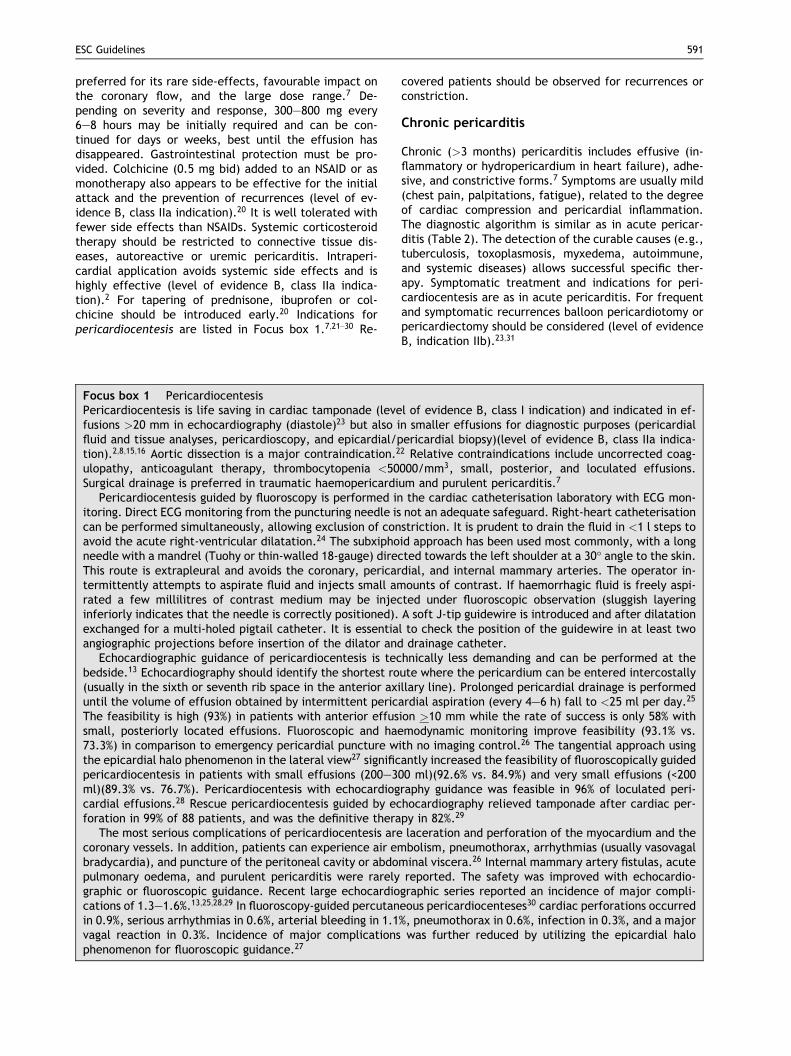
preferred for its rare side-effects, favourable impact onthe coronary flow, and the large dose range.7 De-pending on severity and response, 300–800 mg every6–8 hours may be initially required and can be con-tinued for days or weeks, best until the effusion hasdisappeared. Gastrointestinal protection must be pro-vided. Colchicine (0.5 mg bid) added to an NSAID or asmonotherapy also appears to be effective for the initialattack and the prevention of recurrences (level of ev-idence B, class IIa indication).20 It is well tolerated withfewer side effects than NSAIDs. Systemic corticosteroidtherapy should be restricted to connective tissue dis-eases, autoreactive or uremic pericarditis. Intraperi-cardial application avoids systemic side effects and ishighly effective (level of evidence B, class IIa indica-tion).2 For tapering of prednisone, ibuprofen or col-chicine should be introduced early.20 Indications forpericardiocentesis are listed in Focus box 1.7;21–30 Re-
covered patients should be observed for recurrences orconstriction.
Chronic pericarditis
Chronic (>3 months) pericarditis includes effusive (in-flammatory or hydropericardium in heart failure), adhe-sive, and constrictive forms.7 Symptoms are usually mild(chest pain, palpitations, fatigue), related to the degreeof cardiac compression and pericardial inflammation.The diagnostic algorithm is similar as in acute pericar-ditis (Table 2). The detection of the curable causes (e.g.,tuberculosis, toxoplasmosis, myxedema, autoimmune,and systemic diseases) allows successful specific ther-apy. Symptomatic treatment and indications for peri-cardiocentesis are as in acute pericarditis. For frequentand symptomatic recurrences balloon pericardiotomy orpericardiectomy should be considered (level of evidenceB, indication IIb).23;31
Focus box 1 PericardiocentesisPericardiocentesis is life saving in cardiac tamponade (level of evidence B, class I indication) and indicated in ef-fusions >20 mm in echocardiography (diastole)23 but also in smaller effusions for diagnostic purposes (pericardialfluid and tissue analyses, pericardioscopy, and epicardial/pericardial biopsy)(level of evidence B, class IIa indica-tion).2;8;15;16 Aortic dissection is a major contraindication.22 Relative contraindications include uncorrected coag-ulopathy, anticoagulant therapy, thrombocytopenia <50000/mm3, small, posterior, and loculated effusions.Surgical drainage is preferred in traumatic haemopericardium and purulent pericarditis.7
Pericardiocentesis guided by fluoroscopy is performed in the cardiac catheterisation laboratory with ECG mon-itoring. Direct ECG monitoring from the puncturing needle is not an adequate safeguard. Right-heart catheterisationcan be performed simultaneously, allowing exclusion of constriction. It is prudent to drain the fluid in <1 l steps toavoid the acute right-ventricular dilatation.24 The subxiphoid approach has been used most commonly, with a longneedle with a mandrel (Tuohy or thin-walled 18-gauge) directed towards the left shoulder at a 30� angle to the skin.This route is extrapleural and avoids the coronary, pericardial, and internal mammary arteries. The operator in-termittently attempts to aspirate fluid and injects small amounts of contrast. If haemorrhagic fluid is freely aspi-rated a few millilitres of contrast medium may be injected under fluoroscopic observation (sluggish layeringinferiorly indicates that the needle is correctly positioned). A soft J-tip guidewire is introduced and after dilatationexchanged for a multi-holed pigtail catheter. It is essential to check the position of the guidewire in at least twoangiographic projections before insertion of the dilator and drainage catheter.
Echocardiographic guidance of pericardiocentesis is technically less demanding and can be performed at thebedside.13 Echocardiography should identify the shortest route where the pericardium can be entered intercostally(usually in the sixth or seventh rib space in the anterior axillary line). Prolonged pericardial drainage is performeduntil the volume of effusion obtained by intermittent pericardial aspiration (every 4–6 h) fall to <25 ml per day.25
The feasibility is high (93%) in patients with anterior effusion >10 mm while the rate of success is only 58% withsmall, posteriorly located effusions. Fluoroscopic and haemodynamic monitoring improve feasibility (93.1% vs.73.3%) in comparison to emergency pericardial puncture with no imaging control.26 The tangential approach usingthe epicardial halo phenomenon in the lateral view27 significantly increased the feasibility of fluoroscopically guidedpericardiocentesis in patients with small effusions (200–300 ml)(92.6% vs. 84.9%) and very small effusions (<200ml)(89.3% vs. 76.7%). Pericardiocentesis with echocardiography guidance was feasible in 96% of loculated peri-cardial effusions.28 Rescue pericardiocentesis guided by echocardiography relieved tamponade after cardiac per-foration in 99% of 88 patients, and was the definitive therapy in 82%.29
The most serious complications of pericardiocentesis are laceration and perforation of the myocardium and thecoronary vessels. In addition, patients can experience air embolism, pneumothorax, arrhythmias (usually vasovagalbradycardia), and puncture of the peritoneal cavity or abdominal viscera.26 Internal mammary artery fistulas, acutepulmonary oedema, and purulent pericarditis were rarely reported. The safety was improved with echocardio-graphic or fluoroscopic guidance. Recent large echocardiographic series reported an incidence of major compli-cations of 1.3–1.6%.13;25;28;29 In fluoroscopy-guided percutaneous pericardiocenteses30 cardiac perforations occurredin 0.9%, serious arrhythmias in 0.6%, arterial bleeding in 1.1%, pneumothorax in 0.6%, infection in 0.3%, and a majorvagal reaction in 0.3%. Incidence of major complications was further reduced by utilizing the epicardial halophenomenon for fluoroscopic guidance.27
ESC Guidelines 591

Recurrent pericarditis
The term recurrent pericarditis encompasses (1) theintermittent type (symptom free intervals withouttherapy) and (2) the incessant type (discontinuation ofanti-inflammatory therapy ensures a relapse). Massivepericardial effusion, overt tamponade or constrictionare rare. Evidence for an immunopathological processinclude: (1) the latent period lasting for months; (2) thepresence of anti-heart antibodies; (3) the quick re-sponse to steroid treatment and the similarity and co-existence of recurrent pericarditis with other autoim-mune conditions (lupus, serum sickness, polyserositis,postpericardiotomy/postmyocardial infarction syn-drome, celiac disease, dermatitis herpetiformis, fre-quent arthralgias, eosinophilia, allergic drug reaction,and history of allergy). Potential underlying geneticdisorders were also reported: autosomal dominant in-heritance with incomplete penetrance32 and sex-linkedinheritance (recurrent pericarditis associated with ocu-lar hypertension).33
Symptomatic management relies on exercise restric-tion and the regimen used in acute pericarditis. Col-chicine was effective when NSAIDs and corticosteroidsfailed to prevent relapses.20;34–35 During 1004 months ofcolchicine treatment, only 13.7% new recurrences oc-curred.20 During the 2333 months of follow-up, 60.7% ofthe patients remained recurrence-free. The recom-mended dose is 2 mg/day for one or two days, followedby 1 mg/day (level of evidence B, indication I). Corti-costeroids should be used only in patients with poorgeneral condition or in frequent crises7 (level of evi-dence C, indication IIa). A common mistake is to use adose too low to be effective or to taper the dose toorapidly. The recommended regimen is: prednisone1–1.5 mg/kg, for at least one month. If patients do notrespond adequately, azathioprine (75–100 mg/day) orcyclophosphamide can be added.36 Corticoids should betapered over a three-month period. If symptoms stillrecur, return to the last dose that suppressed themanifestations, maintain that dose for 2–3 weeks andthen recommence tapering. Towards the end of thetaper, introduce anti-inflammatory treatment with col-chicine or NSAID. Renewed treatment should continuefor at least three months. Pericardiectomy is indicatedonly in frequent and highly symptomatic recurrencesresistant to medical treatment (level of evidence B,indication IIa).37 Before pericardiectomy, the patientshould be on a steroid-free regimen for several weeks.Post pericardiectomy recurrences were also demon-strated, possibly due to incomplete resection of thepericardium.
Pericardial effusion and cardiac tamponade
Pericardial effusion may appear as transudate (hydro-pericardium), exudate, pyopericardium or haemoperi-cardium. Large effusions are common with neoplastic,tuberculous, cholesterol, uremic pericarditis, myx-edema, and parasitoses.38 Effusions that develop slowlycan be remarkably asymptomatic, while rapidly accu-
mulating smaller effusions can present with tamponade.Loculated effusions are more common when scarring hassupervened (e.g., postsurgical, posttrauma, purulentpericarditis). Massive chronic pericardial effusions arerare (2–3.5% of all large effusions).39 Cardiac tamponadeis the decompensated phase of cardiac compressioncaused by effusion accumulation and the increased in-trapericardial pressure. In “surgical” tamponade intra-pericardial pressure is rising rapidly, in the matter ofminutes to hours (i.e. haemorrhage), whereas a low-in-tensity inflammatory process is developing days to weeksbefore cardiac compression occurs (“medical” tampon-ade). Heart sounds are distant. Orthopnoea, cough anddysphagia, occasionally with episodes of unconsciousnesscan be observed. Insidiously developing tamponade maypresent with the signs of its complications (renal failure,abdominal plethora, shock liver and mesenteric ische-mia). In 60% of the patients, the cause of pericardialeffusion may be a known medical condition.40 Tampon-ade without two or more inflammatory signs (typicalpain, pericardial friction rub, fever, diffuse ST segmentelevation) is usually associated with a malignant effusion(likelihood ratio 2.9). Electrocardiography may demon-strate diminished QRS and T-wave voltages, PR-segmentdepression, ST-T changes, bundle branch block, andelectrical alternans (rarely seen in the absence of tam-ponade).7 In chest radiography large effusions are de-picted as globular cardiomegaly with sharp margins(“water bottle” silhouette).12 On well-penetrated lateralradiographies, or cine films, pericardial fluid is suggestedby lucent lines within the cardiopericardial shadow(epicardial halo).12;41;42 This sign is useful for the fluo-roscopic guidance of pericardiocentesis.27 The separa-tion of pericardial layers can be detected inechocardiography, when the pericardial fluid exceeds15–35 ml (Fig. 1).43 The size of effusions can be gradedas: (1) small (echo-free space in diastole <10 mm), (2)moderate (10–20 mm), (3) large (P 20 mm), or (4) verylarge (P 20 mm and compression of the heart). In theparasternal long-axis view pericardial fluid reflects at theposterior atrioventricular groove, while pleural fluidcontinues under the left atrium, posterior to the de-scending aorta. In large pericardial effusions, the heartmay move freely within the pericardial cavity (“swingingheart”) inducing pseudo-prolapse and pseudosystolicanterior motion of the mitral valve, paradoxical motionof the interventricular septum, and midsystolic aorticvalve closure.44 Importantly, large effusions generallyindicate more serious disease.7 Intrapericardial bands,combined with a thick visceral or parietal pericardiumare often found after radiation of the chest.45 Rarelytumour masses, sometimes cauliflower-like, are foundwithin or adjacent to the pericardium46 and may evenmasquerade tamponade.47 Other diagnostic pitfalls are:small loculated effusions,48;49 haematoma, cysts, fora-men of Morgagni hernia, hiatus hernia, lipodystrophiawith paracardial fat, inferior left pulmonary vein, leftpleural effusion, mitral annulus calcification, giant leftatrium, epicardial fat (best differentiated in CT), andleft ventricular pseudoaneurysm.46 When bleeding intothe pericardium occurs and thrombosis develops the
592 ESC Guidelines

typical echolucent areas may disappear, so that cardiactamponade may be overlooked. Transesophageal echo-cardiography is here particularly useful58 as well as inidentifying metastases and pericardial thickening.59 CT,spin-echo and cine MRI can also be used to assess the sizeand extent of simple and complex pericardial effusions.51
Effusions measured by CT/MRI tend to be larger than inechocardiography.24;60 Up to one-third of patients withasymptomatic large pericardial chronic effusion developunexpected cardiac tamponade.23 Triggers for tampon-ade include hypovolemia, paroxysmal tachyarrhythmiaand intercurrent acute pericarditis. Diagnostic criteriafor cardiac tamponade are listed in Table 352–60 andFocus box 2.61;62
Pericardiocentesis is not necessary when the diagnosiscan be made otherwise or the effusions are small or re-
solving under anti-inflammatory treatment. Haemody-namic compromise and cardiac tamponade is an absoluteindication for drainage (Focus box 1). Patients with de-hydration and hypovolemia may temporarily improvewith intravenous fluids. Whenever possible, treatmentshould be aimed at the underlying aetiology. Even inidiopathic effusions extended pericardial catheterdrainage (3� 2 days, range 1–13 days) was associatedwith a lower recurrence rates (6% vs. 23%) than in thosewithout catheter drainage during the follow-up of3.8� 4.3 years.25 Resistant neoplastic processes requireintrapericardial treatment,63 percutaneous balloon per-icardiotomy31 or rarely pericardiectomy. Surgical ap-proach is recommended only in patients with very largechronic effusion in whom repeated pericardiocentesisand/or intrapericardial therapy were not successful.64
Constrictive pericarditis
Constrictive pericarditis is a rare but severely disablingconsequence of the chronic inflammation of the peri-cardium, leading to an impaired filling of the ventriclesand reduced ventricular function. Until recently, in-creased pericardial thickness has been considered anessential diagnostic feature of constrictive pericarditis.However, in the large surgical series from the Mayo clinicconstriction was present in 18% of the patients withnormal pericardial thickness.65 Tuberculosis, mediastinalirradiation, and previous cardiac surgical procedures arefrequent causes of the disease, which can present inseveral pathoanatomical forms66 (Fig. 2). Constrictivepericarditis may rarely develop only in the epicardiallayer in patients with previously removed parietal peri-cardium.67 Transient constrictive pericarditis is uncom-mon but important entity, since these patients are notindicated for pericardiectomy.68 Patients complainabout fatigue, peripheral oedema, breathlessness, andabdominal swelling, which may be aggravated by a pro-tein-loosing enteropathy. Typically, there is a long delaybetween the initial pericardial inflammation and theonset of constriction. In decompensated patients venouscongestion, hepatomegaly, pleural effusions, and ascitesmay occur. Haemodynamic impairment of the patientcan be additionally aggravated by a systolic dysfunctiondue to myocardial fibrosis or atrophy. Clinical, echocar-diographic, and haemodynamic parameters can be de-rived from Table 4.50;65;66;69–71 Differential diagnosis hasto include acute dilatation of the heart, pulmonary em-
Focus box 2 Determination of pulsus paradoxusPulsus paradoxus is defined as a drop in systolic blood pressure >10 mmHg during inspiration whereas diastolic bloodpressure remains unchanged. It is easily detected by feeling the pulse.61;62 During inspiration, the pulse may dis-appear or its volume diminishes significantly. Clinically significant pulsus paradoxus is apparent when the patient isbreathing normally. When present only in deep inspiration it should be interpreted with caution. The magnitude ofpulsus paradoxus is evaluated by sphygmomanometry. If the pulsus paradoxus is present, the first Korotkoff sound isheard only during expiration. The blood pressure cuff is therefore inflated above the patient’s systolic pressure.During deflation, the first Korotkoff sound is intermittent. Correlation with the patient’s respiratory cycle identifiesa point at which the sound is audible during expiration, but disappears in inspiration. As the cuff pressure drops,another point is reached when the first blood pressure sound is audible throughout the respiratory cycle. The dif-ference is the measure of pulsus paradoxus.
Fig. 1 Horowitz classification of pericardial effusions.43 Type A: Noeffusion; Type B: Separation of epicardium and pericardium (3–16 ml);Type C 1: Systolic and diastolic separation of epicardium and pericardium(small effusion >16 ml); Type C 2: Systolic and diastolic separation ofepicardium and pericardium with attenuated pericardial motion; Type D:Pronounced separation of epicardium and pericardium with large echo-free space; Type E: Pericardial thickening (>4 mm). Copyrights AmericanHeart Association.
ESC Guidelines 593

Table
3Diagn
osisofca
rdiactamponad
e
Clinical
presentation
Eleva
tedsystemic
venouspressure
a,hyp
otensionb,pulsusparad
oxu
sc,tach
ycardia
d,dyspnoeaortach
ypnoeawith
clear
lungs
Precipitatingfactors
Drugs
(cyclosporine,an
tico
agulants,thrombolytics,etc.),rece
ntca
rdiacsurgery,indwellinginstrumentation,
bluntch
est
trau
ma,
malignan
cies,
connectivetissuedisease,renalfailure,septica
emia
e
ECG
Can
benorm
alornon-specifica
llych
ange
d(ST-T
wav
e),
electrica
lalternans(Q
RS,
rarely
T),
bradycardi(end-stage
),Electromech
anical
dissociation(ago
nal
phase)
Chest
X-ray
Enlargedca
rdiacsilhouettewithclear
lungs.
Mmode/2
Dech
oca
rdiogram
Diastolicco
llap
seofthe(1)an
teriorRVfreewall52f ,
RAco
llap
se53,LA
54andve
ryrarely
LV55
collapse,increasedLV
diastolicwallthickn
ess
“pseudohyp
ertrophy”
56,VCIdilatation(noco
llapse
ininspirium),
“sw
ingingheart”5
7
Doppler
Tricu
spid
flow
increasesan
dmitralflow
decreasesduringinspiration(reve
rsein
exp
iration)
Systolican
ddiastolicflowsarereduce
din
systemic
veinsin
exp
irium
andreve
rseflow
withatrialco
ntractionis
increased58
M-m
odeco
lourDoppler
Largerespiratory
fluctuationsin
mitral/tricuspid
flows5
9
Cardiacca
theterisation
(1)Confirm
ationofthediagn
osisan
dquan
tifica
tionofthehae
modyn
amic
compromise60
RApressure
iseleva
ted(preservedsystolicxdescentan
dabsentordim
inisheddiastolicydescent)
Intrap
erica
rdialpressure
isalso
eleva
tedan
dvirtually
identica
lto
RApressure
(both
pressuresfallin
inspiration)
RVmid-diastolicpressure
eleva
tedan
dequal
totheRAan
dperica
rdialpressures(nodip-and-plateau
configu
ration)
Pulm
onaryartery
diastolicpressure
isslightlyeleva
tedan
dmayco
rrespondto
theRVpressure.
Pulm
onaryca
pillary
wedge
pressure
isalso
eleva
tedan
dnearlyequalto
intraperica
rdialandrigh
tatrialpressure.
LVsystolican
dao
rtic
pressuresmay
benorm
alorreduce
d.
(2)Docu
mentingthat
perica
rdialaspirationis
followedbyhae
modyn
amic
improve
mentg
(3)Detectionoftheco
existinghae
modyn
amic
abnorm
alities(LVfailure,co
nstriction,pulm
onary
hyp
ertension)
(4)Detectionofassociatedca
rdiova
sculardiseases(cardiomyo
pathy,
coronaryartery
disease)
RV/L
Van
giograp
hy
Atrialco
llap
sean
dsm
allhyp
eractiveve
ntricularch
ambers.
Coronaryan
giograp
hy
Coronaryco
mpressionin
diastole.
Computertomograp
hy
Novisualisationofsubepicardialfatalongboth
ventricles,
whichshow
tube-likeco
nfigu
rationan
dan
teriorlydrawn
atrias
LA,left
atrium,LV
,left
ventricle,RA,righ
tatrium,RV,righ
tve
ntricle,VCI,inferiorve
naca
va.
aJu
gularve
nousdistensionis
less
notable
inhyp
ovo
lemic
patients
orin
“surgical
tamponad
e”.
Aninspiratory
increaseorlack
offallofthepressure
intheneck
veins(Kussmaulsign
),whenve
rifiedwith
tamponad
e,orafterperica
rdialdrainag
e,indicateseffusive
-constrictivedisease.
bHeartrate
isusually
>100
beats/min,butmay
belowerin
hyp
othyroidism
andin
uremic
patients.
cPulsusparad
oxu
sis
absentin
tamponad
eco
mplica
tingatrial
septaldefect
61an
din
patients
withsign
ifica
ntao
rtic
regu
rgitation.
dOccasional
patients
arehyp
ertensive
especially
iftheyhav
epre-existinghyp
ertension.6
2
eFe
brile
tamponad
emay
bemisdiagn
osedas
septicshock.
fRightve
ntricularco
llap
seca
nbeab
sentin
eleva
tedrigh
tve
ntricularpressure
andrigh
tve
ntricularhyp
ertrophy6
3orin
righ
tve
ntricularinfarction.
gIfafterdrainag
eofperica
rdialeffusionintrap
erica
rdialpressure
doesnotfallbelow
atrial
pressure,theeffusive
-constrictivediseaseshould
beco
nsidered.
594 ESC Guidelines

bolism, right ventricular infarction, pleural effusion,chronic obstructive lung diseases72 and restrictive car-diomyopathy. The best way to distinguish constrictivepericarditis from restrictive cardiomyopathy is theanalysis of respiratory changes with or without changesof preload by Doppler and/or tissue Doppler echocardi-ography,73 but physical findings, ECG, chest radiography,CT and MRI, haemodynamics, and endomyocardial biopsymay be helpful as well.7
Pericardiectomy is the only treatment for permanentconstriction. The indications are based upon clinicalsymptoms, echocardiography findings, CT/MRI, and heartcatheterisation. There are two standard approaches,both aiming at resecting the diseased pericardium as faras possible:74–77 (1) The antero-lateral thoracotomy (fifthintercostal space) and (2) median sternotomy (fasteraccess to the aorta and right atrium for extracorporealcirculation). A primary installation of cardiopulmonarybypass is not recommended (diffuse bleeding followingsystemic heparinisation). If severe calcified adhesionsbetween peri- and epicardium or a general affection ofthe epicardium (“outer porcelain heart”) are presentsurgery carries a high risk of either incomplete success orsevere myocardial damage. An alternative approach insuch cases may be a “laser shaving” using an Excimerlaser.75 Areas of strong calcification or dense scaring maybe left as islands to avoid major bleeding. Pericardiec-tomy for constrictive pericarditis has a mortality rate of6–12%.75;77 The complete normalization of cardiac hae-modynamics is reported in only 60% of the patients.74;76
The deceleration time (DT) may remain prolonged78 andpostoperative respiratory variations of mitral/tricuspid
flow are found in 9–25%.76;79 Left ventricular ejectionfraction can increase due to a better ventricular fill-ing.76;78 Major complications include acute perioperativecardiac insufficiency and ventricular wall rupture.80 Car-diac mortality and morbidity at pericardiectomy is mainlycaused by the pre-surgically unrecognised presence ofmyocardial atrophy or myocardial fibrosis (Fig. 2).66 Ex-clusion of patients with extensive myocardial fibrosisand/or atrophy reduced the mortality rate for pericardi-ectomy to 5%. Postoperative low cardiac output80 shouldbe treated by fluid substitution and catecholamines, highdoses of digitalis, and intraaortic balloon pump in mostsevere cases. If indication for surgery was establishedearly, long-term survival after pericardiectomy corre-sponds to that of the general population.75;76 However, ifsevere clinical symptoms were present for a longer periodbefore surgery, even a complete pericardiectomy maynot achieve a total restitution.
Pericardial cysts
Congenital pericardial cysts are uncommon; they may beunilocular or multilocular, with the diameter from 1–5cm.81 Inflammatory cysts comprise pseudocysts as wellas encapsulated and loculated pericardial effusions,caused by rheumatic pericarditis, bacterial infection,particularly tuberculosis, trauma and cardiac surgery.Echinococcal cysts usually originate from ruptured hy-datid cysts in the liver and lungs. Most patients areasymptomatic and cysts are detected incidentally onchest roentgenograms as an oval, homogeneous radio-dense lesion, usually at the right cardiophrenic angle.82
Fig. 2 Pathoanatomical forms of constrictive pericarditis vs. restrictive cardiomyopathy. (a) Annular form of pericardial constriction with bilateralthickening of the pericardium along the atrial ventricular grooves with normal configuration of both ventricles and enlargement of both atria. (b) Leftsided form of pericardial constriction with thickened pericardium along the left ventricle and right sided bending of the interventricular septum withtube-like configuration of mainly left ventricle and enlargement of both atria. (lateral sternotomy and partial pericardiectomy is indicated). (c) Rightsided form of pericardial constriction with thickened pericardium along the right ventricle and left sided bending of the interventricular septum withtube-like configuration of mainly right ventricle and enlargement of both atria (median sternotomy and partial pericardiectomy is indicated). (d) My-ocardial atrophy and global form of pericardial constriction with bilateral thickening of the pericardium along both ventricles separated from the rightmyocardial wall by a thin layer of subepicardial fat. Tube-like configuration of both ventricles and enlargement of both atria, however, thinning of theinterventricular septum and posterolateral wall of the left ventricle below 1 cm is suggesting myocardial atrophy (pericardiectomy is contraindicated).(e) Perimyocardial fibrosis and global form of pericardial constriction with bilateral thickening of the pericardium along both ventricles, however, theright sided thickened pericardium cannot be separated from the wave-like thin form of right sided ventricular wall suggesting perimyocardial fibrosis(pericardiectomy is contraindicated). (f) Global form of pericardial constriction with bilateral thickening of the pericardium along both ventriclesseparated from the right myocardial wall by a thin layer of subepicardial fat. Tube-like configuration of both ventricles and enlargement of both atria(median sternotomy and pericardiectomy is indicated). (g) Restrictive cardiomyopathy with normal thin pericardium along both ventricles that shownormal configuration and with enlargement of both atria.
ESC Guidelines 595

Table
4Diagn
ostic
approac
hin
constrictiveperica
rditis
Clinical
presentation
Seve
rech
ronic
systemic
venousco
nge
stionassociatedwithlow
cardiacoutput,
includingjugu
larve
nousdistension,
hyp
otensionwithalow
pulsepressure,ab
dominal
distension,oedemaandmuscle
wasting
ECG
Can
benorm
al,orreve
allow
QRSvo
ltag
e,ge
neralize
dT-w
aveinve
rsion/fl
attening,
LAab
norm
alities,
atrial
fibrillation,
atriove
ntricularblock,intrav
entricularco
nductiondefects,
orrarely
pseudoinfarctionpattern
Chest
X-ray
Perica
rdialca
lcifica
tions,
pleuraleffusions
Mmode/2
Dech
oca
rdiogram
Perica
rdialthicke
ningan
dca
lcifica
tionsa
aswellas
theindirect
sign
sofco
nstriction:
RA&LA
enlargementwithnorm
alap
pearan
ceoftheve
ntricles,
andnorm
alsystolicfunction
Early
pathologica
loutw
ardan
dinwardmove
mentoftheinterventricularseptum
(“dip-plateau
phenomenon”)
72
Flatteringwav
esat
theLV
posteriorwall
LVdiameteris
notincreasingaftertheearly
rapid
fillingphase
VCIan
dthehepatic
veinsaredilatedwithrestrictedrespiratory
fluctuationsb
Doppler
Restrictedfillingofboth
ventricleswithrespiratory
variation>25%ove
rtheAV-valves)
69c
TEE
Measurementoftheperica
rdialthickn
ess
50
CT/M
RI
Thicke
nedan
d/o
rca
lcifiedperica
rdium,tube-likeco
nfigu
rationofoneorboth
ventricles,
narrowingofoneorboth
atrio-
ventriculargroove
s,co
nge
stionoftheca
valve
ins6
6enlargementofoneorboth
atria
Cardiacca
theterisation
“Dip
andplateau
”or“squareroute”sign
inthepressure
curveoftherigh
tan
d/o
rleft
ventricle
EqualisationofLV
/RVend-diastolicpressuresin
therange
of5mmHgorless
72d
RV/L
Van
giograp
hy
ThereductionofRV&LV
size
andincreaseofRA&LA
size
Duringdiastole
arapid
early
fillingwithstopoffurtherenlargement(“dip-plateau
”)
Coronaryan
giograp
hy
Inallpatients
ove
r35
yearsan
din
patients
withahistory
ofmediastinal
irradiation,rega
rdless
oftheag
e
LA,left
atrium,LV
,left
ventricle,RA,righ
tatrium,RV,righ
tve
ntricle,VCI,inferiorve
naca
va,TEE–
tran
soesophag
eal
ech
oca
rdiograp
hy
aThicke
ningoftheperica
rdium
isnotalway
sequal
toco
nstriction(absentin
18%of14
3surgically
prove
nca
ses).Whenclinical,ech
oca
rdiographic,orinva
sive
haemodyn
amic
featuresindicate
constriction,
perica
rdiectomyshould
notbedeniedonthebasis
ofnorm
alperica
rdialthickn
ess.6
5
bDiagn
osisis
difficu
ltin
atrial
fibrillation.Hepatic
diastolicve
inflow
reve
rsal
inexp
irium
isobservedeve
nwhentheflow
velocity
pattern
isinco
nclusive
.69
cPatients
withincreasedatrial
pressuresormixedco
nstrictionan
drestrictiondemonstrate
<25%
respiratory
chan
ges.
72Aprovo
cationtest
withhead-uptiltingorsittingpositionwithdecrease
ofpreloadmay
unmasktheco
nstrictiveperica
rditis.7
0
dIn
theearly
stag
eorin
theoccultform
,these
sign
smay
notbepresentan
dtherapid
infusionof1–
2lofnorm
alsalinemay
benece
ssaryto
establish
thediagn
osis.
Constrictivehaemodyn
amicsmaybemasked
orco
mplica
tedbyva
lvular-
andco
ronaryartery
disease.
eIn
chronic
obstructivelungdiseasemitralin-flow
velocity
willdecreasenearly
100%
duringinspirationan
dincreaseduringexp
iration.ThemitralE-velocity
ishighest
attheendofexp
iration(inco
nstrictive
perica
rditis
mitralE-velocity
ishighest
immediately
afterstartofexp
iration).
71In
addition,superiorve
naca
vaflow
increaseswithinspirationin
chronic
obstructivelungdisease,whereasit
doesnotch
ange
sign
ifica
ntlywithrespirationin
constrictiveperica
rditis.
596 ESC Guidelines
However, the patients can also present with chest dis-comfort, dyspnoea, cough or palpitations, due to thecompression of the heart. Echocardiography is useful,but additional imaging by computed tomography (densityreadings) or magnetic resonance is often needed.83 Thetreatment for congenital and inflammatory cysts is per-cutaneous aspiration and ethanol sclerosis.84;85 If this isnot feasible, video assisted thoracotomy or surgical re-section may be necessary. The surgical excision of ec-chinococcal cysts is not recommended. Percutanousaspiration and instillation of ethanol or silver nitrateafter pre-treatment with Albendazole (800 mg/day 4weeks) is safe and effective.85
Specific forms of pericarditis
Viral pericarditis
Viral pericarditis is the most common infection of thepericardium. Inflammatory abnormalities are due to di-rect viral attack, the immune response (antiviral or an-ticardiac), or both.3;86 Early viral replication inpericardial and epimyocardial tissue elicits cellular andhumoral immune responses against the virus and/or car-diac tissue. Viral genomic fragments in pericardial tissuemay not necessarily replicate, yet they serve as a sourceof antigen to stimulate immune responses. Deposits ofIgM, IgG, and occasionally IgA, can be found in the peri-cardium and myocardium for years.86 Various virusescause pericarditis (entero-, echo-, adeno-, cytomegalo-,Ebstein Barr-, herpes simplex-, influenza, parvo B19,
hepatitis C, HIV, etc). Attacks of enteroviral pericarditisfollow the seasonal epidemics of Coxsackie virus A+B andEchovirus infections.87 Cytomegalovirus pericarditis hasan increased incidence in immunocompromised and HIVinfected hosts.88 Infectious mononucleosis may alsopresent with pericarditis. The diagnosis of viral pericar-ditis is not possible without the evaluation of pericardialeffusion and/or pericardial/epicardial tissue, preferablyby PCR or in-situ hybridisation (level of evidence B, classIIa indication) (Focus boxes 3–4). A four-fold rise in serumantibody levels is suggestive but not diagnostic for viralpericarditis (level of evidence B, class IIb indication).
Treatment of viral pericarditis is directed to resolvesymptoms (see acute pericarditis), prevent complica-tions, and eradicate the virus. In patients with chronic orrecurrent symptomatic pericardial effusion and con-firmed viral infection the following specific treatment isunder investigation: (1) CMV pericarditis: hyperimmu-noglobulin - 1 time per day 4 ml/kg on day 0, 4, and 8; 2ml/kg on day 12 and 16; (2) Coxsackie B pericarditis:Interferon alpha or beta 2,5 Mio. IU/m2 surface area s.c.3� per week; (3) adenovirus and parvovirus B19 peri-myocarditis: immunoglobulin treatment: 10 g intrave-nously at day 1 and 3 for 6–8 hours.113
Pericardial manifestation of human immunodeficiencyvirus (HIV) infection can be due to infective, non-infec-tive and neoplastic diseases (Kaposi sarcoma and/orlymphoma). Infective (myo)pericarditis results from thelocal HIV infection and/or from the other viral (cyto-megalovirus, herpes simplex), bacterial (S. aureus, K.pneumoniae, M. avium, and M. tuberculosis) and fungalcoinfections (Cryptococcus neoformans).114 In progres-
Focus box 3 Analyses of pericardial effusionAnalyses of pericardial effusion can establish the diagnosis of viral, bacterial, tuberculous, fungal, cholesterol, andmalignant pericarditis.7 It should be ordered according to the clinical presentation. Cytology and tumour markers(carcinoembryonic antigen (CEA), alpha-feto protein (AFP), carbohydrate antigens CA 125, CA 72-4, CA 15-3, CA 19-9, CD-30, CD-25, etc.) should be performed in suspected malignant disease. In suspected tuberculosis acid-fastbacilli staining, mycobacterium culture or radiometric growth detection (e.g., BACTEC-460), adenosine deaminase(ADA), interferon (IFN)-gamma, pericardial lysozyme, and as well as PCR analyses for tuberculosis should be per-formed (indication I, level of evidence B).11;89–100 Differentiation of tuberculous and neoplastic effusion is virtuallyabsolute with low levels of ADA and high levels of CEA.94 In addition, very high ADA levels have prognostic value forpericardial constriction.95 However, it should be noted that PCR is as sensitive (75% vs. 83%), but more specific (100%vs. 78%) than ADA estimation for tuberculous pericarditis.99 In suspected bacterial infection at least three culturesof pericardial fluid for aerobes and anaerobes as well as the blood cultures are mandatory (level of evidence B,indication I). PCR analyses for cardiotropic viruses discriminate viral from autoreactive pericarditis (indication IIa,level of evidence B).2 Analyses of the pericardial fluid specific gravity (>1015), protein level (>3.0 g/dl; fluid/serumratio >0.5), LDH (>200 mg/dL; serum/fluid >0.6), and glucose (exudates vs. transudates¼ 77.9� 41.9 vs.96.1� 50.7 mg/dl) can separate exudates from transudates but are not directly diagnostic (class IIb).14 However,purulent effusions with positive cultures have significantly lower fluid glucose levels (47.3� 25.3 vs. 102.5� 35.6mg/dl) and fluid to serum ratios (0.28� 0.14 vs. 0.84� 0.23 mg/dl), than non-infectious effusions.11 White cellcount (WBC) is highest in inflammatory diseases, particularly of bacterial and rheumatologic origin. A very low WBCcount is found in myxedema. Monocyte count is highest in malignant effusions and hypothyroidisms (79� 27% and74� 26%), while rheumatoid and bacterial effusions have the highest proportions of neutrophils (78� 20% and69� 23%). Compared with controls, both bacterial and malignant pericardial fluids have higher cholesterol levels(49� 18 vs. 121� 20 and 117� 33 mg/dl).11
Gram’s stains in pericardial fluid have a specificity of 99%, but a sensitivity of only 38% for exclusion of the in-fection in comparison to bacterial cultures.14 Combination of epithelial membrane antigen, CEA and vimentin im-munocytochemical staining can be useful to distinguish reactive mesothelial and adenocarcinoma cells.101
ESC Guidelines 597
sive disease the incidence of echocardiographically de-tected pericardial effusion is up to 40%.115 Cardiactamponade is rare.116 During the treatment with retro-viral compounds, lipodystrophy can develop (best dem-onstrated by MRI) with intense paracardial fat depositionleading to heart failure. Treatment is symptomatic,while in large effusions and cardiac tamponade pericar-diocentesis is necessary. The use of corticoid therapy iscontraindicated except in patients with secondary tu-berculous pericarditis, as an adjunct to tuberculostatictreatment (level of evidence A, indication I).117
Bacterial pericarditis
Purulent pericarditis in adults is rare (Table 5), but alwaysfatal if untreated.118–121 Mortality rate in treated patientsis 40%, mostly due to cardiac tamponade, toxicity, andconstriction. It is usually a complication of an infectionoriginating elsewhere in the body, arising by contiguousspread or haematogenous dissemination.131 Predisposingconditions are pericardial effusion, immunosuppression,chronic diseases (alcohol abuse, rheumatoid arthritis,etc), cardiac surgery and chest trauma. The disease ap-pears as an acute, fulminant infectious illness with shortduration. Percutaneous pericardiocentesis must bepromptly performed. Obtained pericardial fluid shouldundergo urgent Gram, acid-fast and fungal staining, fol-lowed by cultures of the pericardial and body fluids (levelof evidence B, indication I). Rinsing of the pericardialcavity, combined with effective systemic antibiotictherapy is mandatory (antistaphylococcal antibiotic plusaminoglycoside, followed by tailored antibiotic therapyaccording to pericardial fluid and blood cultures).119 In-trapericardial instillation of antibiotics (e.g., gentamy-cin) is useful but not sufficient. Frequent irrigation of thepericardial cavity with urokinase or streptokinase, usinglarge catheters, may liquefy the purulent exudate,120;121
but open surgical drainage through subxiphoid pericardi-otomy is preferable.118 Pericardiectomy is required inpatients with dense adhesions, loculated and thick pu-rulent effusion, recurrence of tamponade, persistent in-
fection, and progression to constriction.119 Surgicalmortality is up to 8%.
Tuberculous pericarditisIn the last decade TBC pericarditis in the developedcountries has been primarily seen in immunocompro-mised patients (AIDS).123 The mortality rate in untreatedacute effusive TBC pericarditis approaches 85%. Peri-cardial constriction occurs in 30–50%.122;125 The clinicalpresentation is variable: acute pericarditis with orwithout effusion; cardiac tamponade, silent, often largepericardial effusion with a relapsing course, toxicsymptoms with persistent fever, acute constrictivepericarditis, subacute constriction, effusive-constric-tive, or chronic constrictive pericarditis, and pericardialcalcifications.3;89 The diagnosis is made by the identifi-cation of Mycobacterium tuberculosis in the pericardialfluid or tissue, and/or the presence of caseous granulo-mas in the pericardium.3;123 Importantly, PCR can iden-tify DNA of Mycobacterium tuberculosis rapidly from only1 lL of pericardial fluid.127;128 High adenosine deaminaseactivity and interferon gamma concentration in pericar-dial effusion are also diagnostic, with a high sensitivityand specificity (Focus box 3): Both pericardioscopy andpericardial biopsy have also improved the diagnosticaccuracy for TBC pericarditis.15 Pericardial biopsy en-ables rapid diagnosis with better sensitivity than peri-cardiocentesis (100 vs. 33%).
Pericarditis in a patient with proven extracardiac tu-berculosis is strongly suggestive of TBC aetiology (severalsputum cultures should be taken).3;126 The tuberculinskin test may be false negative in 25–33% of tests122 andfalse positive in 30–40% of patients.123 More accurateenzyme-linked immunospot (ELISPOT) test detects T-cells specific for Mycobacterium tuberculosis antigen.132
Perimyocardial TBC involvement is also associated withhigh serum titres of antimyolemmal and antimyosin an-tibodies.133 The diagnostic yield of pericardiocentesis inTBC pericarditis ranges from 30–76% according to themethods applied for the analyses of pericardial effu-sion.122;127 Pericardial fluid demonstrates high specific
Focus box 4 Pericardioscopy and epicardial/pericardial biopsyIntroduction of pericardioscopy and contemporary pathology, virology, and molecular biology techniques haveimproved the diagnostic value of epicardial/pericardial biopsy.2;8;15;16;102–108 Pericardioscopy makes possible to in-spect pericardial surface, select the biopsy site, and take numerous samples safely.16 Targeted pericardial/epi-cardial biopsy during pericardioscopy was particularly useful in the diagnosis of neoplastic pericarditis.15;16;102–104 Nomajor complications occurred in any of the flexible pericardioscopy studies. Mortality reported in the studies withrigid endoscopes was 2.1%,15 and 3.5%103 due to induction of anaesthesia in patients with very large pericardialeffusions.
Histology of epicardial/pericardial biopsies can establish the diagnosis in patients with neoplastic pericarditis andtuberculosis.16;63;102;103 Diagnosis of viral pericarditis can be established by PCR techniques with much higher sen-sitivity and specificity in comparison to viral isolation from fluid and tissue.107–111 Immunohistochemistry, especiallyIgG-, IgM- and IgA- and complement fixation contribute significantly to the diagnostic value of epicardial biopsy.2
Specificity of immunoglobulin fixation in autoreactive pericarditis is 100%. Complement fixation was found primarilyin patients with the autoreactive form and rarely in patients with neoplastic pericarditis.8 Malignant mesotheliomascan be distinguished from pulmonary adenocarcinomas by immunohistochemical staining for CEA, surfactant apo-protein, Lewis a, and Tn antigen.112
598 ESC Guidelines

Table
5Differential
diagn
osisofthespecificform
sofperica
rditis11
8–13
0
Viral
Bac
terial
Tuberculous
Autoreac
tive
Cardiotropic
microbial
agents
Entero-,
ech
o-,
adeno-,
cytomega
lo,
Ebstein
Barr,
herpessimplex,
influenza,parvo
B19
,hepatitis
A,B,C
virus,
HIV
Stap
hyloco
cci,pneumoco
cci,
streptoco
cci,Neisseria,
proteus,
gram
nega
tive
rods,
Legionella
Mycobac
terium
tuberculosis
Autoim
muneproce
ssin
theab
sence
of
viralandbacterialage
nts
Etiologica
levidence
by
PCRorin
situ
hyb
ridisation
(evidence
leve
lB,indicationIIa)
Gram-stain,bac
terial
culture,
PCRforBorreliaan
dch
lamyd
iapneumoniae(evidence
leve
lB,
indicationI)
Ziehl-Neelsen,au
ramin
0stain,
culture,PCR(evidence
leve
lB,
indicationI)
Ig-bindingto
peri-andepicardium,
nega
tive
PCRforca
rdiotropic
agents,
epicarditis
(evidence
leve
lB,
indicationIIa)
Incidence
(%)Western
countries
305–
105per10
0,00
0patients
<4(m
uch
more
inAfricaand
South
America
)20–30
Male:femaleratio
3:1
1:1
1:1
1:1
Predisposition
Unkn
own
Chronic
alco
holab
use,
immuno-suppression,
Alcoholab
use,HIV
infection
Associationto
autoim
munedisorders
Clinical
features
Identica
lto
acute
perica
rditis,
oftensubfebrile
Spikingfeve
r,fulm
inan
t,tach
ycardia,perica
rdialrubs
Subfebrile,ch
ronic
Subfebrile,ch
ronic
Effusionsize
Variable,mostly
small
Variable
Variable,mostly
large
Variable
Tam
ponad
eInfrequent
80%
Frequent
Infrequent
Spontan.Remission
Frequent
None
None
Rare
Recu
rrence
rate
30–50
%Rare
Frequent
Frequent;>25%
Aspect
ofPE
Serous/serosanginous
Purulent
Serosanginous
Serous
Protein
content
>3g/
dL
High
High/interm
ediate
Interm
ediate
Leuko
cyte
count(PE)
>50
00/m
l�10
000/
ml
Interm
ediate>8000
Interm
ediate
<5000
Perica
rdialfluid
analyses
Activatedlymphocytesan
dmac
rophag
es(sparse)
Adenosindeam
inase(ADA)-
nega
tive
Granulocytesan
dmac
rophag
es
(massive
)ADA-nega
tive
Granulocytesan
dmac
rophag
es
(interm
ediate)ADApositive
(>40
U/m
l)
Activatedlymphocytesan
dmac
rophag
es(sparse)ADA-nega
tive
Peri-an
depicardialbiopsy
Lymphocyticperi-/epicarditis,
PCRpositive
forca
rdiotropic
virus
Leuko
cyticepicarditis
Caseousgran
uloma,
PCR
Lymphocyticperi-/epicarditis,PCR
nega
tive
Mortalityifuntreated
Dependingonag
entan
dtamponad
e10
0%85
%In
untreatedtamponad
eIntrap
erica
rdialtreatment
Drainag
e,ifneeded,no
intrap
ercardialco
rticoids
Drainag
ean
drinsing(saline)
gentamycin
80mgi.p.,
Drainag
e,ifneeded
Drainag
e,i.p.triamcinolon(evidence
B,indicationIIa)
Perica
rdiotomy/
perica
rdiectomy
Rarely
needed
Promptlyneeded(evidence
leve
lB,indicationI)
Rarely
needed
Rarely
needed
Systemic
treatment
I.V.im
munoglobulins,
IFN
(inenteroviralperica
rditis)s.c.
I.V.an
tibiotics
Tuberculostatic+prednisone
NSA
IDs,
Colchicine,prednisolone/
azathioprin
Constriction
Rare
Frequent
Frequent(30–
50%)
Rare
ESC Guidelines 599

gravity, high protein levels, and high white-cell count(from 0.7–54� 109/l).123
Various antituberculous drug combinations of differ-ent lengths (6, 9, 12 months) have been ap-plied.94;122;123;126 However, only patients with proven orvery likely TBC pericarditis should be treated. Preventionof constriction in chronic pericardial effusion ofundetermined aetiology by “ex iuvantibus” antitubercu-lar treatment was not successful.134 The use of steroidsremains controversial.126;130;135–137 A meta analysis ofpatients with effusive and constrictive TBC pericardi-tis136;137 suggested that tuberculostatic treatment com-bined with steroids might be associated with fewerdeaths, less frequent need for pericardiocentesis orpericardiectomy (level of evidence A, indicationIIb).126;129 If given, prednisone should be administered inrelatively high doses (1–2 mg/kg per day) since rifam-picin induces its liver metabolism.7 This dose is main-tained for 5–7 days and is progressively reduced todiscontinuation in 6–8 weeks. If, in spite of combinationtherapy, constriction develops pericardiectomy is indi-cated (level of evidence B, class I indication).
Pericarditis in renal failure
Renal failure is a common cause of pericardial disease,producing large pericardial effusions in up to 20% of pa-tients.138 Two forms have been described: (1) Uremicpericarditis – in 6–10% of patients with advanced renalfailure (acute or chronic) before dialysis has been insti-tuted or shortly thereafter.139 It results from inflamma-tion of the visceral and parietal pericardium andcorrelates with the degree of azotemia (BUN >60 mg/dl). (2) Dialysis-associated pericarditis – in up to 13% ofpatients on maintenance haemodialysis,140 and occa-sionally with chronic peritoneal dialysis due to inade-quate dialysis and/or fluid overload.141 Pathologicexamination of the pericardium shows adhesions be-tween the thickened pericardial membranes (“bread andbutter” appearance). The clinical features may includefever and pleuritic chest pain but many patients areasymptomatic. Pericardial rubs may persist even in largeeffusions or may be transient. Due to autonomic im-pairment in uremic patients, heart rate may remain slow(60–80 beats/min) during tamponade, despite fever andhypotension. Anaemia, due to induced resistance toerythropoetin142 may worsen the clinical picture. TheECG does not show the typical diffuse ST/T wave ele-vations observed with other causes of acute pericarditisdue to the lack of the myocardial inflammation.143 If theECG is typical of acute pericarditis, intercurrent infec-tion must be suspected.
Most patients with uremic pericarditis respond rapidlyto haemo- or peritoneal dialysis with resolution of chestpain and pericardial effusion. To avoid haemopericar-dium heparin-free haemodialysis should be used. Hypo-kalemia and hypophosphatemia should be prevented bysupplementing the dialysis solution when appropriate.144
Intensified dialysis usually leads to resolution of thepericarditis within 1–2 weeks.145 Peritoneal dialysis,which does not require heparinisation, may be thera-
peutic in pericarditis resistant to haemodialysis, or ifheparin-free haemodialysis cannot be performed. NSAIDsand systemic corticosteroids have limited success whenintensive dialysis is ineffective.146 Cardiac tamponadeand large chronic effusions resistant to dialysis must betreated with pericardiocentesis. (level of evidence B,class IIa indication). Large, non-resolving symptomaticeffusions should be treated with intrapericardial instil-lation of corticosteroids after pericardiocentesis or sub-xiphoid pericardiotomy (triamcinolone hexacetonide 50mg every 6 h for 2–3 days).140;147 Pericardiectomy is in-dicated only in refractory, severely symptomatic pa-tients due to its potential morbidity and mortality. Afterrenal transplantation, pericarditis has also been reportedin 2.4% of patients, within two months.148 Uraemia orinfection (CMV) may be the causes.
Autoreactive pericarditis and pericardialinvolvement in systemic autoimmune diseases
The diagnosis of autoreactive pericarditis is establishedusing the following criteria:2 (1) increased number oflymphocytes and mononuclear cells >5000/mm3 (auto-reactive lymphocytic), or the presence of antibodiesagainst heart muscle tissue (antisarcolemmal) in thepericardial fluid (autoreactive antibody-mediated); (2)inflammation in epicardial/endomyocardial biopsies byP 14 cells/mm2; (3) exclusion of active viral infectionboth in pericardial effusion and endomyocardial/epi-myocardial biopsies (no virus isolation, no IgM-titeragainst cardiotropic viruses in pericardial effusion, andnegative PCR for major cardiotropic viruses); (4) tuber-culosis, Borrelia burgdorferi, Chlamydia pneumoniae,and other bacterial infection excluded by PCR and/orcultures; (5) neoplastic infiltration absent in pericardialeffusion and biopsy samples; (6) exclusion of systemic,metabolic disorders, and uraemia. Intrapericardialtreatment with triamcinolone is highly efficient with rareside effects.2
Pericarditis occurs in systemic autoimmune diseases:rheumatoid arthritis, systemic lupus erythematosus,progressive systemic sclerosis, polymyositis/ dermato-myositis, mixed connective tissue disease, seronegativespondyloarthropathies, systemic and hypersensitivityvasculitides, Behc�et syndrome, Wegener granulomatosis,and sarcoidosis.7 Intensified treatment of the underlyingdisease and symptomatic management are indicated(evidence level B, indication I).
The post-cardiac injury syndrome:postpericardiotomy syndrome
Post-cardiac injury syndrome develops within days tomonths after cardiac, pericardial injury or both.7;149 Itresembles the post-myocardial infarction syndrome,both appearing to be variants of a common immuno-pathic process. Unlike post-myocardial infarction syn-drome, post-cardiac injury syndrome acutely provokes agreater antiheart antibody response (antisarcolemmaland antifibrillary), probably related to more extensiverelease of antigenic material.149;150 Pericardial effusion
600 ESC Guidelines

also occurs after orthotopic heart transplantation (21%).It is more frequent in patients receiving aminocaproicacid during the operation.151 Cardiac tamponade afteropen heart surgery is more common following valvesurgery than coronary artery bypass grafting (CABG)alone and may be related to the preoperative use ofanticoagulants.152 Constrictive pericarditis may also oc-cur after cardiac surgery. Warfarin administration inpatients with early postoperative pericardial effusionimposes the greatest risk, particularly in those who didnot undergo pericardiocentesis and drainage of the ef-fusion.153 Symptomatic treatment is as in acute peri-carditis (NSAIDs or colchicine for several weeks ormonths, even after disappearance of effusion).154 Longterm (3–6 months) oral corticoids or preferably peri-cardiocentesis and intrapericardial instillation of triam-cinolone (300 mg/m2) are therapeutic options inrefractory forms. Redo surgery and pericardiectomy arevery rarely needed. Primary prevention of postperiocar-diotomy syndrome using short-term perioperative steroidtreatment or colchicine is under investigation.155
Postinfarction pericarditis
Two forms of postinfarction pericarditis can be distin-guished: an “early” form (pericarditis epistenocardica)and a “delayed” form (Dressler’s syndrome).156 Epi-stenocardiac pericarditis, caused by direct exudation,occurs in 5–20% of transmural myocardial infarctions butis clinically discovered rarely. Dressler’s syndrome oc-curs from one week to several months after clinical onsetof myocardial infarction with symptoms and manifesta-tions similar to the post-cardiac injury syndrome. It doesnot require transmural infarction157 and can also appearas an extension of epistenocardiac pericarditis. Its inci-dence is 0.5–5%158 and is still lower in patients treatedwith thrombolytics (<0.5%),159 but was more frequent incases of pericardial bleeding after antithrombotictreatment.156;160 Of note, ECG changes are often over-shadowed by myocardial infarction changes. Stage I ECGchanges are uncommon and suggest “early” post-myo-cardial infarction syndrome whereas failure to evolve or“resurrection” of previously inverted T waves stronglysuggest myocardial infarction pericarditis.161;162 Postin-farction pericardial effusion >10 mm is most frequentlyassociated with haemopericardium, and two thirds ofthese patients may develop tamponade/free wall rup-ture.163 Urgent surgical treatment is life saving. How-ever, if the immediate surgery is not available orcontraindicated pericardiocentesis an intrapericardialfibrin-glue instillation could be an alternative in subacutetamponade.163;164
Hospitalisation to observe for tamponade, differentialdiagnosis, and adjustments of treatment is needed.Ibuprofen, which increases coronary flow, is the agent ofchoice.165 Aspirin, up to 650 mg every 4 hours for 2 to 5days has also been successfully applied. Other nonste-roidal agents risk thinning the infarction zone.164;166
Corticosteroid therapy can be used for refractory symp-toms only but could delay myocardial infarction healing(level of evidence B, class IIa indication).7
Traumatic pericardial effusion andhaemopericardium in aortic dissection
Direct pericardial injury can be induced by accidents oriatrogenic wounds.7;167–170 Blood loss, vasoconstriction,and haematothorax leading to severe hypotension andshock may mask pulses paradoxus.170 Thoracotomy andsurgical repair should be performed.
Iatrogenic tamponade occurs most frequently inpercutaneous mitral valvuloplasty, during or aftertransseptal puncture, particularly, if no biplane cathe-terisation laboratory is available and a small left atriumis present. Whereas the puncture of the interatrial sep-tum is asymptomatic, the passage of the free wall in-duces chest-pain immediately. If high-pressurecontaining structures are punctured, rapid deteriorationoccurs. However, if only the atrial wall is passed, theonset of symptoms and the tamponade may be delayedfor 4 to 6 hours. Rescue pericardiocentesis is successfulin 95–100% with a <1% mortality29 (Table 6).
Transsection of the coronary artery and acute orsubacute cardiac tamponade may occur during percuta-neous coronary interventions.172;173 A breakthrough inthe treatment of coronary perforation is membrane-covered graft stents.177;178 Perforation of the coronaryartery by a guidewire is not infrequent and causes veryrarely a relevant pericardial haemorrhage.
During right ventricular endomyocardial biopsy, dueto the low stiffness of the myocardium, the catheter maypass the myocardium, particularly, when the bioptomehas not been opened before reaching the endocardialborder. The rate of perforation is reported to be in therange of 0.3–5%, leading to tamponade and circulatorycollapse in less than half of the cases.180;181;194 The inci-dence of pericardial haemorrhage in left ventricularendomyocardial biopsy is lower (0.1–3.3%). Frank car-diac perforations seem to be accompanied by suddenbradycardia and hypotension.180 Severe complications,leading to procedure related mortality were reported inonly 0.05% in a worldwide survey of more than 6000cases181 and in none of the 2537 patients from the reg-istry of an experienced reference centre.194
Pacemaker leads penetrating the right ventricle orepicardial electrodes may cause pericarditis with tam-ponade, adhesions, or constriction.190–193 A right bundlebrand block instead of a usually induced left bundlebranch block is a clue.
Blunt chest trauma is the major risk of car accidence.The deceleration force can lead to myocardial contusionwith intrapericardial haemorrhage, cardiac rupture,pericardial rupture, or herniation. Transesophagealechocardiography in the emergency room183 or immedi-ate computed tomography should be performed. Peri-cardial laceration and partial extrusion of the heart intothe mediastinum and pleural space may also occur afterinjury.168
In dissection of the ascending aorta (pericardial ef-fusion can be found in 17–45% of the patients and in 48%of the autopsy cases (Table 6).184 In a clinical series ofaortic dissection, pericardial tamponade was found byCT,185 MRI,186 or echocardiography187 in 17–33% of pa-
ESC Guidelines 601

Table
6Traumatic
perica
rdialeffusion167
–194
Effusiondueto
Incidence
(%)
Mortality(%)
Man
agement
Comment/Reference
Iatroge
nic
Transseptalpuncture
1–3
<1%
Rescueperica
rdioce
ntesis,
ifneeded
Use
biplanean
gio-graphy1
71
Coronaryartery
perforationduringPTCA
(guidewireonly)
Notinfrequent
Notav
ailable
Watch
fulwaitingbywithdrawal
of
guidewire
Reve
rsean
tico
agulation
Coronaryartery
tran
ssectionduringPTCA
0.3–
3.2
Notav
ailable
Sealingbygraftstents
(best)orperfusion
catheters
withballoonocclusionof
perforatedve
ssel,ifperica
rdialpuncture
isneedreinfusionofreco
veredbloodin
vein
avoidsan
aemia.
Surgery
only
if>30
%ofmyo
cardium
atstak
eorbleedingca
nnotbestopped17
2;17
3
Rotablation
0.1–
3Notav
ailable
Seeab
ove
Seeab
ove
172;173
Transluminal
extractionatherectomy
(atheroca
th)
0–2%
Notav
ailable
Seeab
ove
Seeab
ove
Excim
erlaseran
gioplasty
1.7–
3%Notav
ailable
Seeab
ove
Seeab
ove
173
Highpressure
stenting
<2%
(?)
Notav
ailable
Seeab
ove
Seeab
ove
173
Mitralva
lvuloplasty
1–3%
<1%
171;179
Left
ventricularbiopsy
(LV-EMB)
0.1–
3.3%
0%Routineech
oca
rdiograp
hypost
EMB,
perica
rdioce
ntesis,
ifneeded;reve
rse
antico
agulation
180;181;194
Rightve
ntricularbiopsy
(RV-EMB)
0.3–
5%0–
0.05
%Routineech
oca
rdiograp
hypost
EMB,
perica
rdioce
ntesis,
ifneeded;reve
rse
antico
agulation
180;181;194
Pac
emak
erlead
s0–
3–3.1%
0.1%
Routineech
oca
rdiograp
hypost
implantation,perica
rdioce
ntesis,
ifneeded
Perica
rdialeffusionwith/w
ithout
tamponad
e19
0;191,postperica
rdiotomy
syndrome19
2,co
nstrictiveperica
rditis
193
Othercauses
Injury
(direct:e.g.,
stab
bingindirect:
compression,closedch
est
massage
)Notav
ailable
Oftenlethal
Direct:surgery
(seetext)Indirect:
perica
rdioce
ntesisorsurgery
Aortic
dissection
48%post
mortem,
17–45
%in
clinical
series
Lethal
ifnot
operated
Transoesophag
eal
ech
o,CTorMRI,
immediate
surgery
Particu
larlyin
De-Bak
eyI+II¼Stanford
typeA18
4–18
9
602 ESC Guidelines

tients with type I dissection and 18–45% in type II dis-section and 6% in type III dissection.185 Pericardiocente-sis is contraindicated, due to the risk of intensifiedbleeding and extension of the dissection.188;195 Surgeryshould be performed immediately (evidence level B, in-dication I).
Neoplastic pericarditis
Primary tumours of the pericardium are 40 times lesscommon than the metastatic ones.7 Mesothelioma, themost common of the primary tumours, is almost alwaysincurable. The most common secondary malignant tu-mours are lung cancer, breast cancer, malignant mela-noma, lymphomas, and leukemias. Effusions may besmall or large with an imminent tamponade (frequentrecurrences) or constriction. It even may be the initialsign of malignant disease.196 With small malignant effu-sions most patients are asymptomatic. The onset ofdyspnoea, cough, chest pain, tachycardia, jugular venousdistension is observed when the volume of fluid exceeds500 ml. Pulsus paradoxus, hypotension, cardiogenicshock and paradoxical movement of the jugular venouspulse are important signs of cardiac tamponade. Thediagnosis is based on the confirmation of the malignantinfiltration within the pericardium. Of note, in almost 2/3 of the patients with documented malignancy pericar-dial effusion is caused by non-malignant diseases, e.g.,radiation pericarditis, or opportunistic infections.102;103
The chest roentgenogram, CT, and MRI may reveal me-diastinal widening, hilar masses, and pleural effusion.7
The analyses of pericardial fluid, pericardial or epicardialbiopsy are essential for the confirmation of malignantpericardial disease (level of evidence B, indication I)(Focus boxes 3–4).
Treatment of cardiac tamponade is a class I indicationfor pericardiocentesis. The following steps are recom-mended in suspected neoplastic pericardial effusionwithout tamponade: (1) systemic antineoplastic treat-ment as baseline therapy which can prevent recurrencesin up to 67% of cases196 (level of evidence B, class I in-dication); (2) pericardiocentesis to relieve symptoms andestablish diagnosis (level of evidence B, class IIa indica-tion); (3) intrapericardial instillation of cytostatic/scle-rosing agent (level of evidence B, class IIa indication).Pericardial drainage is recommended, in all patients withlarge effusions because of the high recurrence rate(40–70%)(level of evidence B, indication I).197–203 Pre-vention of recurrences may be achieved by intraperi-cardial instillation of sclerosing, cytotoxic agents, orimmunomodulators. Intrapericardial treatment tailoredto the type of the tumour indicates that administrationof cisplatin is most effective in secondary lung cancerand intrapericardial instillation of thiotepa was moreeffective in breast cancer pericardial metastases.204–206
No patient showed signs of constrictive pericarditis (forboth agents level of evidence B, indication IIa). Tetra-cyclines as sclerosing agents also control the malignantpericardial effusion in around 85% of cases, but side ef-fects and complications are quite frequent: fever (19%),chest pain (20%), and atrial arrhythmias (10%) (level of
evidence B, indication IIb).196;202;203 Although classicsclerotherapy after intrapericardial instillation of tetra-cycline, doxycycline, minocycline and bleomycin is aneffective procedure, constrictive pericarditis secondaryto fibrosis remains a severe problem in long-term survi-vors.203
Although intrapericardial administration of radionuc-lides has yielded very good results, it is not widely ac-cepted because of the logistic problems connected withtheir radioactivity207 (level of evidence B, indicationIIa). Radiation therapy is very effective (93%) in con-trolling malignant pericardial effusion (level of evidenceB, indication IIa) in patients with radiosensitive tumourssuch as lymphomas and leukemias. However, radio-therapy of the heart can cause myocarditis and peri-carditis by itself.196 Subxyphoid pericardiotomy isindicated when pericardiocentesis cannot be performed(level of evidence B, indication IIb).208 The procedurecan be carried out in local anaesthesia, but complica-tions include myocardial laceration, pneumothorax, andmortality.196;209–213 Pleuropericardiotomy allows drain-age of malignant pericardial fluid into the pleural space(level of evidence C, indication IIb). It is associated witha higher complications rate and offers no advantageover pericardiocentesis or subxyphoid pericardiotomy.Pericardiectomy is rarely indicated, mainly for pericar-dial constriction or complications of previous proce-dures.196
Percutaneous balloon pericardiotomy creates apleuro-pericardial direct communication, which allowsfluid drainage into the pleural space (level of evidence B,indication IIa). In large malignant pericardial effusionsand recurrent tamponade, it seems to be effective(90–97%) and safe.31;214
Rare forms of pericardial disease
Fungal pericarditisFungal pericarditis occurs mainly in immunocompro-mised patients or in the course of endemic-acquiredfungal infections.215 The clinical picture comprises thefull spectrum of pericardial diseases including fungalmyocarditis.3 Fungal pericarditis is mainly due to en-demic fungi (Histoplasma, Coccidioides), or nonendemic– opportunistic fungi (Candida, Aspergillus, Blastomy-ces) and semifungi (Nocardia, Actinomyces).216–218 Diag-nosis is obtained by staining and culturing pericardialfluid or tissue. Antifungal antibodies in serum are alsohelpful in establishing the diagnosis of fungal infection.3
Antifungal treatment with fluconazole, ketoconasole,itraconasole, amphotericin B, liposomal amphotericin Bor amphotericin B lipid complex is indicated (level ofevidence B, indication I). Corticosteroids and NSAIDs cansupport the treatment with antifungal drugs (level ofevidence C, indication IIa). Patients with pericarditis inthe course of histoplasmosis do not need antifungaltherapy, but respond to nonsteroidal anti-inflammatorydrugs given during 2–12 weeks. Sulfonamides are thedrugs of choice for a nocardiosis infection. Combinationof three antibiotics including penicillin should be givenfor actinomycosis (level of evidence C, indication I).
ESC Guidelines 603

Pericardiocentesis or surgical treatment is indicated forhaemodynamic impairment. Pericardiectomy is indicatedin fungal constrictive pericarditis (evidence level C, in-dication I).
Radiation pericarditisThe probability to develop radiation-induced pericarditisis influenced by the applied source, dose, its fraction-ation, duration, radiation exposed volume, form ofmantel field therapy, and the age of the patients.219
Radiation induced pericarditis may occur already duringthe therapy or months and years later – with latency ofup to 15–20 years. The effusion may be serous orhaemorrhagic, later on with fibrinous adhesions or con-striction, typically without tissue calcification. Thesymptoms may be masked by the underlying disease orthe applied chemotherapy. Imaging should start withechocardiography, followed by cardiac CT or MRI ifnecessary. Pericarditis without tamponade may betreated conservatively or by pericardiocentesis for di-agnostic purposes or if haemodynamic compromise/tamponade occurs. Pericardial constriction may happenin up to 20% of patients, requiring pericardiectomy. Theoperative mortality is high (21%) and the postoperative
five years survival rate is very low (1%)220 mostly due tomyocardial fibrosis.
ChylopericardiumChylopericardium refers to a communication betweenthe pericardial sac and the thoracic duct, as a result oftrauma, congenital anomalies, or as a complication ofopen-heart surgery,221 mediastinal lymphangiomas,lymphangiomatous hamartomas, lymphangiectasis, andobstruction or anomalies of the thoracic duct.222 Infec-tion, tamponade or constriction may aggravate theprognosis.223 The pericardial fluid is sterile, odourless,and opalescent with a milky white appearance and themicroscopic finding of fat droplets. The chylous nature ofthe fluid is confirmed by its alkaline reaction, specificgravity between 1010 and 1021,224;225 Sudan III stain forfat, the high concentrations of triglycerides (5–50 g/l)and protein (22–60 g/l). Enhanced computed tomogra-phy,226 alone or combined with lymphography, canidentify not only the location of the thoracic duct butalso its lymphatic connection to the pericardium.227
Treatment depends on the aetiology and the amount ofchylous accumulation.228 Chylopericardium afterthoracic or cardiac operation is preferably treated by
Table 7 Drug- and toxin-related pericardial disease7;234
A. Drug-induced lupus erythematosusProcainamide Methyldopa IsoniazidTocainide Mesalazine HydantoinsHydralazine Reserpine
B. Hypersensitivity reactionPenicillins Tryptophan Cromolyn sodium
C. Idiosyncratic reaction or hypersensitivityMethysergide Amiodarone CyclophosphamideMinoxidil Streptokinase CyclosporinePractolol p-Aminosalicylic acid MesalazineBromocriptine Thiazides 5-FluorouracilPsicofuranine Streptomycin Vaccines (Smallpox, Yellow fever)Polymer fume inhalation Thiouracils GM-CSFCytarabine Sulfa drugsPhenylbutazone
D. Anthracycline derivativesDoxorubicin Daunorubicin
E. Serum sicknessForeign antisera (e.g., antitetanus) Blood products
F. VenomScorpion fish sting
G. Foreign-substance reactions (direct pericardial application)Talc (Mg silicate) Tetracycline/other
sclerosantsIron in b-thalasssemia
Silicones Asbestos
H. Secondary pericardial bleeding/haemopericardiumAnticoagulants Thrombolytic agents
I. Polymer fume fever – inhalation of the burningfumes of polytetrafluoroethylene (Teflon)
604 ESC Guidelines

pericardiocentesis and diet (medium chain triglyce-rides).229;230 If further production of chylous effusioncontinues, surgical treatment is mandatory (level of ev-idence B, indication I). When conservative treatment andpericardiocentesis fail, pericardio-peritoneal window is areasonable option.231;232 Alternatively, when the courseof the thoracic duct was precisely identified, its ligationand resection just above the diaphragm is the most ef-fective treatment.233 In secondary chylopericardium theunderlying disease should be treated.
Drug- and toxin-related pericarditisPericardial reactions to drugs are rare. However, severalmedications and toxic substances can induce pericardi-tis, tamponade, adhesions, fibrosis, or constriction (Ta-ble 7).7;234 Mechanisms include drug induced lupusreactions, idiosyncrasy, “serum sickness”, foreign sub-stance reactions, and immunopathy. Management isbased on the discontinuation of the causative agent andsymptomatic treatment.
Pericardial effusion in thyroid disordersPericardial effusion occurs in 5–30% of patients withhypothyroidism.7 Fluid accumulates slowly and tampon-ade occurs rarely. In some cases cholesterol pericarditismay be observed. The diagnosis of hypothyroidism isbased on serum levels of thyroxin and thyroid stimulatinghormone. Bradycardia, low-voltage of the QRS and Twave inversion or flattening in the ECG, cardiomegaly inthe roentgenogram and pericardial effusion in echocar-diography, as well as a history of radiation induced thy-roid dysfunction,235 myopathy, ascites, pleural effusionand uveal oedema may be observed.235–239 Therapy withthyroid hormone decreases pericardial effusion (level ofevidence B, indication I).
Pericardial effusion in pregnancyThere is no evidence that pregnancy affects suscepti-bility to pericardial disease. However, many pregnantwomen develop a minimal to moderate clinically silenthydropericardium by the third trimester. Cardiac com-pression is rare.240 ECG changes of acute pericarditis inpregnancy should be distinguished from the slight ST-segment depressions and T-wave changes seen in nor-mal pregnancy.240–241 Occult constriction becomesmanifest in pregnancy due to the increased blood vol-ume.241 Most pericardial disorders are managed as innonpregnant.242;243 Caution is necessary while high-doseaspirin may prematurely close the ductus arteriosus,and colchicine is contraindicated in pregnancy. Peri-cardiotomy and pericardiectomy can be safely per-formed if necessary and do not impose a risk forsubsequent pregnancies.243;244 Foetal pericardial fluidcan be detected by echocardiography after 20 weeks’gestation and is normally 2 mm or less in depth. Morefluid should raise questions of hydrops foetalis, Rh dis-ease, hypoalbuminemia, and immunopathy or mater-nally transmitted mycoplasmal or other infections, andneoplasia.245
Acknowledgements
Members of the Task Force have the pleasure to ac-knowledge participation of Prof. Dr. Annalisa Angelini(Padua, I) and Dr. Steffen Lamparter (Marburg, D) inpreparation of the sections of the Statement on peri-cardial pathology and analyses of pericardial effusion aswell as the kind technical assistance of Ms. VeronicaDean (European Heart House). Special thanks to Profes-sor Elo€isa Arbustini (Pavia, I) for her significant contri-bution to the review process of these guidelines.
References
1. Maisch B, Risti�c AD. The classification of pericardial disease in theage of modern medicine. Curr Cardiol Rep 2002;4(1):13–21.
2. Maisch B, Risti�c AD, Pankuweit S. Intrapericardial treatment ofautoreactive pericardial effusion with triamcinolone: the way toavoid side effects of systemic corticosteroid therapy. Eur Heart J2002;23:1503–8.
3. Spodick DH. Infectious pericarditis. In: Spodick DH, editor. Thepericardium: a comprehensive textbook. New York: Marcel Dekker;1997. p. 260–90.
4. Cottrill CM, Tamaren J, Hall B. Sternal defects associated withcongenital pericardial and cardiac defects. Cardiol Young1998;8(1):100–4.
5. Meunier JP, Lopez S, Teboul J et al. Total pericardial defect: riskfactor for traumatic aortic type A dissection. Ann Thorac Surg2002;74(1):266.
6. Loebe M, Meskhishvili V, Weng Y et al. Use of polytetrafluoroetylenesurgical membrane as a pericardial substitute in the correction ofcongenital heart defects. Tex Heart Inst J 1993;20(3):213–7.
7. Spodick DH. Pericardial diseases. In: Braunwald E, Zippes DP, LibbyP, editors. Heart disease. 6th ed. Philadelphia, London, Toronto,Montreal, Sydney, Tokyo: W.B. Saunders; 2001. p. 1823–76.
8. Maisch B, Bethge C, Drude L, Hufnagel G, Herzum M, Sch€onian U.Pericardioscopy and epicardial biopsy: new diagnostic tools inpericardial and perimyocardial diseases. Eur Heart J 1994;15(SupplC):68–73.
9. Levine MJ, Lorell BH, Diver DJ et al. Implications of echocardio-graphically assisted diagnosis of pericardial tamponade in contem-porary medical patients: detection before hemodynamicembarrassment. J Am Coll Cardiol 1991;17:59–65.
10. Chuttani K, Pandian NG, Mohanty PK et al. Left ventricular diastoliccollapse: an echocardiographic sign of regional cardiac tamponade.Circulation 1991;83:1999–2006.
11. Meyers DG, Meyers RE, Prendergast TW. The usefulness of diagnostictests on pericardial fluid. Chest 1997;111(5):1213–21.
12. Eisenberg MJ, Dunn MM, Kanth N et al. Diagnostic value of chestradiography for pericardial effusion. J Am Coll Cardiol1993;22:588–93.
13. Tsang TS, Enriquez-Sarano M, Freeman WK et al. Consecutive 1127therapeutic echocardiographically guided pericardiocenteses: clin-ical profile, practice patterns, and outcomes spanning 21 years.Mayo Clin Proc 2002;77(5):429–36.
14. Chiles C, Woodard PK, Gutierrez FR et al. Metastatic involvement ofthe heart and pericardium: CT and MR imaging. Radiographics2001;21(2):439–49.
15. Nugue O, Millaire A, Porte H et al. Pericardioscopy in the etiologicdiagnosis of pericardial effusion in 141 consecutive patients.Circulation 1996;94(7):1635–41.
16. Seferovi�c PM, Risti�c AD, Maksimovi�c R et al. Diagnostic value ofpericardial biopsy: improvement with extensive sampling enabledby pericardioscopy. Circulation 2003;107:978–83.
17. Bonnefoy E, Godon P, Kirkorian G, Fatemi M, Chevalier P, Touboul P.Serum cardiac troponin I and ST-segment elevation in patients withacute pericarditis. Eur Heart J 2000;21(10):832–6.
18. Brandt RR, Filzmaier K, Hanrath P. Circulating cardiac troponin I inacute pericarditis. Am J Cardiol 2001;87(11):1326–8.
ESC Guidelines 605

19. Bruch C, Schmermund A, Dagres N et al. Changes in QRS voltage incardiac tamponade and pericardial effusion: reversibility afterpericardiocentesis and after anti-inflammatory drug treatment. JAm Coll Cardiol 2001;38(1):219–26.
20. Adler Y, Finkelstein Y, Guindo J et al. Colchicine treatment forrecurrent pericarditis: a decade of experience. Circulation1998;97:2183–5.
21. Zayas R, Anguita M, Torres F et al. Incidence of specific etiology androle of methods for specific etiologic diagnosis of primary acutepericarditis. Am J Cardiol 1995;75:378–82.
22. Isselbacher EM, Cigarroa JE, Eagle KA. Cardiac tamponade compli-cating proximal aortic dissection: is pericardiocentesis harmful?Circulation 1994;90:2375–9.
23. Sagrista-Sauleda J, Angel J, Permanyer-Miralda G et al. Long-termfollow-up of idiopathic chronic pericardial effusion. N Engl J Med1999;341(27):2054–9.
24. Armstrong WF, Feigenbaum H, Dillon JC. Acute right ventriculardilation and echocardiographic volume overload following pericar-diocentesis for relief of cardiac tamponade. Am Heart J1984;107:1266–70.
25. Tsang TS, Barnes ME, Gersh BJ et al. Outcomes of clinicallysignificant idiopathic pericardial effusion requiring intervention.Am J Cardiol 2002;91(6):704–7.
26. Seferovi�c PM, Risti�c AD, Maksimovi�c R et al. Therapeutic pericar-diocentesis: up-to-date review of indications, efficacy, and risks. In:Seferovi�c PM, Spodick DH, Maisch B, editors, Maksimovi�c R, Risti�cAD, assoc. editors. Pericardiology: contemporary answers to contin-uing challenges, Belgrade, Science 2000;417–26.
27. Maisch B, Risti�c AD. Tangential approach to small pericardialeffusions under fluoroscopic guidance in the lateral view: the halophenomenon [abstract]. Circulation 2001;103(Suppl A):II-730.
28. Tsang TS, Barnes ME, Hayes SN et al. Clinical and echocardiographiccharacteristics of significant pericardial effusions following cardio-thoracic surgery and outcomes of echo-guided pericardiocentesisfor management: Mayo Clinic experience, 1979–1998. Chest1999;116(2):322–31.
29. Tsang TS, Freeman WK, Barnes ME et al. Rescue echocardiograph-ically guided pericardiocentesis for cardiac perforation complicat-ing catheter-based procedures. The Mayo Clinic experience. J AmColl Cardiol 1998;32(5):1345–50.
30. Duvernoy O, Borowiec J, Helmius G et al. Complications ofpercutaneous pericardiocentesis under fluoroscopic guidance. ActaRadiol 1992;33(4):309–13.
31. Ziskind AA, Pearce AC, Lemmon CC et al. Percutaneous balloonpericardiotomy for the treatment of cardiac tamponade and largepericardial effusions: description of technique and report of the first50 cases. J Am Coll Cardiol 1993;21(1):1–5.
32. DeLine JM, Cable DG. Clustering of recurrent pericarditis witheffusion and constriction in a family. Mayo Clin Proc 2002;77(1):39–43.
33. Erdol C, Erdol H, Celik S et al. Idiopathic chronic pericarditisassociated with ocular hypertension: probably an unknown combi-nation. Int J Cardiol 2003;87(2–3):293–5.
34. Guindo J, Rodriguez de la Serna A, Ramie J et al. Recurrentpericarditis - relief with colchicine. Circulation 1990;82:1117–20.
35. Millaire A, de Groote P, De Coulx E, Goullard L, Ducloux G.Treatment of recurrent pericarditis with colchicine. Eur Heart J1994;15:120–4.
36. Asplen CH, Levine HD. Azathioprine therapy of steroid-responsivepericarditis. Am Heart J 1970;80:109–11.
37. Miller JI, Mansour KA, Hatcher CR. Pericardiectomy: currentindication, concept, and results in a university center. Ann ThoracSurg 1982;84:40–5.
38. Merce J, Sagrista-Sauleda J, Permanyer-Miralda G et al. Shouldpericardial drainage be performed routinely in patients who have alarge pericardial effusion without tamponade? Am J Med1998;105:106–9.
39. Soler-Soler J. Massive chronic pericardial effusion. In: Soler-Soler J,Permanyer-Miralda G, Sagrista-Sauleda J, editors. Pericardial dis-eases – old dilemmas and new insights. The Netherlands: Kluwer;1990. p. 153–65.
40. Sagrista-Sauleda J, Merce J, Permanyer-Miralda G et al. Clinicalclues to the causes of large pericardial effusions. Am J Med2000;109(2):95–101.
41. Heinsimer JA, Collins GJ, Burkman MH et al. Supine cross-tablelateral chest roentgenogram for the detection of pericardialeffusion. JAMA 1987;257(23):3266–8.
42. Carsky EW, Mauceri RA, Azimi F. The epicardial fat pad sign: analysisof frontal and lateral chest radiographs in patients with pericardialeffusion. Radiology 1980;137(2):303–8.
43. Horowitz MS, Schultz CS, Stinson EB et al. Sensitivity and specificityof echocardiographic diagnosis of pericardial effusion. Circulation1974;50:239–47.
44. D�aCruz IA, Cohen HC, Prabhu R et al. Diagnosis of cardiactamponade by echocardiography. Changes in mitral valce motionand ventricular dimensions, with special reference to paradoxicalpulse. Circulation 1975;52:460–5.
45. Martin RP, Bowdan R, Filly K et al. Intrapericardial abnormalities inpatients with pericardial effusion: findings by two-dimensionalechocardiography. Circulation 1980;61:568–72.
46. Come P, Riley M, Fortuin N. Echocardiographic mimicry of pericar-dial effusions. Am J Cardiol 1981;47:365–70.
47. Almeda FQ, Adler S, Rosenson RS. Metastatic tumor infiltration ofthe pericardium masquerading as pericardial tamponade. Am J Med2001;111(6):504–5.
48. Kronzon I, Cohen ML, Wines HE. Cardiac tamponade by loculatedpericardial hematoma: limitations of M-mode echocardiography. JAm Coll Cardiol 1983;3:913–5.
49. Berge K, Lanier W, Reeder G. Occult cardiac tamponade detected bytransesophageal echocardiography.Mayo Clin Proc 1992;67:667–70.
50. Ling LH, Oh JK, Tei C, Click RL, Breen JF, Seward JB, Tajik AJ.Pericardial thickness measured with transesophageal echocardiog-raphy: feasibility and potential clinical usefulness. J Am Coll Cardiol1997;29(6):1317–23.
51. Mulvagh SL, Rokey R, Vick GW et al. Usefulness of nuclear magneticresonance imaging for evaluation of pericardial effusions, andcomparison with two-dimensional echocardiography. Am J Cardiol1989;64:1002–9.
52. Reydel B, Spodick DH. Frequency and significance of chambercollapses during cardiac tamponade. Am Heart J 1990;119:1160–3.
53. Kochar GS, Jacobs LE, Kotler MN. Right atrial compression inpostoperative cardiac patients: detection by transesophageal echo-cardiography. J Am Coll Cardiol 1990;16:511–6.
54. Torelli J, Marwick TH, Salcedo EE. Left atrial tamponade: diagnosisby transesophageal echocardiography. J Am Soc Echocardiogr1991;4:413–4.
55. Fresman B, Schwinger ME, Charney R et al. Isolated collapse of left-sided heart chambers in cardiac tamponade. Demonstration by two-dimensional echocardiography. Am Heart J 1991;121:613–6.
56. Di Segni E, Feinberg MS, Sheinowitz M et al. LV pseudohypertrophy incardiac tamponade: an echocardiographic study in cannine model. JAm Coll Cardiol 1993;21:1286–94.
57. Feigneubaum H, Zaky A, Grabham L. Cardiac motion in patients withpericardial effusion: a study using ultrasound cardiography. Circu-lation 1966;34:611–9.
58. Bansal RC, Chandrasekaram K. Role of echocardiography in Dopplertechniques in evaluation of pericardial effusion. Echocardiography1989;6:313–6.
59. Saxena RK, D‘Crus IA, Zitaker M. Color flow Doppler observations onmitral valve flow in tamponade. Echocardiography 1991;8:517–21.
60. Singh S, Wann LS, Schuchard GH et al. Right ventricular and rightatrial collapse in patients with cardiac tamponade–a combinedechocardiographic and hemodynamic study. Circulation1984;70:966.
61. Shabetai R. Pulsus paradoxus: definition, mechanisms, and clinicalassociation. In: Seferovi�c PM, Spodick DH, Maisch B, editors.,Maksimovi�c R, Risti�c AD, assoc. editors. Pericardiology: contempo-rary answers to continuing challenges, Belgrade, Science2000;53–62.
62. Klopfenstein HS, Schuchard GH, Wann LS et al. The relative meritsof pulsus paradoxus and right ventricular diastolic collapse in theearly detection of cardiac tamponade: an experimental echocar-diographic study. Circulation 1985;71:829–33.
63. Maisch B, Risti�c AD, Pankuweit S, Neubauer A, Moll R. Neoplasticpericardial effusion: efficacy and safety of intrapericardial treat-ment with cisplatin. Eur Heart J 2002;23:1625–31.
64. Piehler JM, Pluth JR, Schaff HV et al. Surgical management ofeffusive pericardial disease. Influence of extent of pericardial
606 ESC Guidelines
resection on clinical course. J Thorac Cardiovasc Surg1985;90:506–12.
65. Talreja DR, Edwards WD, Danielson GK et al. Constrictive pericar-ditis in 26 patients with histologically normal pericardial thickness.Circulation 2003;108:1852–7.
66. Rienmuller R, Gurgan M, Erdmann E et al. CT and MR evaluation ofpericardial constriction: a new diagnostic and therapeutic concept.J Thorac Imaging 1993;8(2):108–21.
67. Byrne JG, Karavas AN, Colson YL et al. Cardiac decortication(epicardiectomy) for occult constrictive cardiac physiology afterleft extrapleural pneumonectomy. Chest 2002;122:2256–9.
68. Haley JH, Tajik AJ, Danielson GK et al. Transient constrictivepericarditis: causes and natural history. J Am Coll Cardiol2004;43:271–5.
69. Oh JK, Tajik AJ, Seward JB et al. Diagnostic role of Dopplerechocardiography in constrictive pericarditis. J Am Coll Cardiol1994;23:154–62.
70. Oh JK, Tajik AJ, Appleton CP, Hatle LK, Nishimura RA, Seward JB.Preload reduction to unmask the characteristic Doppler features ofconstrictive pericarditis: a new observation. Circulation1997;95:796–9.
71. Boonyaratavej S, Oh JK, Tajik AJ et al. Comparison of mitral inflowand superior vena cava Doppler velocities in chronic obstructivepulmonary disease and constrictive pericarditis. J Am Coll Cardiol1998;32(7):2043–8.
72. Oh JK, Seward JB, Tajik AJ. The echo manual. 2nd ed. Philadelphia:Lippincott; 1999. pp. 181–194.
73. Rajagopalan N, Garcia MJ, Rodriguez L et al. Comparison of newDoppler echocardiographic methods to differentiate constrictivepericardial heart disease and restrictive cardiomyopathy. Am JCardiol 2001;87(1):86–94.
74. DeValeria PA, Baumgartner WA, Casale AS et al. Current indications,risks, and outcome after pericardiectomy. Ann Thorac Surg1991;52(2):219–24.
75. Ling LH, Oh JK, Schaff HV et al. Constrictive pericarditis in themodern era: evolving clinical spectrum and impact on outcome afterpericardiectomy. Circulation 1999;100(13):1380–6.
76. Senni M, Redfield MM, Ling LH et al. Left ventricular systolic anddiastolic function after pericardiectomy in patients with con-strictive pericarditis: Doppler echocardiographic findings andcorrelation with clinical status. J Am Coll Cardiol 1999;33(5):1182–8.
77. Ufuk Y, Kestelli M, Yilik L et al. Recent surgical experience inchronic constrictive pericarditis. Tex Heart Inst J2003;30(1):27–30.
78. Sun JP, Abdalla IA, Yang XS et al. Respiratory variation of mitral andpulmonary venous Doppler flow velocities in constrictive pericarditisbefore and after pericardiectomy. J Am Soc Echocardiogr 2001;14:119–26.
79. Meijburg HW, Visser CA, Gredee JJ, Westerhof PW. Clinicalrelevance of Doppler pulmonary venous flow characteristics inconstrictive pericarditis. Eur Heart J 1995;16:506–13.
80. Sunday R, Robinson LA, Bosek V. Low cardiac output complicatingpericardiectomy for pericardial tamponade. Ann Thorac Surg1999;67(1):228–31.
81. Satur CM, Hsin MK, Dussek JE. Giant pericardial cysts. Ann ThoracSurg 1996;61(1):208–10.
82. Borges AC, Gellert K, Dietel M et al. Acute right-sided heart failuredue to hemorrhage into a pericardial cyst. Ann Thorac Surg1997;63(3):845–7.
83. Wang ZJ, Reddy GP, Gotway MB, Yeh BM, Hetts SW, Higgins CB. CTand MRI imaging of pericardial disease. Radiographics 2003;23(SpecNo):S167–80.
84. Kinoshita Y, Shimada T, Murakami Y et al. Ethanol sclerosis can be asafe and useful treatment for pericardial cyst. Clin Cardiol1996;19(10):833–5.
85. Simeunovi�c D, Seferovi�c PM, Risti�c AD et al. Pericardial cysts:Incidence, clinical presentations and treatment. In: Seferovi�c PM,Spodick DH, Maisch B, editors. Maksimovi�c R, Risti�c AD, assoc.editors. Pericardiology: contemporary answers to continuing chal-lenges, Belgrade, Science 2000;203–12.
86. Maisch B, Outzen H, Roth D et al. Prognostic determinants inconventionally treated myocarditis and perimyocarditis—focus onantimyolemmal antibodies. Eur Heart J 1991;12:81–7.
87. Saatci U, Ozen S, Ceyhan M, Secmeer G. Cytomegalovirus disease ina renal transplant recipient manifesting with pericarditis. Int UrolNephrol 1993;25:617–9.
88. Campbell P, Li J, Wall T et al. Cytomegalovirus pericarditis: a caseseries and review of the literature. Am J Med Sci 1995;309:229–34.
89. Permanyer-Miralda G, Sagrista-Sauleda J, Soler-Soler J. Primaryacute pericardial disease: A prospective series of 231 consecutivepatients. Am J Cardiol 1985;56:623–30.
90. Garcia LW, Ducatman BS, Wang HH. The value of multiple fluidspecimens in the cytological diagnosis of malignancy. Mod Pathol1994;7(6):665–8.
91. Burgess LJ, Reuter H, Carstens ME, Taljaard JJ, Doubell AF. The useof adenosine deaminase and interferon-gamma as diagnostic toolsfor tuberculous pericarditis. Chest 2002;122(3):900–5.
92. Seo T, Ikeda Y, Onaka H et al. Usefulness of serum CA125measurement for monitoring pericardial effusion. Jpn Circ J1993;57(6):489–94.
93. Fijalkowska A, Szturmowicz M, Tomkowski W et al. The value ofmeasuring adenosine deaminase activity in pericardial effusion fluidfor diagnosing the etiology of pericardial effusion. PneumonolAlergol Pol 1996;64(Suppl 2):174–9.
94. Koh KK, Kim EJ, Cho CH et al. Adenosine deaminase and carcino-embryonic antigen in pericardial effusion diagnosis, especially insuspected tuberculous pericarditis. Circulation 1994;89(6):2728–35.
95. Komsuoglu B, Goldeli O, Kulan K, Komsuoglu SS. The diagnostic andprognostic value of adenosine deaminase in tuberculous pericardi-tis. Eur Heart J 1995;16:1126–30.
96. Aggeli C, Pitsavos C, Brili S et al. Relevance of adenosine deaminaseand lysozyme measurements in the diagnosis of tuberculouspericarditis. Cardiology 2000;94(2):81–5.
97. Dogan R, Demircin M, Sarigul A, Ciliv G, Bozer AY. Diagnostic valueof adenosine deaminase activity in pericardial fluids. J CardiovascSurg (Torino) 1999;40(4):501–4.
98. Burgess LJ, Reuter H, Taljaard JJ, Doubell AF. Role of biochemicaltests in the diagnosis of large pericardial effusions. Chest 2002;121(2):495–9.
99. Lee JH, Lee CW, Lee SG et al. Comparison of polymerase chainreaction with adenosine deaminase activity in pericardial fluid forthe diagnosis of tuberculous pericarditis. Am J Med 2002;113(6):519–21.
100. Burgess LJ, Reuter H, Carstens ME et al. The use of adenosinedeaminase and interferon-gamma as diagnostic tools for tuberculouspericarditis. Chest 2002;122(3):900–5.
101. Chen CJ, Chang SC, Tseng HH. Assessment of immunocytochemicaland histochemical stainings in the distinction between reactivemesothelial cells and adenocarcinoma cells in body effusions. ChungHua Hsueh Tsa Chih Taipei 1994;54(3):149–55.
102. Millaire A, Wurtz A, de Groote P et al. Malignant pericardialeffusions: usefulness of pericardioscopy. Am Heart J1992;124(4):1030–4.
103. Porte HL, Janecki-Delebecq TJ, Finzi L et al. Pericardioscopy forprimary management of pericardial effusion in cancer patients. EurJ Cardiothorac Surg 1999;16(3):287–91.
104. Fujioka S, Koide H, Kitaura Y et al. Molecular detection anddifferentiation of enteroviruses in endomyocardial biopsies andpericardial effusions from dilated cardiomyopathy and myocarditis.Am Heart J 1996;131(4):760–5.
105. Cegielski JP, Devlin BH, Morris AJ et al. Comparison of PCR, culture,and histopathology for diagnosis of tuberculous pericarditis. J ClinMicrobiol 1997;35(12):3254–7.
106. Pankuweit S, Portig I, Eckhardt H et al. Prevalence of viral genomein endomyocardial biopsies from patients with inflammatory heartmuscle disease. Herz 2000;25(3):221–6.
107. Maisch B, Pankuweit S, Brilla C et al. Intrapericardial treatment ofinflammatory and neoplastic pericarditis guided by pericardioscopyand epicardial biopsy – results from a pilot study. Clin Cardiol1999;22(Suppl 1):I17–22.
108. Maisch B, Schonian U, Crombach M et al. Cytomegalovirus associatedinflammatory heart muscle disease. Scand J Infect Dis 1993;88(Suppl 1):135–48.
109. Levy R, Najioullah F, Thouvenot D, Bosshard S, Aymard M, Lina B.Evaluation and comparison of PCR and hybridization methods for
ESC Guidelines 607

rapid detection of cytomegalovirus in clinical samples. J VirolMethods 1996;62(2):103–11.
110. Satoh T. Demonstration of the Epstein-Barr genome by thepolymerase chain reaction and in situ hybridisation in a patientwith viral pericarditis. Br Heart J 1993;69:563–4.
111. Andreoletti L, Hober D, Belaich S, Lobert PE, Dewilde A, Wattre P.Rapid detection of enterovirus in clinical specimens using PCR andmicrowell capture hybridization assay. J Virol Methods1996;62(1):1–10.
112. Noguchi M, Nakajima T, Hirohashi S et al. Immunohistochemicaldistinction of malignant mesothelioma from pulmonary adenocarci-noma with anti-surfactant apoprotein, anti-Lewis a. and anti-Tnantibodies. Hum Pathol 1989;20(1):53–7.
113. Maisch B, Risti�c AD, Seferovi�c PM. New directions in diagnosis andtreatment of pericardial disease: an update by the Taskforce onpericardial disease of the World Heart Federation. Herz2000;25(8):769–80.
114. DeCastro S, Migliau G, Silvestri A et al. Heart involvement in AIDS: aprospective study during various stages of the disease. Eur Heart J1992;13:1452–9.
115. Chen Y, Brennessel D, Walters J et al. Human immunodeficiencyvirus-associated pericardial effusion: report of 40 cases and reviewof literature. Am Heart J 1999;137:516–21.
116. Silva-Cardoso J, Moura B, Martins L et al. Pericardial involvement inhuman immunodeficiency virus infection. Chest 1999;115:418–22.
117. Hakim JG, Ternouth I, Mushangi E et al. Double blind randomisedplacebo controlled trial of adjunctive prednisolone in the treatmentof effusive tuberculous pericarditis in HIV seropositive patients.Heart 2000;84(2):183–8.
118. Sagrista-Sauleda J, Barrabes JA, Permanyer-Miralda G et al. Puru-lent pericarditis: review of a 20-year experience in a generalhospital. J Am Coll Cardiol 1993;22:1661–5.
119. Goodman LJ. Purulent pericarditis. Curr Treat Options CardiovascMed 2000;2(4):343–50.
120. Defouilloy C, Meyer G, Slama M et al. Intrapericardial fibrinolysis: auseful treatment in the management of purulent pericarditis.Intesive Care Med 1997;23:117–8.
121. Ustunsoy H, Celkan MA, Sivrikoz MC et al. Intrapericardial fibrino-lytic therapy in purulent pericarditis. Eur J Cardiothorac Surg2002;22(3):373–6.
122. Sagrista-Sauleda J, Permanyer-Miralda G, Soler-Soler J. Tuberculouspericarditis: ten year experience with a prospective protocol fordiagnosis and treatment. J Am Coll Cardiol 1988;11(4):724–8.
123. Fowler NO. Tuberculous pericarditis. JAMA 1991;266(1):99–103.124. McCaughan BC, Schaff HV, Piehler JM et al. Early and late results of
pericardiectomy for constrictive pericarditis. J Thorac CardiovascSurg 1985;89:340–50.
125. Long R, Younes M, Patton N et al. Tuberculous pericarditis: long-term outcome in patients who received medical therapy alone. AmHeart J 1989;117(5):1133–9.
126. Strang JI, Kakaza HH, Gibson DG et al. Controlled clinical trial ofcomplete open surgical drainage and of prednisolone in treatmentof tuberculous pericardial effusion in Transkei. Lancet1988;2(8614):759–64.
127. Godfrey-Faussett P. Molecular diagnosis of tuberculosis: the needfor new diagnostic tools. Thorax 1995;50(7):709–11.
128. Seino Y, Ikeda U, Kawaguchi K et al. Tuberculosis pericarditispresumably diagnosed by polymerise chain reaction analysis. AmHeart J 1993;126:249–51.
129. Strang JI. Rapid resolution of tuberculous pericardial effusion withhigh dose prednisone and antituberculous drugs. J Infect1994;28:251–4.
130. Alzeer AM, Fitzgerald JM. Corticosteroids and tuberculosis. Risksand use as adjunct therapy. Tuberc Lung Dis 1993;74:6–11.
131. Keersmaekers T, Elshot SR, Sergeant PT. Primary bacterial pericar-ditis. Acta Cardiol 2002;57(5):387–9.
132. Ewer K, Deeks J, Alvarez L et al. Comparison of T-cell-based assaywith tuberculin skin test for diagnosis of Mycobacterium tubercu-losis infection in a school tuberculosis outbreak. Lancet2003;361(9364):1168–73.
133. Maisch B, Maisch S, Kochsiek K. Immune reactions in tuberculous andchronic constrictive pericarditis. Am J Cardiol 1982;50:1007–13.
134. Dwivedi SK, Rastogi P, Saran RK, Narain VS, Puri VK, Hasan M.Antitubercular treatment does not prevent constriction in chronic
pericardial effusion of undetermined etiology: a randomized trial.Indian Heart J 1997;49(4):411–4.
135. Senderovitz T, Viskum K. Corticosteroids and tuberculosis. RespirMed 1994;88:561–5.
136. Mayosi BM, Ntsekhe M, Volmink JA et al. Interventions for treatingtuberculous pericarditis. Cochrane Database Syst Rev2002;(4):CD000526.
137. Ntsekhe M, Wiysonge C, Volmink JA, Commerford PJ, Mayosi BM.Adjuvant corticosteroids for tuberculous pericarditis: promising, butnot proven. Q J Med 2003;96:593–9.
138. Colombo A, Olson HG, Egan J et al. Etiology and prognosticimplications of a large pericardial effusion in men. Clin Cardiol1988;11:389.
139. Rostand SG, Rutsky EA. Pericarditis in end-stage renal disease.Cardiol Clin 1990;8:701–6.
140. Rutsky EA. Treatment of uremic pericarditis and pericardial effu-sion. Am J Kidney Dis 1987;10:2–7.
141. Lundin AP. Recurrent uremic pericarditis: a marker of inadequatedialysis. Semin Dial 1990;3:5–9.
142. Tarng DC, Huang TP. Uraemic pericarditis: a reversible inflamma-tory state of resistance to recombinant human erythropoietin inhaemodialysis patients. Nephrol Dial Transplant 1997;12:1051–7.
143. Gunukula SR, Spodick DH. Pericardial disease in renal patients.Semin Nephrol 2001;21:52–7.
144. Emelife-Obi C, Chow MT, Qamar-Rohail H et al. Use of a phosphorus-enriched hemodialysate to prevent hypophosphatemia in a patientwith renal failure-related pericarditis. Clin Nephrol 1998;50:131–6.
145. Connors JP, Kleiger RE, Shaw RC et al. The indications forpericardiectomy in the uremic pericardial effusion. Surgery1976;80:689–774.
146. Spector D, Alfred H, Siedlecki M et al. A controlled study of theeffect of indomethacin in uremic pericarditis. Kidney Int1983;24:663–7.
147. Wood JE, Mahnensmith RL. Pericarditis associated with renalfailure: evolution and management. Semin Dial 2001;14:61–6.
148. Sever MS, Steinmuller DR, Hayes JM et al. Pericarditis followingrenal transplantation. Transplantation 1991;51:1229–34.
149. Maisch B, Berg PA, Kochsiek K. Clinical significance of immunopath-ological findings in patients with post-pericardiotomy syndrome. I.Relevance of antibody pattern. Clin Exp Immunol 1979;38:189–97.
150. Maisch B, Schuff-Werner P, Berg PA et al. Clinical significance ofimmunopathological findings in patients with post-pericardiotomysyndrome. II. The significance of serum inhibition and rosetteinhibitory factors. Clin Exp Immunol 1979;38(2):198–203.
151. Quin JA, Tauriainen MP, Huber LM et al. Predictors of pericardialeffusion after orthotopic heart transplantation. J Thorac CardiovascSurg 2002;124(5):979–83.
152. Kuvin JT, Harati NA, Pandian NG et al. Postoperative cardiactamponade in the modern surgical era. Ann Thorac Surg2002;74(4):1148–53.
153. Matsuyama K, Matsumoto M, Sugita T et al. Clinical characteristicsof patients with constrictive pericarditis after coronary bypasssurgery. Jpn Circ J 2001;65(6):480–2.
154. Horneffer PJ, Miller RH, Pearson TA et al. The effective treatmentof postpericardiotomy syndrome after cardiac operations. A ran-domized placebo-controlled trial. J Thorac Cardiovasc Surg1990;100(2):292–6.
155. Finkelstein Y, Shemesh J, Mahlab K et al. Colchicine for theprevention of postpericardiotomy syndrome. Herz 2002;27:791–4.
156. Sugiura T, Takehana K, Hatada K et al. Pericardial effusion afterprimary percutaneous transluminal coronary angioplasty in first Q-wave acute myocardial infarction. Am J Cardiol 1998;81:1090–3.
157. Spodick DH. Post-myocardial infarction syndrome (Dressler’s syn-drome). ACC Curr J Rev 1995;4:35–7.
158. Lichstein E. The changing spectrum of post-myocardial infarctionpericarditis. Int Cardiol 1983;4:234–7.
159. Shahar A, Hod H, Barabash GM et al. Disappearance of a syndrome:Dressler’s syndrome in the era of thrombolysis. Cardiology1994;85:255–8.
160. Nagahama Y, Sugiura T, Takehana K et al. The role of infarction-associated pericarditis on the occurrence of atrial fibrillation. EurHeart J 1998;19:287–92.
161. Oliva PB, Hammill SC, Edwards WD. Electrocardiographic diagnosisof postinfarction regional pericarditis: ancillary observations re-
608 ESC Guidelines

garding the effect of reperfusion on the rapidity and amplitude of Twave inversion after acute myocardial infarction. Circulation1993;88:896–904.
162. Oliva PB, Hammill SC, Talano JV. T wave changes consistent withepicardial involvement in acute myocardial infarction: observationsin patients with a postinfarction pericardial effusion withoutclinically recognized postinfarction pericarditis. J Am Coll Cardiol1994;24:1073–7.
163. Figueras J, Juncal A, Carballo J et al. Nature and progression ofpericardial effusion in patients with a first myocardial infarction:relationship to age and free wall rupture. Am Heart J2002;144(2):251–8.
164. Joho S, Asanoi H, Sakabe M et al. Long-term usefulness ofpercutaneous intrapericardial fibrin-glue fixation therapy for oozingtype of left ventricular free wall rupture: a case report. Circ J2002;66(7):705–6.
165. Spodick DH. Safety of ibuprofen for acute myocardial infarctionpericarditis. Am J Cardiol 1986;57(10):896.
166. Jugdutt BI, Basualdo CA. Myocardial infarct expansion duringindomethacin or ibuprofen therapy for symptomatic post infarctionpericarditis. Influence of other pharmacologic agents during earlyremodelling. Can J Cardiol 1989;5(4):211–21.
167. Nagy KK, Lohmann C, Kim DO, Barrett J. Role of echocardiography inthe diagnosis of occult penetrating cardiac injury. J Trauma1995;38:859–62.
168. Buckman RF, Buckman PD. Vertical deceleration trauma: principlesof management. Surg Clin North Am 1991;71:331–40.
169. Asensio JA, Berne JD, Demetriades D et al. Penetrating cardiacinjuries: a prospective study of variables predicting outcomes. J AmColl Surg 1998;186:24–34.
170. Narins CR, Cunningham MJ, Delehantry JM et al. Nonhemorrhagiccardiac tamponade after penetrating chest trauma. Am Heart J1996;132:197–8.
171. Morton MJ, DeMots HL. Complications of transseptal catheterizationand transthoracic left ventricular puncture. In: Kron J, Morton MJ,editors. Complications of cardiac catheterization and angiography.New York: Futura; 1989. p. 77–103.
172. Jungbluth A, D€uber C, Rumpelt HJ et al. Koronararterienmorphol-ogie nach perkutaner transluminaler Koronarangioplasatie (PTCA)mit H€amoperikard. Z Kardiol 1988;77:125–9.
173. Liu F, Erbel R, Haude M, Ge J. Coronary arterial perforation:prediction, diagnosis, management, and prevention. In: Ellis SG,Holmes DR, editors. Strategic approaches in coronary intervention.2nd ed. Philadelphia: Lippincott; 2000. p. 501–14.
174. Nakamura S, Colombo A, Gaglione A et al. Intracoronary ultrasoundobservations during stent implantation. Circulation1994;89(5):2026–34.
175. Erbel R, Clas W, Busch U et al. New balloon catheter for prolongedpercutaneous transluminal coronary angioplasty and bypass flow inoccluded vessels. Cathet Cardiovasc Diagn 1986;12:116–23.
176. Meier B. Benign coronary perforation during percutaneous translu-minal coronary angioplasty. Br Heart J 1985;54:33–5.
177. Welge D, Haude M, von Birgelen C et al. Versorgung einerKoronarperforation nach perkutaner Ballonangioplastie mit einemneuen Membranstent. Z Kardiol 1998;87:948–53.
178. von Birgelen C, Haude M, Herrmann J et al. Early clinical experiencewith the implantation of a novel synthetic coronary stent graft.Cathet Cardiovasc Interv 1999;47:496–503.
179. McKay R, Grossmann W. Balloon valvuloplasty. In: Grossmann W,Baim DS, editors. Cardiac catheterization, angiography and inter-ventions. Philadelphia: Lea & Febiger; 1991. p. 511–33.
180. Levine MJ, Baim DS. Endomyocardial biopsy. In: Grossmann W, BaimDS, editors. Cardiac catheterization, angiography and interventions.Philadelphia: Lea & Febiger; 1991. p. 383–95.
181. Sekiguchi M, Take M. World survey of catheter biopsy of the heart.Clinical, pathological, and theoretical aspects. In: Sekiguchi M,Olsen EGJ, editors. Cardiomyopathy. Baltimore: University ParkPress; 1980. p. 217–25.
182. Bitkover Cy, Al-Khalili F, Ribeiro A et al. Surviving resuscitation:successful repair of cardiac rupture. Ann Thorac Surg1996;61:710–71.
183. Chirillo F, Totis O, Cavarzerani A et al. Usefulness of transthoracicand transesophageal echocardiography in recognition and manage-
ment of cardiovascular injuries after blunt chest trauma. Heart1996;75:301–6.
184. Erbel R. Diseases of the aorta. Heart 2001;86:227–34.185. Hausmann D, Gulba D, Bargheer K et al. Successful thrombolysis of
an aortic arch thrombus in a patient after mesenteric embolism. NEngl J Med 1992;327:500–1.
186. Nienaber CA, von Kodolitsch Y, Nicolas V et al. The diagnosis ofthoracic aortic dissection by noninvasive imaging procedures. N EnglJ Med 1993;328:1–9.
187. Erbel R, Engberding R, Daniel W, Roelandt J, Visser CM, Rennollet H.Echocardiography in diagnosis of aortic dissection. Lancet1989;1:457–61.
188. Erbel R, Alfonso F, Boileau C et al. Diagnosis and management ofaortic dissection. Eur Heart J 2001;22(18):1642–81.
189. Erbel R, Mohr-Kahaly S, Oelert H et al. Diagnostic strategies insuspected aortic dissection: comparison of computed tomography,aortography, and transesophageal echocardiography. Am J CardiacImg 1990;3:157–72.
190. Kiviniemi MS, Pirnes MA, Eranen HJ et al. Complications related topermanent pacemaker therapy. Pacing Clin Electrophysiol1999;22(5):711–20.
191. Matsuura Y, Yamashina H, Higo M, Fujii T. Analysis of complicationsof permanent transvenous implantable cardiac pacemaker relatedto operative and postoperative management in 717 consecutivepatients. Hiroshima J Med Sci 1990;39(4):131–7.
192. Spindler M, Burrows G, Kowallik P et al. Postpericardiotomysyndrome and cardiac tamponade as a late complication afterpacemaker implantation. Pacing Clin Electrophysiol 2001;24(9 Pt1):1433–4.
193. Elinav E, Leibowitz D. Constrictive pericarditis complicating endo-vascular pacemaker implantation. Pacing Clin Electrophysiol2002;25(3):376–7.
194. Maisch B. Myokardbiopsien und Perikardioskopien. In: Hess OM,Simon RWR, editors. Herzkatheter: Einzatz in Diagnostik undTherapie. Berlin-Heidelberg-New York: Springer; 2000. p. 302–49.
195. Mellwig KP, Vogt J, Schmidt HK et al. Acute aortic dissection(Stanford A) with pericardial tamponade–extension of the dissec-tion after emergency pericardial puncture. Z Kardiol1998;87(6):482–6.
196. Vaitkus PT, Herrmann HC, LeWinter MM. Treatment of malignantpericardial effusion. JAMA 1994;272:59–64.
197. Tomkowski W, Szturmowicz M, Fijalkowska A et al. New approachesto the management and treatment of malignant pericardial effu-sion. Support Care Cancer 1997;5:64–6.
198. Tsang TSM, Seward JB, Barnes ME. Outcomes of primary andsecondary treatment of pericardial effusion in patients with malig-nancy. Mayo Clin Proc 2000;75:248–53.
199. Susini G, Pepi M, Sisillo E et al. Percutaneous pericardiocentesisversus subxyphoid pericardiotomy in cardiac tamponade due topostoperative pericardial effusion. J Cardiothorac Vasc Anesthes1993;7:178–83.
200. Fagan SM, Chan KI. Pericardiocentesis. Blind no more. Chest1999;116:275–6.
201. Soler-Soler J, Merce J, Sagrista-Sauleda J. Should pericardialdrainage be performed routinely in patients who have a largepericardial effusion without tamponade? Am J Med1998;105:106–9.
202. DeCamp MM, Mentzer SJ, Swanson SJ et al. Malignant effusivedisease of pleura and pericardium. Chest 1997;112(Suppl):291–5.
203. Zwischenberger JB, Sanker AB, Lee R. Malignant pericardial effu-sion. In: Pass HJ, Mitchell JB, Johnson DH, et al., editors. Lungcancer Principles and practice. Philadelphia: Lippincott, Wiliamsand Wilkins; 2000. p. 1038–46.
204. Bishiniotis TS, Antoniadou S, Katseas G et al. Malignant cardiactamponade in women with breast cancer treated by pericardiocen-tesis and intrapericardial administration of triethylenethiophos-phoramide (thiotepa). Am J Cardiol 2000;86(3):362–4.
205. Colleoni M, Martinelli G, Beretta F et al. Intracavitary chemotherapywith thiotepa in malignant pericardial effusion: an active and welltolerated regimen. J Clin Oncol 1998;16:2371–6.
206. Girardi LN, Ginsberg RJ, Burt ME. Pericardiocentesis and intraper-icardial sclerosis: effective therapy for malignant pericardial effu-sion. Ann Thorac Surg 1997;64:1422–8.
ESC Guidelines 609

207. Dempke W, Firusian N. Treatment of malignant pericardial effusionwith 32 P-colloid. Br J Cancer 1999;80:1955–7.
208. Wilkes JD, Fidias P, Vaickus L et al. Malignancy related pericardialeffusion: 127 cases from Roswell Park Cancer Institute. Cancer1995;76:1377–87.
209. Prager RL, Wilson GH, Bender HW. The subxyphoid approach topericardial disease. Ann Thorac Surg 1982;34:6–9.
210. Krause TJ, Margiotta M, Chandler JJ. Pericardio-peritoneal windowfor malignant pericardial effusion. Surg Gynecol Obstet1991;172:487–8.
211. Griffin S, Fountain W. Pericardio-peritoneal shunt for malignantpericardial effusion. J Cardiovasc Surg 1989;98:1153–4.
212. Ready A, Black J, Lewis R et al. Laparoscopic pericardial fenestra-tion for malignant pericardial effusion. Lancet 1992;339:1609.
213. ShapiraOM,AldeaGS, Fonger JDet al. Video-assisted thoracic surgicaltechniques in the diagnosis andmanagement of pericardial effusion inpatients with advanced lung cancer. Chest 1993;104:1262–3.
214. Risti�c AD, Seferovi�c PM, Maksimovi�c R et al. Percutaneous balloonpericardiotomy in neoplastic pericardial effusion. In: Seferovi�c PM,Spodick DH, Maisch B, editors. Maksimovi�c R, Risti�c AD, assoc.editors. Pericardiology: contemporary answers to continuing chal-lenges, Belgrade; Science 2000;427–38.
215. Canver CC, Patel AK, Kosolcharoen P et al. Fungal purulentconstrictive pericarditis in heart transplant patient. Ann ThoracSurg 1998;65:1792–4.
216. Cishek MB, Yost B, Schaefer S. Cardiac aspergillosis presenting asmyocardial infarction. Clin Cardiol 1996;19:824–7.
217. Wheat J. Histoplasmosis: experience during outbreaks in Indianap-olis and review of the literature. Medicine 1997;76:339–54.
218. Rabinovici R, Szewczyk D, Ovadia P et al. Candida pericarditis:clinical profile and treatment. Ann Thorac Surg 1997;63:1200–4.
219. Kumar PP. Pericardial injury from mediastinal irradiation. J NatlMed Assoc 1980;72(6):591–4.
220. Karram T, Rinkevitch D, Markiewicz W. Poor outcome in radiation-induced constrictive pericarditis. Int J Radiat Oncol Biol Phys1993;25(2):329–31.
221. Kentsch M, D€oring V, Rodemerk U et al. Primary chylopericardium –stepwise diagnosis and therapy of a differential diagnosticallyimportant illness. Z Kardiol 1997;86:417–22.
222. Denfield SW, Rodriguez A, Miller-Hance WC et al. Management ofpostoperative chylopericardium in childhood. Am J Cardiol1989;63:1416–8.
223. Morishita Y, Taira A, Fuori A et al. Constrictive pericarditis secondaryto primary chylopericardium. Am Heart J 1985;109(2):373–5.
224. Akamatsu H, Amano J, Sakamoto T, Suzuki A. Primary chyloperi-cardium. Ann Thorac Surg 1994;58:262–6.
225. Bendayan P, Glock Y, Galinier M et al. Idiopathic chylopericardium.Apropos of a new case. Review of the literature. Arch Mal CoeurVaiss 1991;84:127–30.
226. Svedjeholm R, Jansson K, Olin C. Primary idiopathic chylopericar-dium – a case report and review of the literature. Eur J Cardio-thorac Surg 1997;11:387–90.
227. Kannagi T, Osakada G, Wakabayashi A et al. Primary chylopericar-dium. Chest 1982;81:105–8.
228. Chan BB, Murphy MC, Rodgers BM. Management of chylopericardium.J Pediatr Surg 1990;25:1185–9.
229. Crosby IK, Crouch J, Reed WA. Chylopericardium and chylothorax. JThorac Cardiovasc Surg 1973;65:935–9.
230. Martinez GJ, Marco E, Marin F et al. Chylopericardium after acutepericarditis. Rev Esp Cardiol 1996;49:226–8.
231. Scholten C, Staudacher M, Girsch W et al. A novel therapeuticstrategy for the management of idiopathic chylopericardium andchylothorax. Surgery 1998;123:369–70.
232. Groves LK, Effler DB. Primary chylopericardium. N Engl J Med1954;250:520–3.
233. Furrer M, Hopf M, Ris HB. Isolated primary chylopericardium:treatment by thoracoscopic thoracic duct ligation and pericardialfenestration. J Thorac Cardiovasc Surg 1996;112:1120–1.
234. Spodick DH. Drug- and toxin-related pericardial disease. In: SpodickDH, editor. The pericardium: a comprehensive textbook. New York:Marcel Dekker; 1997. p. 411–6.
235. Tarbell NJ, Thomson L, Mauch P. Thoracic irradiation in Hodgkin’sdisease: disease control and long-term complications. Int J RadiatOncol Biol Phys 1990;18:275–81.
236. Zimmerman J, Yahalom J, Bar-On H. Clinical spectrum of pericardialeffusion as the presenting feature of hypothyroidism. Am Heart J1983;106:770–1.
237. Kerber RE, Sherman B. Echocardiographic evaluation of pericardialeffusion in myxedema. Incidence and biochemical and clinicalcorrelations. Circulation 1975;52:823–7.
238. Hardisty CA, Naik RD, Munro DS. Pericardial effusion in hypothy-roidism. Clin Endocrinol 1980;13:349–54.
239. Parving HH, Hansen JM, Nielsen SV et al. Mechanism of edemaformation in myxedema-increased protein extravasation and rela-tively slow lymphatic drainage. N Engl J Med 1981;301:460–5.
240. Enein M, Aziz A, Zima A et al. Echocardiography of the pericardiumin pregnancy. Obstet Gynecol 1987;69:851–5.
241. Oakley CM. Pericardial disease. In: Oakley CM, editor. Heart diseasein pregnancy. London: BMJ; 1997. p. 226–36.
242. Maisch B, Risti�c AD. Practical aspects of the management ofpericardial disease. Heart 2003;89:1096–103.
243. Risti�c AD, Seferovi�c PM, Ljubi�c A et al. Pericardial disease inpregnancy. Herz 2003;28(3):209–15.
244. Richardson PM, Le Roux BT, Rogers NM, Gotsman MS. Pericardiec-tomy in pregnancy. Thorax 1970;25(5):627–30.
245. Tollens T, Casselman F, Devlieger H et al. Fetal cardiac tamponadedue to an intrapericardial teratoma. Ann Thorac Surg1998;66:59–60.
610 ESC Guidelines