Guidelines for the Use of Echocardiography as a Monitor ... · Guidelines for the Use of...
17
ASE GUIDELINES AND STANDARDS Guidelines for the Use of Echocardiography as a Monitor for Therapeutic Intervention in Adults: A Report from the American Society of Echocardiography Thomas R. Porter, MD, FASE (Chair), Sasha K. Shillcutt, MD, FASE, Mark S. Adams, RDCS, FASE, Georges Desjardins, MD, FASE, Kathryn E. Glas, MD, MBA, FASE, Joan J. Olson, BS, RDCS, RVT, FASE, and Richard W. Troughton, MD, PhD, Omaha, Nebraska; Boston, Massachusetts; Salt Lake City, Utah; Atlanta, Georgia; Christchurch, New Zealand (J Am Soc Echocardiogr 2015;28:40-56.) Keywords: Echocardiography, Monitoring, Therapy, Doppler TABLE OF CONTENTS General Considerations 40 Scope of Work 41 I. Echocardiographic Hemodynamic Monitoring Tools 41 Two-Dimensional Echocardiographic Monitoring Parameters 42 LV Chamber Dimensions 42 Inferior Vena Cava (IVC) Size and Collapsibility 43 Doppler Monitoring Parameters 43 Mitral Inflow 43 TDI 43 Calculated Monitoring Parameters 44 SV, Cardiac Output (CO), and SVR Calculations 44 RV Systolic Function 44 PA Systolic Pressure 45 II. Advantages, Disadvantages, and Recommendations of Echocardiogra- phy as a Monitoring Tool 45 III. Clinical Scenarios 45 Acute CHF Monitoring 45 Critical Care Monitoring 47 Pericardial Tamponade Monitoring 48 Pulmonary Embolism Therapy Monitoring 48 Prosthetic Valve Thrombosis Monitoring 48 Echocardiographic Monitoring in Trauma 48 IV. Perioperative Medicine 49 Echocardiographic Monitoring During Liver, Kidney, and Lung Transplantation 49 Major Vascular Surgery 50 Orthopedic and Spinal Surgery 52 Neurosurgery 52 V. When Has a Meaningful Change in a Monitoring Parameter Occurred? 52 VI. Conclusions Regarding Training and Use of Echocardiography as a Monitoring Tool 52 Notice and Disclaimer 53 Acknowledgments 53 Supplementary data 54 References 54 GENERAL CONSIDERATIONS Recent guidelines have been published providing detailed guidance on specific echocardiographic diagnostic criteria for measurements of diastolic function, chamber dimensions, right ventricular (RV) function, and Doppler measurements. Also, guidelines have been published with respect to requirements for competence in basic and advanced perioperative transesophageal echocardiography (TEE), as well as focused cardiac ultrasound examinations. Increasingly, however, transthoracic echocardiography (TTE) and From the University of Nebraska Medical Center, Omaha Nebraska (T.R.P., J.J.O., S.K.S.); Massachusetts General Hospital, Boston, Massachusetts (M.S.A.); the University of Utah, Salt Lake City, Utah (G.D.); Emory University, Atlanta, Georgia (K.E.G.); and the University of Otago, Christchurch, New Zealand (R.W.T.). The following authors reported no actual or potential conflicts of interest in relation to this document: Mark S. Adams, RDCS, FASE, Georges Desjardins, MD, FASE, Kathryn E. Glas, MD, MBA, FASE, Joan J. Olson, BS, RDCS, RVT, FASE, and Sasha K. Shillcutt, MD, FASE. The following authors reported relationships with one or more commercial interests: Thomas R. Porter, MD, FASE, has received research support from Philips Research North America, GE Healthcare, Astellas Pharma, and Lantheus Medical Imaging; Richard W. Troughton, MD, PhD, has served as a consultant for St. Jude Medical and received research support from St. Jude Medical, Roche Diagnostics, Alere, and Roche Pharmaceuticals. Attention ASE Members: The ASE has gone green! Visit www.aseuniversity.org to earn free continuing medical education credit through an online activity related to this article. Certificates are available for immediate access upon successful completion of the activity. Nonmembers will need to join the ASE to access this great member benefit! Reprint requests: American Society of Echocardiography, 2100 Gateway Centre Boulevard, Suite 310, Morrisville, NC 27560 (Email: [email protected]). 0894-7317/$36.00 Ó 2015 Published by Elsevier Inc. http://dx.doi.org/10.1016/j.echo.2014.09.009 40
Transcript of Guidelines for the Use of Echocardiography as a Monitor ... · Guidelines for the Use of...

ASE GUIDELINES AND STANDARDS
From the Univ
S.K.S.); Mass
University of
Georgia (K.E.G
The following
to this docum
Kathryn E. Gl
Sasha K. Shil
one or more
research supp
Pharma, and
served as a c
St. Jude Med
40
Guidelines for the Use of Echocardiography as aMonitor for Therapeutic Intervention in Adults: A
Report from the American Society ofEchocardiography
Thomas R. Porter, MD, FASE (Chair), Sasha K. Shillcutt, MD, FASE, Mark S. Adams, RDCS, FASE,Georges Desjardins, MD, FASE, Kathryn E. Glas, MD, MBA, FASE, Joan J. Olson, BS, RDCS, RVT, FASE,
and Richard W. Troughton, MD, PhD, Omaha, Nebraska; Boston, Massachusetts; Salt Lake City, Utah; Atlanta,Georgia; Christchurch, New Zealand
(J Am Soc Echocardiogr 2015;28:40-56.)
Keywords: Echocardiography, Monitoring, Therapy, Doppler
TABLE OF CONTENTS
General Considerations 40Scope of Work 41I. Echocardiographic Hemodynamic Monitoring Tools 41
Two-Dimensional Echocardiographic Monitoring Parameters 42LV Chamber Dimensions 42Inferior Vena Cava (IVC) Size and Collapsibility 43
Doppler Monitoring Parameters 43Mitral Inflow 43TDI 43
Calculated Monitoring Parameters 44SV, Cardiac Output (CO), and SVR Calculations 44RV Systolic Function 44PA Systolic Pressure 45
II. Advantages, Disadvantages, and Recommendations of Echocardiogra-phy as a Monitoring Tool 45
III. Clinical Scenarios 45Acute CHF Monitoring 45Critical Care Monitoring 47Pericardial Tamponade Monitoring 48Pulmonary Embolism Therapy Monitoring 48Prosthetic Valve Thrombosis Monitoring 48Echocardiographic Monitoring in Trauma 48
IV. Perioperative Medicine 49Echocardiographic Monitoring During Liver, Kidney, and LungTransplantation 49
ersity of Nebraska Medical Center, Omaha Nebraska (T.R.P., J.J.O.,
achusetts General Hospital, Boston, Massachusetts (M.S.A.); the
Utah, Salt Lake City, Utah (G.D.); Emory University, Atlanta,
.); and the University of Otago, Christchurch, New Zealand (R.W.T.).
authors reported no actual or potential conflicts of interest in relation
ent: Mark S. Adams, RDCS, FASE, Georges Desjardins, MD, FASE,
as, MD, MBA, FASE, Joan J. Olson, BS, RDCS, RVT, FASE, and
lcutt, MD, FASE. The following authors reported relationships with
commercial interests: Thomas R. Porter, MD, FASE, has received
ort from Philips Research North America, GE Healthcare, Astellas
Lantheus Medical Imaging; Richard W. Troughton, MD, PhD, has
onsultant for St. Jude Medical and received research support from
ical, Roche Diagnostics, Alere, and Roche Pharmaceuticals.
Major Vascular Surgery 50Orthopedic and Spinal Surgery 52Neurosurgery 52
V. When Has a Meaningful Change in a Monitoring ParameterOccurred? 52
VI. Conclusions Regarding Training and Use of Echocardiography as aMonitoring Tool 52
Notice and Disclaimer 53Acknowledgments 53Supplementary data 54References 54
GENERAL CONSIDERATIONS
Recent guidelines have been published providing detailed guidanceon specific echocardiographic diagnostic criteria for measurementsof diastolic function, chamber dimensions, right ventricular (RV)function, and Doppler measurements. Also, guidelines have beenpublished with respect to requirements for competence in basicand advanced perioperative transesophageal echocardiography(TEE), as well as focused cardiac ultrasound examinations.Increasingly, however, transthoracic echocardiography (TTE) and
Attention ASE Members:
The ASE has gone green! Visit www.aseuniversity.org to earn free continuing
medical education credit through an online activity related to this article.
Certificates are available for immediate access upon successful completion
of the activity. Nonmembers will need to join the ASE to access this great
member benefit!
Reprint requests: American Society of Echocardiography, 2100 Gateway Centre
Boulevard, Suite 310, Morrisville, NC 27560 (Email: [email protected]).
0894-7317/$36.00
� 2015 Published by Elsevier Inc.
http://dx.doi.org/10.1016/j.echo.2014.09.009
Abbreviations
ASE = American Society ofEchocardiography
CO = Cardiac output
DT = Deceleration time
FAC = Fractional area change
4C = Four-chamber
IVC = Inferior vena cava
LA = Left atrial
LAP = Left atrial pressure
LAX = Long-axis
LV = Left ventricular
LVAD = Left ventricular assist
device
LVIDD = Left ventricular
internal diameter at end-diastole
LVIDS = Left ventricularinternal diameter at end-
systole
LVOT = Left ventricular
outflow tract
ME = Midesophageal
MV = Mitral valve
PA = Pulmonary artery
PW = Pulsed-wave
RAP = Right atrial pressure
RV = Right ventricular
RVIDD = Right ventricular
internal diameter in diastole
RVOT = Right ventricular
outflow tract
SAX = Short-axis
SVR = Systemic vascular
resistance
TAPSE = Tricuspid annular
plane systolic excursion
TDI = Tissue Doppler imaging
TEE = Transesophageal
echocardiography
TTE = Transthoracic
echocardiography
2D = Two-dimensional
VTI = Velocity-time integral
Journal of the American Society of EchocardiographyVolume 28 Number 1
Porter et al 41
TEE are being used to monitorhemodynamics and directtherapy in critically ill patients.Case reports, observationalstudies, and state-of-the-art litera-ture reviews have demonstratedthe potential role of echocardiog-raphy in care and decision mak-ing for medical and surgicalpatients. Intensivists, trauma phy-sicians, cardiologists, and anes-thesiologists are now using TTEand TEE to provide hemody-namic assessments in patientswith life-threatening illnessessuch as sepsis, respiratory failure,congestive heart failure (CHF),shock, and traumatic injuries, aswell as patients with significantrespiratory and cardiac diseasesundergoing noncardiac surgeryand high-risk noncardiac proce-dures.1-10 Ramp tests andweaning protocols usingechocardiographic monitoringare used in left ventricular(LV) assist device (LVAD)optimization and to guide theremoval of assist devices.11,12
The clinical impact on diagnosis,decision making, and manage-ment has led governing bodiesto address the potential value ofechocardiography in unstablemedical and noncardiac surgicalpatients. The recent consensusstatement by the AmericanSociety of Echocardiography(ASE) on focused echo-cardiography13 and the standard-ization of the basic perioperativetransesophageal echocardio-graphic examination8 have ledto the need for guidelinesregarding when, and how, touse echocardiography as a quan-titative monitoring tool. Bydefinition, we propose thatechocardiography is being usedas a monitoring tool if, aftera diagnostic assessment, repeti-tive hemodynamic or anatomicassessments are being madeover a period of minutes, hours,or days in the same patientto guide management. In thiscontext, this covers all areasin which echocardiography is
monitoring a therapeutic cardiac or noncardiac intervention, whetherit is fluid resuscitation, pericardial effusion monitoring, ramp or wean-ing protocols in LVAD cases, or during perioperative care.
SCOPE OF WORK
Multidisciplinary guidelines published by the American Societyof Anesthesiologists and the Society of CardiovascularAnesthesiologists in 2010 recommend the use of TEE in patientswho are undergoing noncardiac surgery and exhibit persistenthypotension or hypoxia despite intervention (category B2 and B3evidence).1 Clinical data exist on the usefulness of TEE and TTEin adult patients in critical care units or emergency departmentswho are hemodynamically unstable or who need noninvasive he-modynamic monitoring.9,10 However, prospective, randomizedclinical trials are lacking on the morbidity, mortality, and cost-effectiveness of echocardiography in this population. Because ofthe ethical and logistic challenges in conducting randomized clinicaltrials on patients who are hemodynamically compromised, expertopinion is heavily relied on for criteria and guidelines. Althoughexpert opinion and a significant body of literature support the useof echocardiography as a tool to guide therapy in patients whoare critically ill, standard guidelines that define when and how echo-cardiography can be used to guide medical and surgical therapyhave not been published.3 This document summarizes the literaturethat supports the use of echocardiography as a monitoring tool inspecific clinical settings. The specific parameters that are used arediscussed first, followed by guidelines for their use in specific clinicalscenarios.
I. ECHOCARDIOGRAPHIC MONITORING TOOLS
Echocardiography has the ability to noninvasively evaluate and trackboth RVand LV hemodynamic status.14,15 In the following section, wediscuss echocardiography-based hemodynamic measurements thatcan be used to serially measure the response to medical interventionssuch as fluid and drug therapy.
Echocardiography can be used to manage the response to fluidresuscitation in critically ill patients who are at risk for heart failure ortissue hypoperfusion.16-18 Traditional monitors, such as centralvenous catheters or pulmonary artery (PA) catheters, have not beenfound to improve survival or decrease length of stay in hospitalizedpatients.19 PA catheters, when used to estimate left atrial (LA) pressure(LAP), can cause PA rupture. They are typically calibrated with saline-filled transducers at the bedside and therefore can be inaccurate in theassessment of LV filling pressures because of waveform artifacts, damp-ing, and airway pressure, especially in ventilated patients.19,20
Furthermore, PA catheters and central venous catheters do notaccurately measure LV diastolic dysfunction, which is more predictiveof mortality in hospitalized patients.21-25 Echocardiography has thepotential to noninvasively measure left-sided filling pressures and guidevolume assessments in hospitalized patients who may be at risk forboth systolic and diastolic heart failure.17 Serial examination of two-dimensional (2D) and Doppler indices can be used to monitor strokevolume (SV) and overall volume status. Several studies have recentlyshown the benefits of goal-directed fluid therapy in surgicalpatients.26-30 In this setting, 2D echocardiography with Doppler canmeasure changes in SV in response to either a fluid bolus or theadministration of a diuretic, while monitoring LAP with transmitraland tissue Doppler imaging (TDI) as well as right atrial pressure(RAP) using vena cava respiratory dynamics. The limitation ofechocardiography in this setting is that it cannot perform continuousmonitoring, and it requires meticulous attention to sample volume

Table 1 Specific echocardiographic monitoring parameters and monitoring values
Monitoring parameter Role Reference System requirements Important technical features
Specific values to use while guiding
interventions
Transmitral E/e0 for LAPNagueh et al.15
Pulsed DopplerTissue Doppler
Doppler alignmentEnd-expiratory acquisition
E/e0 < 8; normal LVEF = normal LAPE/e0 $ 13; normal LVEF = increased
LAP
E/A > 2; DT < 150 msec; depressed
LVEF = increased LAPE/A < 1 andE < 50 cm/sec; depressed
LVEF = normal LAP
IVC size
/collapsibility, for RAPRudski et al.31, Brennan et al.32
2D harmonic Visualization throughout the
respiratory cycle
Size # 2.1 cm; collapses >50%
during sniff = RAP 0–5 mm HgSize > 2.1 cm; collapses >50%during
sniff = 5–10 mm Hg
Size > 2.1; collapses <50% duringsniff = 10–20 mm Hg
LV and RV chamber size, areas,
and volumes for intravascular
volume status and functionLang et al.33
2D harmonic Optimal alignment; endocardial
border visualization*; avoiding
foreshortening
Normal ranges:
LVIDD men 4.2–5.9 cm*
LVIDD women 3.9–5.3 cm*LVEDV 46–106 mL women
LVEDV 62–150 mL men
LVESV 14–42 mL women
LVESV 21–61 mL menRV FAC $ 35%
LVOT stroke distance for
intravascular volume statusRistow et al.34
2D harmonic; pulsed
Doppler
Optimal Doppler alignment;
visualization of aortic valve leafletopening
Normal values
VTI > 18 cm
PASP for right-sided
hemodynamicsLahm et al.5
Pulsed Doppler
Continuous-waveDoppler
Optimal Doppler alignment Normal value: PASP < 35 mm Hg
TAPSE
RV s for RV function during
fluid administrationRudski et al.31
M-mode (TAPSE)
Tissue Doppler (RV s0)Optimal standard 4C view and
alignment with TV annulus and
right ventricle
Normal value: TAPSE $ 16 mm
RV s0 $ 10 cm/sec
LVEDV, LV end-diastolic volume; LVEF, LV ejection fraction LVESV, LV end-systolic volume; PASP, PA systolic pressure; TV, tricuspid valve.
*LV and transmitral Doppler measurements are at the plane of the MV leaflet tips. Please refer to Figure 7 in Lang et al.33 for example images ofbiplane LVEDV and LVESV measurements and Figure 9 in Rudski et al.31 for RV FAC image measurements.
42 Porter et al Journal of the American Society of EchocardiographyJanuary 2015
placement. Current guidelines suggest that specific parameters be usedto detect pressures that are elevated or normal, and not for exactvalues.15 In the setting of advanced decompensated systolic heart fail-ure, using serial TDI measurements may be inaccurate in monitoringfilling pressures.16 Therefore, different recommendations exist formonitoring LAPs in patients with systolic or diastolic heart failure(Table 131,32).
Two-Dimensional Echocardiographic MonitoringParameters
LV Chamber Dimensions. Cardiac chambers can be measuredserially to look for ventricular filling during focused examination ofvolume status. A small LV internal diameter at end-diastole(LVIDD) can be indicative of hypovolemia; care should be taken tonot mistake a low LV internal diameter at end-systole (LVIDS) withhypovolemia. Hypovolemia is best monitored using end-diastolicmeasurements, because a low LVIDS could also depict decreased sys-temic vascular resistance (SVR), increased inotropic state, ordecreased ventricular filling. In hypovolemia, both LVIDD and
LVIDS are decreased, while in the setting of decreased SVR,LVIDD is normal and LVIDS is decreased. Both RV and LV internaldiameters can be measured serially to monitor response to fluids.Measurements should be made in the same echocardiographicview and serially compared. LV dimensions (LVIDD and LVIDS)can be measured in the transthoracic echocardiographic parasternalshort-axis (SAX) or long-axis (LAX) view using either 2D linear mea-surements or M-mode imaging at the LVminor axis, 1 cm distal to themitral valve (MV) annulus at the MV valve leaflet tips.33 The samemeasurements can also be obtained with 2D TEE in the midesopha-geal (ME) two-chamber view at the MV leaflet tips or using M-modeimaging in the transgastric LV SAX view at the midpapillary level. TheSAX or LAX view can be used for LVIDD and LVIDS, and the trans-gastric midpapillary SAX view provides a critical view in monitoringfor the development of regional wall motion abnormalities with anyof the three major epicardial vessels.8 However, the LAX is preferredbecause it may be less prone to improper alignment and thus likely todetect interval changes in dimension size and fractional shortening.Reference ranges for LVIDD are 3.9 to 5.3 cm in women and 4.2to 5.9 cm in men.33

print&web4C=FPO
Figure 1 Change in IVC collapsibility index within 24 hours of pericardiocentesis in a patient who had increased central venous pres-sure (left) before pericardiocentesis and then improved after pericardiocentesis (right). A >10% change in the collapsibility indexshould be considered meaningful.
Journal of the American Society of EchocardiographyVolume 28 Number 1
Porter et al 43
Inferior Vena Cava (IVC) Size and Collapsibility. Hypovolemicpatients can be identified using measurement of both size and collaps-ibility of the IVC for estimation of RAP. Fluid responsiveness of patientscan be measured using 2D or M-mode assessment of IVC parame-ters.29-31 Inspiration in normovolemic, spontaneously breathingpatients causes negative intrathoracic pressure and a decrease in IVCsize. An exaggerated response in IVC collapse occurs in patients inthe hypovolemic state during inspiration.32 Routine measurements insize of the IVC and collapsibility with respiration have been used in pa-tients with shock to reliably guide fluid management decisions.30 Thetransthoracic echocardiographic subcostal window can be used toview the IVC in the sagittal plane by angling and rotating the transducerto the left from the subcostal four-chamber (4C) view.M-mode imagingallows high–frame rate measurements of size changes throughout therespiratory cycle (Figure 1). Care must be taken to ensure that theIVC does not translate out of the imaging plane during portions ofthe respiratory cycle, leading to ‘‘pseudocollapse.’’ Because IVC collapsewill not occur in patients on positive pressure ventilation due toinspiration-induced reductions in venous return, it should not be usedto monitor RAP in this setting.31 Although isolated measurements ofIVC collapsibility have been used to predict response to fluid manage-ment, there are fewer data to support serial measurements of IVCcollapsibility to guide fluid management. Changes in the IVC collaps-ibility index of >10% have been observed with 2-kg weight reductionsafter hemodialysis. In this setting, collapsibility index was better thandry-weight assessments in predicting adverse outcomes associatedwith hemodialysis.32 Values for estimation of RAP using the IVCcollapsibility index are referenced in Table 1 from the guidelines forthe echocardiographic assessment of the right heart in adults.31
Doppler Monitoring Parameters
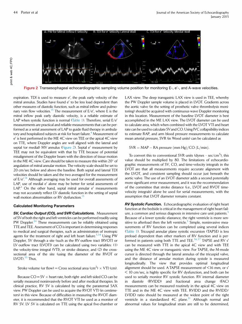
Mitral Inflow. Mitral inflow velocities, both peak early diastolic veloc-ity (E) and late diastolic velocity (A), are commonly used to determinepatterns of diastolic dysfunction and can also be used to seriallymonitor LAP. The mitral E wave represents the LA-LV gradient duringearly diastole and thus is preload dependent. The mitral A wave is theLA-LV gradient during late diastole and is affected by changes in LV dia-stolic function and LA compliance. Mitral inflow velocities (E wave, Awave, DT, and E/A ratio) are measured in the apical 4C viewwith TTEand the ME 4C view with TEE using PWDoppler (Figure 2). The sam-pling volume should be 1 cm distal to the MVannulus or at the leaflettips during diastole, with a sampling gate of 1 to 3 mm.15
Comprehensive explanations of mitral inflow indices for classificationof diastolic function are described in the ASE recommendations forthe evaluation of LV diastolic function.15 It is the recommendation ofthe writing group that the E- and A-wave velocities be used in conjunc-tion with the annular velocities when monitoring for changes in fillingpressures or diastolic function. Figure 2 displays the recommendedsample volume positions for monitoring the tissue Doppler measure-ments of e0 and transmitral measurements of the E and A waves.Although pulmonary venous assessments of systolic filling fractionshave proved feasible for monitoring LAP with TEE in an electivesetting,14 specific cutoffs for normal and abnormal filling pressureshave not been provided, and their feasibility for monitoring LV fillingpressures by TTE has not been demonstrated.4
TDI. PW TDI is a sensitive indicator of LV diastolic function. TDI mea-sures mitral annular velocities during both systole and diastole at end-
print&
web4C=FPO
Figure 2 Transesophageal echocardiographic sampling volume position for monitoring E-, e0-, and A-wave velocities.
44 Porter et al Journal of the American Society of EchocardiographyJanuary 2015
expiration. TDI is used to measure e0, the peak early velocity of themitral annulus. Studies have found e0 to be less load dependent thanother measures of diastolic function, such as mitral inflow and pulmo-nary vein flow velocities.15 The measurement of E/e0, where E is themitral inflow peak early diastolic velocity, is a reliable estimate ofLAP when systolic function is normal (Table 1). Therefore, serial E/e0
measurements are practical and reliablemeasurements that can be per-formed as a serial assessment of LAP to guide fluid therapy in ambula-tory and hospitalized subjects at risk for heart failure.4 Measurement ofe0 is best performed in the ME 4C view on TEE or the apical 4C viewon TTE, where Doppler angles are well aligned with the lateral andseptal (or medial) MV annulus (Figure 2). Septal e0 measurement byTEE may not be equivalent with that by TTE because of potentialmisalignment of the Doppler beam with the direction of tissue motionin theME 4C view. Care should be taken to measure this within 20� ofangulation of mitral annular motion. The velocity scale should be set to20 cm/sec below and above the baseline. Both septal and lateral TDIvelocities should be taken and the two averaged for the measurementof E/e0.14 Although averaging may be used for overall assessments ofLAP, use of medial e0 alone may be better for serial assessments ofLAP.4 On the other hand, septal mitral annular e0 measurementsmay not accurately reflect LV diastolic function in the setting of septalwall motion abnormalities or RV dysfunction.15
Calculated Monitoring Parameters
SV, Cardiac Output (CO), and SVRCalculations. Measurementof SVof both the right and left ventricles can be performed readily usingPW Doppler.34 These measurements can be reliably obtained usingTTE and TEE. Assessment of CO is important in determining responsesto medical and surgical therapies, such as administration of inotropicagents for the treatment of right and left heart failure.5,34 Using PWDoppler, SV through a site (such as the RV outflow tract [RVOT] orLV outflow tract [LVOT]) can be calculated using two variables: (1)the velocity-time integral (VTI), or stroke distance, and (2) the cross-sectional area of the site (using the diameter of the RVOT orLVOT).35 Thus,
Stroke volume (or flow) = Cross sectional area (cm2) � VTI (cm).
Because CO= SV� heart rate, both right- and left-sided CO can beserially measured noninvasively before and after medical therapies. Inclinical practice, RV SV is calculated by using the parasternal SAXview. PW Doppler can be used to acquire the RVOT VTI (in centime-ters) in this view. Because of difficulties in measuring the RVOT diam-eter, it is recommended that the RVOT VTI be used as a monitor ofRV SV. LV SV is calculated on TTE using the apical five-chamber or
LAX view. The deep transgastric LAX view is used in TEE, wherebythe PW Doppler sample volume is placed in LVOT. Gradients acrossthe aortic valve (in the setting of prosthetic valve thrombolysis moni-toring) should be acquired with continuous-wave Doppler monitoringin this location. Measurement of the baseline LVOT diameter is bestaccomplished in the ME LAX view. The LVOT diameter can be usedto calculate area, which when combined with the LVOT VTI and heartrate can be used to calculate SVandCO.Using IVC collapsibility indicesto estimate RAP, and arm blood pressure measurements to calculatemean arterial pressure, SVR (in Wood units) can be calculated as
SVR ¼ MAP� RA pressure ðmm HgÞ=CO ðL=minÞ:To convert this to conventional SVR units (dynes $ sec/cm5), this
value should be multiplied by 80. The limitations of echocardio-graphic measurements of SV, CO, and time-velocity integrals in theLVOT are that all measurements require accurate alignment withthe LVOT, and consistent sampling should occur just beneath theaortic valve. The use of an LVOT diameter adds a second potentiallymore significant error measurement, and it was the recommendationof the committee that stroke distance (i.e., LVOT and RVOT time-velocity integrals) alone be used for serial measurements, with theassumption that LVOT diameter remains constant.
RV Systolic Function. Echocardiographic evaluation of right heartfunction at the bedside is critical in the management of right heart fail-ure, a common and serious diagnosis in intensive care unit patients.5
Because of a lower systolic elastance, the right ventricle is more sen-sitive to afterload then the left ventricle.5 Simple, noninvasive mea-surements of RV function can be completed using several indices(Table 1). Tricuspid annular plane systolic excursion (TAPSE) is lesspreload dependent than other markers of RV function and is per-formed in patients using both TTE and TEE.36,37 TAPSE and RV s0
can be measured with TTE in the apical 4C view and with TEEusing the ME 4C view or transgastric view. For TAPSE, the M-modecursor is directed through the lateral annulus of the tricuspid valve,and the distance of annular motion during systole is measuredlongitudinally. The view that provides optimal longitudinalalignment should be used. A TAPSE measurement of <16 mm, or s0
< 10 cm/sec, is highly specific for RV dysfunction, and both can beused to serially monitor RV systolic function. RV internal diameterin diastole (RVIDD) and fractional area change (FAC)measurements can be measured routinely in the apical 4C view onTTE and in the ME 4C view with TEE. RVIDD and the RVIDD/LVIDD ratio should be measured at the widest point of the rightventricle in a standardized 4C plane.31 Although normal andabnormal values for longitudinal strain are still to be determined,
Table 2 Summary of clinical scenarios in whichechocardiographic monitoring is considered helpful and thelevel of support on the basis of a number of clinical studiesexamining utility in this setting
Clinical scenario
Predominant
monitoring tool Level of data support*
Acute CHF/LVAD TTE B2
Critical care TTE B2
Trauma TTE/TEE D1
Tamponade monitoring TTE B2
Pulmonary embolism TTE B2
Prosthetic valvethrombus
TTE/TEE B2
Kidney/liver/lung
transplantation
TEE Kidney-B3
Liver-B2
Lung-B2
Major vascular surgery TEE B2
Orthopedic/spinal
surgery
TEE B2
Neurosurgery/sitting
position
TEE B2
Journal of the American Society of EchocardiographyVolume 28 Number 1
Porter et al 45
this parameter has been used to monitor RV systolic function duringtherapeutic interventions in pulmonary hypertension.38
PA Systolic Pressure. Besides serial quantification of RV function,pulmonary pressures can also be evaluated by calculating the RV-RAgradient using the modified Bernoulli equation (4V2). Using the peaktricuspid regurgitant jet velocity as V, the RV-RA gradient can becalculated.35 Because RAP can be determined by assessing IVC sizeand collapsibility, PA systolic pressure can be estimated asRAP + RV-RA gradient, where RV-RA gradient is 4 � (peak tricuspidregurgitant jet velocity). The peak tricuspid regurgitant jet velocity ismeasured using continuous-wave Doppler parallel to the tricuspid re-gurgitant jet. This can be performed with TTE in the apical 4C view,parasternal SAX view, or RV inflow view. Doppler through thetricuspid valve in TEE is best performed in either the ME 4C viewor the RV inflow view obtained from transducer angles that alignthe Doppler cursor parallel to the color Doppler jet. An additionaltransesophageal view that is useful for Doppler alignment is obtainedby rotating the viewing angle to 130� to 145� to obtain an apical LAXplane and then rotating clockwise to visualize the tricuspid valve re-gurgitant jet using current guidelines.39 Although measurements aretaken in multiple views with both TTE and TEE, the highest velocitysignal should be used for serial measurements (as this represents themost parallel alignment).
B2, observational studies permit inference of benefit of the guiding
tools listed in Table 3 and clinical outcomes on the basis of noncom-
parative observational studies with associative (e.g. relative risk, cor-relation) or descriptive statistics; B3, observational studies permit
inference of benefit of the monitoring tools in Table 3 on the basis
of case reports only; D1, lack of scientific evidence in the literature
to address whether the monitoring tools in Table 2 affect outcomes.*Please refer to Thys et al.1 for reference to the entire level-of-support
classification.
II. ADVANTAGES, DISADVANTAGES, AND
RECOMMENDATIONS OF ECHOCARDIOGRAPHY AS A
MONITORING TOOL
Patient monitoring is currently performed in most critical care and in-traoperative settings with serial measurements of vital signs, oxygensaturation, end-tidal carbon dioxide monitoring, and occasionallyPA catheters. The use of echocardiographic monitoring is a newconcept that has emerged in several different fields that include cardi-ology, emergency medicine, anesthesiology, and critical care. Asechocardiography becomes more portable, monitoring with echocar-diographic techniques will be used to greater degrees in patient man-agement decisions. Although echocardiographic monitoring hasproved useful in several specific areas outlined in this review, the tech-nique is heavily operator dependent and demands that continuousquality improvement measures be implemented for all practicingechocardiographers within an institution, to ensure that Dopplerand anatomic measurements are following established technicalguidelines that have been published . Second, the data presented inthis document derive for themost part from single-center experiencesand were not rigorously evaluated in prospective studies examiningdifferences in patient outcome as a result of echocardiographicmonitoring. The writing committee has developed a ‘‘level of datasupport’’ that displays the number of studies that have beenperformed for each of the proposed clinical scenarios in which echo-cardiographic monitoring may be useful (Table 2). Note that in thesetting of trauma, there are no clinical comparative studies published,despite numerous reviews suggesting that echocardiographic moni-toring would be useful.
III. CLINICAL SCENARIOS
Expert opinion supports the use of primarily TTE to guide the man-agement of patients in specific clinical settings in which monitoring
of fluid management or drug interventions needs to be assessedrapidly. These are listed in Table 3. Despite this expert opinion, thewriting group could find no formal clinical studies in which moni-toring of echocardiographic Doppler parameters was comparedwith other monitoring modalities in the setting of sepsis, respiratoryfailure, or trauma. Monitoring in CHF, pulmonary embolism, andpericardial tamponade are discussed separately.
Acute CHF Monitoring
In the setting of heart failure, most monitoring applications have dealtwith situations in which Doppler has been used to assess the effects oftherapeutic interventions that will eventually be used for longer termtherapy. Transmitral E- and A-wave velocity ratios, combined with E-wave DT, have been used to assess responsiveness to nitroprussideinfusion and carvedilol therapy, which may also predict prognosis.40
Patients with heart failure and depressed ejection fractions who hadE/A ratios > 1, combined with EDTs < 130 msec, and who did notreverse these parameters with nitroprusside infusion, had the worstprognosis. Changes in mitral filling parameters after leg lifting or nitro-prusside infusion were predictive of tolerance to carvedilol therapyand patient outcome. More recently, studies have evaluated E/e0 tomonitor LAP in response to interventions in patients with symptom-atic heart failure (New York Heart Association classes II and III) in anoutpatient setting and have demonstrated that the E/medial e0 ratiomost accurately reflects changes in LAP.4
Although E/e0 monitoring has been shown to reflect changes inpulmonary capillary wedge pressure in small numbers of patients

Table 3 Specific clinical settings in which echocardiographic monitoring could potentially guide therapeutic interventions
Specific clinical scenario Recommended monitoring parameters
Critical Care Hypotension IVC collapsibility indexRegional wall motion
LVOT VTI response to passive leg raising
CHF IVC collapsibility indexTransmitral E/e0
E/A ratio with EDT
RV s0
TAPSERegional wall motion
Sepsis* IVC collapsibility index
LVIDD, LVIDS
Regional wall motion
Respiratory failure*
Possible pulmonary embolus
IVC collapsibility index
RV s0
TAPSEDoppler PASP
RVOT VTI
Regional wall motion
RVIDD/LVIDD ratio
Pericardial effusion/tamponade Pericardial effusion size
Right ventricular diastolic collapse
IVC collapsibility index
Prosthetic valve dysfunction Transvalvular gradient
Trauma* Blunt trauma
Aortic traumaMyocardial contusion
IVC collapsibility
LVIDD, LVIDSPericardial effusion size
Regional wall motion
Burns IVC collapsibility
LVIDD, LVIDSTransmitral E/A ratio, E/e0
Perioperative Thoracoabdominal cross-clamping Regional wall motion
LVIDD, LVIDS
Liver transplantation LV regional wall motion
LVIDD, LVIDS
RV s0
TAPSERV cavity monitoring for emboli
Renal transplantation LV regional wall motion
LVIDD, LVIDSRV s0
TAPSE
Transmitral E/e0 and E/A ratio
RV cavity monitoring for emboli
Orthopedic/spinal/neurologic surgery RV s0
TAPSE
Transmitral E/e0
RV cavity monitoring for emboli
PASP, PA systolic pressure.*Although potentially useful and used clinically in this setting, no clinical studies have been published examining these echocardiographic param-
eters in monitoring patients in this setting.
46 Porter et al Journal of the American Society of EchocardiographyJanuary 2015
with acute decompensated heart failure,41,42 the writing committeecurrently recommends that E/e0 not be used in monitoring LAP ofpatients with decompensated heart failure with depressed systolicfunction (‘‘cold and wet patients’’), as others have shown that theseparameters do not predict LAPs or guide management in thisclinical setting.16 The primary focus of these studies was to use E/e0
to predict initial pressures, not on E/e0 as amonitoring tool. As a result,
only small subsets of the study population had serial E/e0 measure-ments compared with serial changes in LAPs.
One evolving area in which echocardiographic guidance has becomehelpful in CHF is assessing responsiveness to LVAD therapy.Echocardiographic parameters have been used to serially monitorramped interventions and determine whether patients can be weanedfromLVADtherapy. Rampprotocols are definedas dynamic assessments

print&web4C=FPO
Figure 3 Echocardiographic parameters obtained during the ramp protocol to optimize LVAD settings and assist in the detection ofdevice malfunction. (A) LVIDD changes, (B) changes in aortic valve opening, (C) aortic regurgitation, (D)mitral regurgitation, and (E)changes in RV systolic pressure with each setting change. Reproduced with permission from Uriel et al.11
Journal of the American Society of EchocardiographyVolume 28 Number 1
Porter et al 47
of LV size, hemodynamics, and valvular function with echocardiographyduring incremental device speeds. They have been shown in single-center studies to improve speed optimization and assist in the detectionof device thrombosis. In this setting, the LVAD backup speed is started atthe lowest usable setting (�8,000 rpm) and then increased serially whilemonitoring LVIDD, LVIDS, aortic valve opening, aortic andmitral regur-gitation severity, and RV systolic pressure (Figure 3). Normal resultswould be gradual reductions in LVIDD as the speed is increased to12,000 rpm, while flat responses would indicate device malfunction.11
In theseprotocols, LVIDD is plottedas a functionof change in revolutionsperminute.AnLVIDDslope$�0.16was diagnostic of flowobstructionfrom thrombosis or mechanical obstruction in the LVAD tubing.
Echocardiographic guidance has also been used to determine if pa-tients can be weaned from their LVADs. Once the LVIDD decreasesto <60 mm and mitral regurgitation is reduced in severity on chronicLVAD therapy, the patient is scheduled for an echocardiographicallyguided study in which the LVAD is turned off. LVIDD, LVIDS, and RVsize and function are then assessed; maintenance of LV function(LVEF > 50%) without the development of worsening RV dilatationduring off-pump trials are used as criteria for LVAD removal.12 A lackof change in LVEF or RV size at end-diastole was also associated withgood clinical outcomes after LVAD removal.
Critical Care Monitoring
Although echocardiography plays an invaluable role in assessing thecause of hemodynamic compromise in critically ill patients, its role
for monitoring patients with respiratory failure, sepsis, or unex-plained arrest has not been elucidated. In these settings, there areseveral parameters that could be followed that would be uniqueto echocardiography over other monitoring tools, such as PA cathe-ters or oxygen saturation monitors (Table 4), but to date, no clinicalstudies comparing the techniques have been performed. There areadvantages and disadvantages with either technique. Although serialechocardiography has the advantage of providing anatomic infor-mation regarding changes in systolic and diastolic function, PA cath-eters are more useful when many serial interventions that may affectCO and LV filling pressures are being performed rapidly at thebedside in an acute setting. In the setting of septic shock, goal-directed therapy has been shown to improve patient outcome.43
Although this study used central venous pressure, mean arterialpressure, and central venous oxygen saturation to guide fluid, blood,and vasopressor management, echocardiographic parameters mightbe substituted for most parameters. IVC collapse could be used toassess central venous pressures and LVOTstroke distance to monitorCO. These noninvasive assessments could be combined with bloodpressure monitoring to guide therapy in this setting. In smallnumbers of critically ill patients, an increase in LVOT VTI of>12.5% during passive leg raising predicted increases in SV inresponse to intravenous fluids with 77% sensitivity and 100% spec-ificity.44 The change in LVOT VTI with passive leg raising was moreaccurate than changes in LV dimensions or mitral inflow patterns inpredicting fluid responsiveness.

Table 4 Methods by which critical monitoring parameters are assessed with a PA catheter versus serial echocardiographicmeasurements
Monitoring technique PA catheter Echocardiography Advantage*
Filling pressures RAP, PCWP directly measured RAP, PCWP indirectly measured PA catheter
Cardiac output Thermodilution Doppler derived Equal
Valve assessment Not possible Anatomic/Doppler Echocardiography
Systolic/diastolic function Estimated from PCWP/CO Table 1 parameters Echocardiography
Risks Invasive technique Noninvasive Echocardiography
Speed of assessment Immediate changes detected Operator dependent PA catheter
PCWP, Pulmonary capillary wedge pressure.
*Advantage refers to which monitoring parameter the writing group considered better suited for the particular application.
48 Porter et al Journal of the American Society of EchocardiographyJanuary 2015
Pericardial Tamponade Monitoring
Echocardiographic guidance has played a critical role in decision mak-ing for patients presenting with significant pericardial effusion andguiding pericardiocentesis and postpericardiocentesis management.Echocardiographic monitoring plays a vital role in patients presentingwith pericardial effusion when the rate of accumulation is unknownand the patients have nonspecific symptoms. Approximately 33%of large idiopathic pericardial effusions may suddenly develop tampo-nade physiology.45 Along with monitoring for increase in pericardialeffusion size, monitoring for the development of right atrial collapse(lasting for greater than one-third of the cardiac cycle), early RV dia-stolic collapse, and IVC plethora have been used for determiningwhen pericardiocentesis may be indicated.45
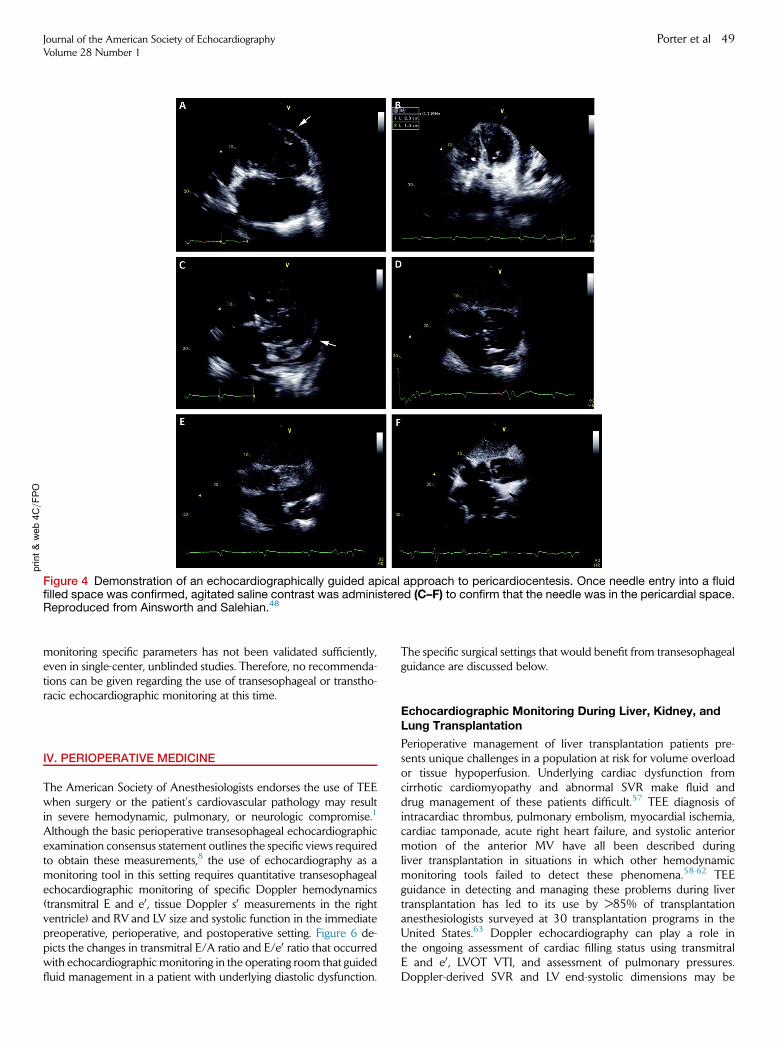
For pericardiocentesis, echocardiographic monitoring has replacedfluoroscopy at many centers because of its ability to detect and guideneedle and catheter placement for loculated effusions.46-48 Usingechocardiographic guidance has resulted in more apical, rather thansubxiphoid, approaches to pericardiocentesis, mainly because thedistance to effusion from the skin surface is smallest in this location.The use of saline contrast administered through thepericardiocentesis needle confirms pericardial entry (Figure 4,Video 1; available at www.onlinejase.com). After this confirmation,echocardiography can be used to confirm both guidewire and pigtailcatheter placement into the pericardial space.48 Drainage then pro-ceeds until there is near disappearance of pericardial effusion onechocardiography (Video 1). The ultrasound transducer can be eithersterilized by placing a gel-filled sterile sleeve over the transducer orplaced in an imaging area that is outside the sterile field. This echocar-diographically guided procedure has a >95% success rate, with a largesingle-site study demonstrating minimal complications.46 Afterdrainage of the effusion, repeat echocardiography within 24 hoursis indicated to examine for reaccumulation. At this point, IVC collapsecan be reevaluated to see if RAPs have decreased or if RAPs remainelevated, as may occur in effusive constrictive pericarditis.45
Pulmonary Embolism Therapy Monitoring
Despite the widespread use of echocardiography in assisting in thediagnosis and initial management of pulmonary embolism, thereare very few published data on the usefulness of monitoring RV sys-tolic function or PA pressures in this setting. Nonetheless, echocardi-ography plays a significant role in making therapeutic decisions inpatients with pulmonary embolism and can facilitate a change inman-agement by identifying those at high risk who might otherwise betreated with less aggressive therapies.49-51 In addition, it can help
assess whether thrombus within the main PA is present, which maywarrant surgical embolectomy. Most important, it is useful tomonitor RV function and PA systolic pressure when thrombolysis isadministered.52
Patients with suspected pulmonary embolism often present withsigns and symptoms that are nonspecific, which can make it difficultto distinguish the diagnosis from other life-threatening disorders.Although not diagnostic of pulmonary embolism, initial TTE canhelp in identifying when pulmonary embolism may be the cause bydetecting RV dilation (RVIDD/LVIDD ratio > 0.9) and assist withruling out other causes, such as pericardial effusion or myocardialinfarction.50,51 Once the diagnosis of pulmonary embolus isestablished, these patients can be risk-stratified according to the ef-fects of elevated RV afterload: hypotensive patients and those withelevated cardiac biomarkers or echocardiographic indices of RV strainare at an increased risk, and thrombolysis is considered a class II indi-cation.52 Patients with massive pulmonary embolism can have serialassessments of RV size and FAC (Figure 5), assessments of RV systolicpressure, and IVC assessments using ASE RV guidelines for normalranges.31 The writing group recommends that considerable attentionbe given to maintaining the same identical imaging plane of the rightventricle when serially examining RV size and FAC, as slight devia-tions in the imaging plane may alter these values.
Prosthetic Valve Thrombosis Monitoring
Both TTE and TEE have been used to detect prosthetic valve throm-bosis and monitor therapy effectiveness.53-56 Fibrinolytic therapy isrecommended if left-sided prosthetic valve thrombus area (planime-tered on a 2D image) is <0.8 cm2, with serial Doppler echocardio-graphic monitoring of mean gradients across the valve to assesseffectiveness of either fibrinolytic or unfractionated heparin treat-ment.55 A significant reduction in the transvalvular gradient at24 hours is indicative of effective therapy. TEE and TTE are comple-mentary in these settings, with serial TEE giving better visualization ofresidual thrombus burden, but both are equally effective at moni-toring for reductions in transvalvular gradients.
Echocardiographic Monitoring in Trauma
TTE and TEE have been proposed as methods tomonitor volume sta-tus and regional and global systolic function in a wide variety of trau-matic settings. Although they have proved useful in the immediateassessment of LV and RV systolic function, volume status, and detec-tion of significant pericardial or aortic pathology, their role in
print&
web4C=FPO
Figure 4 Demonstration of an echocardiographically guided apical approach to pericardiocentesis. Once needle entry into a fluidfilled space was confirmed, agitated saline contrast was administered (C–F) to confirm that the needle was in the pericardial space.Reproduced from Ainsworth and Salehian.48
Journal of the American Society of EchocardiographyVolume 28 Number 1
Porter et al 49
monitoring specific parameters has not been validated sufficiently,even in single-center, unblinded studies. Therefore, no recommenda-tions can be given regarding the use of transesophageal or transtho-racic echocardiographic monitoring at this time.
IV. PERIOPERATIVE MEDICINE
The American Society of Anesthesiologists endorses the use of TEEwhen surgery or the patient’s cardiovascular pathology may resultin severe hemodynamic, pulmonary, or neurologic compromise.1
Although the basic perioperative transesophageal echocardiographicexamination consensus statement outlines the specific views requiredto obtain these measurements,8 the use of echocardiography as amonitoring tool in this setting requires quantitative transesophagealechocardiographic monitoring of specific Doppler hemodynamics(transmitral E and e0, tissue Doppler s0 measurements in the rightventricle) and RV and LV size and systolic function in the immediatepreoperative, perioperative, and postoperative setting. Figure 6 de-picts the changes in transmitral E/A ratio and E/e0 ratio that occurredwith echocardiographic monitoring in the operating room that guidedfluid management in a patient with underlying diastolic dysfunction.
The specific surgical settings that would benefit from transesophagealguidance are discussed below.
Echocardiographic Monitoring During Liver, Kidney, andLung Transplantation
Perioperative management of liver transplantation patients pre-sents unique challenges in a population at risk for volume overloador tissue hypoperfusion. Underlying cardiac dysfunction fromcirrhotic cardiomyopathy and abnormal SVR make fluid anddrug management of these patients difficult.57 TEE diagnosis ofintracardiac thrombus, pulmonary embolism, myocardial ischemia,cardiac tamponade, acute right heart failure, and systolic anteriormotion of the anterior MV have all been described duringliver transplantation in situations in which other hemodynamicmonitoring tools failed to detect these phenomena.58-62 TEEguidance in detecting and managing these problems during livertransplantation has led to its use by >85% of transplantationanesthesiologists surveyed at 30 transplantation programs in theUnited States.63 Doppler echocardiography can play a role inthe ongoing assessment of cardiac filling status using transmitralE and e0, LVOT VTI, and assessment of pulmonary pressures.Doppler-derived SVR and LV end-systolic dimensions may be

print&
web4C=FPO
Figure 5 Serial echocardiograms in a patient with a pulmonary embolus treated with fibrinolytic therapy. FAC improved significantlyafter fibrinolytic therapy, and RVIDD decreased. RV systolic pressure decreased by >50 mm Hg.
50 Porter et al Journal of the American Society of EchocardiographyJanuary 2015
used to monitor dynamic changes in vasodilated states. Detectionof intracardiac thrombus, ventricular function, pericardial effusion,and monitoring for systolic anterior motion of the mitral valve canall be detected with serial 2D TEE monitoring in this population(Figure 7, Video 2; available at www.onlinejase.com), in whichchanges in hemodynamic and prothrombotic conditions occurrapidly.64-67 As long as a patient is not receiving positivepressure ventilation, IVC collapsibility and size can be used topredict fluid responsiveness for postoperative fluid managementalong with ongoing 2D and Doppler hemodynamic and functionassessment. Before considering TEE as a monitoring tool duringliver transplantation, it is important to note that esophagealvarices may be present and that this is considered a relativecontraindication to performing TEE.39 Therefore, the writing grouprecommends lubrication and careful probe insertion to minimizeesophageal variceal bleeding.
Large systematic evaluations of the role of TEE monitoring inkidney transplantation are lacking, and there are only limited datato demonstrate that it can add to central venous pressure monitoringin volume assessment and management of ischemia reperfusioninjury.68 The writing group recommends that the use of TEE as amonitoring tool of LVand RV systolic and diastolic function be consid-ered only if coexisting cardiovascular disease is present.
With regard to echocardiographic monitoring during lung trans-plantation, there is a consensus that TEE is essential to monitoringRV systolic function during and after transplantation.69 Changes in
RV contractility must be identified early, so that inotropic supportor inhaled PAvasodilators can be initiated before overt hemodynamiccompromise occurs. TEE is also used to monitor the pulmonary veinsfor any stenoses that may develop from thrombosis at the anasto-motic sites.70
Major Vascular Surgery
Direct clamping of major vessels causes a sudden significant increasein cardiac loading conditions, which may lead to hemodynamic insta-bility from ventricular failure, myocardial ischemia, and end-organ hy-poperfusion. Echocardiography indices can be used tomonitor effectsof cross-clamping of the aorta or the vena cava on both diastolic andsystolic function.71-75 Previous studies have shown TEE to be moresensitive than PA catheters in the detection of alterations in systolicand diastolic function during cross-clamping of the thoracic and thor-acoabdominal aorta.73,74 Echocardiographic indices that arerecommended to detect these dynamic changes include changes inCO, LV ejection fraction, LV end-diastolic dimensions, regional wallmotion in the transgastric SAX view, and transmitral Doppler flowpatterns.
Complete occlusion of the vena cava can also cause significantchanges in preload and afterload. Intraoperative assessment of ven-tricular filling, wall motion, and both diastolic and systolic functionmay be used to guide medical intraoperative therapy during venacava cross-clamping.

print&web4C=FPO
Figure 6 Intraoperative transesophageal echocardiographic monitoring in two different liver transplantation cases. In the top panel,one sees a normal relatively low E/e0 ratio before liver transplantation, followed by an increase to 8 (C,D) after IVC clamp removal. Thisled to a cessation of intravenous fluid administration. In the bottom panels, one sees a decrease in the E/A ratio during IVC clampingduring liver transplantation (B, bottom panel) but a dramatic increase in the E/A ratio after clamp removal (C, bottom panel). This led toan immediate reduction in fluid administration.
Journal of the American Society of EchocardiographyVolume 28 Number 1
Porter et al 51

print&
web4C=FPO
Figure 7 Transesophageal echocardiographic images during liver transplantation demonstrating acute RV dilation due to emboliza-tion of debris during the initial dissection (left). After the procedure was aborted and anticoagulation given, there was dissolution ofdebris, and RV size decreased (right). See Video 2 (available at www.onlinejase.com).
52 Porter et al Journal of the American Society of EchocardiographyJanuary 2015
Orthopedic and Spinal Surgery
Intraoperative TEE is used in this setting primarily as a rescue proce-dure. Hip arthroplasty, spinal surgery, and knee arthroplasty are allassociated with significant risk for intraoperative cement and fatemboli. Hypotension, ventilation-perfusion mismatch, hypoxemia,pulmonary embolism, and cardiac collapse can all occur during intra-medullary reaming and release of microparticulate matter.76
Intraoperative rescue TEE can be used to monitor microemboli anddetect intracardiac shunting using color-flow Doppler through a pat-ent foramen ovale.77 Both TEE and TTE have been reported to beuseful hemodynamic monitors for lengthy orthopedic and spinal sur-gery.78-81 However, it should be noted that the majority of spinalsurgery is done in the prone position, and TEE is not used. Changesin RV function due to increases in acute pulmonary vascularresistance can be detected using TAPSE, pulmonic valve VTI, andpeak tricuspid regurgitant jet velocity measurements. Fat embolican be visualized with TEE during hip arthroplasty; its identificationhas been associated with neurologic dysfunction and a subsequenthigher American Society of Anesthesiologists physical status($III).81 Despite its limited use, TEE has detected thromboembolicevents during cervical spine surgery.82
Neurosurgery
The vast majority of all neurosurgery (other than spinal) in the UnitedStates is done in the supine position, and TEE monitoring is used pri-marily in a rescue setting. The potential for venous air embolism dur-ing neurosurgery has led to the ‘‘equivocal’’ endorsement of the use ofintraoperative TEE as category B by the American Society ofAnesthesiologists during such procedures.1 Evaluation of the intera-trial septum by color-flow Doppler and agitated saline contrast toassess the risk for paradoxical emboli associated with a patent fora-men ovale can be performed during intraoperative monitoring.Doppler assessment of right-sided pulmonary pressures and 2Dassessment of RV function can detect changes secondary to venousair embolic load, especially in procedures done in the sitting posi-tion.83,84 TEE has been used to guide the placement of right atrialaspiration catheters to an optimal location at the junction of thesuperior vena cava and right atrium.85,86 Ongoing intraoperativeassessment for air entrapment into right-sided cardiac chambers isrecommended when the risk for paradoxical emboli is high.Identification of these complications early, along with careful qualita-tive and quantitative assessments of RV systolic function, may assistsignificantly in preventing hemodynamic deterioration and permit
earlier pharmacologic or surgical interventions. Although TEE moni-toring is useful for neurosurgery in the sitting position, it should benoted that TEE monitoring in the sitting position has been associatedwith posterior tongue edema and even necrosis.87 Further controlledstudies are needed to define the beneficial role of transesophagealmonitoring in this setting.
V. WHEN HAS A MEANINGFUL CHANGE IN A MONITORING
PARAMETER OCCURRED?
On the basis of available evidence, the use of echocardiography formonitoring purposes is justified when categorical changes haveoccurred, such as an increase in LAP from normal to abnormal (an in-crease in E/e0 ratio from <8 to >13 in the setting of normal systolicfunction). It can be used for continuous monitoring in settings inwhich LVIDD, RV FAC, or PA systolic pressure are being reevaluatedin ramp or weaning protocols. In both of these settings, it is importantto note what degree of change must occur before one can say thegiven change is beyond the interobserver variability of the measure-ment (Table 588-93). Although only small numbers of patients areincluded in these studies of variability measurements, they doprovide assistance in determining what cutoffs to use whendeciding whether a change in a parameter is beyond what wouldbe expected on the basis of interobserver variability or coefficientsof variation. In the specific areas of IVC collapsibility index, E/e0, E/A, and PA systolic pressure changes, we have added categoricalchanges (on the basis of guidelines) that should be used whenguiding management. These categorical changes are well within thepublished data regarding coefficient of variation and interobservervariability of the monitoring parameter.
VI. CONCLUSIONS REGARDING TRAINING AND USE OF
ECHOCARDIOGRAPHY AS A MONITORING TOOL
Thewriting committee emphasizes that a minimum of level II trainingexperience94 is required to use echocardiography as a quantitativemonitoring tool, regardless of the clinical scenario for which it is beingapplied. Although level II Core Cardiology Training Symposiumtraining experience in TTE is sufficient to monitor LV dimensionsand IVC collapsibility, additional level III Core Cardiology TrainingSymposium training experience with both TTE and TEE is required

Table 5 Interobserver variability and coefficients of variation for specific echocardiographic monitoring parameters, withrecommended meaningful changes that must occur in a clinical scenario (monitoring setting)
Echocardiographic monitoring
parameter IOV/CV Monitoring setting
Meaningful changes from baseline
in a monitoring setting
IVC collapsibility index Not demonstrated CHF, trauma, perioperative >10%32
Change from <50% to >50%31
E/A ratio
Depressed LV systolic function
6% CV88 CHF, perioperative Change from <1 to 1–2 to 2
E/e0
Normal LV systolic function
8% CV CHF, perioperative >8%89
Change from <8 to 9–14 to$1515
LVOT VTI
LVOT area
6% IOV
4 % IOV
CHF, perioperative setting >6% change in VTI or SV90
PASP 3% IOV Pulmonary embolus,
perioperative, CHF
>3%91
Change from <40 to 40–60 to
>60 mm Hg31
LVIDD, LVIDS 8% IOV Perioperative, CHF ramp/weaning >8%92
RV FAC, S0, and TAPSE RV FAC:10% (IOV)
RV s0: 1.6 mm/sec (IOV)
TAPSE: 1.9 mm (IOV)
Pulmonary embolus
Perioperative
Pulmonary hypertensionCHF LVAD
RV FAC > 10%93
RV s0 > 1.6 mm/sec93
TAPSE > 1.9 mm93
CV, Coefficient of variation; IOV, interobserver variability; PASP, PA systolic pressure.
Journal of the American Society of EchocardiographyVolume 28 Number 1
Porter et al 53
to ensure accurate Doppler and advanced hemodynamic pressuremeasurements in intensive care units, emergency departments, andsurgical suites. Because echocardiographic monitoring is currentlyused in a wide range of clinical settings, it is imperative that the personusing quantitative echocardiography to guide therapeutic decisionmaking (whether an anesthesiologist, a cardiologist, or an emergencyroom physician with level II or level III experience) understand theinterobserver variability of each of the quantitative measurements(as displayed and referenced in Table 5) and have the technical exper-tise required to ensure the serial measurements are obtained accu-rately. Although high-quality images can be obtained by those withlesser experience, as outlined in the basic perioperative transesopha-geal echocardiography consensus statement and focused cardiac ul-trasound recommendations,8,13 the interpretation and use of thequantitative parameters to guide therapeutic decision makingoutlined in this document should be done only by level II and IIItrained personnel. For example, is there a significant global wallmotion abnormality in the left or right ventricle that has developedduring the intraoperative monitoring? Such detection would berequired for basic perioperative or critical care monitoring, but doesnot require a quantitative assessment of LVIDD or LVIDS or theIVC to guide fluid resuscitation and does not require ameasurement of RV FAC in the setting of monitoring or guidinginterventions in pulmonary embolus or intraoperative embolismafter the release of IVC cross-clamping. Quantitative measurements,when used to guide therapeutic decision making, require a minimumof level II training.
In conclusion, a sufficient body of literature exists, originating fromcritical care, anesthesiology, and emergency medicine, demonstratingthe potential role of echocardiographic monitoring. Clinical studieshave demonstrated the role of echocardiographic monitoring inguiding management of pulmonary emboli, pericardial effusions,thrombosed prosthetic valves, and acute heart failure management.However, large-scale clinical trials documenting the effectiveness ofechocardiography as a monitoring tool are lacking in all of these areas,
and basic clinical trials are lacking on the use of echocardiography inguiding trauma management or other critical care and surgical appli-cations. Although a sufficient amount of data have been publishedusing interventional echocardiography to guide percutaneous cardiacinterventions,95 it is a strong consensus recommendation from thewriting group that additional clinical trials be performed that docu-ment the utility of both TTE and TEE as dynamic monitoring modal-ities to aid in the treatment of several acute medical and surgicalconditions.
NOTICE AND DISCLAIMER
This report is made available by the ASE as a courtesy referencesource for its members. This report contains recommendations onlyand should not be used as the sole basis to make medical practice de-cisions or for disciplinary action against any employee. The statementsand recommendations contained in this report are based primarily onthe opinions of experts rather than on scientifically verified data. TheASE makes no express or implied warranties regarding the complete-ness or accuracy of the information in this report, including the war-ranty of merchantability or fitness for a particular purpose. In no eventshall the ASE be liable to you, your patients, or any other third partiesfor any decision made or action taken by you or such other parties inreliance on this information. Nor does your use of this informationconstitute the offering of medical advice by the ASE or create anyphysician-patient relationship between the ASE and your patientsor anyone else.
ACKNOWLEDGMENTS
The writing committee would like to thank Dr. Feng Xie, JulieSommer, and Stacey Therrien for their assistance with manuscriptand figure preparation.

54 Porter et al Journal of the American Society of EchocardiographyJanuary 2015
SUPPLEMENTARY DATA
Supplementary data related to this article can be found at http://dx.doi.org/10.1016/j.echo.2014.09.009.
REFERENCES
1. Thys DM, Abel MD, Brooker RF, CahalanMK, Connis RT, Duke PG, et al.Practice guidelines for perioperative transesophageal echocardiography:an updated report by the American Society of Anesthesiologists and theSociety of Cardiovascular Anesthesiologists task force on transesophagealechocardiography. Anesthesiology 2010;112:1084-96.
2. Hofer CK, Zollinger A, Rak M, Matter-Esner S, Klaghofer R, Pasch Th,Zalunardo MP. Therapeutic impact of intra-operative transesophagealechocardiography during non-cardiac surgery. Anaesthesia 2004;59:3-9.
3. Memtsoudis SG, Rosenberger P, Loffler M, Eltzschig HK, Mizuguchi A,Shernan SK, Fox JA. The usefulness of transesophageal echocardiographyduring intraoperative cardiac arrest in non-cardiac surgery. Anesth Analg2006;102:1653-7.
4. Ritzema JL, RichardsAM,Crozier IG, FramptonCF,Melton IC,DoughtyRN,et al. Serial Doppler echocardiography and tissue Doppler imaging in thedetection of elevated directly measured left atrial pressure in ambulant sub-jects with chronic heart failure. J Am Coll Cardiol Img 2011;4:927-34.
5. Lahm T, McCaslin CA, Wozniak TC, GhummanW, Fadl YY, Obeidat OS,et al. Medical and surgical treatment of acute right ventricular failure. J AmColl Card 2010;56:1435-46.
6. Rydman R, Larsen F, Caidahl K, AlamM. Right ventricular function in pa-tients with pulmonary embolism: early and late findings using Doppler tis-sue imaging. J Am Soc Echocardiogr 2010;23:531-7.
7. Kohli-Seth R, Neuman T, Sinha R, Bassily-Marcus A. Use of echocardiog-raphy and modalities of patient monitoring of trauma patients. Curr OpinAnaesth 2010;23:239-45.
8. Reeves ST, Finley AC, et al. Basic perioperative transesophageal echocar-diography examination: a consensus statement of the American Society ofEchocardiography and the Society of Cardiovascular Anesthesia. J Am SocEchocardiogr 2013;26:443-56.
9. Chimot L, Legrand M, Canet E, Lemiale V, Azoulay E. Echocardiographyin hemodynamic monitoring. Chest 2010;137:501-2.
10. Fox JC, Irwin Z. Emergency and critical care imaging. Emerg Med ClinNorth Am 2008;26:787-812.
11. Uriel N, Morrison KA, Garan AR, Kato TS, Yuzefpolskaya M, Latif F, et al.Development of a novel echocardiography ramp test for speed optimiza-tion and diagnosis of device thrombosis in continuous-flow left ventricularassist devices. J Am Coll Cardiol 2012;60:1764-75.
12. Dandel M, Weng Y, Siniawski H, Stepanenko A, Krabatsch T, Potapov E,et al. Heart failure reversal by ventricular unloading in patients withchronic cardiomyopathy: criteria for weaning from ventricular assist de-vices. Eur Heart Journal 2011;32:1148-60.
13. Spencer KT, Kimura BJ, Korcarz CE, Pellikka PA, Rahko PS, Siegel RJ.Focused cardiac ultrasound: recommendations from the American Soci-ety of Echocardiography. J Am Soc Echocardiogr 2013;26:567-81.
14. Kuecherer HF, Muhiudeen IA, Kusumoto FM, Lee E, Moulinier LE,CahalanMK, et al. Estimation ofmean left atrial pressure from tranesopha-geal pulsed Doppler echocardiography of pulmonary venous flow. Circu-lation 1990;82:1127-39.
15. Nagueh SF, Appleton CP, Gillebert TC, Marino PN, Oh JK, Smiseth OA,et al. Recommendations for the Evaluation of Left Ventricular DiastolicFunction by Echocardiography. J Am Soc Echocardiogr 2009;22:107-33.
16. Mullens W, Borowski AG, Curtin RJ, Thomas JD, Tang WH. TissueDoppler imaging in the estimation of intracardiac filling pressures in de-compensated patients with advanced systolic heart failure. Circulation2009;119:62-70.
17. Mandeville JC, Colebourn CL. Can transthoracic echocardiography beused to predict fluid responsiveness in the critically ill patient? A systematicreview. Crit Care Research Prac 2012. Article ID 513480.
18. BrochO, Renner J, GruenewaldM,Meybohm P, Hocker J, Schottler J, et al.Variation of left ventricular outflow tract velocity and global end-diastolicvolume index reliably predict fluid responsiveness in cardiac surgery pa-tients. J Crit Care 2012;27:e7-325.
19. Shah MR, Hasselblad V, Stevenson LW, Binanay C, O’Connor CM,Sopko G, et al. Impact of the pulmonary artery catheter in critically ill pa-tients. JAMA 2005;294:1664-70.
20. Sandham JD, Hull RD, Brant RF, Knox L, Pineo GF, Doig CJ, et al. A ran-domized, controlled trial of the use of pulmonary artery catheters in high-risk surgical patients. N Engl J Med 2003;348:5-14.
21. Zile MR, Brutsaert DL. New concepts in diastolic dysfunction and diastolicheart failure: part 1: diagnosis, prognosis, and measurements of diastolicfunction. Circulation 2002;105:1387-93.
22. Hammill BG, Curtis LH, Bennett-Guerro E, O’Connor CM, Jollis JG,Schulman KA, et al. Impact of heart failure on patients undergoing majornon-cardiac surgery. Anesthesiology 2008;108:559-67.
23. Redfield MM, Jacobsen SJ, Burnett JC Jr., Mahoney DW, Bailey KR,Rodeheffer RJ. Burden of systolic and diastolic ventricular dysfunctionin the community: appreciating the scope of the heart failure epidemic.JAMA 2003;289:194-202.
24. Swaminathan M, Nicoara A, Phillips-Bute BG, Aeschlimann N,MilanoCA,MackensenGB, et al. Utility of a simple algorithm to grade dia-stolic dysfunction and predict outcome after coronary artery bypass graftsurgery. Ann Thorac Surg 2011;91:1844-51.
25. Groban L, Sanders DM, Houle TT, Antonio BL, Ntuen EC, Zvara DA,et al. Prognostic value of tissue Doppler-derived E/e’ on early morbidevents after cardiac surgery. Echocardiography 2010;27:131-8.
26. Grocott MP, Mythen MG, Gan TJ. Perioperative fluid management andclinical outcomes in adults. Anesth Analg 2005;100:1093-106.
27. Nisanevich V, Felsenstein I, Almogy G, Weissman C, Einav S, Mattot I. Ef-fect of intraoperative fluid management on outcome after intrabdominalsurgery. Anesthesiology 2005;103:25-32.
28. Corcoran T, Rhodes JE, Clarke S, Myles PS, Ho KM. Perioperative fluidmanagement strategies in major surgery: a stratified meta-analysis. AnesthAnalg 2012;112:640-51.
29. Machare-Delgado E, Decaro M, Marik PE. Inferior vena cava variationcompared to pulse contour analysis as predictors of fluid responsiveness:a prospective cohort study. J Int Care Med 2011;26:116-24.
30. LanspaMJ, GrissomCK, et al. Applying dynamic parameters to predict he-modynamic response to volume expansion in spontaneously breathingpatients with septic shock. Shock 2013;39:155-60.
31. Rudski LG, Lai WW, Afilalo J, Hua L, Handschumacher MD,Chandrasekaran K, et al. Guidelines for the echocardiographic assessmentof the right heart in adults: a report from the American Society of Echocar-diography. Endorsed by the European Association of Echocardiography, aregistered branch of the European Society of Cardiology, and the CanadianSociety of Echocardiography. J Am Soc Echocardiogr 2010;23:685-713.
32. Brennan JM, Ronan A, Goonewardena S, Blair JEA, Hammes M, Shah D,et al. Hand carried ultrasound measurement of inferior vena cava forassessment of intravascular volume status in the outpatient hemodialysisclinic. Clin J Am Soc Nephrol 2006;1:749-53.
33. Lang RM, Bierig M, Devereux RB, Flachkampf FA, Foster E, Pellikka PA,et al. Recommendations for chamber quantification: a report from theAmerican Society of Echocardiography’s Guidelines and Standards Com-mittee and the Chamber Quantification Writing Group. J Am Soc Echo-cardiogr 2005;18:1440-63.
34. Ristow B, Na B, Ali S, Whooley MA, Shiller NB. Left ventricular outflowtract and pulmonary artery stroke distances independently predict heartfailure hospitalization and mortality: the Heart and Soul Study. J AmSoc Echocardiogr 2011;24:565-72.
35. Quinones MA, Otto CM, Stoddard M, Wagonner A, Zoqhbi WA, et al.Recommendations for Quantification of Doppler echocardiography: areport from the Doppler quantification task force of the nomenclatureand standards committee of the American society of echocardiography.J Am Soc Echocardiogr 2002;15:167-84.
36. Forfia PR, Fisher MR, Mathai SC, Housten-Harris T, Hemnes AR,Borlauq BA, et al. Tricuspid annular displacement predicts survival in
Journal of the American Society of EchocardiographyVolume 28 Number 1
Porter et al 55
pulmonary hypertension. Am J Respir Crit Care Med 2006;174:1034-41.
37. Meluzin J, Spinarova L, Bkala J, Toman J, Krejci J, Hude P, et al. PulsedDoppler tissue imaging of the velocity of tricuspid annular systolic motion.Eur Heart J 2001;22:340-8.
38. Hardegree EL, Sachdev A, Villarraga HR, Frantz RP, McGoon MD,Kushwaha SS, et al. Role of serial quantitative assessment of right ventric-ular function by strain in pulmonary arterial hypertension. Am J Cardiol2013;111:143-8.
39. Hahn RT, Abraham T, Adams MS, Bruce CJ, Glas KE, Lang RM,Reeves ST, et al. Guidelines for performing a comprehensive transesopha-geal echocardiogram examination: Recommendations from the AmericanSociety of Echocardiography and the Society of Cardiovascular Anesthe-siologists. J Am Soc Echocardiogr 2013;26:921-64.
40. Capomolla S, Pinna GD, Febo O, Caporotondi A, Guazzotti G, LaRovere MT, et al. Echo Doppler mitral flow monitoring: an operatortool to evaluate day to day tolerance to and effectiveness of beta adren-ergic blocking agent therapy in patients with chronic heart failure. J AmColl Cardiol 2001;38:1675-84.
41. Nagueh SF, Bhatt R, Vivo RP, Krim SR, et al. Echocardiographic evaluationof hemodynamics in patients with decompensated heart failure. Circ Car-diovasc Img 2011;4:220-7.
42. Dokainish H, Zogbhi WA, Lakkis NM, Al-Bakshy F, Dhir M,Quinones MA, et al. Optimal noninvasive assessment of left ventricularfilling pressures. A comparison of tissue Doppler echocardiography andB-type natriuretic peptide in patients with pulmonary artery catheters. Cir-culation 2004;109:2432-9.
43. Rivers E, Nguyen B, Havstad S, Ressler J, Muzzin A, Knoblich B, et al. Earlygoal directed therapy of severe sepsis and septic shock. N Engl J Med2001;345:1368-77.
44. Lamia B, Ochagavia A, Monnet X, Chemia D, Richard C, Teboul JL. Echo-cardiographic prediction of volume responsiveness to critically ill patientswith spontaneous breathing activity. Intensive Care Med 2007;33:1125-32.
45. Khandaker MH, Espinosa RE, Nishimura RA, Sinak LJ, Hayes SN,Melduni RM, et al. Pericardial disease: diagnosis and management.Mayo Clin Proc 2010;85:572-93.
46. Tsang TS, Enriquez-Sarano M, Freeman WK, Barnes ME, Sinak LJ,Gersh BJ, et al. Consecutive 1127 therapeutic echocardiographicallyguided pericardiocenteses: clinical profile, practice patterns, and out-comes spanning 21 years. Mayo Clinic Proc 2002;77:429-36.
47. The Task Force on the Diagnosis and Management of Pericardial Diseasesof the European Society of Cardiology. Guidelines on the Diagnosis andManagement of Pericardial Diseases Executive Summary. Eur Heart J2004;25:587-610.
48. Ainsworth CD, Salehian O. Echo-guided pericardiocentesis: let the bub-bles show the way. Circulation 2011;123:e210-1.
49. Cohen R, Loarte P, Navarro V, Mirrer B. Echocardiographic findings inpulmonary embolism: An important guide for the management of the pa-tient. W J Cardiovasc Dis 2012;2:161-4.
50. Price LC, Wort SJ, Finney SJ, Marino PS, Brett SJ. Pulmonary vascular andright ventricular dysfunction in adult critical care: current and emergingoptions for management: a systematic literature review. Critical Care2010;14:R169.
51. Torbicki A, Perrier A, Konstantinides S, Agnelli G, Galie N, Pruszczyk P,et al. Guidelines on the diagnosis and management of acute pulmonaryembolism. Eur Heart J 2008;29:2276-315.
52. Jaff MR,McCurty S, et al. Management of massive and submassive pulmo-nary embolus, ilio-femoral deep venous thrombosis, and chronic throm-boembolic pulmonary hypertension: a scientific statement from theAmerican Heart Association. Circulation 2011;123:1788-838.
53. Vasan RS, Kaul U, Sanqhvi S, Kamlakr T, Neqi PC, Shrivastava S, et al.Thrombolytic therapy for prosthetic valve thrombosis: a study based on se-rial Doppler echocardiographic evaluation. AmHeart J 1992;123:1575-80.
54. Koller PT, Arom KV. Thrombolytic therapy of left-sided prosthetic valvethrombosis. Chest 1995;108:1683-9.
55. Whitlock RP, Sun JC, Fremes SE, Rubens FD, Teoh KH, et al. Antithrom-botic and thrombolytic therapy for valvular disease. Antithrombotic ther-apy and prevention of thrombosis, 9th ed: American College of ChestPhysicians evidence-based clinical practice guidelines. Chest 2012;141(Suppl):e576S-600.
56. Zogbhi WA, Chambers JB, et al. Recommendations for evaluation of pros-thetic valves with echocardiography and Doppler ultrasound. J Am SocEchocardiogr 2009;22:975.
57. Zardi EM, Abbate A, Zardi DM, Dobrina A, Margiotta D, Van Tassel BW,et al. Cirrhotic cardiomyopathy. J Am Coll Cardiol 2010;56:539-40.
58. FeiermanD. Case presentation: transesophageal echocardiography duringorthotopic liver transplantation—not only a different diagnosis, butdifferent management. Liver Transpl Surg 1999;5:340-1.
59. Ellenberger C, Mentha G, Giostra E, Licker M. Cardiovascular collapsedue to massive pulmonary embolism during orthotopic liver transplanta-tion. J Clin Anesth 1999;18:367-71.
60. Planinsic RM, Nicolau-Raducu R, Eqhtesad B, Marcos A. Diagnosis andtreatment of intracardiac thrombosis during orthotopic liver transplanta-tion. Anesth Analg 2004;99:353-6.
61. Sharma A, Pagel PS, Bhatia A. Intraoperative iatrogenic acute pericardialtamponade: use of rescue tranesophageal echocardiography in a patientundergoing orthotopic liver transplantation. J Cardiothorac Vasc Anesth2005;19:364-6.
62. Xia VW, Ho JK, Nourmand H, Wray C, Busuttil RW, Steadman RH. Inci-dental intracardiac thromboemboli during liver transplantation: incidence,risk factors, and management. Liver Transpl 2010;16:1421-7.
63. Wax DB, Torres A, Scher C, Leibowitz AB. Transesophageal echocardiog-raphy utilization in high-volume liver transplant centers in the UnitedStates. J Cardiothor Vasc Anesth 2008;22:811-3.
64. Costa MG, Chiarandini P, Della Rocca G. Hemodynamics during livertransplantation. Tranplant Proc 2007;39:1871-3.
65. Suriani RJ, Cutrone A, Feierman D, Konstadt S. Intraoperative transeso-phageal echocardiography during liver transplantation. J CardiothorVasc Anesth 1996;10:699-707.
66. BurtanshawA. The role of tranesophageal echocardiography for perioper-ative cardiovascular monitoring during orthotopic liver transplantation.Liver Transpl 2006;12:1577-83.
67. De Wolf A. Transesophageal echocardiography and orthotopic liver trans-plantation: general concepts. Liver Transpl Surg 1999;5:339-40.
68. Lin I-H, Lin D-P, Lin F-S, Liu C-C, Hung M-H, Fan S-Z. The role of trans-esophageal echocardiography in transplantation of an adult-sized kidneyto a small child. Acta Anaesthesiologica Taiwanica 2012;50:185-7.
69. Sullivan B, Puskas F, Fernandez-Bustamante A. Transesophageal echocar-diography in non-cardiac thoracic surgery. Anesthesiology Clin 2012;30:657-69.
70. Gonalez-Fernandez C, Gonzalez-Castro A, Rodriguez-Borregan JC,Nistal JF, Martin-Duran R. Pulmonary venous obstruction after lung trans-plantation. Diagnostic advantages of transesophageal echocardiography.Clin Transplant 2009;23:975-80.
71. Aadahl P, Saether OD, Aakhus S, Bjornstad K, Stromholm T, Myhre HO.The importance of transesophageal echocardiography during surgery ofthe thoracic aorta. Eur J Endovasc Surg 1996;12:401-6.
72. Mahmood F, Matyal R, Subramaniam B, Mitchell J, Pomposelli F,Lerner AB, et al. Transmitral flow propagation velocity and assessmentof diastolic function during abdominal aortic aneurysm repair. J Cardio-thorac Vasc Anesth 2007;21:486-91.
73. Maytal R, Hess PE, Asopa A, Zhoa X, Panzica PJ, Mahmmod F. Monitoringthe variation in myocardial function with the Doppler-derived myocardialperformance index during aortic cross-clamp. J Cardiothorac Vasc Anesth2012;26:205-8.
74. Meierhenrich R, Gauss A, Anhaeupl T, Schutz W. Analysis of diastolicfunction in patients undergoing aortic aneurysm repair and impact on he-modynamic response to aortic cross-clamping. J Cardiothorac Vasc Anesth2005;19:165-72.
75. Matyal R, Skubas NJ, Shernan SK, Mahmood F. Perioperative assessmentof diastolic dysfunction. Anesth Analg 2011;113:449-72.
56 Porter et al Journal of the American Society of EchocardiographyJanuary 2015
76. Koessler MJ. The clinical relevance of embolic events detected by transe-sophageal echocardiography during cemented total hip arthroplasty: arandomized clinical trial. Anesth Analg 2001;92:49-55.
77. Shine TSJ, Feinglass NG, Leone BJ, Murray PM. Transesophageal echocar-diography for detection of propagating, massive emboli during prosthetichip fracture surgery. Iowa Ortho Jour 2008;30:211-4.
78. Walker P, Bali K, Van derWall H, BruceW. Evaluation of echogenic emboliduring total knee arthroplasty using transthoracic echocardiography. KneeSurg Sports Trauma 2012;20:2480-8.
79. Pitto RP, Hamer H, Fabiani R, Radespiel-Troeger M, Koessler M. Prophy-laxis against fat and bone-marrow embolism during total hip arthroplastyreduces the incidence of postoperative deep-vein thrombosis. J Bone JointSurg 2002;84:39-48.
80. Soliman DE, Maslow AD, Bokesch PM, Strafford M, Karlin L, Rhodes J,Marx GR. Transoesophageal echocardiography during scoliosis repair:comparison with CVP monitoring. Can J Anaesth 1998;45:925-32.
81. Hill BW, Huang H, Li M. Paradoxical cerebral embolism after total kneearthroplasty. Orthopedics 2012;35:e1659-63.
82. Basaldella L, Ortolani V, Corbanese U, Sorbana C, Longatti P. Massivevenous air embolism in the semi-sitting position during surgery for a cer-vical spinal cord tumor: anatomic and surgical pitfalls. J Clin Neuroscience2009;16:972-5.
83. Feigl GC, Decker K, Wurms M, Krischek B, Ritz R, Unertl K, et al. Neuro-surgical procedures in semi-sitting position: an evaluation of the risk of aparadoxical venous air embolism for patients with a patent foramen ovale.World Neurosurg Jan 2013. online 4.
84. Fathi AR, Eshtehardi P, Meier B. Patent foramen ovale and neurosur-gery in sitting position: a systematic review. Br J Anaesth 2009;102:588-96.
85. Kerr RH, Applegate RL. Accurate placement of right atrial aspiration cath-eter: a descriptive study and prospective trial of intravascular electrocardi-ography. Anesth Analg 2006;103:435-8.
86. Reeves ST, Bevis LA, Bailey BN. Postioning a right atrial aspiration catheterusing transesophageal echocardiography. J Neurosurg Anesthesiol 1996;8:123-5.
87. Hilberath JN, Oakes DA, Shernan SK, Bulwer BE, D’Ambra MN,Eltzschig HK. Safety of transesophageal echocardiography. J Am Soc Echo-cardiogr 2010;23:1115-27.
88. Kangro T, Henriksen G, Jonason T, Nilsson H, Ringqvist I. Factors ofimportance to Doppler indices of left ventricular filling in 50 year oldhealthy subjects. Eur Heart J 1996;17:612-8.
89. Chahal NS, Lim TK, Jain P, Chambers JC, Kooner JS, Senior R. Normativereference values for the tissue Doppler imaging parameters of left ventric-ular function: a population based study. Eur J Echo 2010;11:51-6.
90. Fast JH, van den Merkhof L, Blans W, van Leeuwen K, Uijen G. Determi-nation of cardiac output by single gated pulsed Doppler echocardiogra-phy. Int J Cardiol 1988;21:33-42.
91. Mahjoub H, Levy F, Cassol M, Meimoun P, Peltier M, Rusinaru D, et al.Effects of age on pulmonary artery systolic pressure at rest and during ex-ercise in normal adults. Eur J Echo 2009;10:635-40.
92. Coletta C, DeMarchis E, Lenoli M, Rodato S, Renzi M, Sestili A, et al. Reli-ability of cardiac dimensions and valvular regurgitation assessment by so-nographers using hand-carried ultrasound devices. Eur J Echo 2006;7:275-83.
93. Pinedo M, Villacorta E, Tapia C, Arnold R, Lopez J, Revilla A, Gomez I,Fulquet E, San Roman JA. Inter-and intra-observer variability in the echo-cardiographic evaluation of right ventricular function. Rev Esp Cardiol2010;63:802-9.
94. Ryan T, ArmstrongWF, Khandheria BK. Task Force 4: training in echocar-diography: endorsed by the American Society of Echocardiography. J AmColl Cardiol 2008;51:361-7.
95. Silvestry FE, Kerber RE, Brook MM, Carroll JD, Eberman KM,Goldstein SA, et al. Echocardiography-guided interventions. J Am SocEchocardiogr 2009;22:213-31.