GUIDELINES FOR THE MANAGEMENT OF … · NON-ST SEGMENT ELEVATION ACUTE CORONARY SYNDROME (NSTEACS)...
16
Revised August 2011 First Edition December 2003 GUIDELINES FOR THE MANAGEMENT OF PATIENTS WITH NON-ST SEGMENT ELEVATION ACUTE CORONARY SYNDROME (NSTEACS) INCLUDING UNSTABLE ANGINA AND NON-Q WAVE MYOCARDIAL INFARCTION
Transcript of GUIDELINES FOR THE MANAGEMENT OF … · NON-ST SEGMENT ELEVATION ACUTE CORONARY SYNDROME (NSTEACS)...

Revised August 2011 First Edition December 2003
GUIDELINES FOR THE MANAGEMENT OF
PATIENTS WITH NON-ST SEGMENT ELEVATION
ACUTE CORONARY SYNDROME (NSTEACS)
INCLUDING UNSTABLE ANGINA AND NON-Q WAVE MYOCARDIAL INFARCTION

2
GUIDELINES FOR THE MANAGEMENT OF PATIENTS WITH NON-ST SEGMENT ELEVATION ACUTE CORONARY SYNDROME
(NSTEACS) INCLUDING UNSTABLE ANGINA AND NON-Q WAVE MYOCARDIAL INFARCTION
1.0 INTRODUCTION 1.1 The “acute coronary syndromes” (ACS) are a spectrum of conditions,
which include ∗ Unstable angina (UAP) ∗ Non-ST segment elevation myocardial infarction (NSTEMI) ∗ Acute ST segment elevation MI (STEMI)
1.2 This paper is intended to provide management guidelines for NSTEACS (UAP and NSTEMI) which conform with NICE Clinical Guidelines, are consistent across the Cheshire & Merseyside Cardiac Network area and which allow for equity and best practice within the context of resources currently available to the NHS locally.
1.3 An overview of this NSTEACS guideline is shown in Fig.1 1.3.1 An integrated care pathway for NSTEACS management has been produced to compliment
this guideline and to aid its implementation. It is available for downloading from the CMCN website (www.cmcn.nhs.uk)
These guidelines represent the views of the Cheshire & Merseyside Cardiac Network (CMCN), which were arrived at after consideration of the available evidence, a review of relevant NICE guidelines and the development of consensus. Health professionals are asked to take them into account when exercising their clinical judgement and are encouraged to discuss with colleagues those cases where the assessment of likely benefit from a particular intervention is equivocal. The guidelines do not override the responsibility of health professionals to make appropriate decisions in the circumstances of the individual patient in consultation with the patient and / or guardian or carer.

3
Figure 1 Non-ST Segment Elevation Acute Coronary Syndrome (NSTEACS) Guideline
Pathway
Suspected ACS 2.1 2.2
Confirmation of ACS
2.3 2.4
(≤1.5%)
Initial Treatment of ACS
3.1
(≤1.5%) Risk Stratification
3.2
Coronary Angiography
3.4 4.1
Medical Management
3.3.2
Cardiac Surgery 4.3
PCI 4.2
Cardiac Rehab. Discharge Planning
5.1 5.3

4
2.0 SUSPECTED ACS
2.1 Suspecting an ACS
An ACS should be suspected on clinical grounds based on the occurrence of ischaemic chest pain in a suggestive symptom pattern
2.1.1 The recognition of ischaemic chest pain depends upon a careful consideration of the
following factors (Table 1)
Chest pain features – typical pain
Patient setting – presence of known CV disease and/or risk factors
Examination findings 2.1.2 Patients with NSTEACS usually present with one or more of the following symptom
patterns
New onset (<2months) of severe angina (Canadian Cardiovascular Society,CCS class III or IV)
Crescendo pattern with abrupt worsening of previous angina which has become more frequent more severe, more prolonged and/or less responsive to GTN
Rest angina appearing for the first time; or more prolonged rest angina than usual in patients who normally get some pains at rest.
Post-PCI or post-MI angina appearing in the first few hours or days 2.1.2 Additional helpful diagnostic points are as follows:-
ACS pain/discomfort usually lasts >15 mins
ACS pain/discomfort is often associated with autonomic features (nausea, vomiting, sweating and/or shortness of breath) and haemodynamic instability
DO NOT use response to glyceryl trinitrate (GTN) to suspect ACS
Use the same assessment irrespective of gender and/or ethnicity

5
Table 1 Clinical Basis for Chest Pain Classification Key to Abbreviations IHD - Ischaemic heart disease MI - Myocardial infarction CVA - Cerebrovascular accident (stroke) TIA - Transient cerebral ischaemic event PVD - Peripheral vascular disease M - Male F - Female IDDM - Insulin dependent diabetes mellitus (Type 1) NIDDM - Non-insulin dependent diabetes mellitus (Type 2) AF - Atrial fibrillation SVT - Supraventricular tachycardia VT - Ventricular tachycardia LVH - Left ventricular hypertrophy CCF - Congestive heart failure AR - Aortic regurgitation
BOX 1 - Chest Pain Features Typical Ischaemia All 3 of following present:
Site – Central retrosternal, L Chest
Radiation – across chest, L shoulder/arm, throat, jaw, L side neck
Character – dull, tight, heavy, crushing, ache
Atypical
1-2 of the above typical features and
No positive features of alternative cause
Non-Cardiac
0-1 of the above typical features and/or
Positive features of alternative cause e.g postural, pleuritic, post-prandial, tender
BOX 2 - Patient Setting Evidence of Cardiovascular Disease
Previous/Known IHD, Angina, MI
Previous/Known CVA, TIA
Previous/Known PVD Risk Factors
Age – M > 40 yrs; F >50
Gender – M > F
Family IHD History – especially premature <50 yr M, <60 F
Smoking
Dyslipidaemia
Hypertension
Diabetes Mellitus – IDDM, NIDDM
BOX 3 - Examination Acute Coronary Syndrome
Usually normal
Arrhythmia – AF, SVT, VT, bradycardia
LV dysfunction – S3, pulmonary oedema
Non-Ischaemic Cardiac
Pericardial rub
Valvular disease – especially AS
Cardiomyopathy – LVH, CCF
Aortic dissection – AR, differential arm pulses or BP (R > L), ?TIA or stroke
Non-Cardiac
Musculoskeletal - chest wall tenderness, +ve physical manoeuvres
Respiratory – pleural rub, pneumothorax, consolidation
Other – pyrexia, rash, epigastric tenderness

6
2.2 Management of Suspected ACS
If based on the above an ACS is suspected, start the following:-
Pain relief (GTN and/or an intravenous opioid)
A single loading dose of 300mg aspirin unless the person is allergic
A resting 12-lead ECG.
Other therapeutic interventions as necessary
Pulse oximetry, if available. Offer oxygen: o If oxygen saturation (SpO2) is less than 94% with no risk of hypercapnic
respiratory failure. Aim for SpO2 of 94-98% o To people with chronic obstructive pulmonary disease who are at risk of
hypercapnic respiratory failure. Aim for SpO2 of 88-92% until blood gas analysis is available
Monitor chest pain, pulse, BP, heart rhythm, pulse oximetry, 12 lead ECG (if appropriate)
Decide on Need for Admission on the following basis:-
a) Transfer to the local acute chest pain unit (HEC, HAC, AMU depending on local
designated arrangements) all patients with suspected ACS who have had chest pain within the last 72 hours.
b) Consider discharge and referral to the local Rapid Access Chest pain clinic
patients whose chest pain resolved >72 hours ago in the absence of complications.
2.3 In Hospital Assessment Once admitted to hospital, initial assessment should be as follows:- 2.3.1 History
Chest Pain Features – typical, atypical, non-cardiac (Table 1)
Time Course
Patient setting – known evidence of CVD, risk factors (Table 1)
Symptom pattern – see section 2.1.2 2.3.2 Examination
Signs of complications e.g. pulmonary oedema, arrhythmia
Haemodynamic status
Signs of non-coronary e.g. aortic dissection or non-cardiac e.g. pneumonia causes of acute chest pain (Table 1)
2.3.3 12 Lead ECG
The following patterns should be sought:-
Normal – this does NOT exclude ACS
ST elevation which is persistent between 2 ECGs and which does not resolve with sublingual GTN – implement STEMI guidelines (CMCN PPCI or Thrombolysis guidelines)
Regional ST depression, T wave inversion (especially when deep and symmetrical) or transient ST elevation (which resolves spontaneously or with GTN) suggest NSTEACS
7
Pathological Q waves - consistent with an old infarction and increases suspicion of current ACS
Left bundle branch block – if known to be old is not diagnostically useful; if new (or age unknown) implement STEMI guidelines (thrombolysis, not primary PCI)
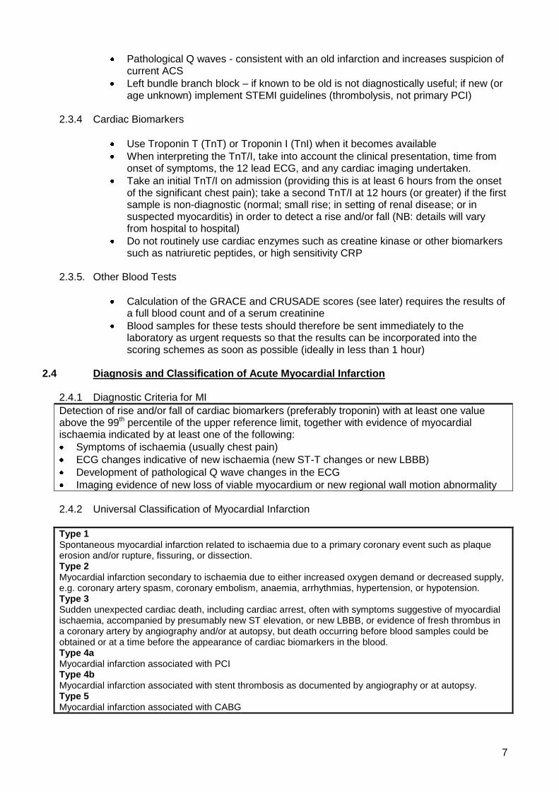
2.3.4 Cardiac Biomarkers
Use Troponin T (TnT) or Troponin I (TnI) when it becomes available
When interpreting the TnT/I, take into account the clinical presentation, time from onset of symptoms, the 12 lead ECG, and any cardiac imaging undertaken.
Take an initial TnT/I on admission (providing this is at least 6 hours from the onset of the significant chest pain); take a second TnT/I at 12 hours (or greater) if the first sample is non-diagnostic (normal; small rise; in setting of renal disease; or in suspected myocarditis) in order to detect a rise and/or fall (NB: details will vary from hospital to hospital)
Do not routinely use cardiac enzymes such as creatine kinase or other biomarkers such as natriuretic peptides, or high sensitivity CRP
2.3.5. Other Blood Tests
Calculation of the GRACE and CRUSADE scores (see later) requires the results of a full blood count and of a serum creatinine
Blood samples for these tests should therefore be sent immediately to the laboratory as urgent requests so that the results can be incorporated into the scoring schemes as soon as possible (ideally in less than 1 hour)
2.4 Diagnosis and Classification of Acute Myocardial Infarction
2.4.1 Diagnostic Criteria for MI
Detection of rise and/or fall of cardiac biomarkers (preferably troponin) with at least one value above the 99th percentile of the upper reference limit, together with evidence of myocardial ischaemia indicated by at least one of the following:
Symptoms of ischaemia (usually chest pain)
ECG changes indicative of new ischaemia (new ST-T changes or new LBBB)
Development of pathological Q wave changes in the ECG
Imaging evidence of new loss of viable myocardium or new regional wall motion abnormality
2.4.2 Universal Classification of Myocardial Infarction
Type 1 Spontaneous myocardial infarction related to ischaemia due to a primary coronary event such as plaque erosion and/or rupture, fissuring, or dissection. Type 2 Myocardial infarction secondary to ischaemia due to either increased oxygen demand or decreased supply, e.g. coronary artery spasm, coronary embolism, anaemia, arrhythmias, hypertension, or hypotension. Type 3 Sudden unexpected cardiac death, including cardiac arrest, often with symptoms suggestive of myocardial ischaemia, accompanied by presumably new ST elevation, or new LBBB, or evidence of fresh thrombus in a coronary artery by angiography and/or at autopsy, but death occurring before blood samples could be obtained or at a time before the appearance of cardiac biomarkers in the blood. Type 4a Myocardial infarction associated with PCI Type 4b Myocardial infarction associated with stent thrombosis as documented by angiography or at autopsy. Type 5 Myocardial infarction associated with CABG

8
3.0 MANAGEMENT OF CONFIRMED NSTEACS
Once a diagnosis of NSTEACS has been reached, management should be along the lines of the algorithms shown in Figs 1 (P3) and 2 (P14) and outlined below.
3.1 Initial Hospital Treatment
Treatment to prevent or reverse intra-coronary thrombosis using anti-platelet and anti-thrombin drugs should be started immediately. However, after the first dose of each has been given, the need for and type of further such treatment should be guided by an individual patient assessment of in-hospital bleeding risk using the CRUSADE bleeding score (See below) which requires the rapid return of initial blood tests, particularly full blood count and creatinine.
3.1.1 Anti-platelet agent – offer a single loading dose of 300mg of aspirin (unless already given)
even if already on regular low dose aspirin; then continue low dose aspirin (usually 75mg) indefinitely. Consider clopidogrel hydrogen sulphate 300mg stat then 75mg daily for patients with aspirin hypersensitivity
3.1.2 Antithrombin
a) Fondaparinux is now the preferred anti-thrombotic agent replacing enoxaparin. It should be given in a dose of 2.5mg subcutaneously once daily for a maximum of 8 days or until discharge whichever is sooner. This is based on the results of the large OASIS-5 trial which showed that fondaparinux was as effective as enoxaparin but caused much fewer major bleeds (2.2 v 4.1%) and on its ease of use. Fondaparinux is a synthetic pentasaccharide which binds to anti-thrombin with greater affinity than either unfractionated heparin (UFH) or low molecular weight heparin (LMWH) and so increases the ability of anti-thrombin to inactivate clotting factor Xa. It has 100% bioavailability after sc administration and has a shelf life much longer than UFH and LMWH. It‟s effects are not reversed by protamine. It has little effect on the activated partial thromboplastin time (aPTT), prothrombin time or bleeding time; does not alter fibrinolysis or platelet functions; and rarely causes thrombocytopaenia as may occur with UHF and LMWH. It is once daily and does not require weight adjustment of dose. Monitoring is not routinely required but can be achieved via an anti –Xa assay calibrated with fondaparinux. Note: Fondaparinux does not afford full anticoagulation and should not be used where this is required e.g. patients needing warfarin, new onset AF, DVT/PE. In these cicumstances use enoxaparin 1mg b.d. sc
b) Fondaparinux is excreted unchanged in the urine and is therefore contra-indicated in
severe renal dysfunction. In the OASIS-5 trial patients with a creatinine >265 mcmoles/L were excluded. Local consensus is that in patients with either a creatinine >265 mcmoles/L, a creatinine clearance <30, or an eGFR <20, sc enoxaparin 1mg/kg o.d. (once daily) should be substituted for fondaparinux. NICE Clinical Guidance 94 recommends UFH rather than LMWH, in patients inappropriate for fondaparinux because of very poor renal function and/or high bleeding risk. THE CMCN working group however considered that the practical problems of IV delivery, adequate control of anticoagulation level and lack of junior doctor familiarity increased the risks of UFH compared to LMWH in the real life setting (as opposed to trials) making the latter a safer option for patients. Individual patient consent should be obtained and documented

9
c) In every patient an assessment of in-hospital bleeding risk should be undertaken using the CRUSADE bleeding score (see below). Where the bleeding risk is high, consider substituting sc enoxaparin as above
3.2 Risk Stratification
Risk stratification should be undertaken in every patient as soon as possible. CMCN no longer recommends the locally developed but unvalidated scoring scheme laid out in the current NSTEACS guidelines. Instead comprehensive risk stratification should be undertaken to quantify the risk of future cardiovascular events using the GRACE 6 month mortality score, the bleeding risk using the CRUSADE bleeding score, and LV function.
3.2.1 GRACE score The GRACE risk calculator requires input of clinical factors, ECG changes, Troponin level
and serum creatinine. The latter two results will not be available immediately so GRACE scoring should be delayed until the urgent blood tests (see Section 2.3.5) return, ideally in less than 1 hour from admission. However, whilst waiting for this score, all patients (irrespective of risk) should be given the basic ACS medical treatment as outlined in Section 3.3.2. Additional medical treatment for ACS (See Section 3.3.3) and the need for coronary angiography (See Section 3.4) should await the GRACE score.
There are a number of related GRACE scores – use the mini – GRACE 6 month mortality score (henceforth referred to as the GRACE score)
It predicts the risk of death due to cardiovascular events and has been validated against the MINAP database which is most relevant to UK practice
It can be accessed in a number of ways using: o A handheld risk calculator (CMCN will supply these on request) o Via the web (PC/MAC/PALM) using the online calculator at www.outcomes-
umassmed.org/GRACE/acs_risk/acs_risk_content.html o Use the Grace score to stratify patients into 5 risk categories as below – each
of which defines a range of 6 month mortalities. o Note that the calculator defaults to creatinine in ng/dl. There is an option to
change the units to micromol/l which should be activated because we report creatinine in the latter units.
Table 2 GRACE Risk Categories based on MINAP Database
Range of mini- GRACE score
Defined by MINAP
Quartiles/octiles
Corresponding
Range of 6 Month
Mortality
% of ACS
Population
CMCN Guideline
Risk Category 6 Month Mortality
<70
0-1.6%
12.5%
Lowest
≤1.5%
71-87
1.6-3.1%
12.5%
Low
>1.5%≤3.0%
88-100
3.1-5.5%
12.5%
Intermediate
>3.0%≤6.0%
101-112
5.5-9.5%
12.5%
High
>6.0%≤9.0%
>112
>9.5%
50.0%
Highest
>9%

10
3.2.2 Bleeding Risk Common risk factors associated with high bleeding risk in clinical trials include:-
Advancing age
Known bleeding complications/conditions
Renal impairment
Low body weight A more quantitative approach to estimating a patients baseline risk of in-hospital major
bleeding during NSTEMI has been reported using the CRUSADE Bleeding Score. A weighted integer score is allocated to each of 8 independent predictors based on its coefficient in a regression model. The CRUSADE Bleeding Score considers baseline patient characteristics (female sex, history of diabetes, peripheral vascular disease), admission clinical variables (heart rate, systolic blood pressure, signs of CHF), and admission laboratory values (haematocrit, glomerular filtration rate, GFR, by Cockcroft-Gault formula) to estimate the patient‟s likelihood of having an in-hospital major bleed event. While treatments increase the likelihood of bleeding, they were not included in the model as they are post-admission variables.
The CRUSADE Bleeding Score calculator is available on-line at:- www.crusadebleedingscore.org To use the CRUSADE Bleeding Score risk calculator input the appropriate range of each
predictor from the drop-down bar. The CRUSADE Bleeding Score and estimated risk of major bleeding will appear below the calculator. The risk of major bleeding derived from CRUSADE should be used as part of a risk/benefit analysis to guide the use of all anti-thrombotic drugs especially in combination.
NOTE: 1) Cockcroft-Gault GFR = (140-age) x (Wt in kg) x constant Serum creatinine in micromol/l where constant = 1.23 for men and 1.04 for women 2) Use of the CRUSADE Risk Calculator required a serum creatinine in order to
calculate the GFR. This will not be available immediately. Scoring should be delayed until it is available (see Section 2.3.5), ideally in less than 1 hour. In the meantime, an initial dose of anti-platelet and anti-thrombin treatment should be given (See Section 3.1) Further treatment should then take into account the CRUSADE bleeding score.
3.2.3 Assessment of LV function
Assess LV function in all patients with acute MI
Consider assessing LV function on all patients with unstable angina
3.3 Further Management Based on GRACE Score Use the GRACE score to determine further treatment taking into account at each stage the
bleeding risk (CRUSADE score) and the patients‟ co-morbidities to guide the risk-benefit balance.
3.3.1 Treatment Algorithm
The treatment/management pathways for each GRACE risk stratum are outlined in Fig 2 (P15) and discussed below

11
3.3.2 Basic Medical Treatment
Anti-Platelet
ASPIRIN 75mg od for all patients without contra-indications
CLOPIDOGREL 75mg od should be substituted for patients who are allergic to aspirin; or intolerant of aspirin because of previous GI bleeding or active/recent PU; or who have significant aspirin – induced dyspepsia
NB: a) Quiescent/old or operated PU, mild dyspepsia, hiatus hernia, or vague indigestion are NOT sufficient reasons to avoid aspirin. B) Where there is active/ongoing gastro-intestinal bleeding neither aspirin nor clopidogrel should be initiated without discussion with a senior clinician. Anti-Thrombin
FONDAPARINUX 2.5mg od sc for a maximum of 8 days or until discharge (whichever is sooner) for all patient without severe renal dysfunction (serum creatinine >265, creatinine clearance < 30 or eGFR <20ml/min) or high bleeding risk (see Initial Treatment) for whom enoxaparin 1mg/kg o d s c should be substituted (Off-licence use; see section 3.3.1)
Anti-Ischaemia
BETA-BLOCKERS IV and/or orally to relieve pain and ischaemia provided there are no contra-indications such as heart failure (Killip >2), hypotension (SBP<100mm) excessive bradycardia (<60bpm) asthma or PVD; a prognostic benefit may also occur
NITRATES should be given initially buccally, IV or sublingually, as appropriate, to relieve pain due to ischaemia. They have no benefit on event rate, or mortality. Subsequently, oral nitrates can be used for continuing angina
CALCIUM BLOCKERS should be used in the following circumstances:-
IN ADDITION to B-blockers if further angina or ischaemia occurs or if hypertension persists. Choose a non-rate limiting drug, usually a dihydropyridine e.g. amlodipine, felodipine.
INSTEAD of a B-blocker if the latter is contra-indicated. Substitute diltiazem or verapamil in the absence of heart block or heart failure
POTASSIUM CHANNEL OPENER Nicorandil 20 mg bd orally has been shown to significantly reduce transient ischaemia, SVT and VT compared to placebo when added to standard therapy. It may therefore be added to nitrates, B-blockers and calcium blockers if there is recurrent ischaemia or angina, although it should be realised that this is currently an unlicensed indication in unstable angina.
Other Drugs
Atorvastatin 80mg o.d. for 3-9 months (with baseline LFT) in all patients without a contraindication. Thereafter simvastatin 40mg nocte should be substituted unless there

12
are overriding reasons why an alternative statin (or other drug) should be chosen. (See CMCN Statin Guidelines)
ACE inhibitors are of proven prognostic benefit for STEMI, anterior MI‟s, large MI‟s with significant LV dysfunction, and for MI with clinical heart failure. They should also be considered for NSTEACS patients with co-existing hypertension, diabetes, LV dysfunction or clinical heart failure; or as part of secondary prevention (HOPE, EUROPA trials). The dose should be up-titrated to a pre-determined target or to the maximum tolerated dose.
3.3.3 Additional Medical Treatment
For all NSTEACS patients except those in the LOWEST RISK GRACE category the following drugs should be considered:-
Clopidogrel 300mg orally stat followed by 75mg o.d. should be given to all such patients in addition to aspirin and fondaparinux. In the absence of a contra-indication or side effect it should be continued for 12 months then stopped. Note: Consider stopping clopidogrel 5 days before CABG in patients with low risk. For patients at intermediate or higher risk, discuss continuing clopidogrel before CABG with the cardiac surgeon. Base the decision on the balance of ischaemic and bleeding risk.
GpIIb/IIIa platelet inhibitors (usually tirofiban or eptifibatide) should be considered as an addition to all of the above for patients in the Intermediate, High and Highest risk categories. They are particularly indicated when chest pain is ongoing or recurrent and especially when PCI is likely (See note at Section 3.3.1).
New anti-platelet agents The CMCN Working Group is aware that a number of new anti-platelet agents either have been approved or are in the process of being assessed for use in ACS patients as an alternative to clopidogrel. These include prasugrel for diabetic ACS and ticagrelor. The place of these agents has not yet been clarified and hence it was decided to omit them from this version of the guideline but to reconsider this issue in an early revision or as a supplement when recommendations are clearer.
NOTE Eptifibatide and tirofiban are licensed for use with aspirin and UFH but do not have UK
marketing authorisation for use with clopidogrel. The recommendation in Table 2 to add these drugs to clopidogrel for Intermediate, High and Highest GRACE risk groups is an off-label use. Informed consent should be obtained and documented before they are used in combination with clopidogrel.
3.4 Coronary Angiography
In the setting of NSTEACS management, coronary angiography is performed partly for diagnostic reasons to confirm the presence of coronary artery disease, but more importantly as a means of identifying patients in whom coronary revascularisation by PCI or CABG would be appropriate. However, it is important to recognise that the assessed risk of death or further cardiac event on its own does not always equate with a patient‟s capacity to benefit from coronary revascularisation. It is very important in making the decision as to whether to embark on the pathway of coronary angiography +/- coronary revascularisation, to take into account other factors such as clinical context, co-morbidity, contraindications and, importantly, patient preference.
Indications
Patients with Intermediate, High or Highest risk according to the GRACE score
Patients who are unstable e.g. cardiogenic shock, pulmonary oedema, arrhythmia irrespective of GRACE score

13
Patients with Low or Lowest risk according to the GRACE score who develop recurrent spontaneous ischaemia or demonstrate ischaemia on non-invasive testing (when indicated clinically)
NOTE: In patients with previous CABG, non-invasive CT coronary angiography BEFORE invasive angiography should be considered to avoid unnecessary invasion in some patients and to clarify the invasive procedure necessary in others.
Timing
Highest risk patients should be considered for urgent transfer to LHCH for further management
Other candidates with indications for coronary angiography should undergo the procedure in a suitable DGH during the same admission within 96 hours or failing that, as soon as possible
Patient Suitability
Patients who are candidates for coronary angiography should have their suitability assessed for that investigation and for the associated coronary revascularisation. This includes a consideration of:
Clinical context – pre-morbid functional status, frailty etc. Co-morbidity including known severe peripheral vascular disease, COPD, renal impairment, previous strokes, co-existent malignancy etc Contra-indications including bleeding risk etc Patient Preference
Where patients are in an appropriate risk category and are suitable as defined above, the final determination in the selection of invasive management strategy should always be that of patient preference. Careful attention should be given to ensuring that patients understand the potential benefits and risks of the treatment options proposed and why other options are not proposed. It is important that realistic estimates of risk, discomfort and benefits are given in order to avoid unrealistic expectations.
Site for Coronary Angiography
The majority of procedures should be undertaken on the DGH side provided adequate facilities and experience are available. The following categories of patients should be considered for transfer to LHCH for their procedure:
o Highest Risk GRACE group o Patients with shock, pulmonary oedema, NHYHA Class IV heart failure o Known or suspected left mainstem disease o Severe valvular disease associated with heart failure o Complex adult congenital heart disease o Where the DGH consultant feels that the risk/benefit ratio is in favour of
transfer to LHCH
In patients with significant renal impairment who are likely to require PCI following coronary angiography, consideration should be given to transfer to an interventional centre (currently LHCH) for both procedures in order to reduce the contrast load and hence the risk of contrast nephropathy. A suggested but arbitrary renal threshold is an eGFR of <30 or creatinine clearance of <45.

14
4.0 Referral to LHCH
4.1 Referral for Coronary Angiography
As soon as a patient has been deemed to need transfer to LHCH for coronary angiography (See section 3.4 Site for Coronary Angiography), the current appropriate arrangements need to be made (see relevant document or contact LHCH).
4.2 Referral for PCI
As soon as a patient has been deemed to need transfer to LHCH for revascularisation by PCI following DGH coronary angiography (See Fig 2), the current appropriate arrangements below need to be made(see relevant document or contact LHCH).
As previously agreed, it remains the expectation that transfer will take place without the need for further discussion. It will be the responsibility of LHCH to prioritorise the patient, locate an available bed, and agree transfer arrangements with the host hospital and with the ambulance service.
4.3 Referral for Cardiac Surgery
As soon as a patient has been deemed to need cardiac surgery (CABG +/- valve surgery) following DGH coronary angiography (See Fig 2), the current appropriate arrangements need to be made(see relevant document or contact LHCH).
5.0 REHABILITATION AND DISCHARGE PLANNING 5.1 Before discharge offer patients information and advice about:
o Their diagnosis and arrangements for follow-up o Cardiac rehabilitation o Management of cardiovascular risk factors and drug therapy for secondary prevention o Lifestyle changes
5.2 Follow guidance in „MI: secondary prevention‟ (NICE clinical guideline 48), „Lipid
modification‟ (NICE clinical guideline 67) and „Brief interventions and referral for smoking cessation in primary care and other settings‟ (NICE public health guidance 1)
5.3 Begin discharge planning as soon as possible after admission. Produce a discharge letter
at point of discharge including diagnosis, investigations undertaken and/or planned, drug treatment given, intervention undertaken and a discharge (TTO) drug list. Follow up arrangements must be clearly stated including outpatient clinic, cardiac rehabilitation etc.

15
Figure 2 MANAGEMENT OF NSTEACS BASED ON GRACE SCORE
Lowest risk (≤1.5%)
Low risk (>1.5-3.0%)
Highest risk >9.0%)
Basic Medical Treatment 3.3.2
Clopidogrel for 12 months 3.3.3
Consider adding a GpIIb/IIIa inhibitor 3.3.3
Medical Treatment
Coronary Angiography 3.4
Refer LHCH for PCI 4.2
Intermediate risk (>3.0-6.0%)
High risk (>6.0-9.0%)
Basic Medical Treatment – 3.3.2
Recurrent spontaneous ischaemia
Consider Ischaemia testing
Ischaemia Demonstrated?
Refer LHCH for CABG +/- valve surgery
4.3
Cardiac Rehabilitation – 5.1
Secondary Prophylaxis – 5.2
Discharge and Follow-up Up Planning – 5.3
Basic Medical Treatment 3.3.2
Clopidogrel for 12 months 3.3.3
Offer coronary angiography in<96hr or as soon as
possible 3.4
Consider referral to LHCH for coronary angiography
3.4, 4.1
No
Yes
Yes
No
Intermediate risk High risk
Highest risk

16
References 1. National Institute for Health and Clinical Excellence Clinical Guideline 94. The early
management of unstable angina and non-ST segment elevation myocardial infarction. March 2010
2. National Institute for Health and Clinical Excellence Clinical Guidelines 95. Assessment and diagnosis of recent onset chest pains or discomfort of suspected cardiac origin. March 2010.
3. Thygesen K, Alpert JS et al. Universal definition of myocardial infarction JACC 2007; 50:2173-95.
4. The Fifth OASIS Investigators, Comparison of Fonadapainux and Enoxaparin in Acute Coronary Syndromes. N Engl J Med 2006; 354: 1464-76
5. Subherwal S, Bach RG et al. Baseline risk of Bleeding in Non-ST segment Elevation Myocardial Infarction. Circulation 2009; 119: 1873-82
6. National Institute for Clinical Excellence Technology Appraisal No 80. Clopidogrel in the treatment of non-ST-elevation acute coronary syndrome. July 2004
7. National Institute for Clinical Excellence Technology Appraisal No. 47. Guidance on the use of glycoprotein IIb/IIIa inhibitors in the treatment of acute coronary syndromes September 2002
8. Intensive versus moderate lipid lowering with statins after acute coronary syndromes (PROVE-IT TIMI 22 Study). N Engl. J Med; 350:1495-504
9. The Heart Outcomes Prevention Evaluation Study Investigators. Effects of an angiotensin-converting-enzyme releasing inhibitor, ramipiril on cardiovascular events in high risk patients N Engl. J Med 2000; 342: 145-53
10. The EURopean trial On reduction of cardiac events with Perindopril in stable coronary Artery disease Investigators. Efficacy of perindopril in reduction of cardiovascular events among patients with stable coronary artery disease: randomised, double-blind, placebo-controlled, multicentre trial. Lancet 2003; 362: 782-88.
.