Guideline Eeg Pediatric 2012
60
Guidelines for Neonatal and Pediatric EEG Monitoring Renée Shellhaas, MD, MS September 22, 2012
-
Upload
chindia-bunga -
Category
Documents
-
view
79 -
download
2
Transcript of Guideline Eeg Pediatric 2012

Guidelines for Neonatal and Pediatric EEG Monitoring
Renée Shellhaas, MD, MS
September 22, 2012

Disclosures
• No conflicts of interest? – I am a neurologist.
• I read EEGs for a living. – I am not a neonatologist.
• I rarely read amplitude-integrated EEGs.
• I am grateful to NICHD, Child Neurology Foundation, and Janette Ferrantino Award for funding my research.

Goals/Objectives
• Identify indications for EEG monitoring among high-risk neonates [and children].
• Discuss relative strengths and weaknesses of conventional EEG monitoring versus amplitude-integrated EEG (aEEG).
• Review preferred methods for neonatal [and pediatric] ICU EEG monitoring.
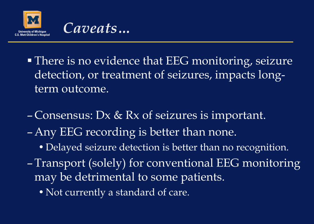
Caveats…
There is no evidence that EEG monitoring, seizure detection, or treatment of seizures, impacts long-term outcome.
– Consensus: Dx & Rx of seizures is important. – Any EEG recording is better than none.
• Delayed seizure detection is better than no recognition. – Transport (solely) for conventional EEG monitoring
may be detrimental to some patients. • Not currently a standard of care.

Neonatal ICU Monitoring

ACNS Critical Care Committee Neonatal Subcommittee Members
Baylor College of Medicine • James J. Riviello, M.D.
Children’s Hospital of Philadelphia • Nicholas Abend, M.D. • Robert R. Clancy, M.D.
Children’s National Medical Center • Taeun Chang, M.D. • Tammy N. Tsuchida, M.D., Ph.D.
Hammersmith Hospital (London) • Courtney Wusthoff, M.D.
Rainbow Babies & Children’s Hospital (Cleveland) • Mark Scher, M.D.
Hospital for Sick Children (Toronto) • Cecil D. Hahn, M.D.
University of Michigan • Renée A. Shellhaas, M.D., M.S.
Guest Participants John Barks, M.D. (U Michigan) Hannah C. Glass, M.D. (UCSF) Terrie Inder, M.D. (Wash U) Sylvie Nguyen, M.D. (Centre Hospitalier Universitaire - Angers) Joseph E. Sullivan, M.D. (UCSF) Steven Weinstein, M.D. (Cornell) Robert White, M.D. (Memorial Hospital, IN)
Shellhaas, et al. ACNS Guideline. J Clin Neurophysiol. 2011 28(6):611-617.

Indications for EEG monitoring
1. High risk for seizures
2. Differential diagnosis of paroxysmal events
3. Monitoring during/after anticonvulsant wean
4. Monitoring pharmacologically-induced burst suppression

Indications: Seizure Detection
• Sick babies often have unusual movements. • Most of these are not seizures.
• >50% of all neonatal seizures are subclinical. • Only detectable with EEG monitoring
• Electroclinical dissociation / uncoupling: • With treatment, clinical signs may vanish while
subclinical electrographic seizures continue.
Clancy, et al. Epilepsia. 1988; 29:256-261. Scher, et al. Pediatrics. 1993;91:128-134. Scher, et al. Pediatric Neurol. 2003;28:277-280.

Seizures and Prognosis
• Babies with neonatal seizures are at high risk for death or neurologic morbidity. – Mortality: 25-40% (may be higher in preterm) – Developmental delay: 67% @ 2-3 yrs – Cerebral palsy: 63% @ 2-3 yrs – Post-neonatal epilepsy: 17-56%
McBride, Neurology 2000;55:506-514. Mizrahi, Epilepsia 2001;42(S7):102. Legido, Pediatrics 1991; 88:583-596. Scher, Pediatr Neurol 1989; 5:17-24 Scher, Pediatrics 1993; 91:128-134.

Clinical vs. electrographic seizures
• Video-EEG monitoring of high-risk infants.
– 9% of electrographic seizures (48/526) had clinical signs recognized and documented by NICU staff.
– 27% of seizures which did have clinical signs (48/179) were recognized and recorded.
– 73% of “seizures” documented by NICU staff had no electrographic correlate (129/177).
Murray, et al. Arch Dis Child Fetal Neonatal Ed. 2008;93:F187-F191.

The literature…read the fine print
• Many studies talk about “seizures” but don’t tell us if they are clinical or electrographic (aEEG or cEEG). – Older studies usually mean clinical seizures…less
applicable now that we know most seizures are subclinical.
– When I say “seizure”, I mean EEG seizure.

BIRDs and Seizures
• BIRD = Brief Intermittent Rhythmic Discharge – <10 seconds – High risk for seizures
• Seizure = sudden, repetitive, evolving and stereotyped ictal pattern with a clear beginning, middle, and ending, an amplitude of at least 2µV, and a minimum duration of 10 seconds

SEIZURE

High risk for seizures High risk of acute brain injury
Demonstrated acute acquired brain injury
Clinically suspected seizures
Prolonged circulatory arrest
Arterial ischemic stroke
Focal clonic or tonic movements
Hypoxic-ischemic encephalopathy
Cerebral sinovenous thrombosis
Eye blinking, gaze-deviation
Infants with sustained hypoxia
Intracranial hemorrhage
Unexplained apnea
Pharmacologically-induced paralysis
Encephalitis Myoclonus
Head trauma with altered mental status
Cerebral edema due to inborn errors of metabolism
Bicycling

Indications: Differential diagnosis
• Most abnormal neonatal movements have no electrographic correlate.
• Neonates don’t have generalized tonic-clonic seizures!
• Heart rate changes during seizures are not consistent.
• Isolated changes in vital signs are rarely due to seizures.
Clinically suspected seizures Focal clonic or tonic movements Eye blinking, gaze-deviation
Unexplained apnea
Myoclonus
Bicycling
Unexplained altered mental status

Indications: During/after anticonvulsant drugs are weaned
• Depends on seizure etiology • Lissencephaly vs. HIE
• How hard were the seizures to control?
• How many meds are you giving?
• What are your goals?

Indications: Burst suppression
– Refractory status-epilepticus: • Titrate medication to optimize burst/interburst
ratios.
– Severe metabolic encephalopathy: • e.g. Discontinuity improves as hyperammonemia
is corrected.
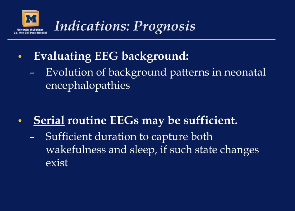
Indications: Prognosis
• Evaluating EEG background: – Evolution of background patterns in neonatal
encephalopathies
• Serial routine EEGs may be sufficient. – Sufficient duration to capture both
wakefulness and sleep, if such state changes exist
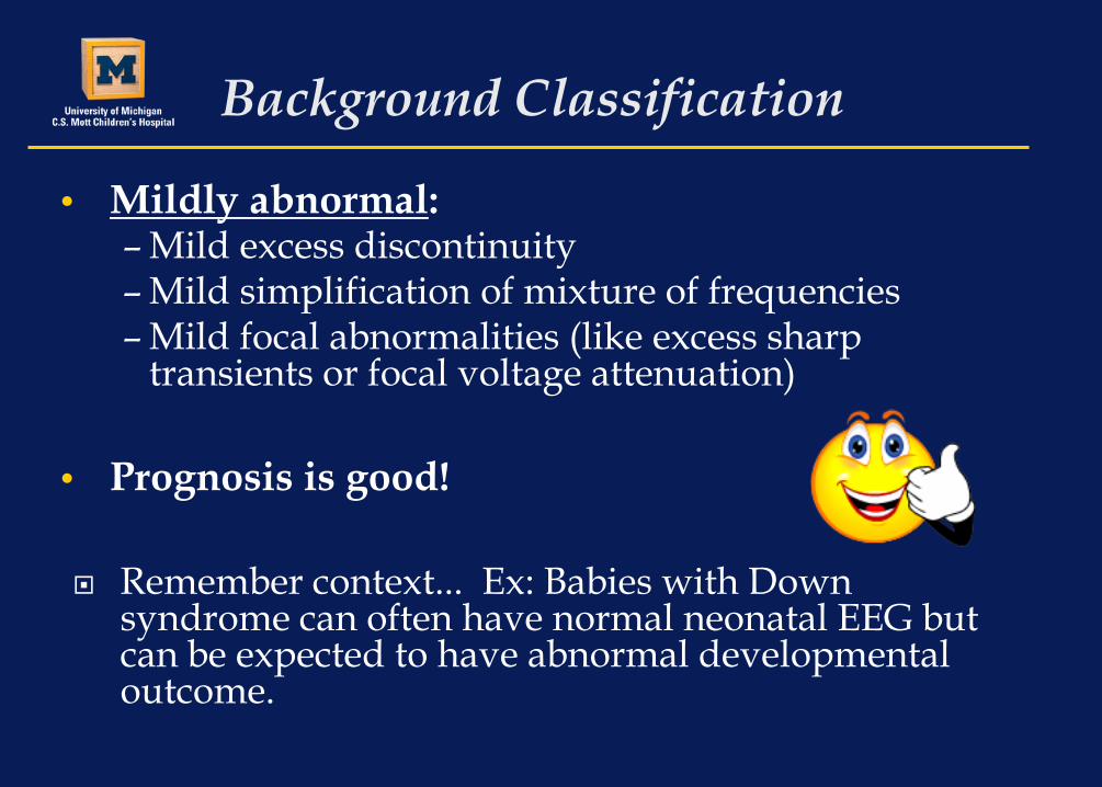
Background Classification
• Mildly abnormal: – Mild excess discontinuity – Mild simplification of mixture of frequencies – Mild focal abnormalities (like excess sharp
transients or focal voltage attenuation)
• Prognosis is good!
Remember context... Ex: Babies with Down syndrome can often have normal neonatal EEG but can be expected to have abnormal developmental outcome.

Background Classification
• Moderately Abnormal – Moderately excessive discontinuity – Moderately excessive asynchrony – Poverty of expected background rhythms – Definite focal abnormalities – Persistent low voltage (<25μV)
• Some do well, others do poorly.

Background Classification
• Markedly abnormal: – Markedly excessive discontinuity (can have some
preservation of age-appropriate background patterns)
– Burst suppression – Gross interhemispheric asynchrony – Extreme low voltage (<5μV) – Depressed and undifferentiated – Isoelectric
• Prognosis is poor.

Neonatal EEG: Technical Aspects
• International 10-20 system – Modified for neonates* – Minimum 9 scalp electrodes
• EKG • Respiratory channel • Eye leads • EMG lead
*Not all laboratories utilize the Pz electrode. Alternate terminology
designates “FP” electrodes as “AF”, and T3/4 as T7/8.

Technical Aspects: Neonatal Montages
• Combined double- and single-distance electrodes

Technical Aspects: Neonatal Montages

Technical Aspects: Event marker
• Event button to be pressed for: – clinical episodes – relevant medication administration – events that could cause cerebral injury – Need the bedside observer to PTFB!!!!
– When you push the button SAY OUT LOUD why you are doing so. • “Look! His left pinky finger twitched.” • “I’m giving the phenobarb bolus now.”

Technical Aspects: Bedside log
• Bedside log to document the time and description of event(s). –Or direct annotation on video EEG record
•Type into computer

Duration of Recording: SCREENING for Seizures
• A routine EEG is inadequate to screen for neonatal seizures.
• Normal EEG background does not preclude seizures.
• Scenarios for routine EEG exams: 1. The baby is having seizures need EEG monitoring 2. We didn’t capture an event need EEG monitoring 3. EEG background looks terrible need EEG monitoring 4. Background looks ok but baby doesn’t need EEG
monitoring 5. See the pattern?

Duration of Recording: SCREENING for Seizures
• Recommend a minimum of 24 hours. – Skip the routine EEG. – If no seizures and EEG background is stable,
stop after 24 hours of monitoring.
– Mean time to seizures: • Neonatal cardiac surgery: 21 hrs (range 10-36 hrs) • HIE with hypothermia: 9.5 hrs (range 5.5-98 hrs). • Heterogeneous high-risk neonates: Always ≤22 hrs.
Clancy et al, Epilepsia. 2005;46:84-90. Laroia et al, Epilepsia. 1998;39:545-51. Wusthoff et al, J Child Neuro. 2011;26:724-728.

Duration of Recording: CONFIRMED Seizures
• Recommend conventional EEG monitoring until 24-hrs seizure-free.
– Unless, in consultation with a neurologist, the decision is made to stop sooner.
– Almost no data…

Duration of Recording: Differential Diagnosis of Events
• Recommend continuing EEG monitoring until multiple typical events are recorded.
– If 3-4 typical events are captured, and are not seizures, and the EEG background remains normal or stable, then monitoring may be discontinued.
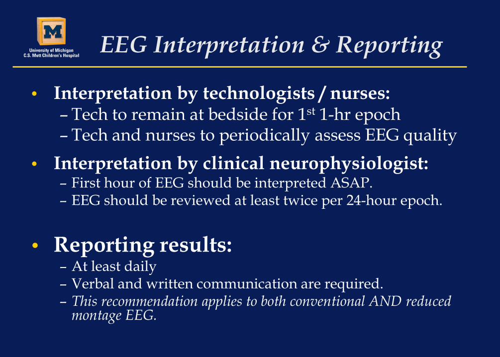
EEG Interpretation & Reporting
• Interpretation by technologists / nurses: – Tech to remain at bedside for 1st 1-hr epoch – Tech and nurses to periodically assess EEG quality
• Interpretation by clinical neurophysiologist: – First hour of EEG should be interpreted ASAP. – EEG should be reviewed at least twice per 24-hour epoch.
• Reporting results: – At least daily – Verbal and written communication are required. – This recommendation applies to both conventional AND reduced
montage EEG.

What about aEEG?

Amplitude-Integrated EEG (aEEG)
• Single channel from biparietal electrodes.
• over watershed zone • many now use 2 aEEG channels plus “raw” EEG
• Leads applied by nurse or neonatologist.
• Interpreted by neonatologists.
• Short- or long-term monitoring.
• Easy to visually track trends.

Standard neonatal EEG
Single channel raw EEG
Amplitude Integrated EEG (aEEG)
Filter <2Hz and >15Hz, rectify, smooth, amplitude-integrate
FP1-T3
T3-O1
FP2-T4
T4-O2
FP1-C3
C3-O1
FP2-C4
C4-O2
T3-C3
C3-CZ
CZ-C4
C4-T4
C3-C4
75 uV2 sec

aEEG monitoring: HIE
• Predicting outcome after HIE: – Early aEEG background – Sleep-wake cycling
• Hypothermia modifies aEEG background: – Normalize by 48 hours good outcome
• 24 hours for normothermia – Lack of sleep-wake cycling = poor prognosis
al Naqeeb. Pediatrics 1999;103:1263-71. Shalak. Pediatrics 2003;111:351-7. Toet. Arch Dis Child Fetal Neonatal Ed 1999;81:19-23. Thoresen. Pediatrics. 2010:126;e131-e139.

aEEG monitoring: preterm infants
• Outcome Prediction • Excessive discontinuity (lower margin amplitude) • Lack of sleep-wake cycling
• predicts adverse outcome for Grade III-IV IVH. • < 29 weeks: excess discontinuity and absence of sleep-
wake poor short term prognosis.
• Abrupt changes in aEEG background can correlate with new pathology.
Bowen. Pediatric Research. 2010; 67(5):538-44. Hellström-Westas. Neuropediatrics. 2001; 32(6):319-24. Niemarkt . Neonatology. 2010; 97(2):175-82. Olischar. Childs Nerv Syst. 2001; 20(1):41-5.

What about aEEG for seizures?

The literature…read the fine print
• Distinguish between “seizure positive” aEEG or EEG records and individual seizure detection. – 8 of 10 patients correctly identified as having seizures
versus 8 of 10 individual seizures correctly detected.

EEG: the Gold Standard
• Conventional Neonatal
EEG is the gold standard for the diagnosis and quantification of neonatal seizures and for assessment of EEG background
10-20 system, modified for neonates

aEEG often used to screen for seizures

Seizures on aEEG
• 125 routine EEGs with seizures (+19 without), recorded from 140 term newborns.
• Created single-channel aEEG traces. – Interpreted by 6 neonatologists with varying expertise.
% of 125 aEEG records with seizures detected
% of 851 individual seizures detected
Mean = 40.3% ± 16.8% (range: 22-57%)
Mean = 25.5% ± 10.6% (range: 12-38%)
Shellhaas, et al. Pediatrics. 2007;120:770-777.
* No false positive records; very few false positive individual seizures.

Seizures on aEEG
• Factors related to seizure detection by aEEG (multivariate analysis*):
• Neonatologists’ level of experience with aEEG • Visibility in C3C4 raw EEG channel • Seizure duration • Peak-to-peak amplitude • Seizure count per hour
*P=<0.001 for all variables
Shellhaas, et al. Pediatrics. 2007;120:770-777.
Inherent features of neonatal seizures (short duration & low amplitude) make detection by aEEG very difficult.

Adding “raw” EEG
• Simultaneously recorded single and dual channel aEEG with conventional EEG in 7 neonates with seizures. – Readers: two experienced neonatologists – Using both two-channel aEEG and the raw
tracings, sensitivity = 76% – Using just aEEG, sensitivity = 27%-56% – 1 false-positive per 39 hours of recording
Shah, et al. Pediatrics. 2008;121:1146-1154.

Confirming aEEG findings
• Brief, infrequent, low amplitude seizures are hardest to detect on aEEG.
• If you see a seizure, you’re probably right. • If you don’t see seizures… doesn’t mean they aren’t
there. • Don’t declare victory based on aEEG.
• Suspect seizures on aEEG conventional EEG. – Confirm diagnosis – Direct treatment

Concurrent EEG and aEEG: Best of both worlds?

Summary: Neonatal EEG monitoring
• Neonatal EEG (cEEG and aEEG) background assessments can assist with estimating prognosis.
• EEG monitoring is the standard for neonatal seizure diagnosis and assessment of treatment response.
• Use aEEG when don’t have EEG. • Document!

Pediatric ICU EEG monitoring

Indications for EEG monitoring
1. High risk for seizures
2. Differential diagnosis of paroxysmal events
3. Monitoring pharmacologically-induced burst suppression

PICU Monitoring: High risk for seizures
• Age < 1(or 2) year(s) • Convulsive seizure or status epilepticus
– ± history of epilepsy
• Acute brain injury – Focal (stroke) or diffuse (HIE) – Traumatic brain injury
Other high risk clinical scenarios: • Ex: Need for pharmacologic paralysis
+ prior seizures / acute brain injury

PICU Monitoring: High risk for seizures
• High-risk EEG features: – Lack of reactivity – Epileptiform abnormalities

PICU Monitoring: Differential diagnosis
• Non-seizure paroxysmal events are common. – ~25% of unselected sample of ICU EEG
monitoring studies had non-epileptic events. Williams. Epilepsia. 2011;52:1130-1136.
• Accurate identification matters
– Avoid unnecessary medications (+ side effects)

PICU Monitoring: Iatrogenic Burst Suppression
• Children requiring pharmacologically-induced burst suppression need continued EEG monitoring. – Evaluate for break-through seizures – Ensure appropriate medication titration

PICU Monitoring: Duration
• 24 hours screening for non-convulsive (or subclinical) seizures:
• ~95% of children with non-convulsive seizures will be detected with 24 hours of EEG.
Abend. Neurology. 2011. • 100% of those with only non-convulsive seizures (no
clinically-apparent seizures) detected in 24 hours . McCoy. Epilepsia. 2011.
• Or until events of interest are recorded and determined not to be seizures.
• And EEG background stable or normal.

PICU Monitoring: Technical aspects
Also EKG; others as needed. See neonatal EEG guidelines re: review, reporting...

Digital Trending
• Compressed display • Highlight epochs of concern • Facilitate timely EEG review
• Need more study!


Conclusions
• Subclinical seizures are common among neonates and children in ICUs. – Can only be diagnosed with EEG.
• EEG monitoring is gold standard for seizure diagnosis and quantification. – Digital trending might help.
• Refer to ACNS guidelines for neonates. • Stay tuned for ACNS pediatric & adult
guidelines.