Guide to neurological examination Professor Yasser metwally
55
A GUIDE TO NEUROLOGICAL EXAMINATION Starting in medical school neurologic examination has remained intimidating for many physicians. The examination has been perfected over many decades thanks to our French and German founding fathers. It has led to eponyms (e.g. - Babinski sign , Hoffmann reflex) different techniques of detecting subtle signs of weakness (e.g. - hand rolling, foot tapping) and even a wide collection of reflex hammers that can be proudly displayed in the office. How is one to summarize a skill or tool that can be directly used in daily practice? As with many medical specialties, the history is dependent on specific knowledge of neurologic disorders. And it has become a cliché to think that neurologic examination is dull and time consuming involving meticulous assessment of reflex asymmetries, sensory deficits and mental function. Some may argue whether a MRI would not suffice. Diagnostic tests have grown tremendously over the last decade and may have left the impression that neurologists, despite their acumen, may be inferior to them. Nothing seems further from the truth. The level of this chapter will be for the medical student but also recently qualified physician in their first years of specialty training. Using the basic tenets of a comprehensive history, examination of the major components of brain, spinal cord, and neuromuscular unit function, a tentative diagnosis should be possible in approximately half of the cases. The principle of neurologic examination is localization of the lesion followed by a differential diagnosis of the cause of the lesion. This is a sacrosanct principle and cannot be ignored despite rapid development in technology. This chapter, however, has to ignore failure of some patients to describe a particular symptom, information forthcoming after repeated history and recognition of malingering. This chapter should have a practical value for the vast majority of physicians seeing patients in the clinic. The History General Issues Sometimes one is not off to a good start. Patients may present their medical history illustrating a plethora of physician encounters, interpretation of diagnostic tests in layman terms, and multiple diagnoses often contradicting each other. As expected, many of the requests for a neurologic examination pertains to the infinite symptoms of dizziness, weakness, fatigue, facial numbness, confusion, and longstanding headache. The physician task is using a series of leading questions to obtain the overriding presenting symptom, onset and time, and its progression. Neurologic diseases may have different evolution over time. The onset can be acute which may be defined in seconds to rapidly progressing in hours, fluctuating typically over weeks, a relenting rapidly progressing course and a fluctuating, remitting and relapsing course such that is seen in multiple sclerosis and myasthenia gravis. In any presenting symptom, associated symptoms are of the utmost importance and many of them may involve systemic features such as fever, nausea and vomiting, or weight loss. The neurologic history remains time consuming but with quick Professor Yasser Metwally www.yassermetwally.com www.yassermetwally.com
Transcript of Guide to neurological examination Professor Yasser metwally

A GUIDE TO NEUROLOGICAL EXAMINATION
Starting in medical school neurologic examination has remained intimidating for manyphysicians. The examination has been perfected over many decades thanks to our Frenchand German founding fathers. It has led to eponyms (e.g. - Babinski sign, Hoffmann reflex)different techniques of detecting subtle signs of weakness (e.g. - hand rolling, foot tapping)and even a wide collection of reflex hammers that can be proudly displayed in the office.How is one to summarize a skill or tool that can be directly used in daily practice? As withmany medical specialties, the history is dependent on specific knowledge of neurologicdisorders. And it has become a cliché to think that neurologic examination is dull and timeconsuming involving meticulous assessment of reflex asymmetries, sensory deficits andmental function. Some may argue whether a MRI would not suffice. Diagnostic tests havegrown tremendously over the last decade and may have left the impression thatneurologists, despite their acumen, may be inferior to them. Nothing seems further fromthe truth.
The level of this chapter will be for the medical student but also recently qualifiedphysician in their first years of specialty training. Using the basic tenets of a comprehensivehistory, examination of the major components of brain, spinal cord, and neuromuscularunit function, a tentative diagnosis should be possible in approximately half of the cases.The principle of neurologic examination is localization of the lesion followed by adifferential diagnosis of the cause of the lesion. This is a sacrosanct principle and cannot beignored despite rapid development in technology. This chapter, however, has to ignorefailure of some patients to describe a particular symptom, information forthcoming afterrepeated history and recognition of malingering. This chapter should have a practical valuefor the vast majority of physicians seeing patients in the clinic.
The History
General Issues
Sometimes one is not off to a good start. Patients may present their medical historyillustrating a plethora of physician encounters, interpretation of diagnostic tests in laymanterms, and multiple diagnoses often contradicting each other. As expected, many of therequests for a neurologic examination pertains to the infinite symptoms of dizziness,weakness, fatigue, facial numbness, confusion, and longstanding headache. The physiciantask is using a series of leading questions to obtain the overriding presenting symptom,onset and time, and its progression. Neurologic diseases may have different evolution overtime. The onset can be acute which may be defined in seconds to rapidly progressing inhours, fluctuating typically over weeks, a relenting rapidly progressing course and afluctuating, remitting and relapsing course such that is seen in multiple sclerosis andmyasthenia gravis. In any presenting symptom, associated symptoms are of the utmostimportance and many of them may involve systemic features such as fever, nausea andvomiting, or weight loss. The neurologic history remains time consuming but with quick
Professor Yasser Metwallywww.yassermetwally.com
www.yassermetwally.com
pointers, one should be able to categorize a reliable account of the presentation. Familyhistory is important in many neurologic disorders. A long list of genetic disorders has beendescribed with a predominant neurologic presentation but many more of the commonneurologic disorders run in families. These include hereditary peripheral neuropathy,multiple sclerosis, but also epilepsy, migraine, and cerebrovascular disease particularlyintracranial aneurysms. The technique of history taking should remain respectful but mayinclude prodding questions, constant verification of the answers, repetitive summaries tothe patient and verification with family members.
Some leading questions are
1. Where in the body did it start?2. When was the last time you were without any symptoms?3. Why did it occur and are triggers known?4. What did you do to relief the symptom?5. How did it progress, evolve, came to a halt or improve?6. Who was helping you out when it happened and what was done?
Specific Complaints
There are many, but only a few are common.
Headache
The cardinal features of headache are quality, severity, localization, relieving andprecipitating factors, associated phenomena such as nausea or scotoma, as well asrecurrence over time. The distinction between an acute and chronic persistent headache isusually easily apparent. Acute headache is often severe in intensity but acute should befurther defined as split second (such as in a ruptured aneurysm), rapidly progressing (suchas in migraine), or jabbing and jolting (such as in cluster headache and trigeminalneuralgia). The severity of pain is difficult to judge but it is important to know whether thepain is throbbing, lancinating, electric, stabbing, radiating, involving the entire head orunilateral, or more circumscript spots such as in a psychogenic headache. Factors that mayaggravate the pain should be identified such as posture, straining, sneezing, coughing,movement, prior alcohol and even consumption of ice cream. Headache that changes withposition is important. Many headaches associated with an increased intracranial pressuredo worse with lying down and are severe at awakening in the morning and headachesassociated with CSF hypotension are worse with standing up and immediately relievedwith lying down. Headaches relieved with knee-chest position may be due to third ventricleobstructive tumors. The severity of the headache is difficult to measure. For example, asevere neck strain from overzealous gardening may look like an acute subarachnoidhemorrhage. Associated symptoms are important. Nausea and vomiting with acute newonset headache is commonly due to a structural lesion. Migraine may have specific featuresthat may further classify the type of migraine such as aura (flickering lights, geometricdistortions and even visual hallucinations), photophobia and sonophobia, and inability toaccept any kitchen smells. Ptosis, nasal stuffing, redness of the conjunctiva may suggest a
Professor Yasser Metwallywww.yassermetwally.com
www.yassermetwally.com

cluster headache. Electric lancinating pain in the cheek, ear, or jaw mimicking a molarabscess may suggest a trigeminal neuralgia. Touching the face with make up, a cold breeze,chewing or toothbrushing may trigger a brief lancinating electric stab as if a finger is put ina socket. It is sometimes a spot diagnosis. A patient who requires a dark room likely hasmigraine or chronic tension headache. A tendency to move, rock back and forth, ischaracteristic of a patient in the middle of a cluster headache, and a patient with trigeminalneuralgia may assume a typical posture in which the palm of the hand is held close to thecheek supported by the other arm but without touching it. It is important to find factorsthat may precipitate or aggravate headache and factors that may provide relief. Certaindrugs may cause immediate relief and can be used as a diagnostic test. This includes severalliters of nasal oxygen for cluster headache, any of the tryptans for classic migraine, andNSAIDs for paroxysmal hemicrania. Nonetheless it is important to mention that a rapidresponse to over-the-counter pain medications and even narcotics do not exclude thepossibility of a more severe disorder such as brain tumor.
Dizziness and Vertigo
Neurologists are sometimes disillusioned when their clinic day only involves patients withdizziness and vertigo. As an isolated symptom it is rarely due to a neurologic cause.Dizziness is often described by the patient as wooziness, giddiness or faintness. Inelucidating these umbrella terms it is important to determine whether the dizziness is truevertigo, in which the patient experiences a true rotational effect, or whether it consists of alight-headedness or presyncopal sensation. It is important to inquire about vertigo withother signs of brain stem dysfunction such as diplopia, dysarthria, dysphagia, hypesthesia,and acute ataxia. On the other hand the presence of ringing in the ear, hearing loss andviolent vomiting may suggest a peripheral (labyrinth) rather than a central (brainstem)cause. vertigo with position change is commonly due to a peripheral cause as well.Intermittent "dizzy spells" are commonly hyperventilation. Failure to perform ahyperventilation test (have the patient breathe in and out for one to two minutes trying toproduce patients own recognizable symptoms) in these patients must be considered amistake. In these dizzy patients who do not have the classic tingling fingers and tight lipsduring the attack a series of expensive MRI and ENT tests have been performed but theyare only waiting to be recognized by an astute physician. Similarly it should be noted thatsome patients describe dizziness to point out a gait disorder and we have seen patients withearly Parkinson’s disease and instability undergo several ENT evaluations until thetendency to pro- or retropulse or cogwheel rigidity is detected.
Sensory Symptoms
Numbness is vague term used by patients and may indicate weakness, pain or itching. Truetingling, in which the patient describes a constant pins and needles sensation should bedifferentiated from a tight band, tight shoe, walking on air or rough surface sensationwhich indicates an abnormality in the posterior columns. Typically numbness has beenpresent for quite some time in extremities. Extremity numbness expanding in a clockwisefashion, although sounding functional, may indicate a cervical spinal cord lesion. Lack oftemperature sense needs to be addressed. Often patients are unable to distinguish hot and
Professor Yasser Metwallywww.yassermetwally.com
www.yassermetwally.com
cold while taking a shower. Failure to recognize objects in a purse or in a pocket may alsoindicate significant loss of proprioception and is commonly found in a cervical spine lesionsuch as cervical spondylosis or syringomyelia. Some of these patients may demonstratespontaneous finger movements in an attempt to orient them in space (pseudoathetosis).
Cognitive Decline
Typically memory decline is a gradual process over years, but an alleged acute worseningmay sometimes prompt early evaluation. Sometimes it is a child from out of state that visitsafter a while, only to find out that personal hygiene has tumbled downhill. In some patientsfamily members are surprised that the patient does not know the date, the name of thepresident or any recent encounter when specifically asked. Memory decline often involvesconsistent difficulty with finding words, names, way to one’s own house, inability todescribe routes to the clinic, but also more subtle problems such as inability to maintain acoherent conversation or failure to complete complex dinners.
A history of nocturnal confusion with wandering through the house and opening ofdrawers without a purpose may be obtained in a patient with advanced Alzheimer’s diseaseor any of the other dementias. Transient focal signs including dysarthria, aphasia, orhemiparesis may point to a multivascular dementia. A recent head trauma, often a caraccident in the months before memory decline may indicate a subdural hematoma, and asexpected it is not volunteered by the patient. Complete loss of memory is known astransient global amnesia and essentially the patient has no recollection of this episode.During this period the patient is constantly asking for where he is, whom he is with, andwhat he has been doing to be in this place. Complete loss of memory with a defined periodof time, (or example, memory loss of three months) is typically psychogenic. Loss of moralbehavior and critical judgment may indicate an advanced stage of dementia but dependingon the underlying personality. Loss of a patient’s own identity is a very late stage ofdementia often emerging when patients become bed - bound. Its early presentation shouldindicate pseudodementia. It is important to inquire about depressive symptoms such asanhedonia, weight loss, and suicidal thoughts to exclude the possibility of a treatabledepression but patients with Alzheimers, particularly high strung individuals, may becomedepressed when defects are becoming noticed.
Speech Disorders
Speech disorders can be grossly distinguished between a dysarthria in which there is amajor disturbance of articulation and aphasia in which the patient’s speech is distortedand words or letters become substituted. A dysarthria is not only slurring of words but alsooutput is hesitant, explosive, and staccato. A fluent aphasia can be so severe ("word salad")that no content is discernible. The patient also then has difficulty with repetition andnaming. A nonfluent aphasia is present when the speech is fragmentary, telegram style,with many substitutions and neologisms. Failure to speak (muteness) is uncommon but mayoccur in extreme advanced forms of Parkinson’s disease and due to bifrontal cerebralinfarcts. Muteness with retained ability to recognize objects by word recognition andcomplete retention of writing is rarely structural and commonly psychogenic.
Professor Yasser Metwallywww.yassermetwally.com
www.yassermetwally.com

Weakness
Weakness may involve one or two extremities. Unilateral weakness has more significancethan a generalized sensation of weakness. In many patients, weakness is progressive;fluctuating weakness should point to the possibility of myasthenia gravis, particularly whenit occurs after fatiguing the muscle. Again the time frame of weakness is important. Rapidonset weakness within days may indicate a Guillain - Barré syndrome, inflammatorymyopathy, or a vasculitis. Unexplained weakness without any sensory symptoms in one ortwo limbs should point to the possibility of ALS and often the patient is not aware ofassociated fasciculations or muscle atrophy and will only indicate weight loss.
Visual abnormalities
Visual abnormalities may involve blindness in one or both eyes, double vision, or blurringof vision. Blurring of vision in itself may indicate an intrinsic ophthalmological cause orinability of the patient to express diplopia. Diplopia is binocular, meaning that covering oneeye will lead to its disappearance. In addition, degree of diplopia increases as the gazeproceeds in the direction of the action of the paralyzed muscle. Monocular blindness can bedue to transient ischemic attack in the retina. This disorder presents with the gradual onset(minutes) of a full gray or black field and sometimes with a small peephole indicatingmacular sparing. In other patients an altitudinal hemianopia is seen in which the definingline between visual loss and normal vision is horizontal. An hemianopia is oftenhomonymous, typically the left eye deals with vision to the left and the right eye to the rightand examination will often delineate the visual fields defect. It is important to additionallyinquire about eye pain. Optic neuritis or painful ophthalmoplegia due to cavernous sinussyndrome or migraine may all present with blindness.
Miscellaneous historical facts
Similar as in other branches of medicine, the occupation of the patient needs to be noted.Several neurologic disorders can be caused by poisons and heavy metal exposure. Lead,arsenic, insecticides, nitric oxide may all cause peripheral neuropathy. Drug-inducedneurologic disorders are rare but many prescription drugs may have neurotoxic sideeffects. (An important reference is Neurotoxic Side Effects of Prescription Drugs.) A historyof a recent infectious diseases, insect bite, tropic travel, and previous hospitalizationsshould be included as well as a survey of the marital history, alcohol and drug use (alcoholcauses subdural hematomas and ectasy or cocaine causes intracranial hemorrhages) whenassessing a patient’s personality. Family history should be scrutinized for possiblehereditary neurologic disorders (e.g. - Charcot - Marie -Tooth polyneuropathy, Huntingtondisease).
Professor Yasser Metwallywww.yassermetwally.com
www.yassermetwally.com

THE NEUROLOGICAL EXAMINATION
Neurologic examination follows a standardized pattern. Experience may tailor the fullexamination and result in focusing more on the most pertinent signs and symptoms. Inaddition often certain abnormalities should be reexamined over and over again to assurethe abnormality.
Consciousness and Evaluation of Cognition
Level of consciousness is measured with the Glasgow Coma Scale. This simple scoringsystem does not indicate the cause of decreased level of consciousness or coma but onlyindicates the depth of coma using three simple components. The spontaneous verbal eyeand motor response is assessed followed by response to voice and pain. The pain stimulus isstandardized using compression of the supraorbital nerve, nailbed, or temporomandibularjoint. These noxious stimuli can produce standardized responses and these are outlined inthe following table.
Table 1 - The Glasgow Coma Score (GCS)
Eye opening Spontaneous (4)
To speech (3)
To pain (2)
Remain closed (1)
Best verbal response Oriented (5)
Confused (4)
Inappropriate words (3)
Incomprehensible words (2)
Mute (1)
Best motor response Obeying commands (6)
Localizing pain (5)
Quick withdrawal to pain (4)
Flexion (coordinated movement to the chest;decorticate) to pain (3)
Extension (endo rotation and stiffening; decerebrate)to pain (2)
None (1)
Professor Yasser Metwallywww.yassermetwally.com
www.yassermetwally.com

Paradoxically the most severe form of coma, persistent vegetative state, the patient has theeyes open and will appear to look about but is unable to track any visual object (awake butnot aware). Apart from determining the depth of coma it is important to evaluate hourlyfluctuations. Fluctuation in level of consciousness may be caused sedative drugs, sleepdeprivation but also by a disorder called nonconvulsive status epilepticus in whichfluctuating level of consciousness is associated with eye lid jerking, staring as well asfumbling with hands and picking at clothes and bed linen. Overt jerking of extremities isnot seen despite continuous electrographic spike and wave activity.
Cognitive function is tested using a series of batteries. Cognitive decline, as alluded toearlier, starts insidiously. In the very old (more than 85) a dividing line between dementiaor some decline in memory function remains often difficult to draw. Several memory scaleshave been developed which test not only memory but also orientation, general knowledge,calculation, abstract thinking and so forth. Table 2 shows the individual components thatare evaluated with a comprehensive bedside mental status examination.
Table 2 - Mental Status Examination (MSE)
1. Orientation: Patient needs to state her full name, address, building, city, and state and currentdate.
2. Attention: is tested by giving a patient a series of numbers. The patient needs to repeat sevendigits forward, usually using a full phone number.
3. Learning: the patient is asked to repeat and memorize four words that are unconnected to eachother, such as, apple, shoe, crying, and Mr. Murphy.
4. Calculation: is tested by subtracting 7 from 100 including other arithmetic problems. Forexample, asking to multiply 5 x 13 or add 11 and 29.
5. Abstraction: is tested by having the patient interpret similarities such as orange, banana,horse, dog, table, bookcase, and to ask the patient to explain common proverbs such as peoplewho live in glass houses shouldn’t throw stones or don’t cry over spilled milk.
6. Judgment: can also be investigated by asking the patient what to do if he would see a personcollapse while crossing the street.
7. Construction: is tested by having a patient draw a cube and a clock showing the handsdirected at a certain time.
Failure to perform any of those tests, but usually a combination, may indicate a cognitivedecline and would justify more extensive psychometric testing. Other investigations ofhigher cortical function are important. Apraxia is due to a disturbance of skilled movementor due to a disconnection of the speech area in the area of cortex that integrates motortasks. Patients are unable to perform tasks such as a salute, form interlocking fingers,comb hair, stick out tongue, or pucker as if to kiss.
Professor Yasser Metwallywww.yassermetwally.com
www.yassermetwally.com
Speech and language
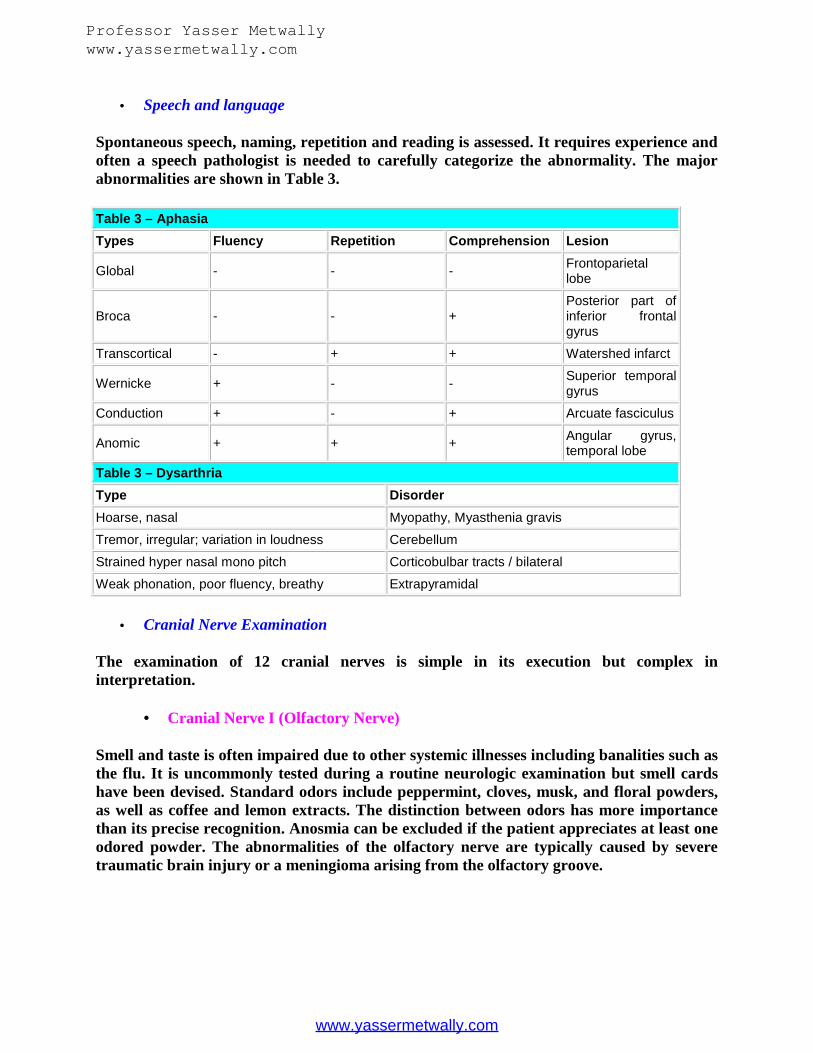
Spontaneous speech, naming, repetition and reading is assessed. It requires experience andoften a speech pathologist is needed to carefully categorize the abnormality. The majorabnormalities are shown in Table 3.
Table 3 – AphasiaTypes Fluency Repetition Comprehension Lesion
Global - - - Frontoparietallobe
Broca - - +Posterior part ofinferior frontalgyrus
Transcortical - + + Watershed infarct
Wernicke + - - Superior temporalgyrus
Conduction + - + Arcuate fasciculus
Anomic + + + Angular gyrus,temporal lobe
Table 3 – DysarthriaType DisorderHoarse, nasal Myopathy, Myasthenia gravis
Tremor, irregular; variation in loudness Cerebellum
Strained hyper nasal mono pitch Corticobulbar tracts / bilateral
Weak phonation, poor fluency, breathy Extrapyramidal
Cranial Nerve Examination
The examination of 12 cranial nerves is simple in its execution but complex ininterpretation.
Cranial Nerve I (Olfactory Nerve)
Smell and taste is often impaired due to other systemic illnesses including banalities such asthe flu. It is uncommonly tested during a routine neurologic examination but smell cardshave been devised. Standard odors include peppermint, cloves, musk, and floral powders,as well as coffee and lemon extracts. The distinction between odors has more importancethan its precise recognition. Anosmia can be excluded if the patient appreciates at least oneodored powder. The abnormalities of the olfactory nerve are typically caused by severetraumatic brain injury or a meningioma arising from the olfactory groove.
Professor Yasser Metwallywww.yassermetwally.com
www.yassermetwally.com

Cranial Nerve II (Optic Nerve)
The optic nerve is examined using several tests starting with visual acuity. Each eye istested separately using Snellen test card. The letters and the line designated 20 should beread at 20 feet recording 20/20 vision. When a refractory error is considered, the patientneeds to view these letters through a pinhole using a piece of paper and creating a hole ofapproximately 1 mm. Marked deterioration of vision is recorded using several standardlandmarks. For example a vision of 1/60 is present when a patient is able to see fingercounting at 1-m distance, 1/200 when moving of the hand is observed. 1/¥ when only lightperception is present and zero when completely blind. These abnormalities are typicallyseen in patients with a lesion of the optic nerve, often due to optic neuritis or anteriorischemic optic neuropathy. The visual fields are tested with a confrontation method inwhich the patient faces the examiner, covers one eye with his hand, and fixes his gaze onthe examiner’s nose. The examiner’s wiggling finger is then brought in along all fourquadrants and mentioned by the patient when it comes into view. Visual field defects arenamed hemianopsia when there is loss of vision in one half field of one eye. Loss vision incorresponding halves of both visual fields is called homonymous hemianopsia. Localizationof a homonymous hemianopsia is typically in the occipital cortex. However, macular(central) sparing may occur due to significant collateral branches from the middle cerebralartery. Lesions in the temporal lobe produce a "pie in the sky" homonymous defect. Alesion in the parietal lobe produces a lower quadrant defect. Examination is followed byfundoscopy in which the optic disk is assessed. Dilatation of the pupil is not needed for mostpurposes but when in doubt a more complete examination with assessment of the maculashould follow. Disk swelling is apparent with loss of the normal venous pulse first followedby loss of sharp temporal or nasal margins. Papilledema in advanced forms assumes theconfiguration of a champagne cork and peripheral hemorrhages are seen. Papilledemaindicates increased intracranial pressure from a mass or due to cerebral venousobstruction. It may also seen in a central venous occlusion and may at times be difficult todistinguish from congenital lesions such as a drusen optic disk or anomalous elevation.
Professor Yasser Metwallywww.yassermetwally.com
www.yassermetwally.com

Figure 1. Keep both yours and the person's eyes Have the patient focus on a distant objectLook at right fundus with your right eye Ophthalmoscope should be close to your eyes.Your head and the scope should move together Set the lens opening at +8 to +10 diopters.With the ophthalmoscope 12-15 inches from the patient's eye, check for the red reflex andfor opacities in lens or aqueous. While adjusting the diopter setting, approach the patientmore closely and systematically inspect the disc, noting the color, shape, margins and cup-to-disc ratio. Inspect the vessels, noting obstruction, caliber and arterial/venous ratio. Notethe presence of arterial/venous nicking and arterial light reflex. Check the background byinspecting for pigmentation, hemorrhages and hard or soft exudates. Next, try to identifythe macula. Have the patient look at light Normal: Disc margins are sharp color: yellowishorange to creamy pink shape: round or oval Cup to disc ratio: less than half Vessels AVratio AV crossing: no indentation No arterial light reflex Fundus background No exudatesor hemorrhages color : red to purplish Macula macula is located 2.5 disc distance temporalto disc no vessels are noted around Macula it may be slightly pigmented .
Figure 2. Position yourself in front of the patient. Test the patient's visual acuity, each eyeseparately, covering one at a time. Snellen's chart is used by Ophthalmologists. Visualacuity is recorded as a fraction. The numerator indicates the distance (in feet) from thechart which the subject can read the line. The denominator indicates the distance at whicha normal eye can read the line. Normal vision is 20/20. A pocket screener is used at thebedside. Hold the pocket screener at a distance of 12-14 inches. At this distance the lettersare equivalent to those on Snellen's chart. In children the techniques used are "E" card: Ifthe child cannot read letters or numbers Fixation and following: In infants. Have the childfollow a toy. Pinhole test: To differentiate refraction errors from organic disease. If visionimproves with pinhole it is refractory error.
Professor Yasser Metwallywww.yassermetwally.com
www.yassermetwally.com
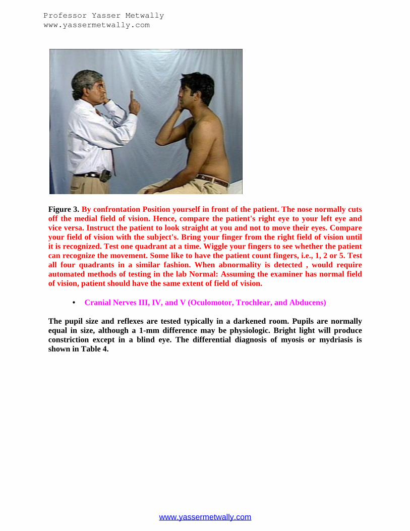
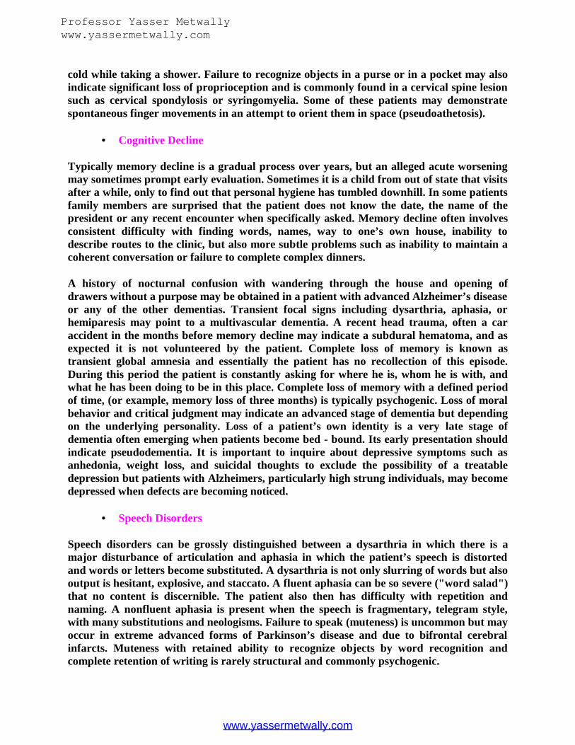
Figure 3. By confrontation Position yourself in front of the patient. The nose normally cutsoff the medial field of vision. Hence, compare the patient's right eye to your left eye andvice versa. Instruct the patient to look straight at you and not to move their eyes. Compareyour field of vision with the subject's. Bring your finger from the right field of vision untilit is recognized. Test one quadrant at a time. Wiggle your fingers to see whether the patientcan recognize the movement. Some like to have the patient count fingers, i.e., 1, 2 or 5. Testall four quadrants in a similar fashion. When abnormality is detected , would requireautomated methods of testing in the lab Normal: Assuming the examiner has normal fieldof vision, patient should have the same extent of field of vision.
Cranial Nerves III, IV, and V (Oculomotor, Trochlear, and Abducens)
The pupil size and reflexes are tested typically in a darkened room. Pupils are normallyequal in size, although a 1-mm difference may be physiologic. Bright light will produceconstriction except in a blind eye. The differential diagnosis of myosis or mydriasis isshown in Table 4.
Professor Yasser Metwallywww.yassermetwally.com
www.yassermetwally.com
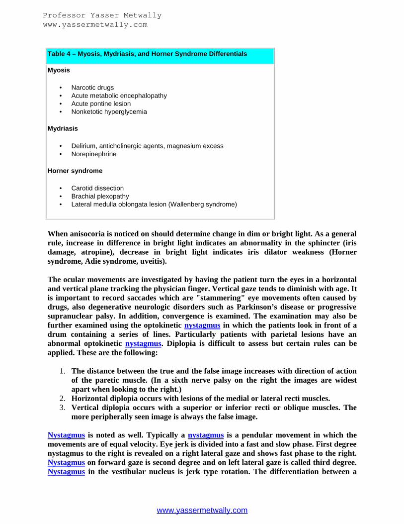
Table 4 – Myosis, Mydriasis, and Horner Syndrome Differentials
Myosis
Narcotic drugs Acute metabolic encephalopathy Acute pontine lesion Nonketotic hyperglycemia
Mydriasis
Delirium, anticholinergic agents, magnesium excess Norepinephrine
Horner syndrome
Carotid dissection Brachial plexopathy Lateral medulla oblongata lesion (Wallenberg syndrome)
When anisocoria is noticed on should determine change in dim or bright light. As a generalrule, increase in difference in bright light indicates an abnormality in the sphincter (irisdamage, atropine), decrease in bright light indicates iris dilator weakness (Hornersyndrome, Adie syndrome, uveitis).
The ocular movements are investigated by having the patient turn the eyes in a horizontaland vertical plane tracking the physician finger. Vertical gaze tends to diminish with age. Itis important to record saccades which are "stammering" eye movements often caused bydrugs, also degenerative neurologic disorders such as Parkinson’s disease or progressivesupranuclear palsy. In addition, convergence is examined. The examination may also befurther examined using the optokinetic nystagmus in which the patients look in front of adrum containing a series of lines. Particularly patients with parietal lesions have anabnormal optokinetic nystagmus. Diplopia is difficult to assess but certain rules can beapplied. These are the following:
1. The distance between the true and the false image increases with direction of actionof the paretic muscle. (In a sixth nerve palsy on the right the images are widestapart when looking to the right.)
2. Horizontal diplopia occurs with lesions of the medial or lateral recti muscles.3. Vertical diplopia occurs with a superior or inferior recti or oblique muscles. The
more peripherally seen image is always the false image.
Nystagmus is noted as well. Typically a nystagmus is a pendular movement in which themovements are of equal velocity. Eye jerk is divided into a fast and slow phase. First degreenystagmus to the right is revealed on a right lateral gaze and shows fast phase to the right.Nystagmus on forward gaze is second degree and on left lateral gaze is called third degree.Nystagmus in the vestibular nucleus is jerk type rotation. The differentiation between a
Professor Yasser Metwallywww.yassermetwally.com
www.yassermetwally.com
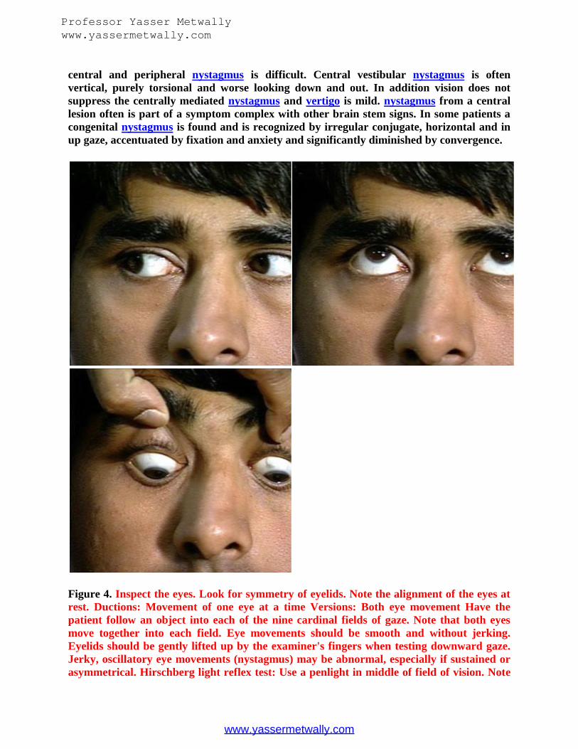
central and peripheral nystagmus is difficult. Central vestibular nystagmus is oftenvertical, purely torsional and worse looking down and out. In addition vision does notsuppress the centrally mediated nystagmus and vertigo is mild. nystagmus from a centrallesion often is part of a symptom complex with other brain stem signs. In some patients acongenital nystagmus is found and is recognized by irregular conjugate, horizontal and inup gaze, accentuated by fixation and anxiety and significantly diminished by convergence.
Figure 4. Inspect the eyes. Look for symmetry of eyelids. Note the alignment of the eyes atrest. Ductions: Movement of one eye at a time Versions: Both eye movement Have thepatient follow an object into each of the nine cardinal fields of gaze. Note that both eyesmove together into each field. Eye movements should be smooth and without jerking.Eyelids should be gently lifted up by the examiner's fingers when testing downward gaze.Jerky, oscillatory eye movements (nystagmus) may be abnormal, especially if sustained orasymmetrical. Hirschberg light reflex test: Use a penlight in middle of field of vision. Note
Professor Yasser Metwallywww.yassermetwally.com
www.yassermetwally.com

light reflection on both cornea. Let patient gaze in different directions, while noting theposition of light reflection in cornea. If they are asymmetrical it indicated there isstrabismus. Look up how to perform cover-uncover test to evaluate non paralyticstrabismus. Normal: Full conjugate eye movements.No nystagmus in any direction
Figure 5. Have the patient look at a distant object Look at size, shape and symmetry ofpupils. Shine a light into each eye and observe constriction of pupil. Flash a light on onepupil and watch it contract briskly. Flash the light again and watch the opposite pupilconstrict (consensual reflex). Repeat this procedure on the opposite eye. Normal: Pupils aresubtle, mild anisocoria (unequal in size) by itself and not necessarily an abnormal findings.Pupil size is 3-5 mm in diameter. They react briskly to light. Both pupils constrictconsensually.
Professor Yasser Metwallywww.yassermetwally.com
www.yassermetwally.com

Figure 6. Ask the patient to followyour finger as you bring it towardthe bridge of his nose. Note theconvergence of the eyes andpupillary constriction. Normal:Convergence should besustainable to within 5-8 cm andboth pupils constrict.
Cranial Nerve V (Trigeminal Nerve)
The trigeminal nerve consists of motor and sensory fibers. The sensory dermatome involvesthe scalp close to the line of the ear to forehead, eye, cheek, and chin. It can be tested bylight touch using a cotton Q-tip, pin, and temperature using hot and cold tubes. Thecorneal reflex is tested using a cotton ball gently striking the outer rim rather thancentrally on the cornea causing a reflective blink. The patient should also indicate touch. Inaddition, the jaw jerk is elicited by tapping on the apex of the jaw. The response is onlysignificant when it is exaggerated and may indicates a brain stem lesion.
Figure 7.Trigeminal nerve has motor and sensory components
Professor Yasser Metwallywww.yassermetwally.com
www.yassermetwally.com

Motor
Have patient clench teeth and feel the Masseters and Temporal muscles and compare sides.Note the strength of contractions. Edentulous patients may not be able to clench "teeth".
Sensory
With a light touch of cotton, check the patient's ability to detect light touch in all areas which are supplied bythe three divisions of the fifth cranial nerve. Instruct the patient to close his eyes and respond by saying "yes"every time he feels the sensation of cotton touching his face.
Compare corresponding contralateral segments of his face. Test pain sensation with a pin in each of the three divisions, comparing both sides.
Test corneal (blink) reflex with a wisp of cotton lightly touched to the edge of the cornea.There should be a consensual eyeblink normally.
Figure 8. A, The corneal reflex, B, examination of the sensory part of the trigeminal nerve
Cranial Nerve VII (Facial Nerve)
This is tested having the patient elevate eyebrows, closing eyelids forcefully in which theeyelashes disappear, and producing a voluntary smile. When a paralysis of the facial nerveexists, pronounciation of sounds that require closure of the lip such as pot and boy isdisturbed. In a peripheral seventh nerve palsy the platysma is also abnormal and can beexamined after the patient draws the lower lip and the angle of the mouth downwards.Taste may be abnormal but only when the lesion is peripheral to its junction with thecordae tympany. It is examined using sugar, salt, and sometimes tartaric acid but theresults are difficult to interpret. A common peripheral facial paralysis called Bell’s palsycan be recognized by involvement of all three branches, inability to blink and close the
Professor Yasser Metwallywww.yassermetwally.com
www.yassermetwally.com

eyelid, tearing, and a so-called Bell’s phenomenon in which with forceful closure of the eyethe globe turns upward.
Figure 9. Inspect the face. Look for asymmetry at rest, during conversation and whentesting various muscles. Ask the patient to wrinkle his forehead or raise his eyebrows,enabling you to test the upper face (frontalis). Next, have the patient tightly close his eyes.Test the strength of the orbicularis oculi by gently trying to pry open the patient's uppereyelid. Instruct him to puff out both cheeks. Check tension by tapping his cheeks with yourfingers. Have the patient smile broadly and show his teeth, testing the lower face. Normal:No facial asymmetry. Wrinkling of the forehead and smiling are equal and symmetrical.
Professor Yasser Metwallywww.yassermetwally.com
www.yassermetwally.com

Cranial Nerve VIII (Acoustic Nerve)
Hearing is tested with a whisper voice. The examiner stands in front of the patient andwhispers words (e.g. - 66, Boston) while covering patient eyes with one hand and blockingthe ear that is not tested with the other hand. Several tuning fork tests are available. TheWeber test is a test in which tuning fork is placed in the middle of the skull in whichhearing normally should be observed in both ears. Lateralization occurs on the same sidein the middle ear involvement, on the opposite side when the cochlear nerve is involved.The Rinne test is performed after placing the vibrating tuning fork against the mastoid andwhen it can no longer be heard it is held in front of the ear. Positive result is when thetuning fork is heard longer by air than bone conduction. An abnormal test is a sign ofmiddle ear defect or a blocking of the external auditory canal. Vestibular function can beexamined with laboratory and caloric testing but also using the Barany test. The patient isseated on examining table and will be reclined backwards with the head hanging over theedge of the table. After a brief interval vertigo will set in and at the same time a briefrotary nystagmus appears. The patient is asked to look downwards. The test is sensitive fora benign positional nystagmus. BPPD is due to dysfunction of the vestibular organ. It iscommon and often misdiagnosed as vertebral - basilar insufficiency.
Figure 10. With eyes closed, the patient should be instructed to acknowledge hearing thegentle rubbing of the examiner's fingers approximately 3-4 inches away from his right andleft ear. A watch, which the examiner can hear at a specific distance from his ear, is placednext to the patient's ear. Ask him to note when the watch sound disappears. Note that theexaminer has to have normal hearing to do this exam (in at least one ear). Normal: In aquiet room, the patient should be able to hear the physician's fingers rubbed lightlytogether 3-4 inches from his ear. With aging Progressive bilateral Presbycusis (oldhearing): Sensory neural loss Difficulty appreciating consonants
Professor Yasser Metwallywww.yassermetwally.com
www.yassermetwally.com
Cranial Nerve IX (Glossopharyngeal Nerve)
This nerve is tested by putting a tongue depressor in the back of the throat which willproduce a gag reflex. Midline elevation of the soft palate occurs. Its significance is dubiousbecause many normal individuals have no gag with stimulation.
Cranial Nerve X (Vagus Nerve)
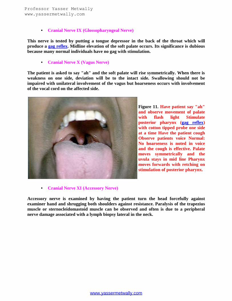
The patient is asked to say "ah" and the soft palate will rise symmetrically. When there isweakness on one side, deviation will be to the intact side. Swallowing should not beimpaired with unilateral involvement of the vagus but hoarseness occurs with involvementof the vocal cord on the affected side.
Figure 11. Have patient say "ah"and observe movement of palatewith flash light Stimulateposterior pharynx (gag reflex)with cotton tipped probe one sideat a time Have the patient coughObserve patients voice Normal:No hoarseness is noted in voiceand the cough is effective. Palatemoves symmetrically and theuvula stays in mid line Pharynxmoves forwards with retching onstimulation of posterior pharynx.
Cranial Nerve XI (Accessory Nerve)
Accessory nerve is examined by having the patient turn the head forcefully againstexaminer hand and shrugging both shoulders against resistance. Paralysis of the trapeziusmuscle or sternocleidomastoid muscle can be observed and often is due to a peripheralnerve damage associated with a lymph biopsy lateral in the neck.
Professor Yasser Metwallywww.yassermetwally.com
www.yassermetwally.com

Figure 12. Inspect Trapezius and Sternocleidomastoid muscles Note muscle size (bulk).Look for asymmetry, atrophy and fasciculation. Determine muscle power by gently tryingto overpower contraction of each group of muscles. Have patient shrug shoulder againstresistance and evaluate strength of Trapezius muscle. Have patient turn head to one sideagainst resistance and evaluate strength and observe contracting sternomastoid muscle
Cranial Nerve XII (Hypoglossal Nerve)
The patient is asked to protrude the tongue and then also the tongue is investigatedcarefully for atrophy and fasciculations. Tongue fasciculations are strong indicators of ALSin the appropriate setting. It may a appear like a bag of worms. The patient then is askedto rapidly move the tongue from left to right and the strength is tested by pushing thetongue against a tongue blade or against the cheek.
Professor Yasser Metwallywww.yassermetwally.com
www.yassermetwally.com

Figure 13. Pay attention to articulation . Inspect Tongue. Note muscle size (bulk). Look forasymmetry, atrophy and fasciculation. Determine muscle power by Having the patient toprotrude the tongue and side to side, up and down. Note the resistance offered by thetongue to your finger pressing on the tongue through each cheek.
Cranial nerve palsy examples
CN III Palsy (a) CN III Palsy (b) Right CN VI Palsy Gaze left
CN VII (Bell's) Palsy (a) CN VII (Bell's) Palsy (b)
Professor Yasser Metwallywww.yassermetwally.com
www.yassermetwally.com
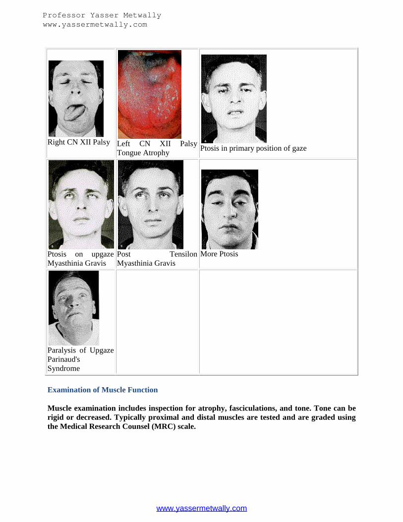
Right CN XII Palsy Left CN XII PalsyTongue Atrophy
Ptosis in primary position of gaze
Ptosis on upgazeMyasthinia Gravis
Post TensilonMyasthinia Gravis
More Ptosis
Paralysis of UpgazeParinaud'sSyndrome
Examination of Muscle Function
Muscle examination includes inspection for atrophy, fasciculations, and tone. Tone can berigid or decreased. Typically proximal and distal muscles are tested and are graded usingthe Medical Research Counsel (MRC) scale.
Professor Yasser Metwallywww.yassermetwally.com
www.yassermetwally.com

Table 5 - MRC Scale
5 = Normal power
4 = Reduced power, but still contracting muscle against resistance
3 = Movement against gravity but not resistance
2 = Movement with gravity eliminated
1 = Flicker of movement only
0 = No movement
Muscle atrophy is seen in many diseases of the peripheral nerve but also in advancedmyopathies. Generally muscle weakness in myopathies involves muscles in a proximaldistribution and peripheral nerve in a distal distribution (hand and foot muscles).
Muscle weakness may involve a nerve root or single nerve. It is summarized in Table 8.Fasciculations are fine twitches in parts of muscle and typically do occur at areas of thelimb that the examiner is not looking at ("the shooting star phenomenon"). Muscle tone isassessed after passive movement of the muscle and often muscle tone becomes clear toresistance. Hypotonia is apparent when a limb is shaken by the examiner documentingsignificant flailing. Loose and toneless muscles not only can be seen in peripheral nerveabnormalities also in the setting of acute cerebellar lesions. Spasticity is diagnosed withincreasing resistance to passive movement followed by a sudden release of resistance,typically called a clasp knife reaction.
Table 6 - Nerve Roots and Peripheral Nerves Supplying Arm / Leg MusclesC4 Levator scapular
C5 -T1
Pectoralis major
C5 -C6
Deltoid (axillary nerve)
Biceps (musculocutaneous nerve)
Brachioradialis (radial nerve)
Supinator (radial nerve)
C6 -C7
Pronator teres (median nerve)
C6 -C7 -C8
Triceps (radial nerve)
Extensor carpi ulnaris (radial nerve)
Flexor carpi ulnaris (median and ulnar nerve)
Professor Yasser Metwallywww.yassermetwally.com
www.yassermetwally.com
C7-8 Digit extensors (radial nerve)
C7-8- T1
Digit flexors (median and ulnar nerves)
C8 -T1
Thenar (median nerve)
Hypothenar (ulnar nerve)
Interossei (ulnar nerve)
L2-3-
4
Iliopsoas (femoral nerve)
Adductor thigh (obturator nerve)
L4-5- S1
Hamstrings (sciatic nerve)
Toe extensors (peroneus nerve)
L2-3-
4
Quadriceps (femoral nerve)
L4-5 Anterior tibial (peroneal nerve)
L5 -S1
Extensor hallucis longus (peroneal nerve)
Peronei (peroneal nerve)
Posterior tibial (tibial nerve)
Toe flexors (tibial nerve)
L5 -S1-2
Gluteus maximus (inferior gluteal nerve)
Gastrocnemius / Soleus (tibial nerve)
Professor Yasser Metwallywww.yassermetwally.com
www.yassermetwally.com

Professor Yasser Metwallywww.yassermetwally.com
www.yassermetwally.com

Professor Yasser Metwallywww.yassermetwally.com
www.yassermetwally.com
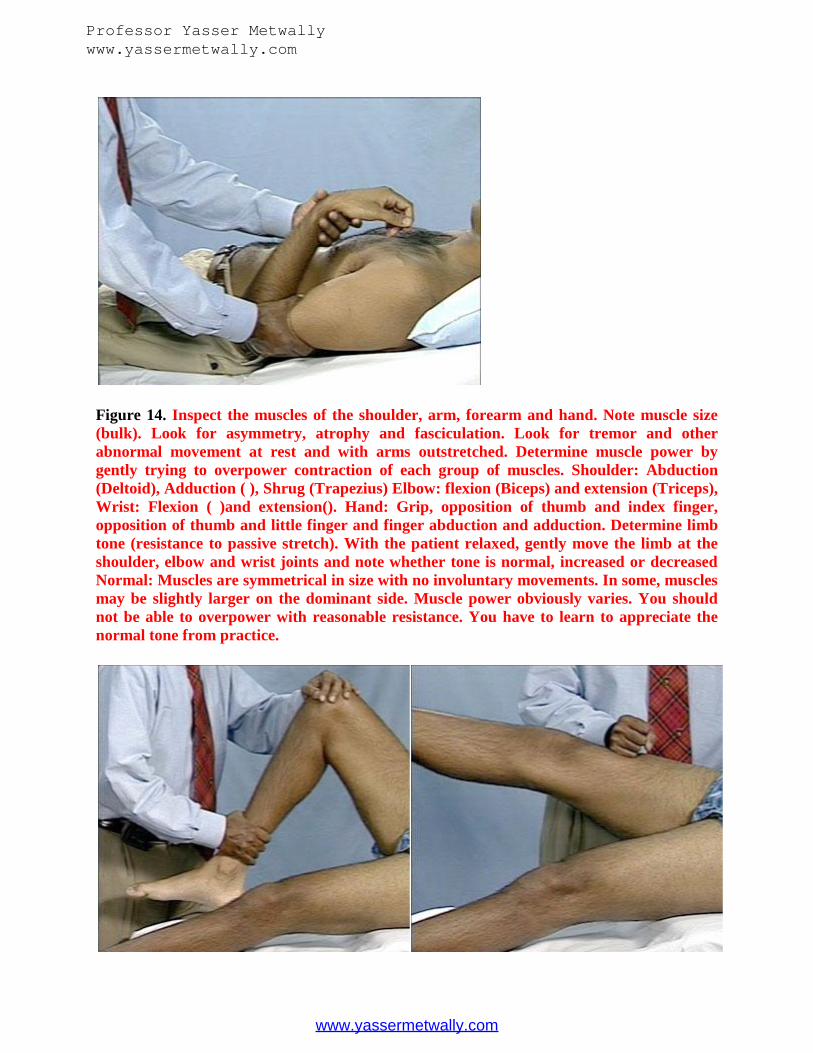
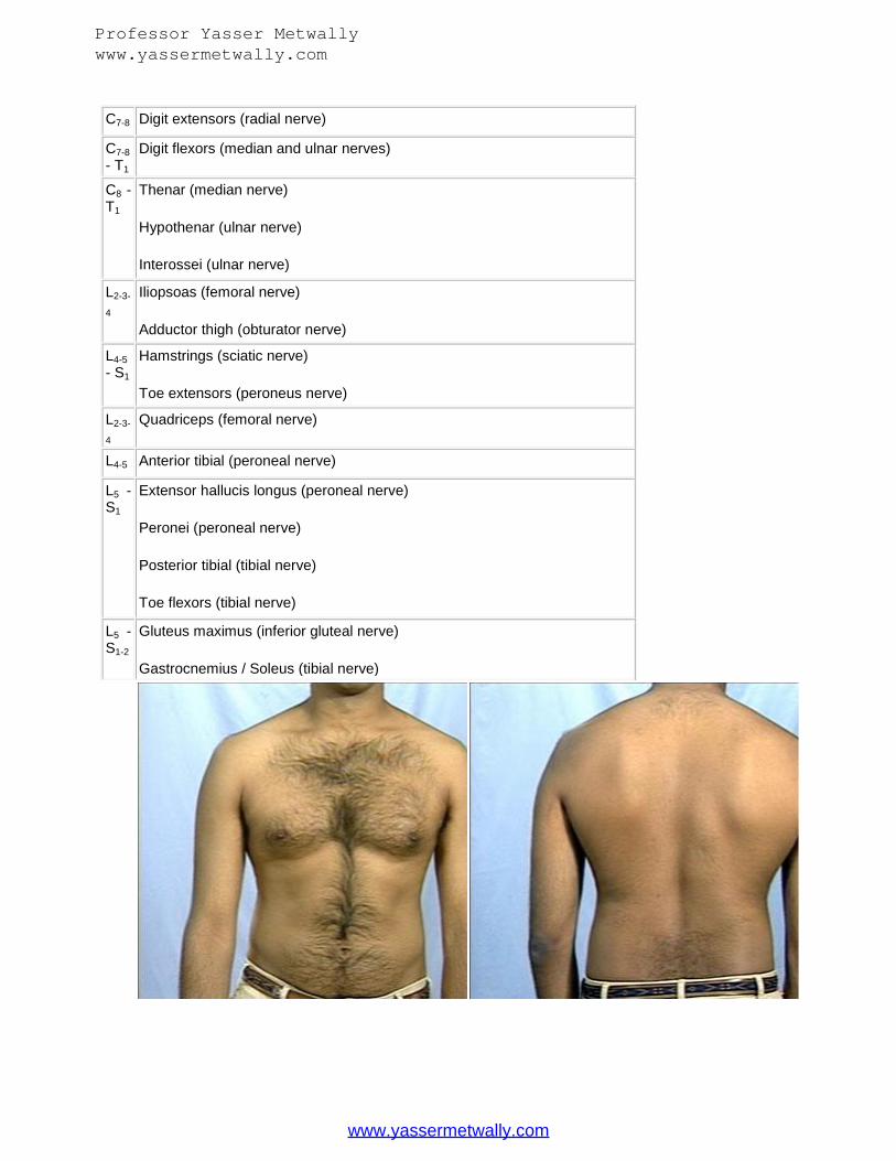
Figure 14. Inspect the muscles of the shoulder, arm, forearm and hand. Note muscle size(bulk). Look for asymmetry, atrophy and fasciculation. Look for tremor and otherabnormal movement at rest and with arms outstretched. Determine muscle power bygently trying to overpower contraction of each group of muscles. Shoulder: Abduction(Deltoid), Adduction ( ), Shrug (Trapezius) Elbow: flexion (Biceps) and extension (Triceps),Wrist: Flexion ( )and extension(). Hand: Grip, opposition of thumb and index finger,opposition of thumb and little finger and finger abduction and adduction. Determine limbtone (resistance to passive stretch). With the patient relaxed, gently move the limb at theshoulder, elbow and wrist joints and note whether tone is normal, increased or decreasedNormal: Muscles are symmetrical in size with no involuntary movements. In some, musclesmay be slightly larger on the dominant side. Muscle power obviously varies. You shouldnot be able to overpower with reasonable resistance. You have to learn to appreciate thenormal tone from practice.
Professor Yasser Metwallywww.yassermetwally.com
www.yassermetwally.com
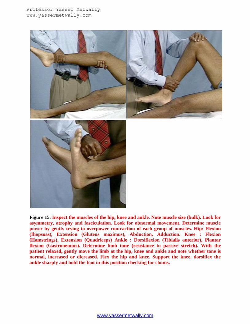
Figure 15. Inspect the muscles of the hip, knee and ankle. Note muscle size (bulk). Look forasymmetry, atrophy and fasciculation. Look for abnormal movement. Determine musclepower by gently trying to overpower contraction of each group of muscles. Hip: Flexion(Iliopsoas), Extension (Gluteus maximus), Abduction, Adduction. Knee : Flexion(Hamstrings), Extension (Quadriceps) Ankle : Dorsiflexion (Tibialis anterior), Plantarflexion (Gastronemius). Determine limb tone (resistance to passive stretch). With thepatient relaxed, gently move the limb at the hip, knee and ankle and note whether tone isnormal, increased or dicreased. Flex the hip and knee. Support the knee, dorsiflex theankle sharply and hold the foot in this position checking for clonus.
Professor Yasser Metwallywww.yassermetwally.com
www.yassermetwally.com

Examination of motor power
Motor examination
Deltoid C5 Axillary N. Biceps C6 MusculocutaneousN.
Triceps C7 Radial N. Brachioradialis C6 Radial N.
Extensor Carpi Ulnaris C7Radial (PosteriorInterossious)
Extensor Digitorum C7Radial (PosteriorInterossious)
First Dorsal Interossious T1Ulner Nerve
Abductor Pollicis Brevis T1Median N.
Psoas L1,2 Hamstring S1 Sciatic Tibialis Anterior L4,5 DeepPeroneal N.
Professor Yasser Metwallywww.yassermetwally.com
www.yassermetwally.com

Ext. Hallucis Longus L5Deep Peroneal N.
Extensor Digitorum BrevisL5 Deep Peroneal N.
Extensor Digitorum LongusL5 Deep Peroneal N.
Gastrocnemious S1 Tibial N.
Reflexes
Tendon reflexes localize to various segments in the spinal cord. The biceps reflex (C5-C6),triceps (C6-C7), knee (L2, L3, and L4), and ankle (L5-S1). Deep tendon reflexes are alsoclassified using a simple grading system (Table 7).
Table 7 - Classification of Deep Tendon Reflexes
0 = Absent
+/- = Present with enforcement
+ = Just present
2+ = Normal reflex
3+ = Brisk reflex, additional beat but still within normal limits
4+ = Pathological brisk reflex and clonus
Reflexes in patients may be depressed without any pathological meaning and in manypatients voluntary contracting a muscle in other limb will facilitate the reflex (Jendrassikmaneuver). Important reflexes are a normal plantar reflex (toes curling down), Babinskisign (unfortunately often called reflex, response or worse "the Babinskis") typically when apiece of metal or wood is applied to lateral surface of the foot or moved in a hockey stickcurve from the heel to the front. It results in flexion of the great toe, spreading of the toes in
Professor Yasser Metwallywww.yassermetwally.com
www.yassermetwally.com
a same response as flexing the knee and contraction of the tensor fascia lata (so called tripleresponse). Other reflexes that need to be examined are abdominal reflexes, stroking thesurface of the abdomen in four segments. Contraction is seen, but in elderly obese, andpatients with lax abdominal muscles, reflexes are most of the time absent. When the twolowest abdominal responses are absent, a localized spinal cord lesion is at the T10 level.Many other reflexes have been described. They include snout reflex by stimulating the lips,grasp reflex with persistent flexion of the fingers after insertion of two fingers in the palm,palmomental reflex with pressure on the palm causing a contraction of the ipsilateralmentalis muscle, all potentially indicating cortical inhibition. The Hoffmann-Tromnerreflex is obtained by snapping the terminal phalanx of the middle finger causing the flexionresponse of all fingers. The abnormality has been falsely considered "the Babinski of thearm" but only asymmetries are of importance. It is often difficult to elicit.
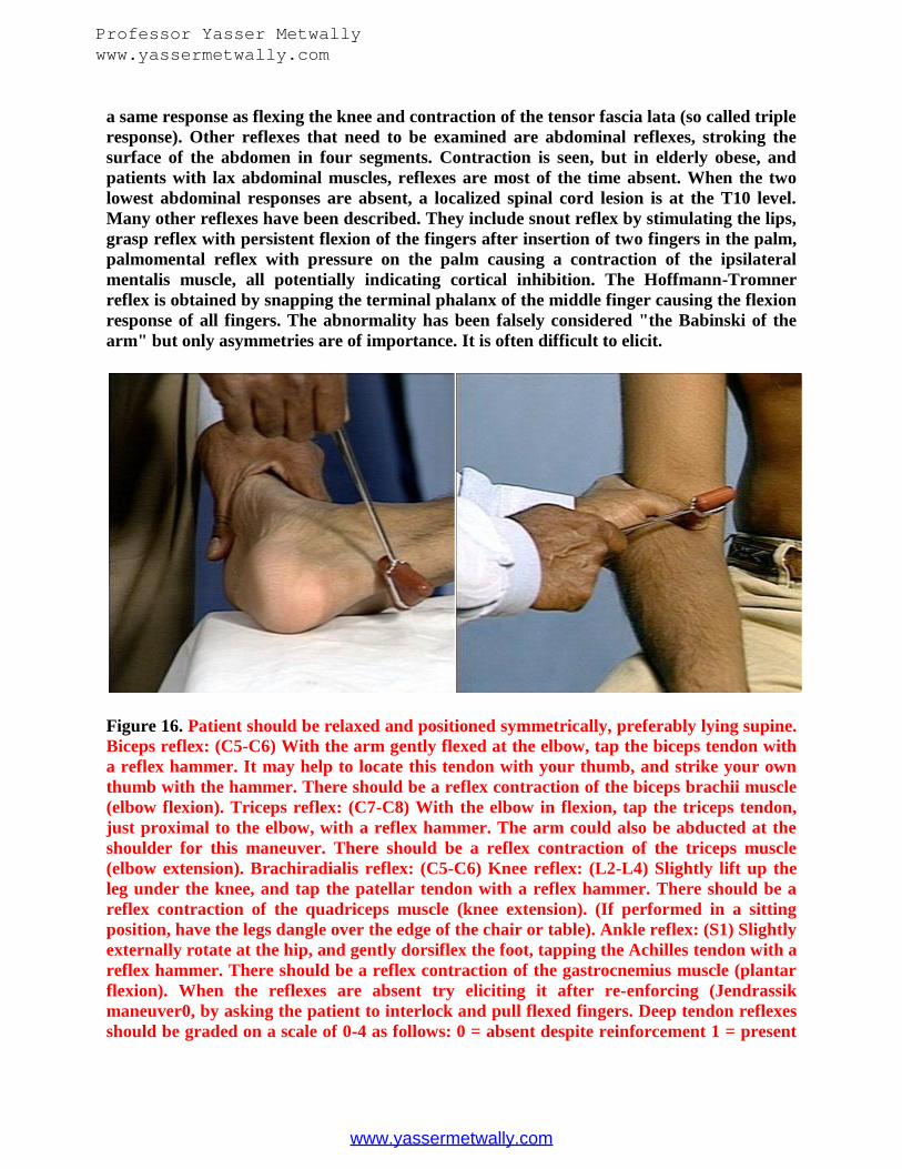
Figure 16. Patient should be relaxed and positioned symmetrically, preferably lying supine.Biceps reflex: (C5-C6) With the arm gently flexed at the elbow, tap the biceps tendon witha reflex hammer. It may help to locate this tendon with your thumb, and strike your ownthumb with the hammer. There should be a reflex contraction of the biceps brachii muscle(elbow flexion). Triceps reflex: (C7-C8) With the elbow in flexion, tap the triceps tendon,just proximal to the elbow, with a reflex hammer. The arm could also be abducted at theshoulder for this maneuver. There should be a reflex contraction of the triceps muscle(elbow extension). Brachiradialis reflex: (C5-C6) Knee reflex: (L2-L4) Slightly lift up theleg under the knee, and tap the patellar tendon with a reflex hammer. There should be areflex contraction of the quadriceps muscle (knee extension). (If performed in a sittingposition, have the legs dangle over the edge of the chair or table). Ankle reflex: (S1) Slightlyexternally rotate at the hip, and gently dorsiflex the foot, tapping the Achilles tendon with areflex hammer. There should be a reflex contraction of the gastrocnemius muscle (plantarflexion). When the reflexes are absent try eliciting it after re-enforcing (Jendrassikmaneuver0, by asking the patient to interlock and pull flexed fingers. Deep tendon reflexesshould be graded on a scale of 0-4 as follows: 0 = absent despite reinforcement 1 = present
Professor Yasser Metwallywww.yassermetwally.com
www.yassermetwally.com

only with reinforcement 2 = normal 3 = increased but normal 4 = markedly hyperactive,with clonus
Figure 17. With the patientsupine, support the weight of thefoot at the ankle.With a pointedobject, stroke the lateral aspect ofthe sole of the foot, from the heelup and across the ball of the foot.Normal: Note plantar flexion ofthe toes.
Sensory Examination
Sensory testing involves assessment of light touch, pinprick, vibration sense, and jointposition sense, and in occasional situations temperature assessment. Light touch involveswisp of cotton ball. The skin is touched, not moved, along it. Pinprick is tested with a sterilepin. Vibration using a tuning fork has similar meaning as joint position sense. Typicallymovement of the toe up or down is assessed or the patient imitates the same movement inthe other limb. When a sensory level is noted by the patient the margins of abnormalityneeds to be carefully localized. Important pointers are shoulders (C4), nipples (TH4) andnavel (TH10). Significant loss of proprioception will cause pseudoathetosis in which thefingers constantly try to orient themselves in space. A 2-point discrimination is alsoassessing the posterior column and normally stimulus separated by 2 mm should bedistinguished.
Professor Yasser Metwallywww.yassermetwally.com
www.yassermetwally.com

Figure 18. Test light touch with a wisp of cotton. Test pain sense with a blunted, disposablesafety pin or splintered cotton tip applicator. For light touch and pain: Have patient closeeyes and report each test stimulus. Test over sides of each foot, leg, thigh, hand, forearmand arm. Compare the right and left and distal with proximal. Test the trunk whereindicated. Test position sense by moving the toe or finger up and down, held by its sides,and have the patient report its position with eyes closed. Vibration sense is tested with avibrating tuning fork placed over bony prominences of the feet (ankles) and hands(knuckles). Ask the patient to report when the vibration sense is lost. Sensation is tested bycomparing the right and left sides In cases of suspected root or nerve lesions, sensation in adermatomal or peripheral nerve distribution is carefully tested. If a spinal cord lesion issuspected, check for sensory loss over the trunk and sacral areas. Normal:Light touch,pinprick, vibration and position sense are intact throughout.
Cerebellar Function
Cerebellar function is tested with a finger-to-nose test or finger-to-finger test typicallyusing additional turning in the wrists to further test coordination. The most commonlyneglected investigation in bed bound patients is a sitting position in which patient may fallto one side with a midline vermis lesion. Dysmetria is noted when the patient cannotsmoothly touch the nose and becomes shaky when reaching target.
Professor Yasser Metwallywww.yassermetwally.com
www.yassermetwally.com

Figure 19. Ask the patient to alternately reach out and touch your extended finger and hisown nose. Test both hands. Ask the (supine) patient to touch his heel to the opposite knee,and slide it smoothly down he shin of that leg. Test both legs. Normal: Patient can touch thetarget and perform movements in a smooth, coordinated manner. Heel to knee to shin areperformed smoothly and accurately.
Gait
A favorite pastime of neurologists is to investigate gait. Typically the patient is asked towalk on the hallway and several components are investigated - stability, stride, initiation ofgait, and turns. Typical abnormalities in a Parkinson’s patient is stooped gait, reduced armswing, fragmentary turning. In a patient with a hemiplegia the affected leg is swungoutwards with a tendency for the foot to catch on the ground. Profound loss of sensoryinformation from the feet is actually heard due a stamping gait.
Table 8 - GaitType FeaturesCerebellar ataxia Wide based, staggering steps, leans forward. Abnormal
turning the corner.
Sensory ataxia Romberg (+); wide based; high stepping.
Frontal lobe ataxia Hesitating start, shuffling, freezing with corners.
Spastic Bouncing wide - based, "tin-man"-like
Akinetic Shuffling, no arm swing, small steps
Professor Yasser Metwallywww.yassermetwally.com
www.yassermetwally.com

Figure 20. Ask the patient to walk back and forth across the room. Observe for equality ofarm swing , balance and rapidity and ease of turning. Next, ask the patient to walk on histiptoes, then on heels. Ask the patient to tandem walk. Test patient's ability to stand withfeet together with eyes open and then closed. (Romberg's test). Reassure patient that youwill support him, in case he becomes unsteady. Normal: Person can walk in balance withthe arms swinging at sides and can turn smoothly. Person should be able to stand with feettogether without falling with eyes open or closed.
Professor Yasser Metwallywww.yassermetwally.com
www.yassermetwally.com

Some abnormal gaits
Steppage Gait Hemiplegic Gait Parkinsonian Gait
Retropulsion
Localization Principles
This document is not designed to give a complete evaluation of the localization techniquesand the reader should be referred to the book by Brazis, Masdeu, and Biller, Localizationin Clinical Neurology, Lippincott Raven, 3rd Edition. Some generalities should bementioned. Lesions of the upper motor neuron will give paralysis, distally more involved inthan the proximal muscles as well as increased reflexes and clonus, and loss of cutaneousreflexes and a Babinski sign. Lesions of the lower motor neuron involve atrophy, flaccidparalysis, fasciculations and weakness is segmental in character. The segmentaldistribution is noticeable. Specific muscles are innervated through a single cord segmentbecause the spinal cord is arranged through separate reflex arcs. Absence of sensoryabnormalities are seen when the lesion is entirely anterior horn. Lesions of theextrapyramidal system will produce bradykinesia with slowness of movement, tremor,shuffling walking with small steps, slow movements, resting tremor, and slowing ofmentation as well as initiating movements. Posterior column syndromes involve ataxia,dysmetria with impossibility of coordinated smooth movements, overshooting the markand increase in symptoms after elimination of vision. Syndromes of cerebellarabnormalities involve decomposition of movement, inability to perform movementssmoothly, hypertonia, excessive rebound, and intention tremor.
Professor Yasser Metwallywww.yassermetwally.com
www.yassermetwally.com
Clinical skills in Neuro Evaluation
Learning Objectives
Upon completion of this course the learner will be able to:
Describe the major components of the neurological exam. Identify 3 common neurological disorders tested during a neurological evaluation. Identify the major components of the motor exam. Describe the two sensory modalities tested using the sensory exam. Describe three common tests used to assess cerebellar dysfunction.
Introduction
The purpose of this course is to summarize the main parts of the neurological exam.Familiarity with this material will allow you to diagnose common neurological disorders,identify neurological emergencies and make referrals to appropriate specialists.
Any health professional faced with the task of assessing a person with an emergent, acuteor even long-standing neurological deficit knows the importance of a quick and reliableneurological exam. Your knowledge of neurological evaluation techniques will allow you togather accurate information about your client's medical condition and help you to createan accurate plan of care. Once you are familiar with the neurological exam, you should beable to complete the exam in 10 to 15 minutes. Additionally you will learn some of the teststhat are used to identify certain types of common neurological dysfunctions. The coursewill cover the following parts of the neurological evaluation:
Index
Tools Patient History Physical Exam Cognitive Assessment/Mental Status Cranial Nerve Assessment Motor Exam Sensory Exam Coordination Exam
Tools
The following tools will be used during the neurological exam:
Reflex hammer (tomahawk model) Penlight Tongue blade Safety pin
Professor Yasser Metwallywww.yassermetwally.com
www.yassermetwally.com

Cotton swab Ophthalmoscope Eye chart Tuning fork Dermatome chart
Patient History
As with all other nursing examinations, the neurological exam begins with the gathering ofan accurate patient history and information about the course of the present injury. Thiswill help to create a baseline as well as providing you with valuable information about thecourse and characteristics of the present illness. The following information is gatheredduring the patient history portion of the neurological exam:
Personal and family history Description of the current problem Past medical history Prior level of function Medication review Review of other major systems
Personal and family history.
The personal history should include a brief personal profile and description of the patient.A brief family history should be included, the source of the information indicated and themental status of the patient noted. Included in this section are the following items:
Date Age Gender Racial background Place of birth Marital status Occupation Religion
Description of the current problem.
A description of the current problem or "chief complaint" and the reason the patient isseeking medical care should be noted. Ask for an explanation of current signs andsymptoms including any physical or psychological changes. Ask about the presence ofdizziness, headaches, visual disturbances, speech or motor control problems. Inquire aboutthe onset and duration of symptoms and remember that in an emergent neurological eventthe progression of symptoms may help to identify the part of the brain that has beenaffected. The following items are included in this section
Professor Yasser Metwallywww.yassermetwally.com
www.yassermetwally.com

Present illness including onset of the problem, the setting it developed in,manifestations and past treatment for the problem.
Analysis of the "main symptom" including location, quality, severity, onset,duration, frequency and factors that aggravate or alleviate the condition.
Past medical history.
Ask about the person's past medical history, previous illnesses and psychological history.Include educational background and any recent change in personality or behavior.Included in this section are the following items:
Childhood illnesses Psychological illnesses Past accidents and injuries Operations Previous hospitalizations Current health Allergies Family history
Prior level of function.
Prior level of function is a critical piece of information that helps to establish the extent ofthe current neurological damage and helps you to differentiate between longstanding andemergent signs and symptoms. Ask about the person's level of daily activities and use ofassistive devices prior to the onset of the current medical problem.
Medication review.
Ask the patient for a list of over-the-counter and prescription medications as well as use ofrecreational drugs and alcohol.
Review of other major systems.
Ask the patient about any problems with the other major systems of the body includingheart, lungs and abdomen.
The Physical Exam
The physical exam includes inspection of the skin and neck, carotid and heart sounds,blood pressure, heart rate and respiratory rate. The following items are inspected in thissection:
Presence of weakness, fatigue or fever The condition of the skin - color, sores, rash, lumps
Professor Yasser Metwallywww.yassermetwally.com
www.yassermetwally.com

Eyes - visual changes, spots, double vision, cataracts, blurred vision, glasses orcontact lenses
Ears - pain, tinnitus, vertigo, discharge or infection Nose and sinuses - presence of cold, stuffiness, discharge or bleeding Mouth and throat - general condition of the teeth and gums, sore throat, bleeding,
sores, hoarseness or dryness Neck - presence of stiffness, pain, lumps or swollen glands Respiratory and cardiac systems Gastrointestinal urinary systems Genitals - presence of pain, discharge, swelling, sensory changes
Cognitive Assessment/Mental Exam
The mental exam starts when the patient enters the medical setting and includes yourobservations and conversations with the patient. If the patient demonstrates cognitivefunctioning that is grossly intact including level of consciousness, alertness, speech, memoryand judgement, there is no need to do a more formal cognitive assessment. If it appearsthat any of the cognitive functions are impaired it will be necessary to do a more detailedcognitive assessment. The following items are included in the cognitive assessment:
Level of consciousness Orientation Speech and language Memory Fund of information Insight and judgement Abstract thought Calculations
Level of consciousness.
There are many acceptable methods to determine level of consciousness including theGlascow coma Scale, the Mini-Mental State or by categorizing the level of consciousnessusing descriptive cognitive scales.
Glascow Coma Scale
A coma is defined by Jennett and Teasdale as "not obeying commands, not uttering wordsand not opening the eyes". The Glascow Coma (table 1) scale was first developed in 1974 asa way to assess and monitor levels of consciousness. A Glascow score of 8 or less out of apossible score of 15 defines coma in 90% of cases. The Glascow Coma scale may be used inconjunction with the Glascow Outcome scale (table 2) to determine level of recovery.
Professor Yasser Metwallywww.yassermetwally.com
www.yassermetwally.com
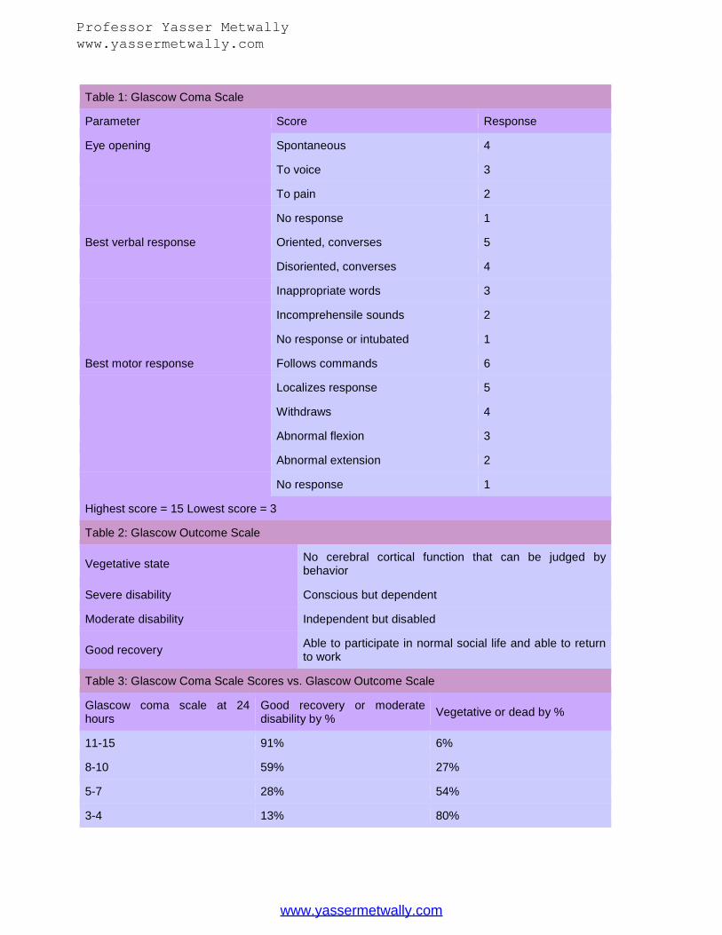
Table 1: Glascow Coma Scale
Parameter Score Response
Eye opening Spontaneous 4
To voice 3
To pain 2
No response 1
Best verbal response Oriented, converses 5
Disoriented, converses 4
Inappropriate words 3
Incomprehensile sounds 2
No response or intubated 1
Best motor response Follows commands 6
Localizes response 5
Withdraws 4
Abnormal flexion 3
Abnormal extension 2
No response 1
Highest score = 15 Lowest score = 3
Table 2: Glascow Outcome Scale
Vegetative state No cerebral cortical function that can be judged bybehavior
Severe disability Conscious but dependent
Moderate disability Independent but disabled
Good recovery Able to participate in normal social life and able to returnto work
Table 3: Glascow Coma Scale Scores vs. Glascow Outcome Scale
Glascow coma scale at 24hours
Good recovery or moderatedisability by % Vegetative or dead by %
11-15 91% 6%
8-10 59% 27%
5-7 28% 54%
3-4 13% 80%
Professor Yasser Metwallywww.yassermetwally.com
www.yassermetwally.com

Another scale used to descriptively assess the level of cognitive functioning in a person witha brain injury is as follows:
Alert Obtunded or confused - a slight but noticeable decrease in alertness with decreased
interest in what is happening in the environment, decreased attention span andmemory.
Stupor - the person appears to be in a deep sleep but can be aroused by noxious orvigorous stimuli.
Coma - eyes closed, no directed motor or verbal activity and unarousable.
Orientation.
Orientation is generally determined by asking the patient to answer a few commonquestions such as the name, place and time. Other questions might include the year, date,day or the name of the president or vice president. Time is often the first part of orientationthat is affected. The inability to remember one's name may be evidence of a psychiatriccondition.
Speech and language.
A detailed assessment of speech and language function is the job of a specialist. During theinitial neurological assessment, however you will be trying to establish the presence of aspeech disorder that did not exist prior to the onset of the current medical problem. Duringthis gross assessment, it is most common to look for the presence of aphasia, a problemwith the understanding of speech or the inability to communicate via speech. Additionally,it is important to note the quality, clarity and fluency of speech. The following items arecontained within a speech and language assessment:
Articulation - look for difficulty with the pronunciation of words, especially wordscontaining "p", "l" and "ch" sounds.
Rate and rhythm - look for changes in the rate and rhythm of speech. Prosody - aprosodia occurs due to a lesion in the right parietal lobe - the part of the
brain that is involved with the tone and musicality of speech. A patient with a lesionto this part of the brain will have flat intonation and a loss of pitch. There will alsobe a change in the accentuation and stress of words and syllables.
Aphasia - aphasia is an acquired communication disorder often caused by vascularinsult that affects a person's ability to speak and/or comprehend the spoken word.Aphasia can affect modalities other than speaking such as writing, gesturing andother non-verbal aspects of communication. For the purpose of a basic neurologicalexam it is sufficient to classify the aphasias as receptive, conductive or expressive.
Professor Yasser Metwallywww.yassermetwally.com
www.yassermetwally.com

Receptive (Wernicke's aphasia).
Speech is often clear and fluent and language is normal in rate, rhythm and melody butthere may be errors in words as well as the presence of added syllables. Language may beexcessive and convey little meaning. Comprehension is usually severely affected.
Conductive aphasia.
Speech is clear, but the patient is unable to repeat words. There is the ability to followcommands because comprehension is usually preserved. Naming and repeating is severelyimpaired. Reading aloud is impaired but reading silently is conserved. Speech is fluent butwith many incorrect words or sounds substituted for correct words.
Expressive (Broca's aphasia).
Comprehension is usually well preserved but speech is unclear and non-fluent. Patientstend to use only key words and omit many nouns and verbs. Neurological damage mayextend to the frontal lobe motor control areas adjoining Broca's area.
Memory.
The portion of the exam that tests memory skills is usually divided into three parts -immediate, recent and past memory.
1. Immediate - ask the patient to recall a few objects over the span of 3 to 5 minutes.2. Recent - ask the patient to recall events within the last several hours to several days.
Common questions might include, "What did you have for breakfast?" "Where doyou live?" and "When did you start to feel ill?"
3. Past - ask about events from childhood or long ago events.
Fund of information.
Ask about current events, name of the president, geography, etc.
Insight and judgement.
Ask about the patient's understanding and awareness of the current illness.
Abstract thought.
Ask the patient to compare and contrast two objects such as a car and a bus or a cucumberand an apple. Ask the patient to interpret a complex concept or political event.
Professor Yasser Metwallywww.yassermetwally.com
www.yassermetwally.com
Calculations.
Ask the patient to do a calculation such as counting backwards from 100 by increments of7 or count upwards by threes. Ask how many dimes are in a dollar or how many weeks intwo years.
The Cranial Nerves
Assessment of the cranial nerves provides information about the function of the nerves inthe head and neck region. With practice this part of the neurological exam can becompleted in just a few minutes. Testing is usually done in numerical order starting withCNI and proceeding to CNXII. The cranial nerves are arranged along the brainstem indescending order from 12 to 1. (CN I is located just above the olfactory epithelium on theinferior surface of the frontal lobe. CN II is located on the inferior surface of the cerebrumbehind the eyes.)
Cranial nerves 12 - 9 are located in the medulla oblongata, the part of the brainstemcontiguous with the spinal cord. Cranial nerves 8 - 5 are located along the pons, the nextportion of the brainstem. Cranial nerves 4 and 3 are located in the area of the midbrain,the uppermost portion of the brainstem.
CN I - The olfactory nerve.
The olfactory nerve is a sensory nerve responsible for smell.
Assessment of CNI is often omitted unless it is suspected that there is damage to theinferior frontal lobe - the area where the olfactory nerve is located. First make sure thenostrils are patent. CN I is usually tested by holding coffee, rubbing alcohol or some otherpungent or aromatic substance under the nose of the patient. Compare one side to theother.
CN II - The optic nerve.
The optic nerve is a sensory nerve responsible for vision. Always test this cranial nervebecause it will give you information about visual acuity and visual fields deficits. Test visualacuity, visual fields and fundi.
1. Visual Acuity. Use the eye chart from your toolkit and test the patient's correctedvision in good light. Have the patient stand 20 feet from the eye chart then read thesmallest line possible. Test both eyes. Compare the results of this test to the patient'sprior level of function.
2. Visual fields. It is important to test for the presence of visual field deficits if yoususpect a disorder that is located in front of the optic chiasm. The visual fields aretested by positioning your finger or a pencil beside the patient's temple in the areaof the peripheral vision. Slowly bring the object forward and ask the patient to saywhen the object becomes visible. Move the pencil or finger up, down, right, left and
Professor Yasser Metwallywww.yassermetwally.com
www.yassermetwally.com
diagonally to the upper right, lower right, upper left and lower left. Keep yourmovements small and slow - it is easier for the eye to detect motion and will makethe test less sensitive to visual field defects. Common visual field deficits include: 1)homonymous hemianopsia in which 1/2 of the visual field on the same side isaffected i.e. the nasal side of the right eye and the temporal side of the left eye, 2)bitemporal hemianopsia in which either the nasal side or the temporal sides of botheyes are affected, and 3) unilateral blindness in which one eye is blinded.
3. Fundi. Look closely at each eye and check for symmetry, clarity, color, contour,retinal abnormalities and the condition of the blood vessels in the eye.
CN III, IV and VI - The oculomotor, trochlear and abducens nerves.
The oculomotor, trochlear and abducens are motor nerves responsible for control of all eyemovements and innervation of all the extraocular eye muscles. CN III controls most of theextraocular eye muscles, eye opening and pupillary constriction. CN IV controls downwardand inward eye movements. CN VI controls lateral eye movements.
These cranial nerves are responsible for motor control of the eye muscles, eyelids and thepupils. They are tested in a group because they work together to control eye movement.Check the eyelids for drooping and symmetry. An eyelid drooping over the pupil (CN III)may indicate the presence of myasthenia gravis or 3rd cranial nerve palsy. Hold yourfinger in front of the patient and ask her to follow your finger as you move it through the 6cardinal fields. The cardinal fields are: 1) lateral and medial along the horizontal plane and2) superior and inferior in far lateral gaze.
Check pupil function for response to light. With the room darkened shine a penlight intoone eye and look for pupil constriction - this is called a direct response. The opposite pupilshould also constrict - this is known as a consensual response.
Next ask the patient to look at a distant object and then at your finger which is held about 4inches in front of the patient's nose. The pupils should constrict and the eyes convergewhen the eyes shift from the distant object to your finger. Dilated or constricted pupils mayindicate neurological disease, glaucoma, drug abuse or reaction to certain medications.
Finally check for the position of the eyes in primary gaze by having the patient lookstraight ahead, then shine a bright light into the pupils. The reflection of the light should bethe same in each pupil. A deviation suggests the presence of a strabismus - the inability toalign the visual axes so they are directed at the same point.
In addition to checking these functions remember to look for the presence of horizontal andvertical nystagmus. Have the patient hold a lateral gaze for 5 to 10 seconds. If a brainstemlesion is suspected, vertical nystagmus is often present and is almost always an indication ofcentral nervous system damage as opposed to vestibular dysfunction.
Professor Yasser Metwallywww.yassermetwally.com
www.yassermetwally.com
CN V - The trigeminal nerve.
The trigeminal nerve is a mixed motor and sensory nerve that innervates the muscles ofmastication and sensation from the skin, muscles and joints in the face, mouth and teethand lateral jaw movement. Both motor and sensory components should be assessed.
The motor portion of the trigeminal nerve is tested by palpating the masseter muscles whilethe patient clenches the jaw and by testing the strength of the jaw while opening andclosing the mouth. If a weakness is present the jaw will deviate to the weak side.
The sensory portion of the trigeminal nerve can be tested for temperature sensation usingthe metal surface of your tuning fork or using test tubes filled with warm and cold water.
The corneal reflex should be tested in a patient with suspected brainstem or hemisphericlesions by having the patient look up and away from the examiner. Approaching the eyefrom the side, lightly touch the cornea of one eye with a piece of cotton. Repeat in the othereye. A symmetric blink reflex should be present. Absence of the blink reflex indicates a 5thor 7th cranial nerve lesion.
CN VII - The facial nerve.
The facial nerve is mixed motor and sensory. The motor portion innervates the muscles offacial expression, the lacrimal and salivary glands. The sensory portion is responsible fortaste sensation from the anterior 2/3rds of the tongue and the skin of the external ear.
The facial nerve can be tested by asking the patient to smile, wrinkle the forehead, puff outthe cheeks and close the eyes tightly. Look for symmetry from side to side and differencesbetween the upper two-thirds and the lower one-third of the face. Weakness in the upperpart of the face may indicate a central nervous system lesion while weakness in the lowerone third may indicate a peripheral nervous system problem.
CN VIII - The vestibulocochlear nerve.
The vestibulocochlear nerve is sensory for hearing, balance and orientation in space.
Cranial nerve VIII is responsible for two main functions - control of vestibular functionand hearing. It is usually only tested in patients with symptoms of vertigo, imbalance orsuspected lesions in the cerebellopontine angle or brainstem.
Hearing can be screened initially by asking whether of not the patient hears your questions.To test hearing acuity, cover one ear and test the other with a watch held close to the ear orby whispering.
The vestibular portion of CN VIII is more complicated but should be tested if the patientreports symptoms of nausea, vertigo, anxiety or if there are signs of nystagmus, sweating,hypotension, vomiting, hypotension or postural deviations. Use the caloric test in which the
Professor Yasser Metwallywww.yassermetwally.com
www.yassermetwally.com

patient is placed in supine with the head elevated to 30 degrees. Irrigate the ear canal withcold water. If the vestibular pathways are intact and the patient is awake the caloric testwill cause nausea, horizontal nystagmus and vertigo to the irrigated side.
CN IX and X - The glossopharyngeal and vagus nerves.
The glossopharyngeal and vagus nerves are both mixed motor and sensory nerves. CN IXis involved with control of swallowing, sensation to the posterior 2/3rds of the tongue andinnervation of the parotid gland. CN X innervates the smooth muscle of the heart, bloodvessels, trachea, bronchi, esophagus, stomach and intestines. The motor portion innervatesthe muscles of the larynx, pharynx, palate and muscles of speech. The sensory portioninnervates visceral sensation from the pharynx, larynx, thorax and abdomen.
These cranial nerves supply motor control to the larynx as well as sensory control to thetongue. Observe the position and symmetry of the palate at rest and as the patient pants ofsays "ahhh". Test the gag reflex by lightly touching the posterior wall of the pharynx oneach side with a tongue blade - this is an indication of both glossopharygeal and vagusfunction. In addition, listen for a soft breathy voice and ask if the patient has had difficultywith swallowing. Hoarse sounding speech may indicate vocal cord paralysis while nasalspeech indicates paralysis of the palate.
CN XI - The spinal accessory nerve.
The spinal accessory nerve is a motor nerve that innervates the trapezius andsternocleidomastoid muscles.
To test the trapezius muscles, ask the patient to shrug the shoulders upward while theexaminer applies a downward pressure. To test the sternocleidomastoid muscles, have thepatient turn her head to the right while the examiner applies a gentle pressure in theopposite direction. During both of these tests look for symmetry of motion from side to sideand palpate the muscles to look for atrophy, abnormal tone or fasciculations in the muscle.Also note the strength of the contractions.
Slow, alternating motion may indicate the presence of a central nervous system lesion whileatrophy and fasciculations may indicate the presence of a peripheral nervous system lesion.
CN XII - The hypoglossal nerve.
The hypoglossal nerve is responsible for motor control of the intrinsic muscles of thetongue.
Check the position of the tongue within the mouth then look for smoothness of movementand symmetry as the patient protrudes the tongue and moves it from side to side.
The Motor Exam
Professor Yasser Metwallywww.yassermetwally.com
www.yassermetwally.com

The motor portion of the neurological exam includes observation of gross motor functionssuch as gait, extremity strength, reflexes, abnormal movement and abnormal tone -especially weakness and floppiness in the distal extremities. Begin by checking the generalappearance of the patient - preferably with the clothes removed. Inspect the musclesvisually and by palpation and note the presence of muscle fasciculations, abnormalities inmuscle bulk and tenderness.
Tone.
The term muscle tone refers to the force with which a muscle resists lengthening. Tone ispresent because of two factors - the mechanical elasticity of the muscle fibers and a neuralcomponent called the stretch reflex. Both of these factors resist uncontrolled lengthening ofthe muscle and contribute to the natural tone of the muscle.
Tone is assessed clinically by passively flexing and extending the patient's limbs and notingthe resistance of the muscles. Look for the presence of hypotonicity, hypertonicity, rigidityor spasticity. When checking for tone, make sure the patient is fully relaxed then take holdof the wrist and shake it back and forth. Raise the arm and drop it onto a soft surface.Grasp the ankle and shake it from side to side. Finally, with the patient in supine, raise theknee quickly and observe the movement of the ankle. Normally, when no abnormal tone ispresent, the foot will slide along the bed without lifting into the air.
Strength.
Muscle strength is usually assessed on a scale of 0-5 with 0 being no movement and 5 fullmovement against strong resistance. The following muscle groups are typically tested:
Ask the patient to squeeze your pointer and middle fingers Elbow flexion Elbow extension Thumb opposition Hip flexion Knee flexion Knee extension Ankle dorsiflexion (L4, L5) Ankle plantarflexion (S1)
Professor Yasser Metwallywww.yassermetwally.com
www.yassermetwally.com
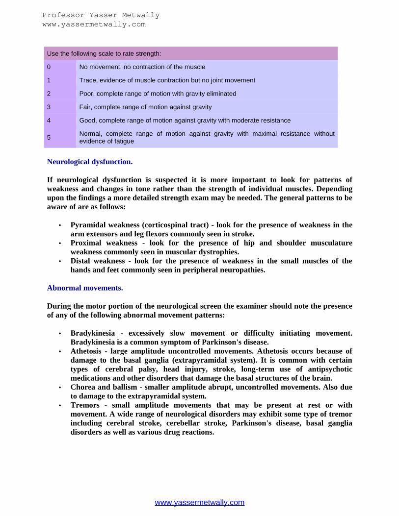
Use the following scale to rate strength:
0 No movement, no contraction of the muscle
1 Trace, evidence of muscle contraction but no joint movement
2 Poor, complete range of motion with gravity eliminated
3 Fair, complete range of motion against gravity
4 Good, complete range of motion against gravity with moderate resistance
5 Normal, complete range of motion against gravity with maximal resistance withoutevidence of fatigue
Neurological dysfunction.
If neurological dysfunction is suspected it is more important to look for patterns ofweakness and changes in tone rather than the strength of individual muscles. Dependingupon the findings a more detailed strength exam may be needed. The general patterns to beaware of are as follows:
Pyramidal weakness (corticospinal tract) - look for the presence of weakness in thearm extensors and leg flexors commonly seen in stroke.
Proximal weakness - look for the presence of hip and shoulder musculatureweakness commonly seen in muscular dystrophies.
Distal weakness - look for the presence of weakness in the small muscles of thehands and feet commonly seen in peripheral neuropathies.
Abnormal movements.
During the motor portion of the neurological screen the examiner should note the presenceof any of the following abnormal movement patterns:
Bradykinesia - excessively slow movement or difficulty initiating movement.Bradykinesia is a common symptom of Parkinson's disease.
Athetosis - large amplitude uncontrolled movements. Athetosis occurs because ofdamage to the basal ganglia (extrapyramidal system). It is common with certaintypes of cerebral palsy, head injury, stroke, long-term use of antipsychoticmedications and other disorders that damage the basal structures of the brain.
Chorea and ballism - smaller amplitude abrupt, uncontrolled movements. Also dueto damage to the extrapyramidal system.
Tremors - small amplitude movements that may be present at rest or withmovement. A wide range of neurological disorders may exhibit some type of tremorincluding cerebral stroke, cerebellar stroke, Parkinson's disease, basal gangliadisorders as well as various drug reactions.
Professor Yasser Metwallywww.yassermetwally.com
www.yassermetwally.com

Reflexes
Two categories of reflexes are tested:
1. The stretch or deep tendon reflexes2. The superficial or cutaneous reflexes
Stretch reflexes.
The stretch reflex is a type of spinal reflex also referred to as the deep tendon or myotaticreflex. Stretch reflexes trigger a contraction when the muscle is stretched or lengthened.Typically, the following stretch reflexes are tested with the patient in a seated or supineposition:
Biceps reflex (C5, C6) Triceps reflex (C6, C7) Brachioradialis reflex (C5, C6) Patellar reflex (L2,3,4) Achilles reflex (S1)
Plantar or Babinski reflex (L5, S1)
The Babinski or plantar reflex is a non-specific reflex but is a useful screen for the presenceof a central nervous system lesion. In a normal response there will be a downward flexingof the toes. If the great toe lifts and the other toes fan upward, the Babinski is positive andmay indicate the presence of upper motor neuron damage. If the Babinski reflex is positive,a more thorough motor exam should be done.
Scoring of Deep Tendon Reflexes
Grade Response
0 No response
1+ Sluggish or diminished
2+ Active or expected response
3+ More brisk than expected, slightly hyperactive
4+ Brisk, hyperactive, with intermittent or transient clonus
A hyperactive reflex response suggests upper motor neuron damage while a decreasedresponse suggest lower motor neuron, spinal damage or a disease of the neuromuscularjunction or muscles.
Superficial reflexes.
The superficial reflexes are also referred to as cutaneous reflexes and include the following:
Professor Yasser Metwallywww.yassermetwally.com
www.yassermetwally.com

Abdominal reflex. The abdominal reflex is tested by scratching the skin near theumbilicus in a diagonal manner. In a normal response the umbilicus will pull in thedirection of the stimulation. Loss of this reflex may indicate a central nervoussystem lesion or an injury to the nerves that supply the umbilical region (T7 to L1).
Cremasteric reflex. The cremasteric reflex is tested by stroking the skin along theinner thigh and looking for a lifting of the scrotum on the stimulated side.
Anal wink. The anal wink is tested by stroking the perianal area and looking for acontraction of the anal sphincter.
Pharyngeal or gag reflex. Test the gag reflex by touching the posterior wall of thepharynx with a tongue blade - this is an indication of both glossopharygeal andvagus function.
Gait
Gait is the single most important part of the motor exam because it allows to examiner toassess muscle strength, coordination, balance and timing - all vital higher corticalfunctions. Gait is also a vital functional skill and gives the examiner an idea of the ability ofthe patient to perform a complex motor task.
Ask the patient to walk down a hallway. Observe for symmetry, rhythm and speed whilewalking. Look for limping, scissoring, staggering, weight bearing and foot clearance duringthe swing phase. Ask the patient to walk on the heels and then on the toes - these are goodstests for peroneal/tibialis and gastrocnemius muscle function. Observe the person walkingtoe to heel and note any abnormalities with balance and coordination during this task. Ifthe patient has difficulty with any of these tasks involving gait or balance a more thoroughbalance evaluation should be done by a physical therapist.
Note any of the following common gait disorders:
Ataxic gait - a possible indication of cerebellar dysfunction. Festinating gait - a possible sign of Parkinson's disease. Hemiplegic gait with one-sided weakness - a possible sign of cerebral stroke. Spastic gait - a possible sign of cerebral palsy.
The Sensory Exam
The sensory portion of the neurological exam is very useful if the patient is cooperative andalert and able to give accurate responses to your questions. If the patient is uncooperativeor unreliable the sensory exam is often skipped. In general keep the following principles inmind during the sensory portion of the exam:
Compare distal sensation to proximal sensation
Professor Yasser Metwallywww.yassermetwally.com
www.yassermetwally.com

Look for symmetry by comparing one side to the other Test each of the dermatomes by moving the stimulation
The examiner will need a standard dermatome chart to accurately map sensory function.Begin by applying a light sensation to one side of the body, then compare to the other side.Gradually increase the intensity of the stimulation. The exam should focus on the suspectedlesion. For example if a central nervous system lesion is suspected the tests should bedirected towards central nervous system sensory tests. Sensory testing is usually dividedinto two parts:
1. Primary sensory modalities such as pain, light touch, temperature, vibration andjoint position sense.
2. Cortical sensory modalities such as stereognosis, two-point discrimination,graphesthesia and double simultaneous stimulation.
Primary sensory modalities.
The purpose of this part of the neurological exam is to test the sensory pathways thatascend via the spinal cord from the periphery to the sensory processing centers of thebrain. The following sensory modalities are commonly tested:
Pain - have the patient identify a sharp or dull sensation by alternating the point ofa sterile needle with the end of a blunt object such as the rounded end of a paperclip. The patient should be able to differentiate between sharp and dull.
Light touch - lightly touch an area of the skin with your finger or a small piece ofcotton. The patient should be able to identify the location of the touch.
Vibration - place the stem of the tuning fork on the bony prominence of the ankle,shin, wrist, elbow, shoulder and sternum. The patient should be able to name thelocation of the vibration. Loss of vibration sense suggests the presence of aperipheral neuropathy.
Joint position sense - hold the joint lightly in a neutral position between your twofingers. Raise or lower the digit or extremity and ask the patient to identify thedirection of the movement.
Cortical sensory modalities.
Cortical sensory input is processed by the parietal lobes. Processing of sensory input at thecortical level involves some degree of cognitive processing and control. Because the parietallobes receive their sensory input from the primary sensory modalities, cortical modalitiesshould be tested only if primary modalities are intact. The following sensory modalitiesshould be tested:
Stereognosis - ask the patient to identify an object placed in the hand. Compare theresults of one hand to the other.
Two-point discrimination - the ability to identify two closely placed stimuli.Stimulate the skin using an object with two points and ask the patient to identify the
Professor Yasser Metwallywww.yassermetwally.com
www.yassermetwally.com

when two points of stimulus are felt versus one point of stimulus. Different parts ofthe body will have different thresholds for two-point discrimination.
Double simultaneous discrimination - with the eyes closed, touch one or both handsand have the patient identify the location of the stimulation. If a parietal lobe lesionis present the patient will be unable to identify simultaneous stimulation on thecontralateral side of the body.
Minimal Distances for Two Point Discrimination by Location
Body part Minimal distance (mm)
Tongue 1 mm
Fingertips 2-8 mm
Toes 3-8 mm
Palms of hands 8-12 mm
Chest and forearms 40 mm
Back 40 to 70 mm
Upper arms and thighs 75 mm
Coordination
Coordination refers to the ability of the nervous system to organize multiple systems intoorganized and efficient patterns of movement. Coordination is affected by a variety ofmedical and physiologic factors including medical condition, medications, strength,alignment, timing and scaling of movements. Alignment refers to the arrangement of bodysegments on one another and is affected by both musculoskeletal and neurological factors.Timing is the ability to apply force with the appropriate speed and precision to prevent lossof balance or dyscoordination. Scaling a movement properly means that the force output ofthe muscles is appropriate to the amplitude of the instability.
The following items are commonly assessed to determine if there are problems withcoordination:
Observe the patient at rest and note the presence of abnormal postures, tremor,chorea, athetosis or dystonia.
Have the patient hold both arms outstretched with eyes closed and note anyabnormal movement such as tremor, weakness or posturing.
Have the patient perform a simple functional task such as buttoning a shirt orwriting and observe the smoothness and rhythm of the movement.
Have the patient move from sit to stand without use of the hands and observe forpostural instability and loss of balance.
Finally, a series of simple coordination tests should be performed to evaluate cerebellardysfunction:
Professor Yasser Metwallywww.yassermetwally.com
www.yassermetwally.com

Finger tap - tap the index finger against the thumb or a firm surface. Look forsmoothness of movement.
Finger to nose - ask the patient to touch her nose and then touch the examiner'soutstretched finger. Observe the movement for accuracy, smoothness and presenceof tremor or oscillations.
Rapid alternating hand movement - have the patient pronate and supinate herhands as quickly as possible. Observe the rhythm and accuracy of the movement aswell as the patient's ability to stop the movement and change direction.
Heel to shin test - while in sitting ask the patient to run the heel of one foot up anddown along the shin of the opposite leg. The patient should be able to perform thismovement smoothly and repeat it easily.
Conclusion
Completion of a neurological evaluation on a person with a suspected neurological injurywill give you a picture of the extent and possible location of the nervous system damage.This is especially important with sudden onset or life-threatening diseases in which theoutcome is dependent on an accurate referral or timely treatment. With practice you willrefine your evaluation skills and find that you will be able to complete the neurologicalexam quickly and efficiently.
References
1. Alpers and Mancall's Essentials of the Neurological Examination, Edition 2.2. Clinical Examination in Neurology Mayo Clinic and Mayo Foundation, 7th Edition.3. Brazis, Masdeu, and Biller. Localization Clinical Neurology, Lippincott and
Williams.4. Brust. Neurotoxic Side Effects of Prescription Drugs. Butterworth-Heinemann.5. Chipps EM, Clanin NJ and Campbell VG. Neurological disorders. Missouri:
Mosby-Year Book; 1992. p. 19-34.6. Jennett B, Teasdale G and Braakman R, et. al. Predicting outcome in individual
patients after severe head injury. Lancet: 1976; 1 p. 1031-1034.7. Kandel ER, Schwartz JH and Jessell TM. Principles of neural science. Connecticut:
Appleton and Lange; 1991.8. Lowenstein DH. Fundamentals of the neurological exam [course notes]. San
Francisco (CA): Univ. of California San Francisco; 1992.9. Magee DJ. Orthopedic physical assessment. Philadelphia: The WB Saunders
Company. 1987.10. Malasanos L, Barkauskas V and Stoltenberg-Allen, K. Health assessment, ed St
Louis: Mosby-Year Book. 1990.11. Minor M and Minor S. Patient evaluation methods for the health professional.
Virginia: Reston Publishing Company, Inc. 1985.12. Seidel HM, et. al.: Mosby's guide to physical examination, ed 2. St Louis: Mosby-
Year Book. 1991.
Professor Yasser Metwallywww.yassermetwally.com
www.yassermetwally.com

13. Shumway-Cook A and Woollacott MH. Motor control: theory and practicalapplications. Baltimore: Williams and Wilkins. 1995.
Professor Yasser Metwallywww.yassermetwally.com
www.yassermetwally.com


![White Lesions_Part II [Lecture by Dr.Eman Metwally @AmCoFam]](https://static.fdocuments.us/doc/165x107/547aed33b37959892b8b4c31/white-lesionspart-ii-lecture-by-dreman-metwally-amcofam.jpg)


![White Lesions_Part III [Lecture by Dr.Eman Metwally @AmCoFam]](https://static.fdocuments.us/doc/165x107/577d27c41a28ab4e1ea4c57a/white-lesionspart-iii-lecture-by-dreman-metwally-amcofam.jpg)