Growth of the mandible and biological characteristics of ...
12
Review article Growth of the mandible and biological characteristics of the mandibular condylar cartilage Itaru Mizoguchi * , Naoko Toriya, Yuya Nakao Division of Orthodontics and Dentofacial Orthopedics, Department of Oral Growth and Development, School of Dentistry, Health Sciences University of Hokkaido, 1757 Kanazawa, Ishikari-tobetsu, Hokkaido 061-0293, Japan Received 11 May 2013; received in revised form 26 July 2013; accepted 30 July 2013 Contents 1. Introduction . . . . . . . . . . . . . . . . . . . . . . . . . . . . . . . . . . . . . . . . . . . . . . . . . . . . . . . . . . . . . . . . . . . . . . . . . . . . . 140 2. Relationship between maxillofacial skeletal morphology and occlusion . . . . . . . . . . . . . . . . . . . . . . . . . . . . . . . . 140 3. Relationships between growth pattern of the mandible and maxillofacial skeletal morphology . . . . . . . . . . . . . . 141 Japanese Dental Science Review (2013) 49, 139—150 KEYWORDS Mandible; Growth; Maxillofacial morphology; Condylar cartilage; Extracellular matrix; Collagen Summary Mandibular condylar cartilage is the center of greatest growth in the craniofacial complex, and is associated with maxillofacial skeleton morphogenesis and temporomandibular joint function. The condylar process grows in a wide range of directions from anterosuperior to posterior, resulting in highly diverse mandibular growth and morphology. Condylar growth direction is closely related to mandibular displacement direction and vertical jaw deviations (i.e., high or low angle). Condylar cartilage, which is ontogenetically designated secondary cartilage, differs from other primary cartilage (e.g., articular cartilage and growth plate of a long bone cranial base cartilage, nasal septal cartilage) in the following ways. (1) Condylar cartilage is a heterogeneous tissue containing fibroblasts, osteochondral progenitor cells, and chondrocytes. (2) Type I collagen, which is derived from progenitor cells, and cartilage-characteristic type II collagen are colocalized in the cartilaginous cell layer. Colocalization of both collagen types may be an adaptation to the complex biomechanical environments of condylar cartilage. (3) Periph- eral condylar cartilage contains chondroid bone, a specialized calcified tissue with morphological properties intermediate between those of bone and cartilage. This hybrid tissue may play an important role in regulating different rates of bone formation in intramembranous and endo- chondral ossification, allowing for highly diverse growth directions and condylar and maxillofacial morphology. # 2013 Japanese Association for Dental Science. Published by Elsevier Ltd. All rights reserved. * Corresponding author. Tel.: +81 133 23 2975; fax: +81 133 23 3048. E-mail address: [email protected] (I. Mizoguchi). Available online at www.sciencedirect.com jo u rn al ho m epag e: ww w.els evier .c om /lo cat e/jds r 1882-7616/$ — see front matter # 2013 Japanese Association for Dental Science. Published by Elsevier Ltd. All rights reserved. http://dx.doi.org/10.1016/j.jdsr.2013.07.004
Transcript of Growth of the mandible and biological characteristics of ...
Review article
Growth of the mandible and biologicalcharacteristics of the mandibular condylarcartilage
Itaru Mizoguchi *, Naoko Toriya, Yuya Nakao
Division of Orthodontics and Dentofacial Orthopedics, Department of Oral Growth and Development, School ofDentistry, Health Sciences University of Hokkaido, 1757 Kanazawa, Ishikari-tobetsu, Hokkaido 061-0293, Japan
Received 11 May 2013; received in revised form 26 July 2013; accepted 30 July 2013
Contents
1. Introduction . . . . . . . . . . . . . . . . . . . . . . . . . . . . . . . . . . . . . . . . . . . . . . . . . . . . . . . . . . . . . . . . . . . . . . . . . . . . . 140
2. Relationship between maxillofacial skeletal morphology and occlusion . . . . . . . . . . . . . . . . . . . . . . . . . . . . . . . . 140
3. Relationships between growth pattern of the mandible and maxillofacial skeletal morphology . . . . . . . . . . . . . . 141
Japanese Dental Science Review (2013) 49, 139—150
KEYWORDSMandible;Growth;Maxillofacial morphology;Condylar cartilage;Extracellular matrix;Collagen
Summary Mandibular condylar cartilage is the center of greatest growth in the craniofacialcomplex, and is associated with maxillofacial skeleton morphogenesis and temporomandibularjoint function. The condylar process grows in a wide range of directions from anterosuperior toposterior, resulting in highly diverse mandibular growth and morphology. Condylar growthdirection is closely related to mandibular displacement direction and vertical jaw deviations(i.e., high or low angle). Condylar cartilage, which is ontogenetically designated secondarycartilage, differs from other primary cartilage (e.g., articular cartilage and growth plate of a longbone cranial base cartilage, nasal septal cartilage) in the following ways. (1) Condylar cartilage isa heterogeneous tissue containing fibroblasts, osteochondral progenitor cells, and chondrocytes.(2) Type I collagen, which is derived from progenitor cells, and cartilage-characteristic type IIcollagen are colocalized in the cartilaginous cell layer. Colocalization of both collagen types maybe an adaptation to the complex biomechanical environments of condylar cartilage. (3) Periph-eral condylar cartilage contains chondroid bone, a specialized calcified tissue with morphologicalproperties intermediate between those of bone and cartilage. This hybrid tissue may play animportant role in regulating different rates of bone formation in intramembranous and endo-chondral ossification, allowing for highly diverse growth directions and condylar and maxillofacialmorphology.# 2013 Japanese Association for Dental Science. Published by Elsevier Ltd. All rights reserved.
* Corresponding author. Tel.: +81 133 23 2975; fax: +81 133 23 3048.E-mail address: [email protected] (I. Mizoguchi).
Available online at www.sciencedirect.com
jo u rn al ho m epag e: ww w.els evier . c om / lo cat e/ jds r
1882-7616/$ — see front matter # 2013 Japanese Association for Dental Science. Published by Elsevier Ltd. All rights reserved.
http://dx.doi.org/10.1016/j.jdsr.2013.07.004
4. Role of the condylar cartilage in mandibular growth . . . . . . . . . . . . . . . . . . . . . . . . . . . . . . . . . . . . . . . . . . . . . . 141
5. Histological organization of mandibular condylar cartilage . . . . . . . . . . . . . . . . . . . . . . . . . . . . . . . . . . . . . . . . . 144
5.1. Classification of condylar cartilage cell layers . . . . . . . . . . . . . . . . . . . . . . . . . . . . . . . . . . . . . . . . . . . . . . 144
5.2. Fibrous layer . . . . . . . . . . . . . . . . . . . . . . . . . . . . . . . . . . . . . . . . . . . . . . . . . . . . . . . . . . . . . . . . . . . . . . . 145
5.3. Proliferative cell layer . . . . . . . . . . . . . . . . . . . . . . . . . . . . . . . . . . . . . . . . . . . . . . . . . . . . . . . . . . . . . . . . 145
5.4. Chondrocytic cell layer. . . . . . . . . . . . . . . . . . . . . . . . . . . . . . . . . . . . . . . . . . . . . . . . . . . . . . . . . . . . . . . . 145
5.5. Hypertrophic cell layer . . . . . . . . . . . . . . . . . . . . . . . . . . . . . . . . . . . . . . . . . . . . . . . . . . . . . . . . . . . . . . . . 145
6. Types I and II collagen in condylar cartilage . . . . . . . . . . . . . . . . . . . . . . . . . . . . . . . . . . . . . . . . . . . . . . . . . . . . . 146
7. Bone formation in the condyle: Presence of a third bone formation process . . . . . . . . . . . . . . . . . . . . . . . . . . . . 147
References . . . . . . . . . . . . . . . . . . . . . . . . . . . . . . . . . . . . . . . . . . . . . . . . . . . . . . . . . . . . . . . . . . . . . . . . . . . . . . 148
140 I. Mizoguchi et al.
Figure 1 Nine types of maxillofacial skeletal morphology.Reproduced with permission from Sugawara and Kawamura [20],with vertical deviation names changed from those in the original.
1. Introduction
Growth of the craniofacial skeleton largely influences occlu-sal relationships, jaw relationships, and orofacial functions[1—8]. In the growth of the craniofacial skeleton, cartilagi-nous tissues, including those of the sphenooccipital synchon-drosis in the cranial base, the nasal septal cartilage in thenasomaxillary complex, and the condylar cartilage in themandible, play important roles as major growth sites for therespective anatomical components [8—10]. Among these, thecondylar cartilage acts as the center of greatest growth in thecraniofacial complex [3,11] and is associated with morpho-genesis of the craniofacial skeleton and temporomandibularjoint function [1—8,12—14].
Condylar cartilage, which is designated as secondarycartilage [15—18], differs from other primary cartilage inhistological organization; modes of proliferation, differen-tiation and calcification; and response to environmentalfactors (e.g., biomechanical stress, hormones and growthfactors) [15]. The condylar cartilage is a unique and inter-esting tissue among cartilaginous tissues in the human body.The present article reviews the relationship between max-illofacial morphology and mandible growth behavior from aclinical viewpoint, as well as biological characteristics of thecondylar cartilage, with particular focus on the extracellularmatrix (ECM).
2. Relationship between maxillofacialskeletal morphology and occlusion
To diagnose maxillofacial skeletal morphology, two-dimen-sional analysis is performed using a lateral cephalogram[19,20]. Based on the anteroposterior jaw relationship, threetypes of skeletal relationship may be defined: (1) a normalrelationship between the maxilla and mandible (Class I); (2)distal position of the mandible relative to the maxilla due to aprotruded maxilla and/or retruded mandible (Class II); and(3) mesial position of the mandible relative to the maxilla dueto a retruded maxilla and/or protruded mandible (Class III)(Fig. 1). In contrast, based on the vertical jaw relationship,three types of jaw relationship may be defined: (1) mediumangle (normal face, mesiofacial pattern), (2) low angle (shortface, brachyfacial pattern, skeletal deep bite and hypodi-vergent type), and (3) high angle (long face, dolichofacialpattern, skeletal open bite and hyperdivergent type) [19—26]. In patients with low angles, small anterior facial heightsand mandibular plane angles (angle between the mandibularplane and the FH plane, and angle between the mandibular
plane to the cranial base) and large mandibular ramus lengthsand shallow antegonial notches can be observed; the oppo-site features are present in patients with high angles [19—26].
These vertical deviations are closely associated with ante-rior overbite; patients with low angles tend to have deepbites and those with high angles tend to have open bites [19—26]. In addition, vertical deviations largely influence not onlyoverbite, but also anteroposterior occlusal relationships[27]. The following observations were made after comparingthe occlusal conditions of two patients with almost the sameanteroposterior jaw relationship, but opposite vertical jawdeviations. The patient with a low angle had an anterior deepbite, and Class I canine and molar relationships, whereas thepatient with a high angle showed a small overbite, and ClassIII canine and molar relationships (Fig. 2). The question arisesas to why the vertical jaw deviations influence anteroposter-ior occlusal relationships, as in these patients. The verticaljaw relationships are closely associated with the occlusalplane angles (angle between the occlusal plane and the FH

Figure 2 Facial and oral photographs and cephalometric tracing of two patients with similar anteroposterior jaw relationships, asshown by ANB angles, but opposite vertical jaw deviations. (a—c) A patient with a low angle and skeletal Class III relationship. (d—f) Apatient with a high angle and skeletal Class III relationship. Note that the Class III dental relationship is more severe in the patient with ahigh angle.
Mandibular growth and condylar cartilage 141
plane, and angle between the occlusal plane to the cranialbase); patients with low angles tend to have small occlusalplane angles and the opposite is true of those with high angles[21,22]. According to mathematical model analysis, steepen-ing of the occlusal plane results in the posteriorization of themaxillary dentition relative to the mandibular dentition, i.e.,a shift from a Class II to Class III [27]. Thus, the low anglepatients with flatter occlusal planes tend to have Class IIocclusions, and the high angle patients with steeper occlusalplanes tend to have Class III occlusions.
3. Relationships between growth pattern ofthe mandible and maxillofacial skeletalmorphology
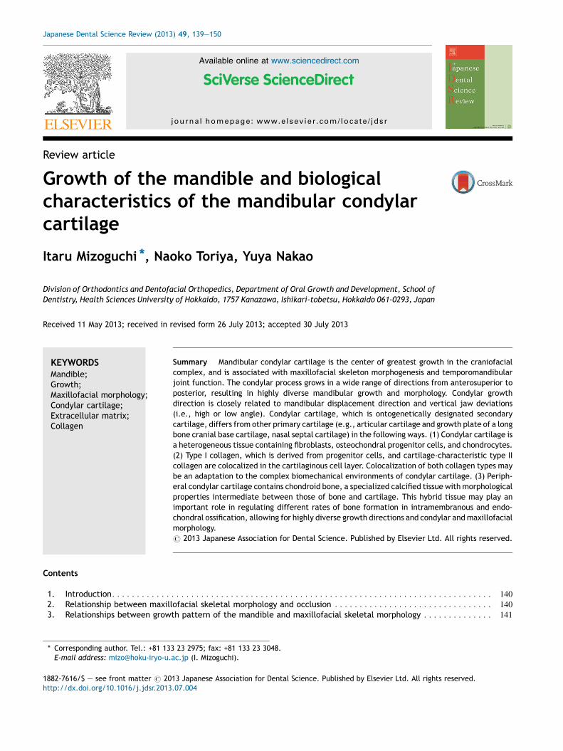
Growth of the mandibular condyle contributes not only toincreased mandible size, but also to anteroinferior displace-ment (transposition) of the mandible [1—8]. Using longitu-dinal cephalometric studies with tantalum implants, Bjorkand coworkers [2—4] provided variable information aboutindividual variation in the growth pattern of the mandible.Whereas the length of a long bone increases in a rectilineardirection along its long axis, the condylar process grows in awide range of directions from anterosuperior to posterior(Fig. 3). This divergent growth allows for highly diversegrowth and morphology of the mandible. Condylar growth
direction is closely related to the displacement (transposi-tion) direction of the mandible and vertical jaw deviations[2—4]. In individuals with low angles, mandibular growth ischaracterized by anterosuperior growth of the condyle,absorption of the inferior gonial border, and anterior displa-cement of the mandible [2—4] (Figs. 3 and 4a). In contrast,individuals with high angles show posterosuperior growth ofthe condyle, apposition at the inferior gonial border, andinferoposterior displacement of the mandible [2—4] (Figs. 3and 4b).
4. Role of the condylar cartilage inmandibular growth
In a long bone, two spatially separated cartilages (i.e.,articular cartilage and growth plate) exist during the growthstage [28,29]. The articular cartilage functions as a shockabsorber against mechanical loading and the growth platefunctions as a growth site. In contrast, only a single cartilage,the mandibular condylar cartilage, exists in the mandiblethroughout life, and plays roles in articulating function andgrowth. Therefore, the condylar cartilage is an ‘‘all-in-onetype tissue’’ [28,29]. The disturbance of condylar growthgreatly influences maxillofacial morphology and occlusalrelationships [12—14]. When the bilateral condyles areaffected, the mandible rotates in the posteroinferior
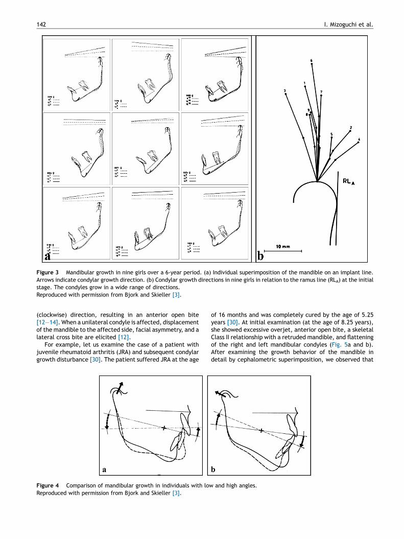
Figure 3 Mandibular growth in nine girls over a 6-year period. (a) Individual superimposition of the mandible on an implant line.Arrows indicate condylar growth direction. (b) Condylar growth directions in nine girls in relation to the ramus line (RLA) at the initialstage. The condyles grow in a wide range of directions.Reproduced with permission from Bjork and Skieller [3].
142 I. Mizoguchi et al.
(clockwise) direction, resulting in an anterior open bite[12—14]. When a unilateral condyle is affected, displacementof the mandible to the affected side, facial asymmetry, and alateral cross bite are elicited [12].
For example, let us examine the case of a patient withjuvenile rheumatoid arthritis (JRA) and subsequent condylargrowth disturbance [30]. The patient suffered JRA at the age
Figure 4 Comparison of mandibular growth in individuals with lowReproduced with permission from Bjork and Skieller [3].
of 16 months and was completely cured by the age of 5.25years [30]. At initial examination (at the age of 8.25 years),she showed excessive overjet, anterior open bite, a skeletalClass II relationship with a retruded mandible, and flatteningof the right and left mandibular condyles (Fig. 5a and b).After examining the growth behavior of the mandible indetail by cephalometric superimposition, we observed that
and high angles.
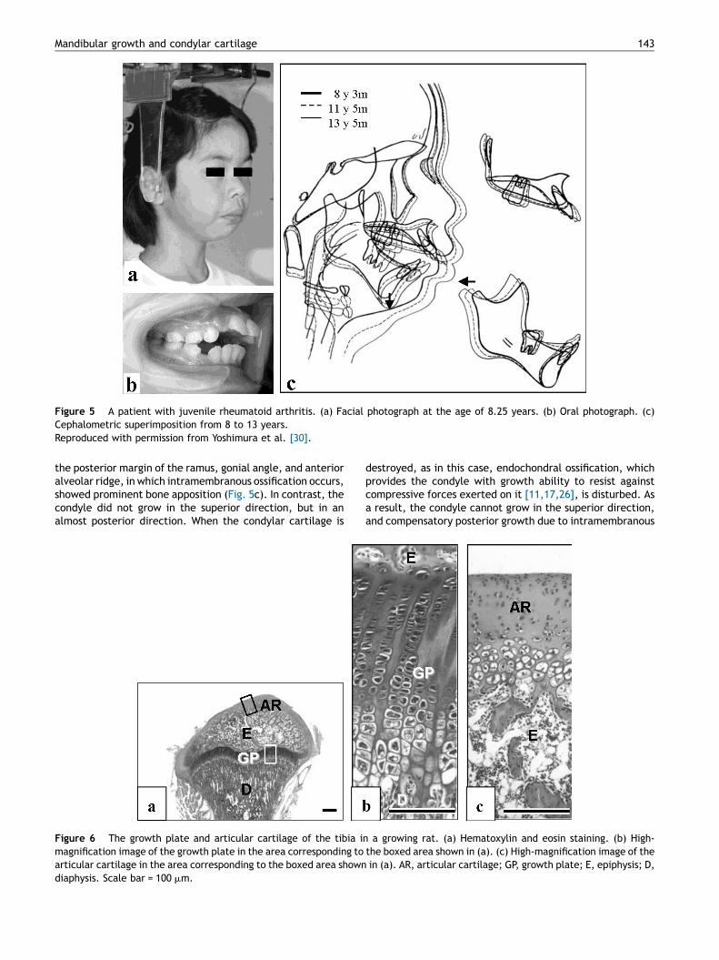
Figure 5 A patient with juvenile rheumatoid arthritis. (a) Facial photograph at the age of 8.25 years. (b) Oral photograph. (c)Cephalometric superimposition from 8 to 13 years.Reproduced with permission from Yoshimura et al. [30].
Mandibular growth and condylar cartilage 143
the posterior margin of the ramus, gonial angle, and anterioralveolar ridge, in which intramembranous ossification occurs,showed prominent bone apposition (Fig. 5c). In contrast, thecondyle did not grow in the superior direction, but in analmost posterior direction. When the condylar cartilage is
Figure 6 The growth plate and articular cartilage of the tibia imagnification image of the growth plate in the area corresponding toarticular cartilage in the area corresponding to the boxed area showndiaphysis. Scale bar = 100 mm.
destroyed, as in this case, endochondral ossification, whichprovides the condyle with growth ability to resist againstcompressive forces exerted on it [11,17,26], is disturbed. Asa result, the condyle cannot grow in the superior direction,and compensatory posterior growth due to intramembranous
n a growing rat. (a) Hematoxylin and eosin staining. (b) High- the boxed area shown in (a). (c) High-magnification image of the
in (a). AR, articular cartilage; GP, growth plate; E, epiphysis; D,

Figure 7 Mandibular condylar cartilage cell layers in a growingrat (hematoxylin and eosin staining). F, fibrous layer; P, prolifer-ative cell layer; C, chondrocytic cell layer; H, hypertrophic celllayer. Scale bar = 50 mm.
144 I. Mizoguchi et al.
ossification at the posterior condylar margin becomes pre-dominant, which is one characteristic of the high-angle type[2,26]. These results suggest that the balance betweenintramembranous and endochondral ossification in the con-dyle may be a factor determining divergent condylar growthdirection.
5. Histological organization of mandibularcondylar cartilage
5.1. Classification of condylar cartilage celllayers
Primary cartilage, such as articular cartilage and growthplates in a long bone, synchondroses in the cranial base,and nasal septal cartilage, consists of a chondrocyte popula-tion (Fig. 6). In contrast, condylar cartilage (i.e., secondarycartilage) is a heterogeneous tissue containing cells at var-ious stages of chondrogenic maturation [31—41] (Fig. 7).Classifications and terminology related to condylar cartilagecell layers differ among investigators (Table 1). Cell layerclassification depends on animal species and growth stage,histological method, and molecular markers used in a givenstudy. In this paper, a classification comprising four cell layersis used to explain the characteristics of each cell layerbecause four cell layers can be easily distinguished fromeach other based on type I and II collagen localization.
Table 1 Overview of terminology of the cell layers of the mandibular condylar cartilage.
Authors Non-cartilaginous (perichondrial) Cartilaginous
Layer 1 Layer 2 Layer 3 Layer 4 Layer 5 Layer 6
Greenspanet al. [31]
Articular Proliferating Hypertrophic(premineralizing)
Hypertrophic(mineralizing)
Folke andStallard [44]
Fibrous Embryonic Intermediate Vesicular Erosion
Carlsonet al. [33]
Articular Prechondroblastic(proliferative)
Chondroblastic(maturation)
Chondroblastic(hypertrophy)
Morita [39] Fibrous Proliferative cell Mature cell Hypertrophic cellTerashima [40] Fibrous Subfibrous cell Osteochondro
progeniorChondroblastic a Chondrocytic Hypertrophic cell
Coprayet al. [29]
Articlular Proliferative Transitional Hypertrophic
Silbermannet al. [36]
Articular Progenitor cell Chondroblastic Hypertrophic
Luderet al. [38]
Articular Polymorphic cell Flattened cell Upper hypertrophiccell
Lower hypertrophiccell
Kantomaaet al. [50]
Articular cell Polymorphic cell Flattened cell Upper hypertrophiccell
Lower hypertrophiccell
Strausset al. [48]
Perichondralcell
Progenitor Maturechondroblasts
Hypertrophicchondroctes
Petrovic [35] Fibrouscapsule
Skeletoblasts andprechondroblasts
Functionalchondroblasts
Hypertrophicchondroblasts
Shibukawaet al. [58]
Fibroblastic Polymorphic Flattenedchondrocytes
Hypertrophicchondrocytes
This paper Fibrous Proliferative cell Chondrocytic Hypertrophic cell
a It is unknown whether this layer contains chondrocytes or cells at a stage prior to chondrocytes.

Mandibular growth and condylar cartilage 145
5.2. Fibrous layer
The most superficial layer of the condylar cartilage consistsof dense fibrous connective tissue with scattered cells, andits periphery is continuous with the outer layer of the peri-osteum (Fig. 7). The cells are flat and surrounded by densecollagen bundles [38—40]. This layer is not related to deeperchondrogenic differentiation, but functions as a protectivecovering for the underlying cartilaginous tissue [38].Recently, Ohno et al. [42] revealed that superficial zoneprotein, also known as proteoglycan-4 and lubricant, isrestricted to the superficial part of the condylar cartilageand functions as a joint boundary lubricant.
5.3. Proliferative cell layer
Based on cellular morphology, this layer is further dividedinto two sublayers: the upper sublayer (i.e., polymorphic celllayer), where irregular polygonal cells with large roundnuclei are densely packed; and the lower sublayer (i.e.,flattened cell layer), where flattened cells are oriented withtheir long axes parallel to the articular surface [38] (Fig. 7).The cells in the upper sublayer have poorly developed cyto-plasmic organelles, extend thin cell processes to the adja-cent cells, and form gap junctions [38]. Autoradiographicstudies using 3H-thymidine and 3H-proline have indicatedthat proliferative activities are limited almost exclusivelyto cells in the polymorphic cell layer and the upper part of thelower flattened cell layer, and that collagen synthesis activitywas low in the proliferative cell layer [32,38,43—47].
One unique characteristic of the condylar cartilage is thatthe cells in the proliferative layer have multilineage poten-tial and can differentiate into osteoblasts or chondrocytes(osteochondral progenitors), and more differentiated cellscommitted to becoming chondrocytes (chondroprogenitors)or fat progenitor cells [32—41]. Their differentiation pathwayis regulated by biomechanical force. Under physiologicalconditions, progenitor cells differentiate into chondrocytes.Under non-functional conditions, however, such as in vitro
Figure 8 Immunohistochemical localization of types I and II collaggrowing rat. (a, c) Localization of type I collagen. b, d Localization
cartilage; E, epiphysis. Scale bar = 100 mm.
organ culture and in vivo immobilization experiments, orunder excessive tensile loading, progenitor cells undergoingchondrogenic differentiation are replaced by intramembra-nous bone, along with a phenotypic switch from type II totype I collagen [17,36,37,41,48—52]. In addition, a consider-able amount of information has recently become availableregarding local regulatory factors of osteochondrogenic dif-ferentiation, including growth factors and signaling mole-cules, such as bone morphogenetic proteins (BMPs) [53,54],transforming growth factor-b [55], interleukin 1 [56], Indianhedgehog [57,58], and parathyroid hormone-related protein[58—60], and transcription factors such as SOX9, RUNX2, andOsterix [61,62].
5.4. Chondrocytic cell layer
The chondrocytic cell layer contains chondrocytes at variousstages of maturation. Cellular morphology changes fromflattened to spherical with progressive depth (Fig. 7). TheECM in this layer has an increased area and shows hematox-ylin-philic staining and metachromatic staining with tolui-dine blue, indicating active deposition of cartilage-characteristic matrices [63]. Synthetic activity of collagens,proteoglycans, and sulfated glycosaminoglycans peak in thiscell layer [38,46,61,62,64].
5.5. Hypertrophic cell layer
Hypertrophy, the terminal differentiation stage of the chon-drogenic lineage, is required for the replacement of cartilagewith bone (endochondral ossification) (Fig. 7). With advan-cing hypertrophy, chondrocytes increase in volume, initiatecalcification of the surrounding matrix, and are invaded bythe bone marrow vasculature accompanied by chondroclasticand osteogenic precursor cells [65,66]. The calcified carti-laginous matrix is degraded by differentiated chondroclasts,and newly secreted bone matrix is then deposited onto thecartilage remnants [65]. In the hypertrophic cell layer,although synthetic activity of matrix synthesis is decreased,
en in the growth plate (a, b) and articular cartilage (c, d) of aof type II collagen. GP, growth plate; D, diaphysis; AR, articular
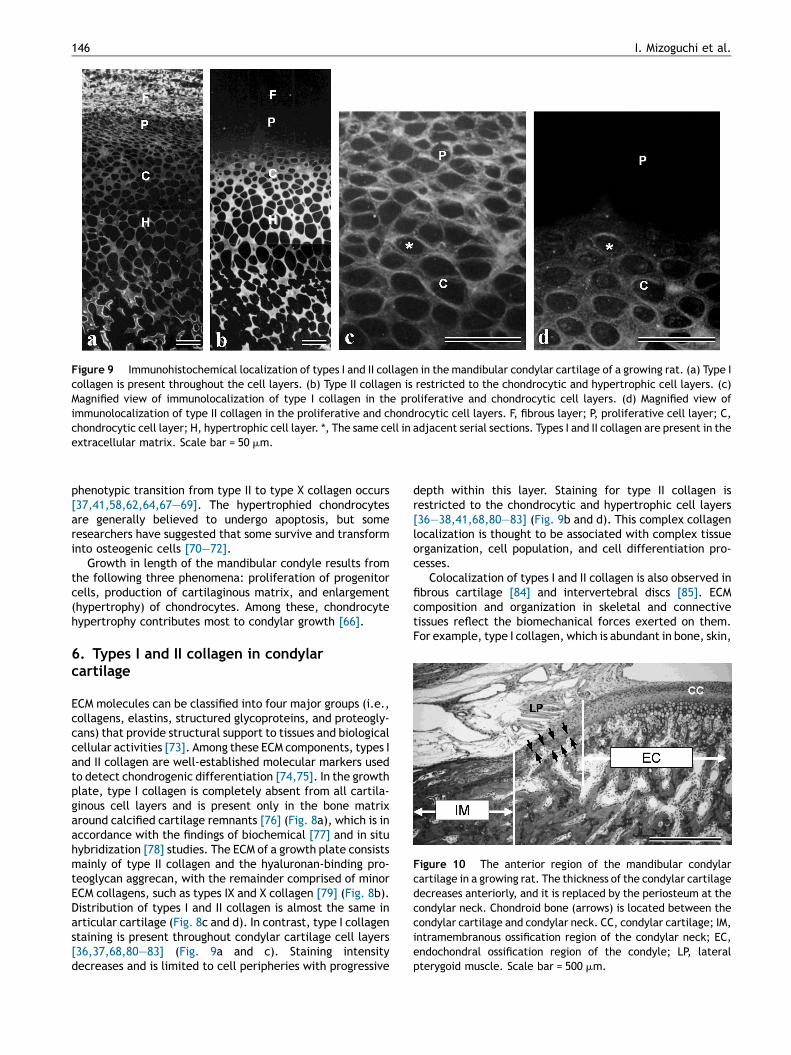
Figure 9 Immunohistochemical localization of types I and II collagen in the mandibular condylar cartilage of a growing rat. (a) Type Icollagen is present throughout the cell layers. (b) Type II collagen is restricted to the chondrocytic and hypertrophic cell layers. (c)Magnified view of immunolocalization of type I collagen in the proliferative and chondrocytic cell layers. (d) Magnified view ofimmunolocalization of type II collagen in the proliferative and chondrocytic cell layers. F, fibrous layer; P, proliferative cell layer; C,chondrocytic cell layer; H, hypertrophic cell layer. *, The same cell in adjacent serial sections. Types I and II collagen are present in theextracellular matrix. Scale bar = 50 mm.
Figure 10 The anterior region of the mandibular condylarcartilage in a growing rat. The thickness of the condylar cartilagedecreases anteriorly, and it is replaced by the periosteum at thecondylar neck. Chondroid bone (arrows) is located between thecondylar cartilage and condylar neck. CC, condylar cartilage; IM,intramembranous ossification region of the condylar neck; EC,endochondral ossification region of the condyle; LP, lateralpterygoid muscle. Scale bar = 500 mm.
146 I. Mizoguchi et al.
phenotypic transition from type II to type X collagen occurs[37,41,58,62,64,67—69]. The hypertrophied chondrocytesare generally believed to undergo apoptosis, but someresearchers have suggested that some survive and transforminto osteogenic cells [70—72].
Growth in length of the mandibular condyle results fromthe following three phenomena: proliferation of progenitorcells, production of cartilaginous matrix, and enlargement(hypertrophy) of chondrocytes. Among these, chondrocytehypertrophy contributes most to condylar growth [66].
6. Types I and II collagen in condylarcartilage
ECM molecules can be classified into four major groups (i.e.,collagens, elastins, structured glycoproteins, and proteogly-cans) that provide structural support to tissues and biologicalcellular activities [73]. Among these ECM components, types Iand II collagen are well-established molecular markers usedto detect chondrogenic differentiation [74,75]. In the growthplate, type I collagen is completely absent from all cartila-ginous cell layers and is present only in the bone matrixaround calcified cartilage remnants [76] (Fig. 8a), which is inaccordance with the findings of biochemical [77] and in situhybridization [78] studies. The ECM of a growth plate consistsmainly of type II collagen and the hyaluronan-binding pro-teoglycan aggrecan, with the remainder comprised of minorECM collagens, such as types IX and X collagen [79] (Fig. 8b).Distribution of types I and II collagen is almost the same inarticular cartilage (Fig. 8c and d). In contrast, type I collagenstaining is present throughout condylar cartilage cell layers[36,37,68,80—83] (Fig. 9a and c). Staining intensitydecreases and is limited to cell peripheries with progressive
depth within this layer. Staining for type II collagen isrestricted to the chondrocytic and hypertrophic cell layers[36—38,41,68,80—83] (Fig. 9b and d). This complex collagenlocalization is thought to be associated with complex tissueorganization, cell population, and cell differentiation pro-cesses.
Colocalization of types I and II collagen is also observed infibrous cartilage [84] and intervertebral discs [85]. ECMcomposition and organization in skeletal and connectivetissues reflect the biomechanical forces exerted on them.For example, type I collagen, which is abundant in bone, skin,
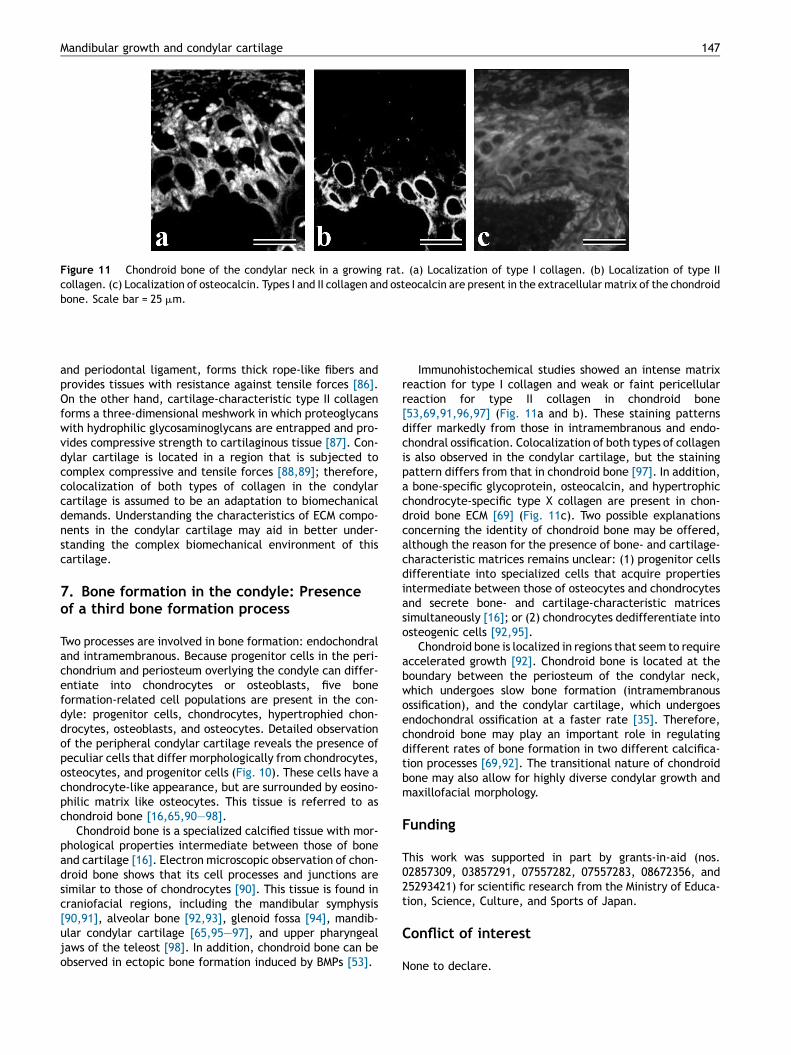
Figure 11 Chondroid bone of the condylar neck in a growing rat. (a) Localization of type I collagen. (b) Localization of type IIcollagen. (c) Localization of osteocalcin. Types I and II collagen and osteocalcin are present in the extracellular matrix of the chondroidbone. Scale bar = 25 mm.
Mandibular growth and condylar cartilage 147
and periodontal ligament, forms thick rope-like fibers andprovides tissues with resistance against tensile forces [86].On the other hand, cartilage-characteristic type II collagenforms a three-dimensional meshwork in which proteoglycanswith hydrophilic glycosaminoglycans are entrapped and pro-vides compressive strength to cartilaginous tissue [87]. Con-dylar cartilage is located in a region that is subjected tocomplex compressive and tensile forces [88,89]; therefore,colocalization of both types of collagen in the condylarcartilage is assumed to be an adaptation to biomechanicaldemands. Understanding the characteristics of ECM compo-nents in the condylar cartilage may aid in better under-standing the complex biomechanical environment of thiscartilage.
7. Bone formation in the condyle: Presenceof a third bone formation process
Two processes are involved in bone formation: endochondraland intramembranous. Because progenitor cells in the peri-chondrium and periosteum overlying the condyle can differ-entiate into chondrocytes or osteoblasts, five boneformation-related cell populations are present in the con-dyle: progenitor cells, chondrocytes, hypertrophied chon-drocytes, osteoblasts, and osteocytes. Detailed observationof the peripheral condylar cartilage reveals the presence ofpeculiar cells that differ morphologically from chondrocytes,osteocytes, and progenitor cells (Fig. 10). These cells have achondrocyte-like appearance, but are surrounded by eosino-philic matrix like osteocytes. This tissue is referred to aschondroid bone [16,65,90—98].
Chondroid bone is a specialized calcified tissue with mor-phological properties intermediate between those of boneand cartilage [16]. Electron microscopic observation of chon-droid bone shows that its cell processes and junctions aresimilar to those of chondrocytes [90]. This tissue is found incraniofacial regions, including the mandibular symphysis[90,91], alveolar bone [92,93], glenoid fossa [94], mandib-ular condylar cartilage [65,95—97], and upper pharyngealjaws of the teleost [98]. In addition, chondroid bone can beobserved in ectopic bone formation induced by BMPs [53].
Immunohistochemical studies showed an intense matrixreaction for type I collagen and weak or faint pericellularreaction for type II collagen in chondroid bone[53,69,91,96,97] (Fig. 11a and b). These staining patternsdiffer markedly from those in intramembranous and endo-chondral ossification. Colocalization of both types of collagenis also observed in the condylar cartilage, but the stainingpattern differs from that in chondroid bone [97]. In addition,a bone-specific glycoprotein, osteocalcin, and hypertrophicchondrocyte-specific type X collagen are present in chon-droid bone ECM [69] (Fig. 11c). Two possible explanationsconcerning the identity of chondroid bone may be offered,although the reason for the presence of bone- and cartilage-characteristic matrices remains unclear: (1) progenitor cellsdifferentiate into specialized cells that acquire propertiesintermediate between those of osteocytes and chondrocytesand secrete bone- and cartilage-characteristic matricessimultaneously [16]; or (2) chondrocytes dedifferentiate intoosteogenic cells [92,95].
Chondroid bone is localized in regions that seem to requireaccelerated growth [92]. Chondroid bone is located at theboundary between the periosteum of the condylar neck,which undergoes slow bone formation (intramembranousossification), and the condylar cartilage, which undergoesendochondral ossification at a faster rate [35]. Therefore,chondroid bone may play an important role in regulatingdifferent rates of bone formation in two different calcifica-tion processes [69,92]. The transitional nature of chondroidbone may also allow for highly diverse condylar growth andmaxillofacial morphology.
Funding
This work was supported in part by grants-in-aid (nos.02857309, 03857291, 07557282, 07557283, 08672356, and25293421) for scientific research from the Ministry of Educa-tion, Science, Culture, and Sports of Japan.
Conflict of interest
None to declare.

148 I. Mizoguchi et al.
References
[1] Proffit WR, Fields Jr HW. Later stages of development.In: Contemporary orthodontics. St. Louis: Mosby Year Book;1986. p. 87—104.
[2] Bjork A. Variations in the growth pattern of the growing mandi-ble. J Dent Res 1963;42:406—32.
[3] Bjork A, Skieller V. Facial growth and tooth eruption: an implantstudy at the age of puberty. Am J Orthod 1972;64:339—83.
[4] Bjork A, Skieller V. Normal and abnormal growth of the mandi-ble. A synthesis of longitudinal cephalometric implant studiesover a period 25 years. Eur J Orthod 1983;5:1—46.
[5] Mathews JR, Ware WH. Longitudinal mandibular growth inchildren with tantalum implants. Am J Orthod 1978;74:633—55.
[6] Baumrind S, Ben-Bassat Y, Korn EL, Bravo LA, Curry S. Mandibu-lar remodeling measured on cephalograms, 1. Osseous changesrelative to superimposition on metallic implants. Am J OrthodDentofacial Orthop 1992;102:134—42.
[7] Buschang PH, Gandini Jr LG. Mandibular skeletal growth andmodeling between 10 and 15 years of age. Eur J Orthod2002;24:69—79.
[8] Enlow DH. Book of facial growth. Philadelphia, London, Toronto:W.B. Saunders; 1975: 10—146.
[9] Kvinnsland S, Kvinnsland S. Growth in craniofacial cartilagesstudied by 3H-thymidine incorporation. Growth 1975;39:305—14.
[10] Takano-Yamamoto T, Soma S, Nakagawa K, Kobayashi Y, Kawa-kami M, Sakuda M. Comparison of the effects of hydrostaticcompressive force on glycosaminoglycan synthesis and prolifer-ation in rabbit chondrocytes from mandibular condylar carti-lage, nasal septum, and spheno-occipital synchondrosis in vitro.Am J Orthod Dentofacial Orthop 1991;99:448—55.
[11] Tingey TF, Shapiro PA. Selective inhibition of condylar growth inthe rabbit mandible using intra-articular papain. Am J Orthod1982;81:455—64.
[12] Schellhas KP, Pollei SR, Wilkes CH. Pediatric internal derange-ment of the temporomandibular joint: effect on facial devel-opment. Am J Orthod 1993;104:51—9.
[13] Arnett GW, Milam SB, Gottesman L. Progressive mandibularretrusion-idiopathic condylar resorption. Part I. Am J OrthodDentofacial Orthop 1996;110:8—15.
[14] Bryndahl F, Eriksson L, Legrell PE, Isberg A. Bilateral TMJ diskdisplacement induces mandibular retrognathia. J Dent Res2006;85:1118—23.
[15] Durkin JF, Heeley JD, Irving JT. The cartilage of the mandibularcondyle. In: Noble HW, Durkin JD, De Boever J, editors. Oralsciences review 2. Toronto: Munkusgaard; 1973. p. 22—99.
[16] Beresford WA. Chondroid bone, secondary cartilage and meta-plasia. Munchen: Urban & Schwarzenberg; 1981.
[17] Hall BK. Selective proliferation and accumulation of chondro-progenitor cells as the mode of action of biomechanicalfactors during secondary chondrogenesis. Teratology 1979;20:81—92.
[18] Fang J, Hall BK. Chondrogenic cell differentiation from mem-brane bone periostea. Anat Embryol 1997;196:349—62.
[19] Sassouni V. Clacification of skeletal facial types. Am J Orthod1969;55:109—23.
[20] Sugawara J, Kawamura J. CDS analysis–—two dimensional mor-phologic analysis of dentofacial deformities using cephalomet-ric drawing. Clin Orthod 1997;2:43—58 (in Japanese).
[21] Opdenbeek H, Bell WH. The short face syndrome. Am J Orthod1978;73:499—511.
[22] Schendel SA, Eisenfeld J, Bell WH, Epker BN, Mishelevich DJ.The long face syndrome; vertical maxillary excess. Am J Orthod1976;70:398—408.
[23] Lowe AA. Correlations between orofacial muscle activity andcraniofacial morphology in a sample of control and anterioropen-bite subjects. Am J Orthod 1980;78:89—98.
[24] Ricketts R, Bench R, Gugino C, Hilgers J, Schulhof R. Biopro-gressive therapy. Denver: Rocky Mountain Orthodontics; 1979:55—9.
[25] Nezu H, Nagata K. Orthodontic diagnosis and treatment inbioprogressive therapy. Tokyo: Rocky Mountain Morita; 1988 :22—116 (in Japanese).
[26] Haskell B, Day M, Tetz J. Computer-aided modeling in theassessment of the biomechanical determination of diverseskeletal patterns. Am J Orthod 1986;89:363—82.
[27] Braun S, Legan HL. Changes in occlusion related to the cant ofthe occlusal plane. Am J Orthod 1997;111:184—8.
[28] Symons NBB. Studies on the growth and form of the mandible.Dent Rec 1951;71:41—53.
[29] Copray JCVM, Jansen HWB, Duterloo HS. Growth and growthpressure of mandibular condylar and some primary cartilages ofthe rat in vitro. Am J Orthod Dentofacial Orthop 1986;90:19—28.
[30] Yoshimura K, Mizoguchi I, Sugawara J, Mitani H. Craniofacialgrowth changes associated with juvenile rheumatoid arthritis —a case report from 8 to 13 years of age-. J Jpn Orthod Soc1992;51:398—404 (in Japanese with English abstract).
[31] Greenspan JS, Blackwood HJ. Histochemical studies of chon-drocyte function in the cartilage of the mandibular condyle ofthe rat. J Anat 1966;100:615—26.
[32] Charlier JP, Petrovic A, Stutzmann J. Effects of mandibularhyperpropulsion on the prechondroblastic zone of young ratcondyle. Am J Orthod 1969;55:71—4.
[33] Carlson DS, McNamara Jr JA, Jaul DH. Histological analysis ofthe growth of the mandibular condyle in the Rhesus monkey(Macaca mulatta). Am J Anat 1978;151:103—17.
[34] McNamara Jr JA. Quantitative analysis of temporomandibularjoint adaptations to protrusive function. Am J Orthod1979;76:593—611.
[35] Stutzmann JJ, Petrovic AG. Bone cell histogenesis: the skeleto-blasts as a stem-cell for preosteoblasts and for secondary-typeprechondroblasts. In: Dixon AD, Sarnat BG, editors. Factors andmechanisms influencing bone growth. New York: Alan R Liss Inc;1982. p. 29—43.
[36] Silbermann M, Reddi AH, Hand AR, Leapman RD, von der Mark K,Franzen A. Further characterization of the extracellular matrixin the mandibular condyle in neonatal mice. J Anat1987;151:169—88.
[37] Silbermann M, von der Mark K. An immunohistochemicalstudy of the distribution of matrical proteins in the mandibularcondyle of neonatal mice I. Collagens. J Anat 1990;170:11—22.
[38] Luder HU, Leblond CP, von der Mark K. Cellular stages incartilage formation as revealed by morphometry, radioautogra-phy and type II collagen immunostaining of the mandibularcondyle from weanling rats. Am J Anat 1988;182:197—214.
[39] Morita S. Ultrastructual and cytochemical studies on the ratmandibular condyle with advancing age. J Jpn Orthod Soc1982;41:171—215 (in Japanese with English abstract).
[40] Terashima T. Observation on the structure of the mandibularcondyle of the prenatal rabbits. J Stomatol Soc Jpn 1985;154—202 (in Japanese with English abstract).
[41] Chen J, Utreja A, Kalajzic Z, Sobue T, Rowe D, Wadhwa S.Isolation and characterization of murine mandibular condylarcartilage cell populations. Cells Tissues Organs 2012;195:232—43. http://dx.doi.org/10.1177/0022034510390810.
[42] Ohno S, Schmid T, Tanne Y, Kamiya T, Honda K, Ohno-NakaharaM, et al. Expression of superficial zone protein in mandibularcondyle cartilage. Osteoarthritis Cartilage 2006;14:807—13.
[43] Blackwood HJ. Growth of the mandibular condyle of the ratstudied with tritiated thymidine. Arch Oral Biol 1966;11:493—500.
[44] Folke LE, Stallard RE. Cellular kinetics within the mandibularjoint. Acta Odontol Scand 1967;25:469—89.

Mandibular growth and condylar cartilage 149
[45] Oberg T, Jajers CM, Lohmander S, Friberg U. Autoradiographicstudies with H3-thymidine on cell proliferation and differentia-tion in the mandibular joint of young guinea pigs. Odontol Revy1967;18:327—44.
[46] Hinton RJ. Effect of altered masticatory function on [3H]-thy-midine and [35S]-sulphate incorporation in the condylar carti-lage of the rat. Acta Anat 1988;131:136—9.
[47] Pirttiniemi P, Kantomaa T, Tuominen M. Increased condylargrowth after experimental relocation of the glenoid fossa. JDent Res 1993;72:1356—9.
[48] Strauss PG, Closs EI, Schmidt J, Erfle V. Gene expression duringosteogenic differentiation in mandibular condyles in vitro. JCell Biol 1990;110:1369—78.
[49] Kantomaa T, Hall BK. Mechanism of adaptation in the mandibu-lar condyle of the mouse. An organ culture study. Acta Anat1988;132:114—9.
[50] Kantomaa T, Tuominen M, Pirttiniemi P. Effect of mechanicalforces on chondrocyte maturation and differentiation in themandibular condyle of the rat. J Dent Res 1994;73:1150—6.
[51] Takahashi I, Mizoguchi I, Nakamura M, Kagayama M, Mitani H.Effects of hyperactivities of lateral pterygoid muscle on thegrowth of the mandibular condylar cartilage of the rats. AnatRec 1995;241:328—36.
[52] Takahashi I, Mizoguchi I, Nakamura M, Kagayama M, Mitani H.Effects of expansive forces on the mid-palatal suture cartilageof the rats. Bone 1996;18:314—48.
[53] Sasano Y, Mizoguchi I, Takahashi I, Kagayama M, Saitoh T, KubokiY. BMPs induce endochondral ossification in rats when implantedwithin a carrier made of fibrous glass membrane. Anat Rec1997;247:472—8.
[54] Wang EA, Rosen V, D’Alessandro JS, Bauduy M, Cordes P, HaradaT, et al. Recombinant human bone morphogenetic proteininduces bone formation. Proc Natl Acad Sci USA1990;87:2220—4.
[55] Joyce ME, Roberts AB, Sporn MB, Bolander ME. Transforminggrowth factor-beta and the initiation of chondrogenesis andosteogenesis in the rat femur. J Cell Biol 1990;110:2195—207.
[56] Goldring MB, Birkhead J, Sandell LJ, Kimura T, Krane SM.Interleukin 1 suppresses expression of cartilage-specific typesII and IX collagens and increases types I and III collagens inhuman chondrocytes. J Clin Invest 1988;82:2026—37.
[57] Ochiai TY, Shibukawa M, Nagayama C, Mundy T, Yasuda T, OkabeK, et al. Indian hedgehog roles in post-natal TMJ developmentand organization. J Dent Res 2010;89:349—54. http://dx.doi.org/10.1177/0022034510363078.
[58] Shibukawa Y, Young B, Wu C, Yamada S, Long F, Pacifici M, et al.Temporomandibular joint formation and condyle growth requireIndian Hedgehog signalling. Dev Dyn 2007;236:426—34.
[59] Suda N, Shibata S, Yamazaki K, Kuroda T, Senior PV, Beck F, et al.Parathyroid hormone-related protein regulates proliferation ofcondylar hypertrophic chondrocytes. J Bone Miner Res1999;14:1838—47.
[60] Rabie AB, Tang GH, Xiong H, Hagg U. PTHrP regulates chondro-cyte maturation in condylar cartilage. J Dent Res 2003;82:627—31.
[61] Shibata S, Suda N, Suzuki S, Fukuoka H, Yamashita Y. An in situhybridization study of Runx2, Osterix, and Sox9 at the onset ofcondylar cartilage formation in fetal mouse mandible. J Anat2006;208:169—77.
[62] Shibata S, Suda N, Yoda S, Fukuoka H, Ohyama K, Yamashita Y,et al. Runx2-deficient mice lack mandibular condylar cartilageand have deformed Meckel’s cartilage. Anat Embryol2004;208:273—80.
[63] Kantomaa T, Tuominen M, Pirttiniemi P, Ronning O. Weaning andthe histology of the mandibular condyle in the rat. Acta Anat1992;144:311—5.
[64] Fukada K, Shibata S, Suzuki S, Ohya K, Kuroda T. In situhybridisation study of type I, II, X collagens and aggrecan mRNAs
in the developing condylar cartilage of fetal mouse mandible. JAnat 1999;195:321—9.
[65] Luder HU, Schroeder HE. Light and electron microscopic mor-phology of the temporomandibular joint in growing and maturecrab-eating monkeys (Macaca fascicularis): the condylar calci-fied cartilage. Anat Embryol 1992;185:189—99.
[66] Luder HU. Perichondrial and endochondral components of man-dibular condylar growth: morphometric and autoradiographicquantitation in rats. J Anat 1994;185:587—98.
[67] Salo LA, Hoyland J, Ayad S, Kielty CM, Freemont A, Pirttiniemi P,et al. The expression of types X and VI collagen and fibrillin in ratmandibular condylar cartilage. Response to mastication forces.Acta Odontol Scand 1996;54:295—302.
[68] Ohashi N, Ejiri S, Ozawa E. Changes in type I, II and X collagenimmunoreactivity of the mandibular condylar cartilage in anaturally aging rat model. J Bone Miner Metab 1997;15:77—83.
[69] Mizoguchi I, Takahashi I, Sasano Y, Kagayama M, Kuboki Y, MitaniH. Localization of types I, II and X collagen and osteocalcin inintramembranous, endochondral and chondroid bone of rats.Anat Embryol 1997;196:291—7.
[70] Yoshioka C, Yagi T. Electron microscopic observations on thefate of hypertrophic chondrocytes in condylar cartilage of ratmandible. J Craniofac Genet Dev Biol 1988;8:253—64.
[71] Thesingh CW, Groot CG, Wassenaar AM. Transdifferentiation ofhypertrophic chondrocytes into osteoblasts in murine fetalmetatarsal bones, induced by co-cultured cerebrum. BoneMineral 1991;12:25—40.
[72] Roach HI, Erenpreisa J, Aigner T. Osteogenic differentiation ofhypertrophic chondrocytes involves asymmetric cell divisionsand apoptosis. J Cell Biol 1995;131:483—94.
[73] Kreis T, Vale R. Guidebook to the extracellular matrix andadhesion proteins. Oxford: A Sanbrook & Tooze Publication atOxford University Press; 1993: 3—16.
[74] Kosher RA, Kulyk WM, Gay SW. Collagen gene expression duringlimb cartilage differentiation. J Cell Biol 1986;102:1151—6.
[75] Linsenmayer TF, Toole BP, Trelstad RL. Temporal and spatialtransitions in collagen types during embryonic chick limb de-velopment. Dev Biol 1973;35:232—9.
[76] Horton WA, Dwyer C, Goering R, Dean DC. Immunohistochemis-try of types I and II collagen in undecalcified skeletal tissues. JHistochem Cytochem 1983;31:417—25.
[77] von der Mark. Immunological studies on collagen type transitionin chondrogenesis. Curr Top Dev Biol 1980;14:199—225.
[78] Sandberg M, Vuorio E. Localization of types I, II, and III collagenmRNAs in developing human skeletal tissues by in situ hybrid-ization. J Cell Biol 1987;104:1077—84.
[79] van der Rest M, Mayne R, Ninomiya Y, Seidah NG, Chretien M,Olsen BR. The structure of type IX collagen. J Biol Chem1985;260:220—5.
[80] Milam SB, Klebe RJ, Triplett RG, Herbert D. Characterization ofthe extracellular matrix of the primate temporomandibularjoint. J Oral Maxillofac Surg 1991;49:381—91.
[81] Mizoguchi I, Nakamura M, Takahashi I, Kagayama M, Mitani H. Animmunohistochemical study of localization of type I and type IIcollagens in mandibular condylar cartilage compared with tibialgrowth plate. Histochemistry 1990;93:593—9.
[82] Mizoguchi I, Nakamura M, Takahashi I, Kagayama M, Mitani H. Acomparison of immunohistochemical localization of type I andtype collagens in craniofacial cartilages of the rat. Acta Anat1992;144:59—64.
[83] Salo L, Kantomaa T. Type II collagen expression in the mandibu-lar condyle during growth adaptation: an experimental study inthe rabbit. Calcif Tissue Int 1993;52:465—9.
[84] Eyre DR. Biochemistry of the intervertebral disc. Int Rev Con-nect Tissue Res 1979;8:227—91.
[85] Beard HK, Ryvar R, Brown R, Muir H. Immunochemical localiza-tion of collagen types and proteoglycan in pig intervertebraldiscs. Immunology 1980;41:491—501.

150 I. Mizoguchi et al.
[86] Danielson KG, Baribault H, Holmes DF, Graham H, Kadler KE,Iozzo RV. Targeted disruption of decorin leads to abnormalcollagen fibril morphology and skin fragility. J Cell Biol1997;136:729—43.
[87] Scott JE. Cartilage is held together by elastic glycan strings.Physiological and pathological implications. Biorheology2008;45:209—17.
[88] Kimura A. Stress analysis of the temporomandibular joint by finiteelement method. Jpn Oral Surg 1990;36:1180—96 (in Japanese).
[89] Hart RT, Hennebel VV, Thongpreda N, Van Buskirk WC, AndersonRC. Modeling the biomechanics of the mandible: a three-dimen-sional finite element study. J Biomech 1992;25:261—86.
[90] Goret-NicaiseM,DhemA.Electronmicroscopicstudyofchondroidtissue in the cat mandible. Calcif Tissue Int 1987;40:219—23.
[91] Goret-Nicaise M. Identification of collagen type I and type II inchondroid tissue. Calcif Tissue Int 1984;36:682—9.
[92] Enlow DH. A study of the post-natal growth and remodeling ofbone. Am J Anat 1962;110:79—101.
[93] Akimoto S, Sasa R, Segawa K, Takiguchi R. Morphological char-acterization of chondroid bone on the alveolar crest of theneonatal rat mandible. Jpn Oral Biol 1991;33:396—9.
[94] Wright DM, Moffett Jr BC. The postnatal development of thehuman temporomandibular joint. Am J Anat 1974;141:235—50.
[95] Hall BK. Immobilization and cartilage transformation into bonein the embryonic chick. Anat Rec 1972;173:391—404.
[96] Silbermann M, Reddi AH, Hand AR, Leapman RD, von der Mark K,Franzen A. Chondroid bone arises from mesenchymal stem cellsin organ culture of mandibular condyles. J Craniofac Genet DevBiol 1987;7:59—79.
[97] Mizoguchi I, Nakamura M, Takahashi I, Sasano Y, Kagayama M,Mitani H. Presence of chondroid bone on rat mandibular condy-lar cartilage; an immunohistochemical study. Anat Embryol1993;187:9—15.
[98] Huysseune A, Verraes W. Chondroid bone on the upper pharyn-geal jaws and neurocranial base in the adult fish Astatotilapiaelegans. Am J Anat 1986;177:527—35.