Gross morphological studies on the prenatal developed ... · PDF fileKey words: Adrenal...
21
SCVMJ, IVX (1) 2009 267 GROSS MORPHOLOGICAL STUDIES ON THE PRENATAL DEVELOPED ADRENAL GLAND IN THE ONE HUMPED CAMEL (CAMELUS DROMEDARIUS) Sanaa M. El-Nahla; Imam,H.M.E; Moussa,E.M. and Elsayed, A.K. Department of Anatomy and Embryology, Faculty of Veterinary Medicine, Suez Canal University, Ismailia – EGYPT ABSTRACT The current investigation was conducted on 60 camel fetuses of 5-117 cm CVRL of both sexes collected from both El-Basateen and El-Qureen slaughter houses (Cairo and El-sharqeua governorates, respectively).The shape, color, weight, location and dimensions of both right and left adrenal glands were examined Obtained results reveald that The weight of each gland increased with advancing age of camel fetuses. The left adrenal weights were more heavier than that of the right ones throughout the stages of the development. The length of the right adrenal glands were more than that of the left adrenal in contrary to the breadth and thickness. Both glands were located at the same level at the early stages of development but with advancing age, the right adrenal was situated more cranially than the left one. Key words: Adrenal cortex, Adrenal medulla, morphology, prenatal, development, camel. INTRODUCTION The adrenal glands (Glandula supr- arenalis) are of the most important en- docrine glands in the body whose shape, size and exact location vary from one species to another (Frandson, 1970). It is a vital endocrine gland in the body which is divided into the adrenal medulla and cortex. The adrenal cortex produces a complex array of steroid hor- mones (Glucocorticoids, mineralocortic- oids, as well as, accessory sex hormones), while the medulla secretes epinephrine and norepinephrine (Bone, 1982; Vrezas, Will- enberg and Bornstein, 2004 and Kempna & Flück, 2008). The importance of the gland extends to the prenatal life as the steroid hormones which are produced by the fetal adrenal cortex. These hormones promote the maturation of fetal organ systems, inc- luding the lung, liver, thyroid and gut, wh- ich are necessary for extrauterine life
Transcript of Gross morphological studies on the prenatal developed ... · PDF fileKey words: Adrenal...

SCVMJ, IVX (1) 2009 267
GROSS MORPHOLOGICAL STUDIES ON THE PRENATAL
DEVELOPED ADRENAL GLAND IN THE ONE HUMPED CAMEL
(CAMELUS DROMEDARIUS)
Sanaa M. El-Nahla; Imam,H.M.E; Moussa,E.M. and Elsayed, A.K.
Department of Anatomy and Embryology, Faculty of Veterinary Medicine,
Suez Canal University, Ismailia – EGYPT
ABSTRACT
The current investigation was conducted on 60 camel fetuses of 5-117 cm CVRL of
both sexes collected from both El-Basateen and El-Qureen slaughter houses (Cairo and
El-sharqeua governorates, respectively).The shape, color, weight, location and
dimensions of both right and left adrenal glands were examined Obtained results reveald
that The weight of each gland increased with advancing age of camel fetuses. The left
adrenal weights were more heavier than that of the right ones throughout the stages of the
development. The length of the right adrenal glands were more than that of the left
adrenal in contrary to the breadth and thickness. Both glands were located at the same
level at the early stages of development but with advancing age, the right adrenal was
situated more cranially than the left one.
Key words: Adrenal cortex, Adrenal medulla, morphology, prenatal, development, camel.
INTRODUCTION
The adrenal glands (Glandula supr-
arenalis) are of the most important en-
docrine glands in the body whose shape,
size and exact location vary from one
species to another (Frandson, 1970).
It is a vital endocrine gland in the
body which is divided into the adrenal
medulla and cortex. The adrenal cortex
produces a complex array of steroid hor-
mones (Glucocorticoids, mineralocortic-
oids, as well as, accessory sex hormones),
while the medulla secretes epinephrine and
norepinephrine (Bone, 1982; Vrezas, Will-
enberg and Bornstein, 2004 and Kempna
& Flück, 2008).
The importance of the gland
extends to the prenatal life as the steroid
hormones which are produced by the fetal
adrenal cortex. These hormones promote
the maturation of fetal organ systems, inc-
luding the lung, liver, thyroid and gut, wh-
ich are necessary for extrauterine life
268 Sanaa et al.,
(Liggins, 1976). In some species (Sheep,
Goat and Rabbit), cortisol is produced by
the fetal adrenal for regulation the timing
of parturition (Liggins, Fairclough, Gri-
eves, Kendall and Knox, 1973; Liggins,
1980 and Mesiano & Jaffe, 1997).
Therefore, the objective of this
study was to investigate the prenatal deve-
lopment of the adrenal gland throughout
the whole fetal life of the one–humped
camel. Moreover, to establish a base for
further academic and applied studies in
such valuable and unique animal.
MATERIALS & METHODS
The current study was carried out on 60
camel fetuses with crown vertebral rump
length (CVRL) ranged from 5 to 117 cm.
of both sexes (Table 1). The fetuses were
collected from both El-Basateen (in Cairo)
and El-qureen (in El-sharkia) Slaughter-
houses. The collected fetuses were wei-
ghed (Table 2).The collected fetuses were
examined either freshly ( 20-117 cm
CVRl) or after immersed in 10% Formalin
(5-20 cm CVRL). The fetuses were
dissected ventrally, laterally and coronally.
The shape, color, size, position, relations
and measurments of the adrenal glands
were thoroughly described either by the
naked eyes or magnifying lens then were
imaged using digital camera (8.1 mega
pixel).
The adrenal glands of the pre-
viously dissected fetuses, were gently and
carefully removed and their weight were
recorded using digital balance (Sartorios)
(Table 2). The measurements (length, Bre-
adth and thickness) of the resected glands
were recorded as shown in fig. (1A&B)
using fine threads, ruler and Varnier's scale
caliber and then tabulated (Table 3).
The nomenclatures used in this
study were in accordance to Nomina Anat-
omica Veterinaria (2005) and Nomina
Embryologica Veterinaria (2006) whene-
ver possible.
SCVMJ, IVX (1) 2009 269
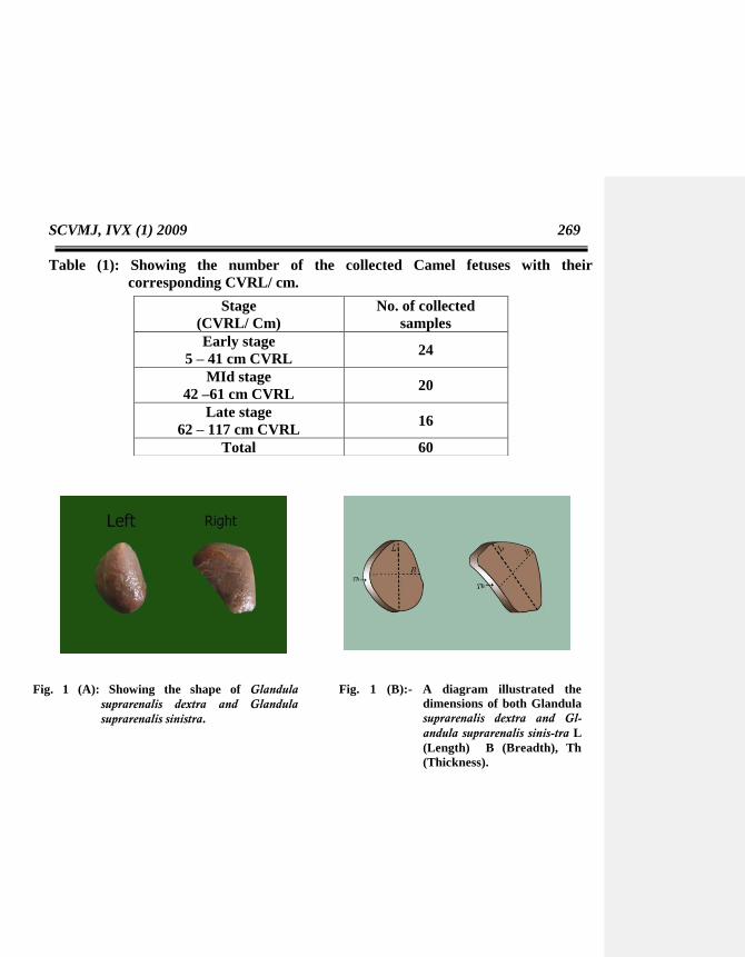
Table (1): Showing the number of the collected Camel fetuses with their
corresponding CVRL/ cm.
Fig. 1 (A): Showing the shape of Glandula
suprarenalis dextra and Glandula
suprarenalis sinistra.
Fig. 1 (B):- A diagram illustrated the
dimensions of both Glandula
suprarenalis dextra and Gl-
andula suprarenalis sinis-tra L
(Length) B (Breadth), Th
(Thickness).
Stage
(CVRL/ Cm)
No. of collected
samples
Early stage
5 – 41 cm CVRL 24
MId stage
42 –61 cm CVRL 20
Late stage
62 – 117 cm CVRL 16
Total 60

270 Sanaa et al.,
RESULTS
The adrenal glands (Glandulae ad-
renals) were two retroperitoneal organs
(fig, 2), situated dorsally at the sublumbar
region and covered with fat (fig, 3). They
were related to the cranial poles of the
corresponding kidney at their junction with
the medial border.
The right and left adrenals were
located nearly at the same level at the 2nd
and 3rd
lumbar vertebrae in the early
embryonic life (at 5 cm CVRL) (fig, 4). As
the development progressed, the left
adrenal gland of 10 cm CVRL camel
fetuses was placed under the level of the
transverse process of 3rd
lumbar vertebra
(fig, 5) while the right one became more
cranial below the transverse process of 2nd
lumbar vertebra (fig, 6).
From 18 cm CVRL till the end of
gestation, the right adrenal gland was
located under the transverse process of the
2nd
lumbar vertebra and reached between
2nd
& 3rd
lumbar transverse processes,
while the left one situated below the level
of the transverse process of the 3rd
lumbar
vertebra and extended between the 3rd
and
4th
lumbar vertebra at the end of gestation(
Fig 7).
Glandula adrenalis dexter :
The right adrenal gland had two
surfaces (dorsal and ventral) and three
borders (cranial, lateral and medial). It was
embedded between caudate lobe of liver
and right kidney. The cranial border was
related to the caudate lobe of the liver, the
lateral one was in close to cranial pole of
right kidney while the medial border was
related to caudal vena cava. Fig (11,
12&13).
Glandula adrenalis sinister:
The left adrenal had two surfaces,
the dorsal surface was related to the left
crus of diaphragm and the ventral surface
was related to the jejunum and left lobe of
pancreas. It was related cranially to the
rumen and laterally to the concave border
of the spleen near its dorsal extremity
infront of the cranial pole of the left kidney
and related medially to the abdominal
Aorta. Fig (2,9&14).
The shape of adrenal glands was
difficult to be determined at the early
embryonic stages, as it was very small in
size. It was shown that both glands were
small ovoid in the camel fetuses less than
7cm CVRL. The left adrenal was still
ovoid or nearly circular till the end of
gestation period. The right adrenal was
slightly elongated in the camel fetuses of
12 cm CVRL and then became triangular
with rounded angles at 15 cm CVRL till
the full term fetus. Fig (10).
Both glands were pale rosy red in
color throughout the different stages of
embryonic development and they were
firm in consistency.
The Weight of right and left glands
increased with advancing of CVRL.
During the different developmental
stages (ranged from 5 to 117 cm CVRL),
the weight of the left adrenal (ranged from
0.0011±0.0004 gm to 0.8160±0.04 gm )

SCVMJ, IVX (1) 2009 271
which was more than that of the right one
(ranged from 0.0008±0.0003 to 0.7110±0.02
gm) as showed in table (2).
Increase of the weight of both
adrenals could be divided into two stages
according to its rate as shown in fig. (15).
The first stage from 5-40 cm CVRL camel
fetuses, the weight of the right gland ranged
from (0.0008±0.0003 to 0.0740±0.006 gm)
and the weight of the left one ranged from
(0.0011±0.0004 to 0.0850±0.007gm) by
rate of 0.002 gm/ 1cm CVRL for both
glands. In the second stage from 42 –117
cm CVRL, the right adrenal increased
10.95 times more than the first stage as it
increased from (0.1130±0.06 to 0.7110±0.02
gm) by rate of 0.006 gm / 1cm CVRL and
the left adrenal increased 10.33 times more
than the first stage as increased from
(0.1240±0.005 to 0.8160±0.04gm) by rate
of 0.007 gm/ 1cm CVRL. (Table 2)
The percentage between the weight
of the right & left adrenal glands and the
weight of their fetuses were variable in the
different developmental stages. At 5 cm
CVRL camel fetus, the right adrenal re-
presented 0.004% and the left one
represented 0.005% from the weight of the
fetus. This percentage increased till reach
its maxi-mum at 26 cm CVRL where the
right adrenal represented 0.019% from
weight of fetus while the left represented
0.020%. After that, there was decreasing in
percentage to 0.005% for right adrenal and
0.006% for the left one at 62 cm CVRL
stage, then the percentage of both right and
left adrenals weight to the weight of fetus
ranged from ( 0.005% to 0.007%) till the
end of gestation period ( from 62 – 117 cm
CVRL). Table (2) & fig. (16).
The dimensions of both glands of
embryos less than 15 cm CVRL was very
small to the extent that it couldn't be
measured, but it was shown that the
adrenals at CVRL less than 10 cm had the
size of sesame seed. After 15cm CVRL,
length, breadth and thickness were
measurable in both glands as shown in fig
(1 A&B).
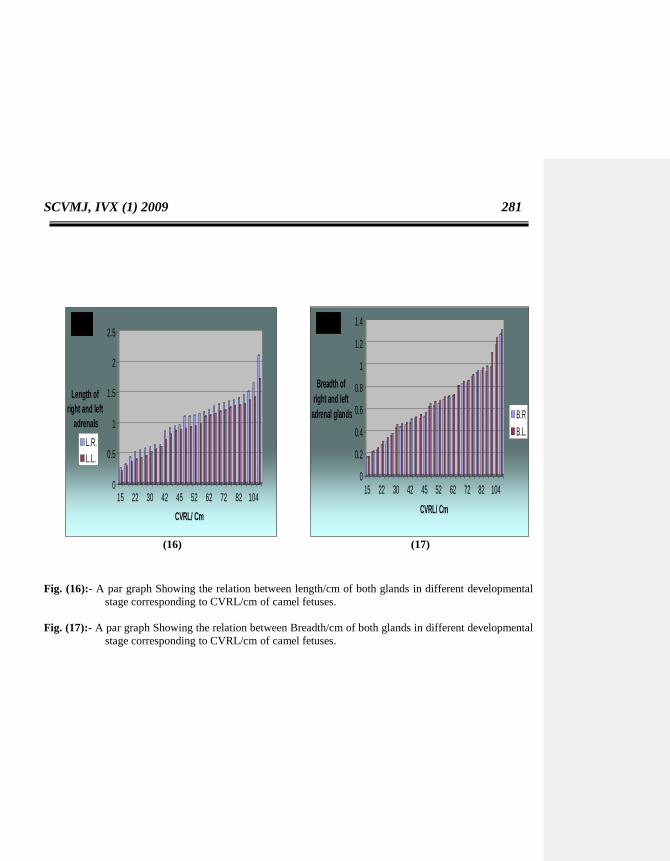
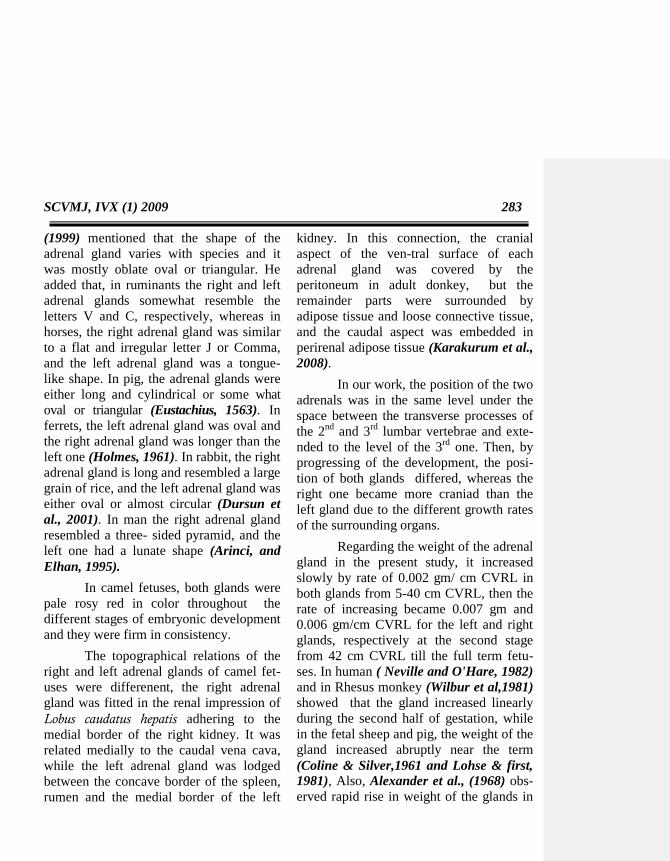
Comparing the two glands in
different stages of deve-lopment (15 to 117
cm CVRL camel fet-uses), it was shown
that the length of the right one (ranged
from 0.26±0.01 to 2.11±0.4 cm) was
markedly more than that of the left one
(ranged from 0.22±0.01 to 1.72±0.4 cm),
in contrast to the breadth which was
slightly more in left adrenal (ranged from
0.17±0.02 to 1.32±0.3 cm) than right one
(ranged from 0.17±0.02 to 1.28±0.4 cm).
The thickness also was sli-ghtly increased
in the left adrenal (ranged from 0.13±0.01
to 0.87±0.2 cm) than that of the right one
(ranged from 0.12±0.03 to 0.75±0.2 cm).
as in Table (3) and Fig. (17, 18 &19).
On cross section, each gland con-
sisted of two parts, cortex and medulla.
The outer smaller cortex was reddish bro-
wn in color and occupied about 1/5 of the
adrenal gland. The inner larger medulla
was dark brown in color and occupied
about 4/5 of the gland. Fig (20).

272 Sanaa et al.,
Table (2) :Showing the mean weight of both right and left adrenal glands with its
percentage to the corresponding camel fetuses weight. ± SD.
CV
RL
/cm
weight
of fetus/ gm
weight of
right
adrenal/gm
weight of
left
adrenal/gm
Ratio of
right adrenal
weight
to fetus weight
Ratio of left
adrenal weight to
fetus weight
5 22.17±2.7 0.0008±0.000
3 0.0011±0.0004 0.004 0.005
7.5 44.31±2.5 0.0027±0.000
4 0.0032±0.0003 0.006 0.007
9.5 62.50±2.3 0.0045±0.000
6 0.0074±0.0005 0.007 0.012
11.5 70.00±2.1 0.0083±0.000
4 0.0088±0.0006 0.012 0.013
13 78.00±1.9 0.0098±0.000
5 0.0105±0.0007 0.013 0.013
15 98.00±1.7 0.0109±0.001 0.0120±0.003 0.011 0.012
16 128.5±2.5 0.0110±0.001 0.0130±0.002 0.009 0.010
18 140.00±3.9 0.0140±0.003 0.0160±0.004 0.010 0.011
19 160.00±2.3 0.0169±0.006 0.0175±0.007 0.011 0.011
22 190.00±1.9 0.0321±0.007 0.0342±0.008 0.017 0.018
26 200.00±2.5 0.0384±0.001 0.039±0.003 0.019 0.020
27 290.00±2.5 0.0433±0.003 0.0467±0.004 0.015 0.016
30 380.00±2.6 0.0485±0.006 0.0493±0.007 0.013 0.013
31 470.00±3.9 0.0498±0.007 0.0510±0.006 0.011 0.011
33 560.00±5 0.0530±0.004 0.0640±0.005 0.009 0.011
40 986.00±4 0.0740±0.006 0.0850±0.007 0.010 0.011
42 1050.00±5.5 0.1130±0.06 0.1240±0.005 0.011 0.012
43 1400.00±9.4 0.1200±0.08 0.1270±0.006 0.009 0.009
44 1550.00±10 0.1240±0.05 0.1310±0.004 0.008 0.008
46 1700.00±11 0.1290±0.04 0.1340±0.008 0.008 0.008
50 2000.00±9 0.1320±0.07 0.1380±0.007 0.007 0.007
51 2150.00±12 0.1430±0.08 0.1440±0.02 0.007 0.007
53 2500.00±8 0.1490±0.07 0.1560±0.03 0.006 0.006
55 2750.00±15 0.1660±0.05 0.1730±0.05 0.006 0.006
56.5 3100.00±17 0.1760±0.04 0.1820±0.02 0.006 0.006
62 3850.00±11 0.1930±0.04 0.2240±0.04 0.005 0.006
67 4900.00±13 0.2230±0.02 0.2410±0.02 0.005 0.005
71 5300.00±15 0.2460±0.03 0.2520±0.03 0.005 0.005
72 5750.00±10 0.3010±0.04 0.3040±0.04 0.005 0.005
78 6200.00±11 0.3890±0.02 0.4110±0.03 0.006 0.007
80 6850.00±12 0.4250±0.02 0.4430±0.02 0.006 0.006
82 7150.00±12 0.4460±0.01 0.4680±0.02 0.006 0.007
84 7450.00±13 0.4650±0.02 0.4960±0.01 0.006 0.007
92 9350.00±11 0.5120±0.01 0.5620±0.03 0.005 0.006
104 12250.00±15 0.5870±0.03 0.6210±0.02 0.005 0.005
117 16500.00±17 0.7110±0.02 0.8160±0.04 0.005 0.005
Formatted ...
Formatted ...
Formatted ...
Formatted ...
Formatted ...
Formatted ...
Formatted ...
Formatted ...
Formatted ...
Formatted ...
Formatted ...
Formatted ...
Formatted ...
Formatted ...
Formatted ...
Formatted ...
Formatted ...
Formatted ...
Formatted ...
Formatted ...
Formatted ...
Formatted ...
Formatted ...
Formatted ...
Formatted ...
Formatted ...
Formatted ...
Formatted ...
Formatted ...
Formatted ...
Formatted ...
Formatted ...
Formatted ...
Formatted ...
Formatted ...
Formatted ...
Formatted ...
SCVMJ, IVX (1) 2009 273
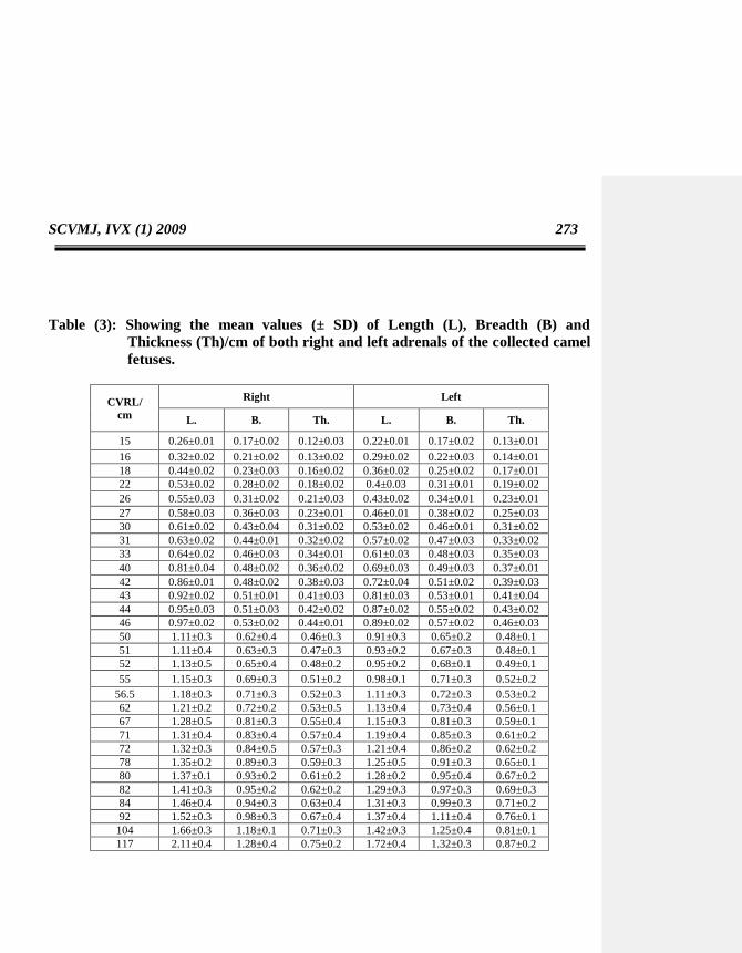
Table (3): Showing the mean values (± SD) of Length (L), Breadth (B) and
Thickness (Th)/cm of both right and left adrenals of the collected camel
fetuses.
CVRL/
cm
Right Left
L. B. Th. L. B. Th.
15 0.26±0.01 0.17±0.02 0.12±0.03 0.22±0.01 0.17±0.02 0.13±0.01
16 0.32±0.02 0.21±0.02 0.13±0.02 0.29±0.02 0.22±0.03 0.14±0.01
18 0.44±0.02 0.23±0.03 0.16±0.02 0.36±0.02 0.25±0.02 0.17±0.01
22 0.53±0.02 0.28±0.02 0.18±0.02 0.4±0.03 0.31±0.01 0.19±0.02
26 0.55±0.03 0.31±0.02 0.21±0.03 0.43±0.02 0.34±0.01 0.23±0.01
27 0.58±0.03 0.36±0.03 0.23±0.01 0.46±0.01 0.38±0.02 0.25±0.03
30 0.61±0.02 0.43±0.04 0.31±0.02 0.53±0.02 0.46±0.01 0.31±0.02
31 0.63±0.02 0.44±0.01 0.32±0.02 0.57±0.02 0.47±0.03 0.33±0.02
33 0.64±0.02 0.46±0.03 0.34±0.01 0.61±0.03 0.48±0.03 0.35±0.03
40 0.81±0.04 0.48±0.02 0.36±0.02 0.69±0.03 0.49±0.03 0.37±0.01
42 0.86±0.01 0.48±0.02 0.38±0.03 0.72±0.04 0.51±0.02 0.39±0.03
43 0.92±0.02 0.51±0.01 0.41±0.03 0.81±0.03 0.53±0.01 0.41±0.04
44 0.95±0.03 0.51±0.03 0.42±0.02 0.87±0.02 0.55±0.02 0.43±0.02
46 0.97±0.02 0.53±0.02 0.44±0.01 0.89±0.02 0.57±0.02 0.46±0.03
50 1.11±0.3 0.62±0.4 0.46±0.3 0.91±0.3 0.65±0.2 0.48±0.1
51 1.11±0.4 0.63±0.3 0.47±0.3 0.93±0.2 0.67±0.3 0.48±0.1
52 1.13±0.5 0.65±0.4 0.48±0.2 0.95±0.2 0.68±0.1 0.49±0.1
55 1.15±0.3 0.69±0.3 0.51±0.2 0.98±0.1 0.71±0.3 0.52±0.2
56.5 1.18±0.3 0.71±0.3 0.52±0.3 1.11±0.3 0.72±0.3 0.53±0.2
62 1.21±0.2 0.72±0.2 0.53±0.5 1.13±0.4 0.73±0.4 0.56±0.1
67 1.28±0.5 0.81±0.3 0.55±0.4 1.15±0.3 0.81±0.3 0.59±0.1
71 1.31±0.4 0.83±0.4 0.57±0.4 1.19±0.4 0.85±0.3 0.61±0.2
72 1.32±0.3 0.84±0.5 0.57±0.3 1.21±0.4 0.86±0.2 0.62±0.2
78 1.35±0.2 0.89±0.3 0.59±0.3 1.25±0.5 0.91±0.3 0.65±0.1
80 1.37±0.1 0.93±0.2 0.61±0.2 1.28±0.2 0.95±0.4 0.67±0.2
82 1.41±0.3 0.95±0.2 0.62±0.2 1.29±0.3 0.97±0.3 0.69±0.3
84 1.46±0.4 0.94±0.3 0.63±0.4 1.31±0.3 0.99±0.3 0.71±0.2
92 1.52±0.3 0.98±0.3 0.67±0.4 1.37±0.4 1.11±0.4 0.76±0.1
104 1.66±0.3 1.18±0.1 0.71±0.3 1.42±0.3 1.25±0.4 0.81±0.1
117 2.11±0.4 1.28±0.4 0.75±0.2 1.72±0.4 1.32±0.3 0.87±0.2

274 Sanaa et al.,
(2) (3)
Fig. (2): A photograph of the abdominal contents after removal of the left lateral abdominal wall of 28 cm
CVRL camel fetus showing that :-
Glandula suprarenalis sinestra with the corresponding kidney located retroperitoneal (arrow head).
1 - Ren sinister 4 – Costa ΧΙΙ
2 - Lien 5 - Intestinum tenue
3 - Rumen
- T1, T2, T3 (Processus transversus ad Vertebrae lumbales Ι, ΙΙ et ΙΙΙ)
- Black arrow (Glandula suprarenlais sinistra).
Fig. (3):-A photograph of the abdominal contents after removal of the left lateral abdominal wall of 60 cm
CVRL camel fetus showing:-
Glandula suprarenalis sinestra (arrow) surrounded by fat (arrow head).
1 - Ren sinister 4 – Costa ΧΙΙ
2 - Lien 5 - Intestinum tenue
3 - Rumen
- T1, T2, T3 (Processus transversus ad Vertebrae lumbales Ι, ΙΙ et ΙΙΙ)).

SCVMJ, IVX (1) 2009 275
(4) (5)
Fig. (4):- A photograph showing the topographical relations of Glandula suprarenalis dextra and Glandula
suprarenalis sinistra in 5 cm CVRL camel fetus showing :-
1- Ren dexter.
2- Ren sinister.
- Arrow (Glandula suprarenalis dextra).
- Arrow head (Glandula suprarenalis sinistra)
- T1, T2, T3 (Processus transversus of ad Vertebrae lumbales Ι, ΙΙ et ΙΙΙ)).
Fig. (5):- A photograph showing the topographical relations of Glandula suprarenalis sinistra in 10 cm
CVRL camel fetus showing:-
1- Ren sinister
2- Ren dexter
- Arrow (Glandula suprarenalis sinistra).
276 Sanaa et al.,
- T1, T2, T3 (Processus transversus ad Vertebrae lumbales Ι, ΙΙ et ΙΙΙ)).
(6) (7)
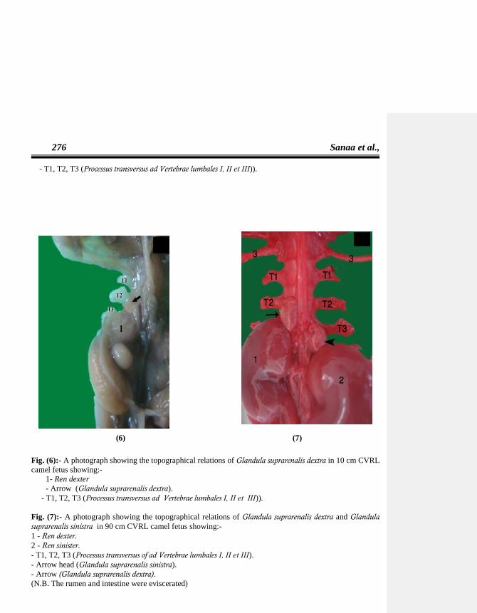
Fig. (6):- A photograph showing the topographical relations of Glandula suprarenalis dextra in 10 cm CVRL
camel fetus showing:-
1- Ren dexter
- Arrow (Glandula suprarenalis dextra).
- T1, T2, T3 (Processus transversus ad Vertebrae lumbales Ι, ΙΙ et ΙΙΙ)).
Fig. (7):- A photograph showing the topographical relations of Glandula suprarenalis dextra and Glandula
suprarenalis sinistra in 90 cm CVRL camel fetus showing:-
1 - Ren dexter.
2 - Ren sinister.
- T1, T2, T3 (Processus transversus of ad Vertebrae lumbales Ι, ΙΙ et ΙΙΙ).
- Arrow head (Glandula suprarenalis sinistra).
- Arrow (Glandula suprarenalis dextra).
(N.B. The rumen and intestine were eviscerated)
SCVMJ, IVX (1) 2009 277
(8) (9)
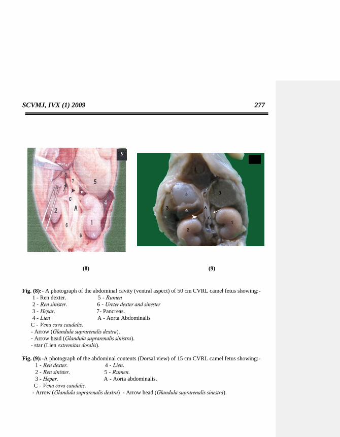
Fig. (8):- A photograph of the abdominal cavity (ventral aspect) of 50 cm CVRL camel fetus showing:-
1 - Ren dexter. 5 - Rumen
2 - Ren sinister. 6 - Ureter dexter and sinester
3 - Hepar. 7- Pancreas.
4 - Lien A - Aorta Abdominalis
C - Vena cava caudalis.
- Arrow (Glandula suprarenalis dextra).
- Arrow head (Glandula suprarenalis sinistra).
- star (Lien extremitas dosalis).
Fig. (9):-A photograph of the abdominal contents (Dorsal view) of 15 cm CVRL camel fetus showing:-
1 - Ren dexter. 4 - Lien.
2 - Ren sinister. 5 - Rumen.
3 - Hepar. A - Aorta abdominalis.
C - Vena cava caudalis.
- Arrow (Glandula suprarenalis dextra) - Arrow head (Glandula suprarenalis sinestra).

278 Sanaa et al.,
(10) (11)
Fig. (10):- A photograph of the abdominal contents after removal of the right lateral abdominal wall of 80
cm CVRL camel fetus showing:-
1 - Ren dexter.
2 - Hepar.
3 – Costa ΧΙΙ.
4 - Intestinum tenue.
- T1, T2 (Processus transversus ad Vertebrae lumbales Ι, ΙΙ et ΙΙΙ ).
- Arrow (Glandula suprarenalis dextra).
Fig. (11):- A photograph of showing the topographic relationship of Glandula suprarenalis dextra in 90 cm
CVRL camel fetus showing:-
1- Ren dexter.
2 - Hepar.
C - Vena cava cadalis.
- T1,T2 (Processus transversus ad Vertebrae lumbales Ι, et ΙΙ).
- Arrow (Glandula suprarenalis dextra).
(N.B. The rumen and intestine were eviscerated).
SCVMJ, IVX (1) 2009 279
(12) (13)
(12) (13)
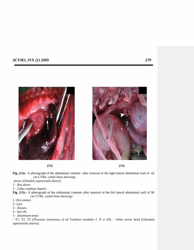
Fig. (12):- A photograph of the abdominal contents after removal of the right lateral abdominal wall of 42
cm CVRL camel fetus showing:-
arrow (Glandula suprarenalis dextra)
1 - Ren dexter
2 - Lobus caudatus hepatis.
Fig. (13):- A photograph of the abdominal contents after removal of the left lateral abdominal wall of 90
cm CVRL camel fetus showing:-
1- Ren sinister.
2- Lien.
3 - Rumen.
4 - last rib.
5 - Intestinum tenue.
- T1, T2, T3 (Processus transversus of ad Vertebrae lumbales Ι, ΙΙ et ΙΙΙ). - white arrow head (Glandula
suprarenalis sinistra).

280 Sanaa et al.,
Fig. (14):- A par graph showing the relation between the weight/gm of both right and left adrenal
glands with its corresponding CVRL/cm of camel fetuses.
Fig. (15) : A Histogram showing the percent pf both right adrenal glands weight to the total fetal
weight in different stages corresponding to CVRL/cm of camel fetuses.
0
0.1
0.2
0.3
0.4
0.5
0.6
0.7
0.8
0.9
5 9 13.5 16 19 22 27 31 40 43 45 51 55 62 71 78 82 92 117
CVRL/Cm
weig
ht
of
rig
ht
an
d left
ad
ren
al g
lan
ds/ g
m
Right Left
percentage of adrenal gland weight to the weight of the fetus
0.000%
0.005%
0.010%
0.015%
0.020%
0.025%
5 9.5 13 16 19 22 27 31 40 43 46 51 55 62 71 78 82 92 117
CVRL
pe
rce
nt
right left
SCVMJ, IVX (1) 2009 281
(16) (17)
Fig. (16):- A par graph Showing the relation between length/cm of both glands in different developmental
stage corresponding to CVRL/cm of camel fetuses.
Fig. (17):- A par graph Showing the relation between Breadth/cm of both glands in different developmental
stage corresponding to CVRL/cm of camel fetuses.
0
0.5
1
1.5
2
2.5
Length of
right and left
adrenals
15 22 30 42 45 52 62 72 82 104
CVRL/ Cm
L.R.
L.L.
0
0.2
0.4
0.6
0.8
1
1.2
1.4
Breadth of
right and left
adrenal glands
15 22 30 42 45 52 62 72 82 104
CVRL/ Cm
B.R
B.L.

282 Sanaa et al.,
(18) (19)
Fig. (18):- A par graph Showing the relation between thickness/cm of both glands in different
developmental stage corresponding to CVRL/cm of camel fetuses.
Fig (19):- A photograph of cross section of Glandula suprarenalis at 65 cm CVRL showing an outer thin
cortex (C) and inner thick medulla (M).
DISCUSSION
According to the available literat-
ures, there was no adequate information on
the shape, color and localization of adrenal
gland during the prenatal life in domestic
animals. Therefore, the findings of the
current study was discussed by comparing
these items with that of some adult
domestic animals.
The shape of adrenal glands was
difficult to be determined at the early
embryonic stages as it was very small in
size. It was shown that both glands were
small ovoid in the camel fetuses, less than
7cm CVRL. The left adrenal was still
ovoid or nearly spheroid till the end of
gestation period. The right adrenal was
slightly elongated in the camel fetuses of
12 cm CVRL and then became triangular
with rounded angles at 15 cm CVRL till
the full term fetus. In this respect, Dursun
0
0.1
0.2
0.3
0.4
0.5
0.6
0.7
0.8
0.9
Thickness of
right and left
adrenal
glands
15 22 30 42 45 52 62 72 82 104
CVRL
Th.R.
Th.L.
SCVMJ, IVX (1) 2009 283
(1999) mentioned that the shape of the
adrenal gland varies with species and it
was mostly oblate oval or triangular. He
added that, in ruminants the right and left
adrenal glands somewhat resemble the
letters V and C, respectively, whereas in
horses, the right adrenal gland was similar
to a flat and irregular letter J or Comma,
and the left adrenal gland was a tongue-
like shape. In pig, the adrenal glands were
either long and cylindrical or some what
oval or triangular (Eustachius, 1563). In
ferrets, the left adrenal gland was oval and
the right adrenal gland was longer than the
left one (Holmes, 1961). In rabbit, the right
adrenal gland is long and resembled a large
grain of rice, and the left adrenal gland was
either oval or almost circular (Dursun et
al., 2001). In man the right adrenal gland
resembled a three- sided pyramid, and the
left one had a lunate shape (Arinci, and
Elhan, 1995).
In camel fetuses, both glands were
pale rosy red in color throughout the
different stages of embryonic development
and they were firm in consistency.
The topographical relations of the
right and left adrenal glands of camel fet-
uses were differenent, the right adrenal
gland was fitted in the renal impression of
Lobus caudatus hepatis adhering to the
medial border of the right kidney. It was
related medially to the caudal vena cava,
while the left adrenal gland was lodged
between the concave border of the spleen,
rumen and the medial border of the left
kidney. In this connection, the cranial
aspect of the ven-tral surface of each
adrenal gland was covered by the
peritoneum in adult donkey, but the
remainder parts were surrounded by
adipose tissue and loose connective tissue,
and the caudal aspect was embedded in
perirenal adipose tissue (Karakurum et al.,
2008).
In our work, the position of the two
adrenals was in the same level under the
space between the transverse processes of
the 2nd
and 3rd
lumbar vertebrae and exte-
nded to the level of the 3rd
one. Then, by
progressing of the development, the posi-
tion of both glands differed, whereas the
right one became more craniad than the
left gland due to the different growth rates
of the surrounding organs.
Regarding the weight of the adrenal
gland in the present study, it increased
slowly by rate of 0.002 gm/ cm CVRL in
both glands from 5-40 cm CVRL, then the
rate of increasing became 0.007 gm and
0.006 gm/cm CVRL for the left and right
glands, respectively at the second stage
from 42 cm CVRL till the full term fetu-
ses. In human ( Neville and O'Hare, 1982)
and in Rhesus monkey (Wilbur et al,1981)
showed that the gland increased linearly
during the second half of gestation, while
in the fetal sheep and pig, the weight of the
gland increased abruptly near the term
(Coline & Silver,1961 and Lohse & first,
1981), Also, Alexander et al., (1968) obs-
erved rapid rise in weight of the glands in

284 Sanaa et al.,
ovine fetus during the last week of
gestation. Yamauchi (1979) recorded that
the foal fetal adrenal gland began to
increase in weight from about the end of
4th
month of pregnancy when fetus had
CRL of about 20 cm. On the same point,
the weight of the right and left adrenals in
camel fetuses at 117 cm CVRL were
0.811±0.02 and 0.816± 0.04 respectively,
while in the fetal lamb, Boshier et al.,
(1980) observed that the gland weight was
doubled in the period from 136 days of
pregnancy till birth (0.45-0.80 gm). In this
respect, Durand et al., (1978) recorded
that in the second growth period just
before birth, the weight of the adrenal was
doubled and reached 300 mg. On the other
hand, fetal rat adrenal growth rate decr-
eased as the term approach (Cohen, 1963).
The size of the adrenal glands in
the camel fetuses increased steadily throu-
ghout the developmental stages, while in
fetal lamb there was increasing of the gla-
nd size from 136 to 147 days (Boshier et
al., 1980). Also, the ovine adrenal size
showed a rapid increase through two gro-
wth phases, the first phase of gradual onset
from 123 to 137 days and the second phase
from 143 days to term as recorded by
Durand et al., (1978). In camel fetuses, the
size of the gland increased due to the
differentiation of the cortical zones and
hyperplasia of the zona fasciculata, as well
as, the highly proliferation of the adrenal
medulla. Yamauchi (1979) restricted his
observations on the thickness of the med-
ulla in the fetal horse. In the same point,
Boshier et al., (1980) mentioned that the
increase of size of the ovine gland resulted
from differential growth of the inner
cortical zone, for neither zona glomerulosa
nor medulla. Also Durand et al., (1978)
accounted that the increase in the ovine
adrenal size in the second growth phase
was mainly due to the cellular
hypertrophy.
Eustachius (1563) observed the
color of the adult adrenal cortex as it was
flesh colored, cream or light yellow depen-
ding upon its lipid contents. He added that,
in horse, dog, cat and chicken, the adrenal
cortex was cream or light yellow in color
because of high lipid content, while in
ruminant and swine, the adrenals were
flesh colored because of a poor lipid con-
tent, as well as the camel fetuses, it appea-
red reddish brown. He added that the color
of the medulla in adult adrenals was redd-
ish brown because of the presence of abu-
ndance of blood in the medullary veins.
These observations were in a line with our
findings in the camel fetuses.
REFERENCES
Alexander, D.P; Britton, H.G; James,
V.H; Nixon, D.A; Parker, R.A; Wintour, E.M. and Wright, R.D. (1968):Steroid
secretion by the adrenal gland of foetal and
neonatal sheep.Endocrinology, 40 (1): 1-13.
Arinci, K., Elhan, A. (1995): Anatomi –
1. Cilt. Güneş Yayınevi, Ankara; 445-448.

SCVMJ, IVX (1) 2009 285
Bone, J.F. (1982):Animal anatomy and
physiology. 2nd
Ed. Pp. 325-326. Reston
publishing company,Inc.Reston.Vinzinia.
Boshier, D.P. and Holloway, H. (1989): Morphometric analyses of adrenal gland
growth in fetal and neonatal sheep. I. The
adrenal cortex. J.Anat, 167: 1-14.
Cohen, A. (1963): Corrélations entre l'hy-
pophyse et le cortex surrénal chez le foetus
de rat. Le cortex surrénal du nouveau-né.
Archives d'anatomie microscoique et de
morphologie expérimentale, 52: 277-407.
Comline, R.S. and Silver, M. (1961): The
release of adrenaline and nor adrenaline
from the adrenal gland of the foetal sheep.
J. physiol, 156: 424 – 444.
Durand, P.H.; Bosc, M. and Nicolle, A. (1978): roissance des surrenales de foetus
ovin en fin de gestation: evolution de
1’AND et des proteines membranaires.
CR Acad Sci Paris, 287: 297-300.
Dursun, N (1999): Veteriner Anatomi II.
Medisan Yayınevi, Ankara. 184-185.
Dursun, N., Bozkurt, E.U. Ozgel, O. (2001): The suprarenal glands and their arterial va-
scularization in New Zealand rabbit (Oryc-
tolagus cuniculus L.). Ankara ـniv. Vet.
Fak. Derg.; 48: 21-25. (article in Turkish
with an abstract in English).
Eustachius, B. (1563): Opuscula Anato-
mica. Libellus de Renibus venet.
Frandson, R.D. (1970): Anatomy and ph-
ysiology of the farm animals. 5th
Ed. Pp.
426-430. Lea& Febiger.philadelphia.
Holmes, R.L. (1961): The adrenal glands
of the ferret, (Mustela putorius). J. Anat.;
95: 325-336.
Karakurum, E. Dursun, Z. N (2008): Morphology and Arterial Vasculature of
Donkey (Equus asinus L.) Adrenal Gland.
Turk. J. Vet. Anim. Sci.
Kempna, P. and Flück, C.E. (2008): Best
Practice & Research Clinical Endocrin-
ology & Metabolism. Vol. 22, No. 1, pp.
77–93.
Liggins, G.C. (1976): Adrenocortical-rela-
ted maturational events in the fetus. Am J
Obstet Gynecol, (126):931–941
Liggins, G.C. (1980): Endocrinology of
parturition. In: Novy MJ, Resko JA (eds)
Fetal Endocrinology. Academic Press,
Inc., New York, Pp 211–237.
Liggins, G.C.; Fairclough, R.J; Grieves,
S.A; Kendall, J.Z and Knox, B.S. (1973): The mechanism of initiation of parturition
in the ewe. Recent Prog Horm Res, (29):111–
159.
Lohse, J.K. and First, N.L. (1981): Deve-

286 Sanaa et al.,
lopment of the procine fetal adrenal in late
gestation. Biol Reprod, 251 (1): 181 – 190.
Mesiano, S. and Jaffe, R.B. (1997): De-
velopmental and Functional Biology of the
Primate Fetal Adrenal Cortex. Endocrine
Reviews, 18: 378–403.
Neville, A.M. and O'Hare, M.J. (1982): The human adrenal cortex. Berlin:springer-
verlag.
Nomina Anatomica Veterinaria (2005): 5
th Ed. Prepared by the international com-
mittee on Veterinary Gross Anatomical
Nomenclature (I.C.V.G.A.N.) and publi-
shed by Editorial Committee. Hannover,
Columbia, Gent and sapporo.
Nomina Embryologica Veterinaria (2006):
2nd
Ed. Prepared by the international com-
mittee on Veterinary Embryological Nom-
enclature (I.C.V.E.N.) and published in
Ghent (Belgium)
Vrezas,I; Willenberg,H.S. and Born-stein, S.R. (2004): Adrenal cortex, Ana-
tomy. Encyclopedia of Endocrine diseases,
volume 1. University of Düsseldorf, Ger-
many.
Wilbur, P.; Miles, J. and Scott, W. (1981): Fetal and Postnatal Development
of the Adrenal Glandsin Macaca mulatta.
Biology of reproduction, (25): 1079-1089
Yamauchi, S. (1979): Histological deve-
lopment of the equine fetal adrenal gland. J
Reprod Fertil Suppl, (27):487-91.
العربي الملخص
وحيد السنام قبل الوالدة في الجمل( فوق الكلوية)دراسات ظاهرية على تطور الغدة الكظرية
، احمد كامل السيد ، عيد على موسى ، هشام محمد إمام ثناء مختار النحلة جامعة قناة السويس – الطــــــب البيطـــرى كلية -ح واالجنة قسم التشري
سم وتم الحصول عليهم 111الى 5جنينا من أجنة الجمال يتراوح اطوالها بين 06تم اجراء هذه الدراسة على عدد
.من مجزر البساتين بالقاهرة ومجزر القرين بالشرقيةسىم 06يزيد بزيادة العمر ولكنة في المراحل األولية حتىى طىول أوضحت الدراسة الظاهرية أن وزن الغدة الكظرية
واتضىح أيضىا أن وزن الغىدة اليسىرى أكبىر مىن . سىم تكىون الزيىادة ملحوظىة 111-01تكون الزيادة طفيفة بينما من سىرى اليمنى فى جميع مراحل العمر المختلفة أما بالنسبة للطول فقد اتضىح أن طىول الغىدة اليمنىى اكبىر مىن الغىدة الي
.اليسرى أكثر سمكا وعرضا من اليمنى عكس فان الغدةالوعلى .لنخاعا –القشرة -الغدة الكظرية: لكلمات المرشدة