Granular Cell Tumor: A Rare Breast Lesion Tumor de Células ...
4
CASO CLÍNICO Revista Científica da Ordem dos Médicos www.actamedicaportuguesa.com 61 REFERÊNCIAS 1. Arnold DM, Nazi I, Warkentin TE, Smith JW, Tolti LJ, George JN, et al. Approach to the diagnosis and management of drug-induced immune thrombocytopenia. Transfus Med Rev. 2013;27:137-45. 2. Kenney B, Stack G. Drug induced thrombocytopenia. Arch Pathol Lab Med. 2009;133:309-14. 3. FDA Adverse Events Reporting System (AERS). New Hampshire: Food and Drug Administration Website; 2008. [consultado 2018 set 29] DIsponível em: https://www.accessdata.fda.gov/drugsatfda_docs/ label/2008/021350s008lbl.pdf. 4. De Silva E, Kim H. Focus on platelet apoptosis. Chem Biol Interac. 2018;284:1–11. 5. Arnold DM, Kukaswadia S, Nazi I, Esmail A, Dewar L, Smith JW, et al. A systematic evaluation of laboratory testing for drug-induced immune thrombocytopenia. J Thromb Haemost. 2013;11:169–76. 6. George JN, Aster RH. Drug-induced thrombocytopenia: pathogenesis, evaluation, and management. Hematology Am Soc Hematol Educ Program. 2009:153-8. 7. Visentin GP, Liu CY. Drug induced thrombocytopenia. Hematol Oncol Clin of North Am. 2007;21:685-96. 8. Agapakis DI, Massa EV. A case of fenofibrate-induced immune thrombocytopenia: first report. P R Health Sci J. 2015;34:170-3. 9. Van den Bemt PM, Meyboom RH, Egberts AC. Drug-induced immune thrombocytopenia. Drug Saf. 2004;27:1243-52. RESUMO Os tumores de células granulares são tumores benignos raros que podem mimetizar lesões malignas nos exames de imagem mamária. Podem originar-se em qualquer localização, contudo, os de origem mamária representam uma percentagem mínima de todos os tumores de células granulares. Os autores apresentam um caso de uma mulher de 54 anos de idade com um tumor de células granulares da mama que se apresentou como uma lesão suspeita, tanto clinicamente, como nos exames de imagem de mamografia, ecografia e ressonância magnética. Realizou-se biópsia com agulha grossa, guiada por ecografia, cuja histologia revelou achados consistentes com um tumor de células granulares, que foi confirmado posteriormente pelo exame histológico da peça cirúrgica de tumorectomia. Os autores expõem este caso pela sua raridade e para enfatizar esta hipótese diagnóstica, muitas vezes desafiante, e que mimetiza frequentemente lesões mamárias malignas. Palavras-chave: Neoplasias da Mama/diagnóstico; Neoplasias da Mama/diagnóstico por imagem; Neoplasias da Mama/patologia; Tumor de Células Granulares/diagnóstico; Tumor de Células Granulares/diagnóstico por imagem; Tumor de Células Granulares/ patologia Granular Cell Tumor: A Rare Breast Lesion Tumor de Células Granulares: Uma Rara Lesão na Mama Sofia DUTRA 1 , José Carlos MARQUES 2 Acta Med Port 2020 Jan;33(1):61-64 ▪ https://doi.org/10.20344/amp.10279 ABSTRACT Granular cell tumors are uncommon, and are usually benign neoplasms that can mimic malignancy on breast imaging tests. These tumors can originate anywhere in the body and the breast accounts for only a few cases of all granular cell tumors. We report a case of a 54-year-old woman with a granular cell tumor of the breast presenting clinically on breast imaging (ultrasound, mammography and magnetic resonance) as a suspicious lesion. Core needle biopsy was performed for tissue diagnosis and was consistent with granular cell tumor that was confirmed in the histopathological report of the surgical specimen following a breast lumpectomy. Given the rarity of this tumor, we present this case to highlight this diagnostic hypothesis, that can be challenging and frequently confused with breast carcinoma. Keywords: Breast Neoplasms/diagnosis; Breast Neoplasms/diagnostic imaging; Breast Neoplasms/pathology; Granular Cell Tumor/ diagnosis; Granular Cell Tumor/diagnostic imaging; Granular Cell Tumor/pathology 1. Radiology Department. Hospital do Divino Espírito Santo. Ponta Delgada. Portugal. 2. Radiology Department. Instituto Português de Oncologia Francisco Gentil. Lisboa. Portugal. Autor correspondente: Sofia Dutra. Sofi[email protected] Recebido: 22 de janeiro de 2018 - Aceite: 14 de janeiro de 2019 | Copyright © Ordem dos Médicos 2020 INTRODUCTION A granular cell tumor (GCT) is an uncommon tumor that may appear anywhere in the body, and it was first described by Abrikossoff in 1926. 1,2 The most common site of origin is the tongue. 3 A GCT involving the breast parenchyma is a very rare lesion, and its ability to mimic breast carcinoma may be challenging for clinicians, radiologists and patholo- gists. 4 We report a case of a granular cell tumor of the breast, mimicking a breast carcinoma on ultrasonography, mam- mography and magnetic resonance, in which the diagnosis was made by core-needle-biopsy and confirmed by histo- logical examination of the corresponding surgical speci- men. CASE REPORT A 54-year-old woman was referred to our breast center by the family physician, because of a suspicious left breast mass visible in breast imaging tests, after she had noticed a lump during breast self-examination. She had no personal or family history of malignancy and there was no prior his- tory of breast trauma or infection. The physical examination showed a 3 cm firm, painless Dutra S, et al. Granular cell tumor: a rare breast lesion, Acta Med Port 2020 Jan;33(1):61-64
Transcript of Granular Cell Tumor: A Rare Breast Lesion Tumor de Células ...
CA
SO C
LÍN
ICO
Revista Científica da Ordem dos Médicos www.actamedicaportuguesa.com 61
REFERÊNCIAS 1. Arnold DM, Nazi I, Warkentin TE, Smith JW, Tolti LJ, George JN, et al.
Approach to the diagnosis and management of drug-induced immune thrombocytopenia. Transfus Med Rev. 2013;27:137-45.
2. Kenney B, Stack G. Drug induced thrombocytopenia. Arch Pathol Lab Med. 2009;133:309-14.
3. FDA Adverse Events Reporting System (AERS). New Hampshire: Food and Drug Administration Website; 2008. [consultado 2018 set 29] DIsponível em: https://www.accessdata.fda.gov/drugsatfda_docs/label/2008/021350s008lbl.pdf.
4. De Silva E, Kim H. Focus on platelet apoptosis. Chem Biol Interac. 2018;284:1–11.
5. Arnold DM, Kukaswadia S, Nazi I, Esmail A, Dewar L, Smith JW, et al.
A systematic evaluation of laboratory testing for drug-induced immune thrombocytopenia. J Thromb Haemost. 2013;11:169–76.
6. George JN, Aster RH. Drug-induced thrombocytopenia: pathogenesis, evaluation, and management. Hematology Am Soc Hematol Educ Program. 2009:153-8.
7. Visentin GP, Liu CY. Drug induced thrombocytopenia. Hematol Oncol Clin of North Am. 2007;21:685-96.
8. Agapakis DI, Massa EV. A case of fenofibrate-induced immune thrombocytopenia: first report. P R Health Sci J. 2015;34:170-3.
9. Van den Bemt PM, Meyboom RH, Egberts AC. Drug-induced immune thrombocytopenia. Drug Saf. 2004;27:1243-52.
RESUMOOs tumores de células granulares são tumores benignos raros que podem mimetizar lesões malignas nos exames de imagem mamária. Podem originar-se em qualquer localização, contudo, os de origem mamária representam uma percentagem mínima de todos os tumores de células granulares. Os autores apresentam um caso de uma mulher de 54 anos de idade com um tumor de células granulares da mama que se apresentou como uma lesão suspeita, tanto clinicamente, como nos exames de imagem de mamografia, ecografia e ressonância magnética. Realizou-se biópsia com agulha grossa, guiada por ecografia, cuja histologia revelou achados consistentes com um tumor de células granulares, que foi confirmado posteriormente pelo exame histológico da peça cirúrgica de tumorectomia. Os autores expõem este caso pela sua raridade e para enfatizar esta hipótese diagnóstica, muitas vezes desafiante, e que mimetiza frequentemente lesões mamárias malignas. Palavras-chave: Neoplasias da Mama/diagnóstico; Neoplasias da Mama/diagnóstico por imagem; Neoplasias da Mama/patologia; Tumor de Células Granulares/diagnóstico; Tumor de Células Granulares/diagnóstico por imagem; Tumor de Células Granulares/patologia
Granular Cell Tumor: A Rare Breast Lesion
Tumor de Células Granulares: Uma Rara Lesão na Mama
Sofia DUTRA1, José Carlos MARQUES2
Acta Med Port 2020 Jan;33(1):61-64 ▪ https://doi.org/10.20344/amp.10279
ABSTRACTGranular cell tumors are uncommon, and are usually benign neoplasms that can mimic malignancy on breast imaging tests. These tumors can originate anywhere in the body and the breast accounts for only a few cases of all granular cell tumors. We report a case of a 54-year-old woman with a granular cell tumor of the breast presenting clinically on breast imaging (ultrasound, mammography and magnetic resonance) as a suspicious lesion. Core needle biopsy was performed for tissue diagnosis and was consistent with granular cell tumor that was confirmed in the histopathological report of the surgical specimen following a breast lumpectomy. Given the rarity of this tumor, we present this case to highlight this diagnostic hypothesis, that can be challenging and frequently confused with breast carcinoma. Keywords: Breast Neoplasms/diagnosis; Breast Neoplasms/diagnostic imaging; Breast Neoplasms/pathology; Granular Cell Tumor/diagnosis; Granular Cell Tumor/diagnostic imaging; Granular Cell Tumor/pathology
1. Radiology Department. Hospital do Divino Espírito Santo. Ponta Delgada. Portugal.2. Radiology Department. Instituto Português de Oncologia Francisco Gentil. Lisboa. Portugal. Autor correspondente: Sofia Dutra. [email protected]: 22 de janeiro de 2018 - Aceite: 14 de janeiro de 2019 | Copyright © Ordem dos Médicos 2020
INTRODUCTION A granular cell tumor (GCT) is an uncommon tumor that may appear anywhere in the body, and it was first described by Abrikossoff in 1926.1,2 The most common site of origin is the tongue.3 A GCT involving the breast parenchyma is a very rare lesion, and its ability to mimic breast carcinoma may be challenging for clinicians, radiologists and patholo-gists.4
We report a case of a granular cell tumor of the breast, mimicking a breast carcinoma on ultrasonography, mam-mography and magnetic resonance, in which the diagnosis was made by core-needle-biopsy and confirmed by histo-
logical examination of the corresponding surgical speci-men.
CASE REPORT A 54-year-old woman was referred to our breast center by the family physician, because of a suspicious left breast mass visible in breast imaging tests, after she had noticed a lump during breast self-examination. She had no personal or family history of malignancy and there was no prior his-tory of breast trauma or infection. The physical examination showed a 3 cm firm, painless
Dutra S, et al. Granular cell tumor: a rare breast lesion, Acta Med Port 2020 Jan;33(1):61-64
CA
SO C
LÍNIC
O
62Revista Científica da Ordem dos Médicos www.actamedicaportuguesa.com
Dutra S, et al. Granular cell tumor: a rare breast lesion, Acta Med Port 2020 Jan;33(1):61-64
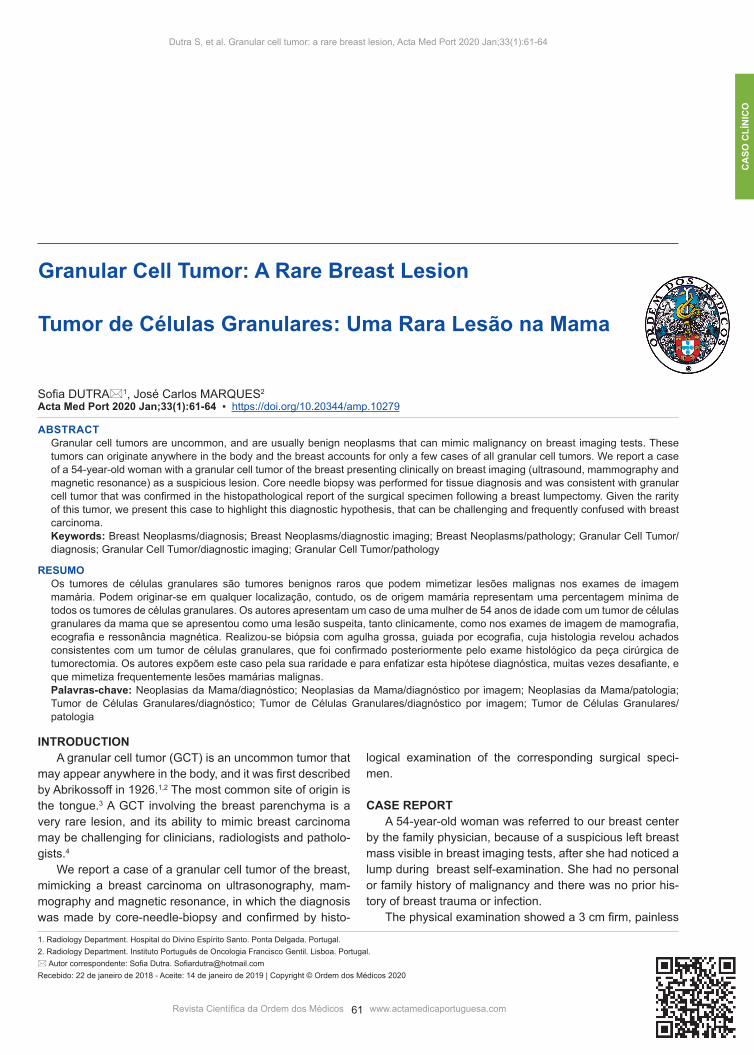
Figure 1 – (A) Mediolateral oblique mammographic view shows a deep, round dense mass, only partially visualized, in the upper quadrants of the left breast. (B) Ultrasound reveals a suspicious-appearing lesion that corresponds to the mammographic abnormality.
A B
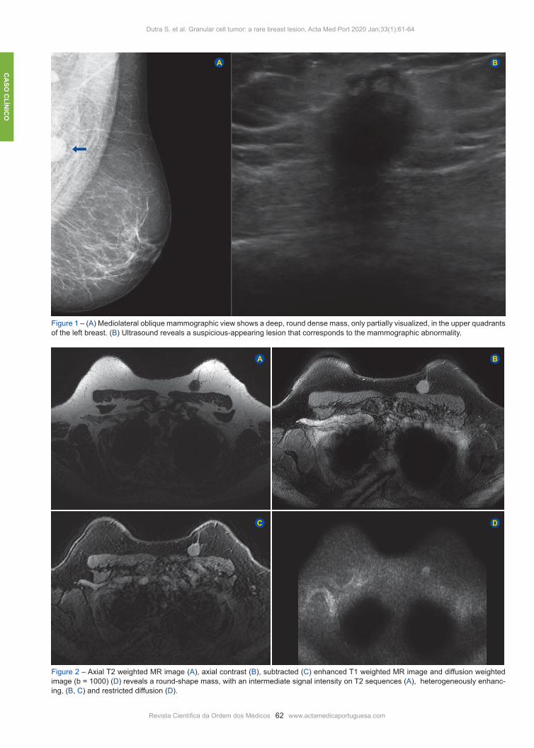
Figure 2 – Axial T2 weighted MR image (A), axial contrast (B), subtracted (C) enhanced T1 weighted MR image and diffusion weighted image (b = 1000) (D) reveals a round-shape mass, with an intermediate signal intensity on T2 sequences (A), heterogeneously enhanc-ing, (B, C) and restricted diffusion (D).
A
C
B
D

CA
SO C
LÍN
ICO
Revista Científica da Ordem dos Médicos www.actamedicaportuguesa.com 63
Dutra S, et al. Granular cell tumor: a rare breast lesion, Acta Med Port 2020 Jan;33(1):xxx-xxx
mass, located in the superomedial quadrant of the left breast, that was fixed to the deep planes. There were no axillary nodes, nipple retraction or skin changes. The me-diolateral oblique mammographic view revealed a deep, round dense mass, with spiculated margins, partially visu-alized, in the upper quadrants of the left breast (BIRADS 4c) (Fig. 1A). There was no associated calcification or skin thickness. The ultrasound showed an hypoechoic mass, heterogeneous, with microlobulated borders and posterior acoustic shadowing, located in the upper inner quadrant (UIQ) of the left breast, approximately 28 mm in largest diameter and oriented perpendicular to the skin (Fig. 1B). Breast MRI was performed in a 1.5T available system. It revealed a deep round-shaped mass, located immediately anterior to the pectoralis major muscle, of intermediate sig-nal intensity on T2 weighted imaging, when compared with the adjacent muscles (Fig. 2A). Post-gadolinium T1-weight-ed images revealed a nonhomogeneous enhancing mass, with a more pronounced enhancement in the periphery of the lesion (Figs. 2B, 2C). The kinetic contrast enhancement profile showed a rapid initial enhancement followed by a gradual wash-out in the delayed phase. There was diffusion restriction (Fig. 2D).
The mass was suspicious for malignancy andultra-sound-guided core biopsy using a 14-gauge automated
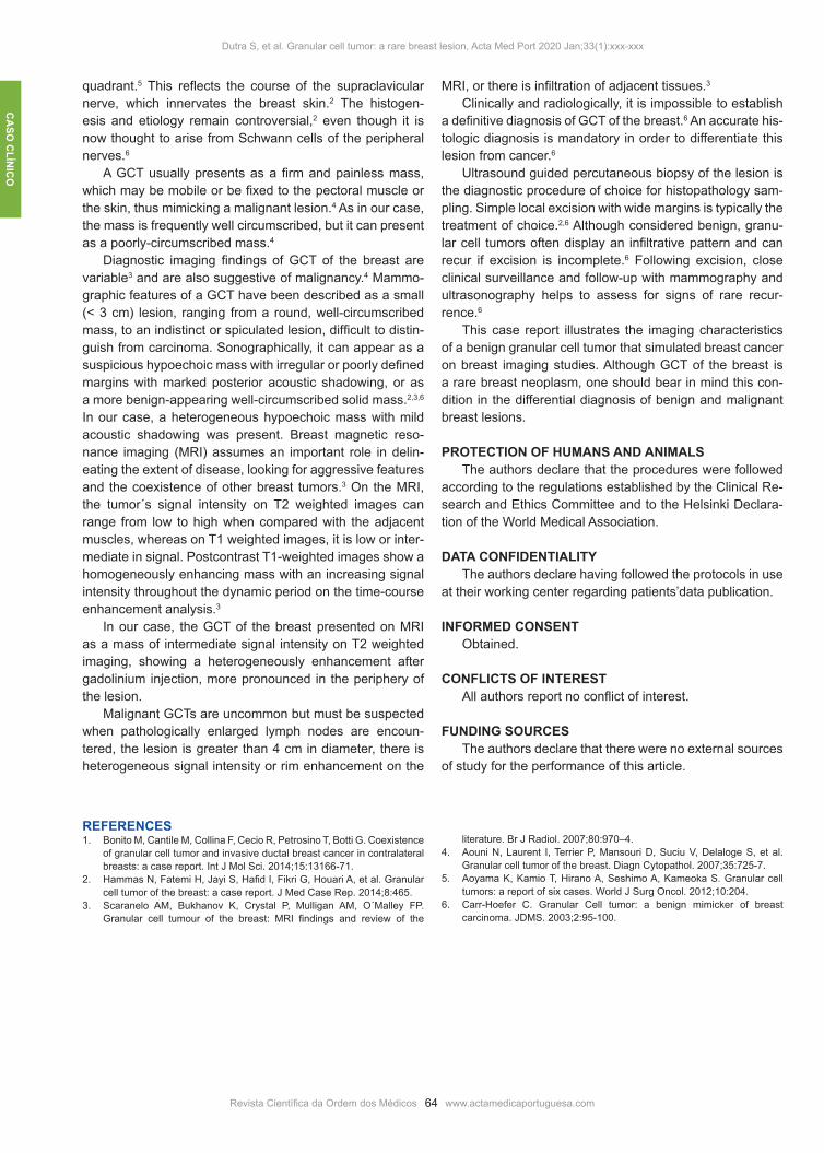
needle was performed, for histological diagnosis. The find-ings were consistent with granular cell tumor. Although the lesion was considered benign, the treat-ment recommendation was surgical excision and a lumpec-tomy was performed with adequate margins. On gross ex-amination, the surgically excised specimen consisted of a white solid hard mass, 28 x 22 x 20 mm in diameter, with specimen margins clear of tumor cells. The histological ex-amination revealed a group of cells with abundant granular, pale pink cytoplasm and inconspicuous cytoplasmic bor-ders, with small and bland nuclei, confirming the diagnosis of granular cell tumor (Fig. 3). The patient tolerated the sur-gical procedure well with full recovery. Annual radiological follow-up has been performed for five years now, without evidence of local recurrence until the date.
DISCUSSION Granular cell tumors account for between 5% and 8% of all GCT cases.3 It occurs in a wide range of ages, with a slight preponderance in middle-aged premenopausal sub-jects and especially in African-American women.4
Granular cell tumors of the breast arise from intra-lob-ular breast stroma.2 In contrast to most of the other breast tumors, which occur predominantly in the upper outer quad-rant, GCT are most frequently found in the upper inner
Figure 3 – Histopathological examination showed that the tumor cells consist of groups of cells with abundant granular, pale pink cyto-plasm and inconspicuous cytoplasmic borders. Nuclei are small and bland.
CA
SO C
LÍNIC
O
64Revista Científica da Ordem dos Médicos www.actamedicaportuguesa.com
Dutra S, et al. Granular cell tumor: a rare breast lesion, Acta Med Port 2020 Jan;33(1):xxx-xxx
quadrant.5 This reflects the course of the supraclavicular nerve, which innervates the breast skin.2 The histogen-esis and etiology remain controversial,2 even though it is now thought to arise from Schwann cells of the peripheral nerves.6 A GCT usually presents as a firm and painless mass, which may be mobile or be fixed to the pectoral muscle or the skin, thus mimicking a malignant lesion.4 As in our case, the mass is frequently well circumscribed, but it can present as a poorly-circumscribed mass.4 Diagnostic imaging findings of GCT of the breast are variable3 and are also suggestive of malignancy.4 Mammo-graphic features of a GCT have been described as a small (< 3 cm) lesion, ranging from a round, well-circumscribed mass, to an indistinct or spiculated lesion, difficult to distin-guish from carcinoma. Sonographically, it can appear as a suspicious hypoechoic mass with irregular or poorly defined margins with marked posterior acoustic shadowing, or as a more benign-appearing well-circumscribed solid mass.2,3,6 In our case, a heterogeneous hypoechoic mass with mild acoustic shadowing was present. Breast magnetic reso-nance imaging (MRI) assumes an important role in delin-eating the extent of disease, looking for aggressive features and the coexistence of other breast tumors.3 On the MRI, the tumor´s signal intensity on T2 weighted images can range from low to high when compared with the adjacent muscles, whereas on T1 weighted images, it is low or inter-mediate in signal. Postcontrast T1-weighted images show a homogeneously enhancing mass with an increasing signal intensity throughout the dynamic period on the time-course enhancement analysis.3
In our case, the GCT of the breast presented on MRI as a mass of intermediate signal intensity on T2 weighted imaging, showing a heterogeneously enhancement after gadolinium injection, more pronounced in the periphery of the lesion. Malignant GCTs are uncommon but must be suspected when pathologically enlarged lymph nodes are encoun-tered, the lesion is greater than 4 cm in diameter, there is heterogeneous signal intensity or rim enhancement on the
MRI, or there is infiltration of adjacent tissues.3
Clinically and radiologically, it is impossible to establish a definitive diagnosis of GCT of the breast.6 An accurate his-tologic diagnosis is mandatory in order to differentiate this lesion from cancer.6
Ultrasound guided percutaneous biopsy of the lesion is the diagnostic procedure of choice for histopathology sam-pling. Simple local excision with wide margins is typically the treatment of choice.2,6 Although considered benign, granu-lar cell tumors often display an infiltrative pattern and can recur if excision is incomplete.6 Following excision, close clinical surveillance and follow-up with mammography and ultrasonography helps to assess for signs of rare recur-rence.6
This case report illustrates the imaging characteristics of a benign granular cell tumor that simulated breast cancer on breast imaging studies. Although GCT of the breast is a rare breast neoplasm, one should bear in mind this con-dition in the differential diagnosis of benign and malignant breast lesions.
PROTECTION OF HUMANS AND ANIMALS The authors declare that the procedures were followed according to the regulations established by the Clinical Re-search and Ethics Committee and to the Helsinki Declara-tion of the World Medical Association.
DATA CONFIDENTIALITY The authors declare having followed the protocols in use at their working center regarding patients’data publication.
INFORMED CONSENT Obtained.
CONFLICTS OF INTEREST All authors report no conflict of interest.
FUNDING SOURCES The authors declare that there were no external sources of study for the performance of this article.
REFERENCES1. Bonito M, Cantile M, Collina F, Cecio R, Petrosino T, Botti G. Coexistence
of granular cell tumor and invasive ductal breast cancer in contralateral breasts: a case report. Int J Mol Sci. 2014;15:13166-71.
2. Hammas N, Fatemi H, Jayi S, Hafid I, Fikri G, Houari A, et al. Granular cell tumor of the breast: a case report. J Med Case Rep. 2014;8:465.
3. Scaranelo AM, Bukhanov K, Crystal P, Mulligan AM, O´Malley FP. Granular cell tumour of the breast: MRI findings and review of the
literature. Br J Radiol. 2007;80:970–4. 4. Aouni N, Laurent I, Terrier P, Mansouri D, Suciu V, Delaloge S, et al.
Granular cell tumor of the breast. Diagn Cytopathol. 2007;35:725-7.5. Aoyama K, Kamio T, Hirano A, Seshimo A, Kameoka S. Granular cell
tumors: a report of six cases. World J Surg Oncol. 2012;10:204.6. Carr-Hoefer C. Granular Cell tumor: a benign mimicker of breast
carcinoma. JDMS. 2003;2:95-100.