
Absorption and reflection of infrared radiation by polymers ...
LETTERSPUBLISHED ONLINE: 1 NOVEMBER 2009 | DOI: 10.1038/NMAT2564
Gold nanocages covered by smart polymers forcontrolled release with near-infrared lightMustafa S. Yavuz*, Yiyun Cheng*, Jingyi Chen*, Claire M. Cobley, Qiang Zhang, Matthew Rycenga,Jingwei Xie, Chulhong Kim, Kwang H. Song, Andrea G. Schwartz, Lihong V. Wang and Younan Xia†
Photosensitive caged compounds have enhanced our ability toaddress the complexity of biological systems by generatingeffectors with remarkable spatial/temporal resolutions1–3. Thecaging effect is typically removed by photolysis with ultravioletlight to liberate the bioactive species. Although this techniquehas been successfully applied to many biological problems, itsuffers from a number of intrinsic drawbacks. For example, itrequires dedicated efforts to design and synthesize a precursorcompound for each effector. The ultraviolet light may causedamage to biological samples and is suitable only for in vitrostudies because of its quick attenuation in tissue4. Herewe address these issues by developing a platform based onthe photothermal effect of gold nanocages. Gold nanocagesrepresent a class of nanostructures with hollow interiorsand porous walls5. They can have strong absorption (for thephotothermal effect) in the near-infrared while maintaininga compact size. When the surface of a gold nanocage iscovered with a smart polymer, the pre-loaded effector canbe released in a controllable fashion using a near-infraredlaser. This system works well with various effectors withoutinvolving sophisticated syntheses, and is well suited for in vivostudies owing to the high transparency of soft tissue in thenear-infrared region6.
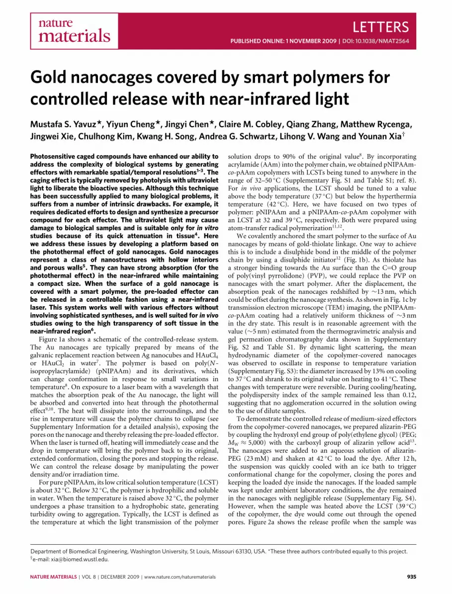
Figure 1a shows a schematic of the controlled-release system.The Au nanocages are typically prepared by means of thegalvanic replacement reaction between Ag nanocubes and HAuCl4or HAuCl2 in water7. The polymer is based on poly(N -isopropylacrylamide) (pNIPAAm) and its derivatives, whichcan change conformation in response to small variations intemperature8. On exposure to a laser beam with a wavelength thatmatches the absorption peak of the Au nanocage, the light willbe absorbed and converted into heat through the photothermaleffect9,10. The heat will dissipate into the surroundings, and therise in temperature will cause the polymer chains to collapse (seeSupplementary Information for a detailed analysis), exposing thepores on the nanocage and thereby releasing the pre-loaded effector.When the laser is turned off, heating will immediately cease and thedrop in temperature will bring the polymer back to its original,extended conformation, closing the pores and stopping the release.We can control the release dosage by manipulating the powerdensity and/or irradiation time.
For pure pNIPAAm, its low critical solution temperature (LCST)is about 32 ◦C. Below 32 ◦C, the polymer is hydrophilic and solublein water. When the temperature is raised above 32 ◦C, the polymerundergoes a phase transition to a hydrophobic state, generatingturbidity owing to aggregation. Typically, the LCST is defined asthe temperature at which the light transmission of the polymer
Department of Biomedical Engineering, Washington University, St Louis, Missouri 63130, USA. *These three authors contributed equally to this project.†e-mail: [email protected].
solution drops to 90% of the original value8. By incorporatingacrylamide (AAm) into the polymer chain, we obtained pNIPAAm-co-pAAm copolymers with LCSTs being tuned to anywhere in therange of 32–50 ◦C (Supplementary Fig. S1 and Table S1; ref. 8).For in vivo applications, the LCST should be tuned to a valueabove the body temperature (37 ◦C) but below the hyperthermiatemperature (42 ◦C). Here, we have focused on two types ofpolymer: pNIPAAm and a pNIPAAm-co-pAAm copolymer withan LCST at 32 and 39 ◦C, respectively. Both were prepared usingatom-transfer radical polymerization11,12.
We covalently anchored the smart polymer to the surface of Aunanocages by means of gold-thiolate linkage. One way to achievethis is to include a disulphide bond in the middle of the polymerchain by using a disulphide initiator12 (Fig. 1b). As thiolate hasa stronger binding towards the Au surface than the C=O groupof poly(vinyl pyrrolidone) (PVP), we could replace the PVP onnanocages with the smart polymer. After the displacement, theabsorption peak of the nanocages redshifted by ∼13 nm, whichcould be offset during the nanocage synthesis. As shown in Fig. 1c bytransmission electron microscope (TEM) imaging, the pNIPAAm-co-pAAm coating had a relatively uniform thickness of ∼3 nmin the dry state. This result is in reasonable agreement with thevalue (∼5 nm) estimated from the thermogravimetric analysis andgel permeation chromatography data shown in SupplementaryFig. S2 and Table S1. By dynamic light scattering, the meanhydrodynamic diameter of the copolymer-covered nanocageswas observed to oscillate in response to temperature variation(Supplementary Fig. S3): the diameter increased by 13% on coolingto 37 ◦C and shrank to its original value on heating to 41 ◦C. Thesechanges with temperature were reversible. During cooling/heating,the polydispersity index of the sample remained less than 0.12,suggesting that no agglomeration occurred in the solution owingto the use of dilute samples.
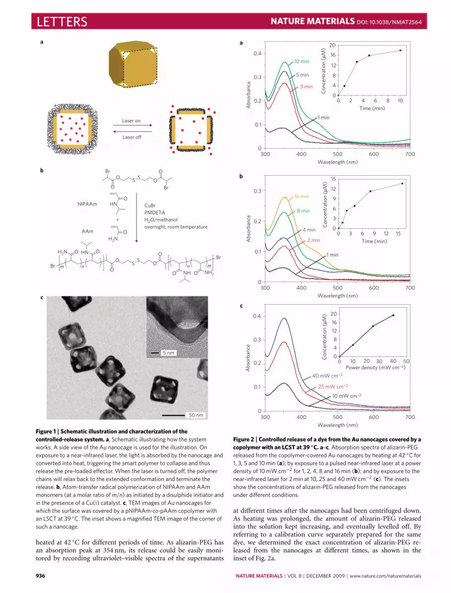
To demonstrate the controlled release of medium-sized effectorsfrom the copolymer-covered nanocages, we prepared alizarin-PEGby coupling the hydroxyl end group of poly(ethylene glycol) (PEG;MW ≈ 5,000) with the carboxyl group of alizarin yellow acid13.The nanocages were added to an aqueous solution of alizarin-PEG (23mM) and shaken at 42 ◦C to load the dye. After 12 h,the suspension was quickly cooled with an ice bath to triggerconformational change for the copolymer, closing the pores andkeeping the loaded dye inside the nanocages. If the loaded samplewas kept under ambient laboratory conditions, the dye remainedin the nanocages with negligible release (Supplementary Fig. S4).However, when the sample was heated above the LCST (39 ◦C)of the copolymer, the dye would come out through the openedpores. Figure 2a shows the release profile when the sample was
NATUREMATERIALS | VOL 8 | DECEMBER 2009 | www.nature.com/naturematerials 935
LETTERS NATUREMATERIALS DOI: 10.1038/NMAT2564
Laser on
Laser off
O
O
O NH2
mnO NH
SS
O
O
n
O
O
H2N HN
Br
O
OSSO
O
Br
CuBrPMDETAH2O/methanolovernight, room temperature
NIPAAmO
HN
+
H2NOAAm
Br
Br
m
5 nm
50 nm
a
c
b
Figure 1 | Schematic illustration and characterization of thecontrolled-release system. a, Schematic illustrating how the systemworks. A side view of the Au nanocage is used for the illustration. Onexposure to a near-infrared laser, the light is absorbed by the nanocage andconverted into heat, triggering the smart polymer to collapse and thusrelease the pre-loaded effector. When the laser is turned off, the polymerchains will relax back to the extended conformation and terminate therelease. b, Atom-transfer radical polymerization of NIPAAm and AAmmonomers (at a molar ratio of m/n) as initiated by a disulphide initiator andin the presence of a Cu(I) catalyst. c, TEM images of Au nanocages forwhich the surface was covered by a pNIPAAm-co-pAAm copolymer withan LSCT at 39 ◦C. The inset shows a magnified TEM image of the corner ofsuch a nanocage.
heated at 42 ◦C for different periods of time. As alizarin-PEG hasan absorption peak at 354 nm, its release could be easily moni-tored by recording ultraviolet–visible spectra of the supernatants
0.4
10 min
5 min
3 min
16 min
8 min
4 min
40 mW cm¬2
2 min
25 mW cm¬2
1 min
1 min
10 mW cm¬2
20
Con
cent
ratio
n (µ
M)
Con
cent
ratio
n (µ
M)
Con
cent
ratio
n (µ
M)
16
12
8
4
0
Abs
orba
nce
Abs
orba
nce
Abs
orba
nce
0.3
0.2
0.1
0
0.3
0.2
0.1
0
0.4
0.3
0.2
0.1
0300 400 500
Wavelength (nm)
600 700
300 400 500
Wavelength (nm)
600 700
300 400 500
Wavelength (nm)
600 700
0 2 4 6
Time (min)
8 10
Time (min)
0 3 6 9 12 15
Power density (mW cm¬2)0 10 20 30 40 50
0
3
6
9
12
15
20
16
12
8
4
0
a
c
b
Figure 2 | Controlled release of a dye from the Au nanocages covered by acopolymer with an LCST at 39 ◦C. a–c, Absorption spectra of alizarin-PEGreleased from the copolymer-covered Au nanocages by heating at 42 ◦C for1, 3, 5 and 10 min (a); by exposure to a pulsed near-infrared laser at a powerdensity of 10 mW cm−2 for 1, 2, 4, 8 and 16 min (b); and by exposure to thenear-infrared laser for 2 min at 10, 25 and 40 mW cm−2 (c). The insetsshow the concentrations of alizarin-PEG released from the nanocagesunder different conditions.
at different times after the nanocages had been centrifuged down.As heating was prolonged, the amount of alizarin-PEG releasedinto the solution kept increasing, and eventually levelled off. Byreferring to a calibration curve separately prepared for the samedye, we determined the exact concentration of alizarin-PEG re-leased from the nanocages at different times, as shown in theinset of Fig. 2a.
936 NATUREMATERIALS | VOL 8 | DECEMBER 2009 | www.nature.com/naturematerials
NATUREMATERIALS DOI: 10.1038/NMAT2564 LETTERSa
b
0
0
100
80
60
40C-1 C-2 2 min 5 min
2 4 6 8 10
1
2
3
4
Con
cent
ratio
n (µ
M)
Cel
l via
bilit
y (%
)
Time (min)
5
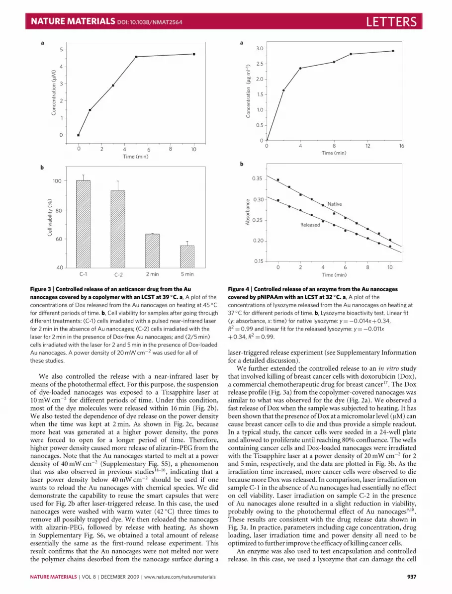
Figure 3 | Controlled release of an anticancer drug from the Aunanocages covered by a copolymer with an LCST at 39 ◦C. a, A plot of theconcentrations of Dox released from the Au nanocages on heating at 45 ◦Cfor different periods of time. b, Cell viability for samples after going throughdifferent treatments: (C-1) cells irradiated with a pulsed near-infrared laserfor 2 min in the absence of Au nanocages; (C-2) cells irradiated with thelaser for 2 min in the presence of Dox-free Au nanocages; and (2/5 min)cells irradiated with the laser for 2 and 5 min in the presence of Dox-loadedAu nanocages. A power density of 20 mW cm−2 was used for all ofthese studies.
We also controlled the release with a near-infrared laser bymeans of the photothermal effect. For this purpose, the suspensionof dye-loaded nanocages was exposed to a Ti:sapphire laser at10mWcm−2 for different periods of time. Under this condition,most of the dye molecules were released within 16min (Fig. 2b).We also tested the dependence of dye release on the power densitywhen the time was kept at 2min. As shown in Fig. 2c, becausemore heat was generated at a higher power density, the poreswere forced to open for a longer period of time. Therefore,higher power density caused more release of alizarin-PEG from thenanocages. Note that the Au nanocages started to melt at a powerdensity of 40mWcm−2 (Supplementary Fig. S5), a phenomenonthat was also observed in previous studies14–16, indicating that alaser power density below 40mWcm−2 should be used if onewants to reload the Au nanocages with chemical species. We diddemonstrate the capability to reuse the smart capsules that wereused for Fig. 2b after laser-triggered release. In this case, the usednanocages were washed with warm water (42 ◦C) three times toremove all possibly trapped dye. We then reloaded the nanocageswith alizarin-PEG, followed by release with heating. As shownin Supplementary Fig. S6, we obtained a total amount of releaseessentially the same as the first-round release experiment. Thisresult confirms that the Au nanocages were not melted nor werethe polymer chains desorbed from the nanocage surface during a
a
b
3.0
2.5
2.0
1.5
1.0
0.5
0
0.35
0.30
0.25
0.20
0.15
0 4 8Time (min)
Abs
orba
nce
Con
cent
ratio
n (
µg m
l¬1 )
Time (min)
12 16
0 2
Released
Native
4 6 8 10
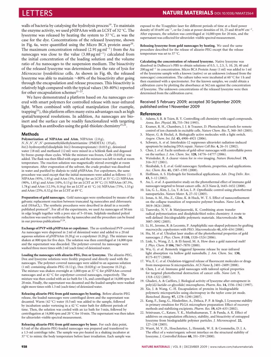
Figure 4 | Controlled release of an enzyme from the Au nanocagescovered by pNIPAAmwith an LCST at 32 ◦C. a, A plot of theconcentrations of lysozyme released from the Au nanocages on heating at37 ◦C for different periods of time. b, Lysozyme bioactivity test. Linear fit(y: absorbance, x: time) for native lysozyme: y=−0.014x+0.34,R2=0.99 and linear fit for the released lysozyme: y=−0.011x
+0.34, R2=0.99.
laser-triggered release experiment (see Supplementary Informationfor a detailed discussion).
We further extended the controlled release to an in vitro studythat involved killing of breast cancer cells with doxorubicin (Dox),a commercial chemotherapeutic drug for breast cancer17. The Doxrelease profile (Fig. 3a) from the copolymer-covered nanocages wassimilar to what was observed for the dye (Fig. 2a). We observed afast release of Dox when the sample was subjected to heating. It hasbeen shown that the presence ofDox at amicromolar level (µM) cancause breast cancer cells to die and thus provide a simple readout.In a typical study, the cancer cells were seeded in a 24-well plateand allowed to proliferate until reaching 80% confluence. The wellscontaining cancer cells and Dox-loaded nanocages were irradiatedwith the Ti:sapphire laser at a power density of 20mWcm−2 for 2and 5min, respectively, and the data are plotted in Fig. 3b. As theirradiation time increased, more cancer cells were observed to diebecause more Dox was released. In comparison, laser irradiation onsample C-1 in the absence of Au nanocages had essentially no effecton cell viability. Laser irradiation on sample C-2 in the presenceof Au nanocages alone resulted in a slight reduction in viability,probably owing to the photothermal effect of Au nanocages9,18.These results are consistent with the drug release data shown inFig. 3a. In practice, parameters including cage concentration, drugloading, laser irradiation time and power density all need to beoptimized to further improve the efficacy of killing cancer cells.
An enzyme was also used to test encapsulation and controlledrelease. In this case, we used a lysozyme that can damage the cell
NATUREMATERIALS | VOL 8 | DECEMBER 2009 | www.nature.com/naturematerials 937
LETTERS NATUREMATERIALS DOI: 10.1038/NMAT2564
walls of bacteria by catalysing the hydrolysis process19. To maintainthe enzyme activity, we used pNIPAAmwith an LCST of 32 ◦C. Thelysozyme was released by heating the system to 37 ◦C, as was thecase for the dye. Concentrations of the released lysozyme, shownin Fig. 4a, were quantified using the Micro BCA protein assay20.The maximum concentration released (2.91 µgml−1) from the Aunanocages was close to the value (3.88 µgml−1) calculated fromthe initial concentration of the loading solution and the volumeratio of Au nanocages to the suspension medium. The bioactivityof the released lysozyme was determined from the rate of lysis forMicrococcus lysodeikticus cells. As shown in Fig. 4b, the releasedlysozyme was able to maintain ∼80% of the bioactivity after goingthrough the encapsulation and release processes. This bioactivity isrelatively high compared with the typical values (30–80%) reportedfor other encapsulation schemes21–23.
We have demonstrated a platform based on Au nanocages cov-ered with smart polymers for controlled release with near-infraredlight. When combined with optical manipulation (for example,trapping24), this platform offers many extra advantages such as highspatial/temporal resolutions. In addition, Au nanocages are bio-inert and the surface can be readily functionalized with targetingligands such as antibodies using the gold-thiolate chemistry25,26.
MethodsPolymerization of NIPAAm and AAm. NIPAAm (2.0 g),N ,N ,N ′,N ′,N ′-pentamethyldiethylenetriamine (PMDETA) (35 µl),bis(2-hydroxyethyl)disulphide bis(2-bromopropionate) (0.015 g), deionizedwater (18ml) and methanol (12ml) were mixed in a Schlenk flask and degassedby freeze–pump–thaw cycles. While the mixture was frozen, CuBr (0.010 g) wasadded. The flask was then filled with argon and the mixture was left to melt at roomtemperature. The reaction solution was magnetically stirred overnight at roomtemperature. After evaporation of the solvent, the crude product was dissolvedin water and purified by dialysis to yield pNIPAAm. For copolymers, the sameprocedure was used except that the initial monomers were added as follows: (1)NIPAAm (95%, 1.93 g) and AAm (5%, 0.64 g) for an LCST at 35 ◦C; (2) NIPAAm(90%, 1.83 g) and AAm (10%, 0.13 g) for an LCST at 39 ◦C; (3) NIPAAm (87.5%,1.78 g) and AAm (12.5%, 0.16 g) for an LCST at 41 ◦C; (4) NIPAAm (75%, 1.53 g)and AAm (25%, 0.32 g) for an LCST at 49 ◦C.
Preparation of gold nanocages. Gold nanocages were synthesized by means of thegalvanic replacement reaction between truncated Ag nanocubes and chloroauricacid (HAuCl4). The synthetic procedures were described in detail in a recentlypublished protocol27. For all of the experiments, we used Au nanocages of 50 nmin edge length together with a pore size of 5–10 nm. Sulphide-mediated polyolreduction was used to synthesize the Ag nanocubes and the procedure can be foundin our previous publications27,28.
Exchange of PVPwith pNIPAAmor copolymer. The as-synthesized PVP-coveredAu nanocages were dispersed in 2ml of deionized water and added to a 20mlaqueous solution of pNIPAAm (0.20 g) or a copolymer (0.20 g). The mixture wasshaken at 800 rpm for five days. The solution was then centrifuged at 14,000 rpmand the supernatant was discarded. The polymer-covered Au nanocages werewashed threemore times with 2ml (each time) of deionized water.
Loading the nanocages with alizarin-PEG, Dox or lysozyme. The alizarin-PEG,Dox and lysozyme solutions were freshly prepared and directly used with thenanocages. The polymer-covered nanocages were added to an aqueous solution(1ml) containing alizarin-PEG (0.12 g), Dox (0.020 g) or lysozyme (0.25 g).The mixture was shaken overnight at 1,000 rpm at 37 ◦C for pNIPAAm-coverednanocages and at 42 ◦C for copolymer-covered nanocages, respectively. Themixture was then cooled with an ice bath for 1 h, and centrifuged at 14,000 rpm for20min. Finally, the supernatant was decanted and the loaded samples were washedeight more times with 1.5ml (each time) of deionized water.
Releasing alizarin-PEG from gold nanocages by heating. Before alizarin-PEGrelease, the loaded nanocages were centrifuged down and the supernatant wasdecanted. Warm (42 ◦C) water (0.5ml) was added to the sample, followedby incubation under vortexing in a 42 ◦C water bath for different periods oftime. Then, the solution was cooled with an ice bath for 5min, followed bycentrifugation at 14,000 rpm and 20 ◦C for 10min. The supernatant was then takenfor ultraviolet–visible spectral measurement.
Releasing alizarin-PEG from gold nanocages by laser. For each data point,0.5ml of the alizarin-PEG-loaded nanocages was prepared and transferred toa 1.5-ml centrifuge tube. The sample was pre-heated in a shaking incubator at37 ◦C to mimic the body temperature before laser irradiation. Each sample was
exposed to the Ti:sapphire laser for different periods of time at a fixed powerdensity of 10mWcm−2, or for 2min at power densities of 10, 25 and 40mWcm−2.After exposure, the solution was centrifuged at 14,000 rpm for 20min, and thesupernatant was collected for ultraviolet–visible spectralmeasurement.
Releasing lysozyme from gold nanocages by heating. We used the sameprocedure described for the release of alizarin-PEG except that the releasetemperature was set to 37 ◦C.
Calculating the concentration of released lysozyme. Native lysozyme wasdissolved in Dulbecco’s PBS to obtain solutions of 0.5, 1, 2.5, 5, 10, 20, 40 and200 µgml−1 in concentration. Micro BCA Protein Assay (1ml) was added to 1mlof the lysozyme sample with a known (native) or an unknown (released from thenanocages) concentration. The culture tubes were incubated at 60 ◦C for 1 h andthen examined with a spectrometer. For the known samples, we could obtain acalibration curve by plotting the absorbance at 562 nm against the concentrationof lysozyme. The unknown concentrations of the released lysozyme were thendetermined from the calibration curve.
Received 5 February 2009; accepted 30 September 2009;published online 1 November 2009
References1. Adams, S. R. & Tsien, R. Y. Controlling cell chemistry with caged compounds.
Annu. Rev. Physiol. 55, 755–784 (1993).2. Kramer, R. H., Chambers, J. J. & Trauner, D. Photochemical tools for remote
control of ion channels in excitable cells. Nature Chem. Bio. 7, 360–365 (2005).3. Mayer, G. & Heckel, A. Biologically active molecules with a light switch.
Angew. Chem. Int. Ed. 45, 4900–4921 (2006).4. Schwarz, A. et al. Interleukin-12 suppresses ultraviolet radiation-induced
apoptosis by inducing DNA repair. Nature Cell Bio. 4, 26–31 (2002).5. Chen, J. et al. Facile synthesis of gold-silver nanocages with controllable pores
on the surface. J. Am. Chem. Soc. 128, 14776–14777 (2006).6. Weissleder, R. A clearer vision for in vivo imaging. Nature Biotechnol. 19,
316–317 (2001).7. Skrabalak, S. E. et al. Gold nanocages: Synthesis, properties, and applications.
Acc. Chem. Res. 41, 1587–1595 (2008).8. Hoffman, A. S. Hydrogels for biomedical applications. Adv. Drug Deliv. Rev.
43, 3–12 (2002).9. Au, L. et al. A quantitative study on the photothermal effect of immuno gold
nanocages targeted to breast cancer cells. ACS Nano 2, 1645–1652 (2008).10. Liu, G. L., Kim, J., Lu, Y. & Lee, L. P. Optofluidic control using photothermal
nanoparticles. Nature Mater. 5, 27–32 (2005).11. Jonas, A. M., Hu, Z., Gline, K. & Huck, W. T. S. Effect of nanoconfinement
on the collapse transition of responsive polymer brushes. Nano Lett. 8,3819–3824 (2008).
12. Tsarevsky, N. V. & Matyjaszewski, K. Combining atom transferradical polymerization and disulphide/thiol redox chemistry: A route towell-defined (bio)degradable polymeric materials. Macromolecules 38,3087–3092 (2005).
13. Li, H., Jerome, R. & Lecomte, P. Amphiphilic sun-shaped polymers by graftingmacrocyclic copolyesters with PEO.Macromolecules 41, 650–654 (2008).
14. Hu, M. et al. Ultrafast laser studies of the photothermal properties of goldnanocages. J. Phys. Chem. B 110, 1520–1524 (2006).
15. Link, S., Wang, Z. L. & El-Sayed, M. A. How does a gold nanorod melt?J. Phys. Chem. B 104, 7867–7870 (2000).
16. Wu, G. et al. Remotely triggered liposome release by near-infraredlight absorption via hollow gold nanoshells. J. Am. Chem. Soc. 130,8175–8177 (2008).
17. Wu, E. C. et al. Oxidation-triggered release of fluorescent molecules or drugsfrom mesoporous Si microparticles. ACS Nano 2, 2401–2409 (2008).
18. Chen, J. et al. Immuno gold nanocages with tailored optical propertiesfor targeted photothermal destruction of cancer cells. Nano Lett. 7,1318–1322 (2007).
19. Ghaderi, A. & Carlfors, J. Biological activity of lysozyme after entrapment inpoly(d,l-lactide-co-glycolide) microspheres. Pharm. Res. 14, 1556–1562 (1997).
20. Xie, J. & Wang, C.-H. Encapsulation of proteins in biodegradablepolymeric microparticles using electrospray in the taylor cone-jet mode.Biotechnol. Bioeng. 97, 1278–1290 (2007).
21. Kang, F., Jiang, G., Hinderliter, A., Deluca, P. P. & Singh, J. Lysozyme stabilityin primary emulsion for PLGA microsphere preparation: Effect of recoverymethods and stabilizing excipients. Pharm. Res. 19, 629–633 (2002).
22. Srinivasan, C., Katare, Y. K., Muthukumaran, T. & Panda, A. K. Effect ofadditives on encapsulation efficiency, stability, and bioactivity of entrappedlysozyme from biodegradable polymer particles. J. Microencapsul. 22,127–138 (2005).
23. Weert, M. V. D., Hoechstetter, J., Hennink, W. E. & Crommelin, D. J. A.The effect of a water/organic solvent interface on the structural stability oflysozyme. J. Controlled Release 68, 351–359 (2000).
938 NATUREMATERIALS | VOL 8 | DECEMBER 2009 | www.nature.com/naturematerials
NATUREMATERIALS DOI: 10.1038/NMAT2564 LETTERS24. Hansen, P. M., Bhatia, V. K., Harrit, N. & Oddershede, L. Expanding
the optical trapping range of gold nanoparticles. Nano Lett. 5,1937–1942 (2005).
25. Love, J. C., Estroff, L. A., Kriebel, J. K., Nuzzo, R. G. & Whitesides, G. M.Self-assembled monolayers of thiolates on metals as a form of nanotechnology.Chem. Rev. 105, 1103–1170 (2005).
26. Chen, J. et al. Gold nanocages: Bioconjugation and their potential use as opticalimaging contrast agents. Nano Lett. 5, 473–477 (2005).
27. Skrabalak, S. E., Au, L., Li, X. & Xia, Y. Facile synthesis of Ag nanocubes andAu nanocages. Nature Protocols 2, 2182–2190 (2007).
28. Siekkinen, A. R., McLellan, J. M., Chen, J. & Xia, Y. Rapid synthesisof small silver nanocubes by mediating polyol reduction with a traceamount of sodium sulfide or sodium hydrosulfide. Chem. Phys. Lett. 432,491–496 (2006).
AcknowledgementsThis work was supported by a 2006 Director’s Pioneer Award from the NIH (DP1OD000798). Part of the work was carried out at the Nano Research Facility (NRF),a member of the National Nanotechnology Infrastructure Network (NNIN),
which is supported by the NSF under award no. ECS-0335765. NRF is part of School ofEngineering and Applied Science atWashingtonUniversity in St Louis.
Author contributionsM.S.Y. and Y.C. synthesized the alizarin-PEG dye and polymers, carried out the loadingand controlled-release experiments and did data analysis. J.C., C.M.C. and A.G.S.carried out the synthesis, surface modification and characterization of Au nanocages.C.M.C. and Q.Z. synthesized the Ag nanocubes. M.R. analysed the mechanism forlaser-triggered release. C.K., K.H.S. and L.V.W. were involved in the planning oflaser-triggered release experiments and helped with the analysis on Au nanocagemelting. J.C. and J.X. conducted the cell viability, protein assay and enzyme activityassays. Y.X. conceived the strategy, supervised the experiments and prepared differentversions of the manuscript.
Additional informationSupplementary information accompanies this paper on www.nature.com/naturematerials.Reprints and permissions information is available online at http://npg.nature.com/reprintsandpermissions. Correspondence and requests for materials should beaddressed to Y.X.
NATUREMATERIALS | VOL 8 | DECEMBER 2009 | www.nature.com/naturematerials 939


![Laurence W. McKeen, PhD - Pentasil Used in Medical Devices.pdf · of branched polymers include star polymers, comb polymers, brush polymers, dendronized polymers [1], ladders, and](https://static.fdocuments.us/doc/165x107/5fd30108783da00f76371237/laurence-w-mckeen-phd-pentasil-used-in-medical-devicespdf-of-branched-polymers.jpg)

