Glycosylation of Human Milk Lactoferrin exhibits dynamic changes
30
Glycosylation of Human Milk Lactoferrin exhibits dynamic changes during early lactation enhancing its role in pathogenic bacteria-host interactions Mariana Barboza 1,2 , Janneth Pinzon 2,3 , Saumya Wickramasinghe 7 , John W. Froehlich 1,2 Isabelle Moeller 1 , Jennifer T. Smilowitz 2,5 , L. Renee Ruhaak 1 , Jincui Huang 1 , Bo Lönnerdal 4 , J. Bruce German 2,5 , Juan F. Medrano 7 , Bart C. Weimer 2,3 and Carlito B. Lebrilla 1,2,6 Department of Chemistry 1 , Functional Glycobiology Program, Food for Health Institute 2 , Department of Population Health and Reproduction, School of Veterinary Medicine 3 , Department of Nutrition 4 , Department of Food Science and Technology 5 , School of Medicine 6 , Department of Animal Science 7 , University of California Davis. One Shields Ave, California 95616 Running title: dynamic changes in glycosylation of human milk lactoferrin To whom correspondence should be addressed: Carlito B. Lebrilla University of California, Davis Department of Chemistry One Shields Avenue Davis, CA, 95616 USA Email: [email protected] Phone: 1-530-752-0504 FAX: 1-530-754-8995 MCP Papers in Press. Published on January 19, 2012 as Manuscript M111.015248 Copyright 2012 by The American Society for Biochemistry and Molecular Biology, Inc. by guest on December 13, 2018 http://www.mcponline.org/ Downloaded from
Transcript of Glycosylation of Human Milk Lactoferrin exhibits dynamic changes

Glycosylation of Human Milk Lactoferrin exhibits dynamic changes during early lactation
enhancing its role in pathogenic bacteria-host interactions
Mariana Barboza1,2, Janneth Pinzon2,3, Saumya Wickramasinghe7, John W. Froehlich1,2 Isabelle
Moeller1, Jennifer T. Smilowitz2,5, L. Renee Ruhaak1, Jincui Huang1, Bo Lönnerdal4, J. Bruce
German2,5, Juan F. Medrano7, Bart C. Weimer2,3 and Carlito B. Lebrilla1,2,6
Department of Chemistry1, Functional Glycobiology Program, Food for Health Institute2,
Department of Population Health and Reproduction, School of Veterinary Medicine3,
Department of Nutrition4, Department of Food Science and Technology5, School of Medicine6 ,
Department of Animal Science7, University of California Davis. One Shields Ave, California
95616
Running title: dynamic changes in glycosylation of human milk lactoferrin
To whom correspondence should be addressed:
Carlito B. Lebrilla
University of California, Davis
Department of Chemistry
One Shields Avenue
Davis, CA, 95616
USA
Email: [email protected]
Phone: 1-530-752-0504
FAX: 1-530-754-8995
MCP Papers in Press. Published on January 19, 2012 as Manuscript M111.015248
Copyright 2012 by The American Society for Biochemistry and Molecular Biology, Inc.
by guest on Decem
ber 13, 2018http://w
ww
.mcponline.org/
Dow
nloaded from

2
ABBREVIATIONS
BSSL bile-salt stimulated lipase
DHB 2,5-dihydroxybenzoic acid
FUT fucosyltransferase
hmLF human milk lactoferrin
IRMPD infrared multiphoton dissociation
MWCO molecular weight cut off
OST oligosaccharide transferase
PNGase F peptide N-glycosidase F
RPKM reads per kilo base per million mapped reads
SEM standard error of the mean
SWIFT stored-waveform inverse Fourier transform
TFA trifluoroacetic acid
by guest on Decem
ber 13, 2018http://w
ww
.mcponline.org/
Dow
nloaded from

3
SUMMARY Human milk lactoferrin (hmLF) is the most abundant glycoprotein present in human milk and
displays a broad range of protective functions in the gut of newborn infants. hmLF is N-
glycosylated, but little is known about the lactation stage-related development of the
glycosylation phenotype. hmLF glycosylation from milk samples from five donors during the
first ten weeks of lactation was assessed and observed to be more diverse than previously
reported. During this period dynamic changes in glycosylation were observed corresponding to a
decrease in glycosylation in the second week followed by an increase in total glycosylation as
well as higher order fucosylation thereafter. Gene expression analysis was performed in milk
somatic cells from a sixth subject. It was found that fucosyltransferase expression increased
during entire period, while expression of genes for the oligosaccharyl transferase complex
decreased in the second week. The effect of hmLF glycosylation was examined for the protein’s
ability to affect bacterial binding to epithelial cells. hmLF significantly inhibited pathogen
adhesion and purified hmLF glycans significantly reduced Salmonella invasion of colonic
epithelial cells to levels associated with non-invasive deletion mutants. This study indicates that
hmLF glycosylation is tightly regulated by gene expression and that glyco-variation is involved
in modulating pathogen association.
by guest on Decem
ber 13, 2018http://w
ww
.mcponline.org/
Dow
nloaded from

4
INTRODUCTION
Human milk constitutes the first source of nutrients for the newborn infant, but it has also
evolved to endow several key physiological advantages to the neonate. Other than to provide the
neonate with energy and amino acid building blocks, proteins possess a wide range of biological
activities that promote the normal development and maturation of specific organs in the
newborn, specifically, the functions of the gut mucosa and the growth of gut microbiota (1).
Human milk proteins also display a protective effect against infectious diseases via antimicrobial
and immuno-modulatory activities that confer passive immunity to the breast-fed infant (1-3).
Many of these proteins are post-translationally modified and the possible roles of such
modifications in mediating demonstrated bioactivities are largely unexplored.
Lactoferrin (LF) is an iron-binding glycoprotein found in milk from most species, but
human milk LF (hmLF) is the most abundant glycoprotein present in colostrum and mature milk
(6-8 mg/ml and 2-4 mg/ml, respectively) (1, 4). The presence of glycans on hmLF is long known
(5), but so far, the only role identified is to protect the molecule from proteolysis (6).
Glycosylation is a common but complex type of post-translational modification of
proteins, directly affecting glycoprotein structure, trafficking, recognition and biological
functions (7-10). Carbohydrate structures attached to proteins play key roles in mediating cell
signaling and cell-cell recognition events (11, 12). Changes in protein glycosylation have been
related to the onset and/or progression of several diseases such as different types of cancer,
immunological disorders as well as congenital disorders (13-19). Additionally, glycosylation and
glycan diversity are directly related to modulating microbial adhesion and invasion during
infection (9). Indeed, the first step in bacterial infection is the recognition of host glycans by
bacterial lectins or vice versa. Thus, glycans attached to human milk glycoproteins may act to
by guest on Decem
ber 13, 2018http://w
ww
.mcponline.org/
Dow
nloaded from

5
block or modulate pathogen association to the epithelial surface, which in turn may also be a key
role in the protection of breast-fed infants against gastrointestinal tract infections.
Despite numerous studies establishing the variation in total protein concentration as well
as composition of human milk, there has been little effort characterizing the variation in
glycosylation of milk glycoproteins over the course of lactation. Only one protein, namely bile-
salt stimulated lipase (BSSL) was shown to have a dynamic glycosylation pattern over the course
of lactation (20). The biological significance of these findings is, however, so far unclear and
unexplored.
Given the central role of lactoferrin in infant development and health and its status as the
most abundant glycoprotein in milk, we examined the changes in glycosylation during the first
months of lactation with the hypothesis that glycan variation is common over the course of
lactation as a mechanism to block pathogen association during breastfeeding. hmLF binds
several pathogenic Gram positive (21, 22) and Gram negative (23, 24) bacteria to exert
antimicrobial activity due to either iron-depletion and/or bacterial membrane disruption; the
latter being triggered by a short sequence of amino acids found in the N-terminal domain of the
protein, known as lactoferricin (25, 26). In addition, hmLF may inhibit infections caused by
viruses, yeast, fungi, parasites and other eukaryotic microbes (27). However, to date, the role of
glycosylation of hmLF in these antimicrobial, antiviral, antifungal and antiparasitic activities has
not been elucidated. We determined the N-glycan profile of human milk lactoferrin by mass
spectrometry analysis in individual samples of hmLF purified from five donors during the first
72 days of lactation. The expression of genes associated with glycosylation in milk somatic cells
was assessed to evaluate the regulation of the dynamic glycosylation. The biological and/or
functional significance of glycans found in hmLF was determined using in vitro studies of host-
by guest on Decem
ber 13, 2018http://w
ww
.mcponline.org/
Dow
nloaded from

6
microbe interactions with colonic epithelial cells and gastrointestinal bacterial pathogens in the
presence of hmLF glycoforms and released N-glycans.
EXPERIMENTAL PROCEDURES
hmLF N-glycan analysis
A purified human milk lactoferrin standard was obtained from Sigma Aldrich (St. Louis,
MO), Heparin-Sepharose 6 fast flow was purchased from GE Healthcare (Pittsburgh, PA) and 10
ml econopack columns were purchased from Bio-Rad (Richmond, CA). Glycerol free peptide N-
glycosidase F (PNGase F) was purchased from New England Biolabs (Ipswich, MA). α-1-3/4
fucosidase (from Xantomonas sp.) was obtained from Calbiochem (San Diego, CA) and β-1-4
galactosidase from Glyco (Novato, CA). Recombinant α-2-3/6 sialidase was a kind gift from Dr.
David Mills (Department of Viticulture and Enology, UC Davis). Solid-phase-extraction
graphitized-carbon and C8 cartridges were purchased from Glygen corporation (Columbia, MD)
and Supelco (Bellefonte, PA), respectively and Microcon centrifugal filter devices (ultracel YM-
10) were from Millipore Corporation (Bedford, MA). Acetonitrile and trifluoroacetic acid (TFA)
were ACS quality or higher.
Human milk samples. Samples were donated by five healthy women from Reno, NV,
who gave birth to term infants ( > 38 weeks). Overall, human milk samples collected on days 1,
5, 10, 15, 30, 44, 58 and 72 postpartum were interrogated in this study. All milk samples were
manually expressed and immediately frozen. Samples were then transferred to a -80ºC freezer
within 3 hours and stored until analysis.
Lactoferrin purification from human milk samples. LF purification from individual milk
samples was performed in parallel following a procedure described by Lonnerdal et al. (37) with
by guest on Decem
ber 13, 2018http://w
ww
.mcponline.org/
Dow
nloaded from

7
slight modifications, as follows. Briefly, whole human milk samples (0.5 ml) were centrifuged at
max speed, for 30 min, at 4°C. The lower aqueous phase was recovered in a new tube and a
CaCl2 solution (pH 4.6) was added to a final concentration of 60 mM. The mixture was
incubated 1 h at room temperature (~25˚C), and further centrifuged at 6750 g for 20 min at room
temperature. Empty columns were packed with 1 ml of heparin-Sepharose resin and equilibrated
with 50 mM Tris HCl pH 8.0 (running buffer). The whey fractions obtained were loaded onto the
columns and the flow-through was collected and reloaded onto the column twice. Columns were
closed and the samples were allowed to interact with the resin for 3 h at room temperature.
Upon washing with 15 ml of running buffer, weakly bound proteins were eluted using 5 column
volumes of 50 mM Tris HCl pH 8, 0.3 M NaCl2 (EBI), and 1 ml fractions were collected. LF
bound to heparin-Sepharose and was eluted with 5 column volumes of 50 mM Tris HCl pH 8, 1
M NaCl (EBII). Fractions were collected, dialyzed against 10 mM ammonium bicarbonate,
concentrated and stored at -20°C. Protein concentration was determined using the Bradford assay
and 5 µl aliquots were assayed by SDS-PAGE.
N-glycan release and purification. Commercially available LF and LF purified from
single samples from all donors during the course of lactation (20 µg) were reduced and alkylated
in 50 mM NH4CO3. PNGase F (1 µl, or 500 NEB units) was added to all samples and glycans
were released by incubation at 37°C for 16 h. Released glycans were purified by solid phase
extraction using porous graphitized carbon cartridges. The cartridges were conditioned with three
volumes of deionized water, followed by three volumes of 80% acetonitrile in 0.1% aqueous
trifluoroacetic acid (TFA) (v/v) and another three volumes of deionized water. The
oligosaccharide samples were loaded onto the cartridge, incubated for 10 min at room
temperature, and washed with three volumes of deionized water. Elution was performed to
fractionate the oligosaccharides mixture into two fractions previous to MS analysis in order to
by guest on Decem
ber 13, 2018http://w
ww
.mcponline.org/
Dow
nloaded from

8
minimize suppression. Glycans were eluted with three volumes of 20% acetonitrile in water
(v/v), followed by 3 volumes of 40% acetonitrile, 0.1% TFA in water (v/v) and dried in vacuo.
Glycans were reconstituted in 10 µL of deionized water.
MALDI FTICR-MS analysis. All mass spectra were acquired on an HiRes MALDI–
FTICR MS instrument with an external MALDI source, a 355-nm pulsed Nd:YAG laser, a
quadrupole ion guide, and a 7.0 Tesla superconducting magnet (IonSpec, Irvine, CA, USA). The
analyte–matrix deposit was prepared with 1 µl of sample being spotted on the MALDI plate,
followed by the addition of 0.1 µl of 0.1mM NaCl as a dopant and 1 µl of 0.4 M DHB. The spots
were allowed to dry under vacuum before analysis. For detection, ion excitation was performed
through an arbitrary waveform with amplitude of 150.0 V (base to peak) at a rate of 2 MHz for a
scan range of m/z 216–4500 and 1024K data points. Five acquisition scans were performed on
each sample in the positive ion mode. The instrument was externally calibrated with a malto-
oligosaccharide mixture, and glycans were identified by accurate mass with a mass tolerance < 5
ppm.
For MS/MS analysis, individual glycan ions were selected within the ICR cell using stored-
waveform inverse Fourier transform (SWIFT) isolation prior to collision-induced-dissociation
(CID) and infrared multiphoton dissociation (IRMPD). For IRMPD experiments, the infrared
radiation was supplied by a 10.6 µm, 20 W CO2 laser (Parallax Laser, Inc., Waltham, MA).
Fragmentation was optimized by varying the IRMPD laser pulse time between 500 and 1500 ms.
Irradiation time was increased until the majority of the precursor ion was dissociated.
Creation of hmLF glycoforms. Sialic acid-, fucose- and galactose-free hmLF glycoforms
were created by treatment of standard hmLF with the corresponding exoglycosidases, followed
by ultrafiltration. For each treatment 2 mg of hmLF was reconstituted in 1 ml of appropriate
exoglycosidase digestion buffer. Digestion with α-2-3/6-sialidase (recombinant, gift from Dr.
by guest on Decem
ber 13, 2018http://w
ww
.mcponline.org/
Dow
nloaded from

9
David Mills, UC Davis) was carried out in 50 mM ammonium phosphate pH 6, treatment with
α−1-3/4-fucosidase (Calbiochem, San Diego, CA) in 50 mM sodium phosphate pH 5.5, and
digestion with β−1−4-galactosidase (Glyco, Novato, CA) was performed in 50 mM Tris-HCl pH
7. The corresponding enzymes (3 µl) were added to each reaction tube and incubated with
agitation for 24 h at 37°C in a dry oven. After digestion, the samples were frozen to inactivate
the enzymes and released monosaccharides were removed from hmLF glycoforms by
ultrafiltration using deionized water and Microcon centrifugal devices with a MWCO of 10 kD.
LF glycoforms were recovered in 1 ml of water and protein concentration was determined using
Bradford. Free hmLF N-glycans were prepared from 2 mg of reduced and alkylated LF with
PNGase F as described above and subsequentially separated from the protein by solid phase
extraction using a C8 cartridge conditioned with six volumes of acetonitrile followed by six
volumes of water. Sample was loaded and glycans were recovered in 9 ml of water, followed by
drying in vacuo.
RNA extraction, sequencing and data analysis for gene expression studies
Fresh milk sample collection. Fresh milk samples were obtained from a healthy female
on days 4, 15, 30 and 40 postpartum who gave birth to a term infant (> 38 weeks). In the early
morning period, the donor manually pumped one breast until emptied into a collection bag, and
immediately delivered on cold-packs to the lab for processing. Samples were divided into two
aliquots of approximately 20 ml for oligosaccharide profile analysis and RNA extraction from
somatic cells. The Institutional Review Board of University of California, Davis, approved the
project. The research was conducted in accordance with the ethical standards outlined in the
Helsinki Declaration, with all participants providing written informed consent.
by guest on Decem
ber 13, 2018http://w
ww
.mcponline.org/
Dow
nloaded from

10
RNA extraction for gene expression studies. Somatic cells were pelleted by adding 50 µl
of 0.5 M EDTA to 20 ml of fresh milk and centrifuged at 1800 rpm at 4˚C for 10 min. The pellet
of cells was washed with 10 ml of PBS at pH 7.2 and 10 µl of 0.5 M EDTA (final conc. 0.5 mM)
and filtered through sterile cheesecloth to remove any debris. The cells were then centrifuged
again at 1800 rpm, 4˚C for 10 min. The supernatant was decanted and RNA was extracted from
the milk somatic cell pellet using Trizol (Invitrogen, Carlsbad, CA) according to the
manufacturer’s instructions. RNA was quantified by an ND-1000 spectrophotometer (Fisher
Thermo, Wilmington, MA) and the quality and integrity was assessed by the spectrophotometer
260/280 ratio, gel electrophoresis and by capillary electrophoresis with an Experion bio-analyzer
(Bio-Rad, Hercules, CA).
RNA sequencing and data analysis. Gene expression analysis was conducted on fresh
milk samples collected on days 4, 15, 30 and 40 postpartum by RNA sequencing (RNA-Seq).
Messenger RNA was isolated and purified using RNA-Seq sample preparation Kit (Illumina, San
Diego, CA and NuGen, San Carlos, CA). Subsequently, mRNA was fragmented to
approximately 200 bp fragments and first and second strand cDNA were synthesized, followed
by end repair and adapter ligation. The fragments were purified and sequenced at the UC Davis
Genome Center DNA Technologies Core Facility using the Illumina Genome Analyzer (GAII).
Short sequence reads of 36-40 bp were assembled, and analyzed in RNA-Seq and expression
analysis application of CLC Genomics workbench 3.7 (CLC Bio, Aarhus, Denmark). Human
Genome, GRCh37.1 (http://www.ncbi.nlm.nih.gov/genome/guide/human/index.html) was
utilized as the reference genome for the assembly. Data were normalized by calculating the
‘reads per kilo base per million mapped reads’ (RPKM) for each gene (38) and annotated with
NCBI human genome assembly (35,489 unique genes).
by guest on Decem
ber 13, 2018http://w
ww
.mcponline.org/
Dow
nloaded from

11
In vitro host-microbe interactions assays in the presence of lactoferrin glycoforms.
In vitro cell culture. Intestinal epithelial cells (Caco-2; ATCC HTB-37) were grown as
per the manufacturer’s instructions in T-25 flasks at 37˚C with 5% CO2. Subsequently, for
compound treatment, cells were seeded to a density of 105 cells/cm2 in a 96-well plate using
DMEM/High Modified (Thermo Scientific, Rockford, IL) with 16.6% fetal bovine serum (FBS)
(HyClone Laboratories, Logan, UT), non-essential amino acids (Thermo Scientific), 10 mM
MOPS (Sigma, St. Louis, MO), 10 mM TES (Sigma), 15 mM HEPES (Sigma) and 2 mM
NaH2PO4 (Sigma). Cells were incubated at 37˚C in 5% CO2 atmosphere for 14-days post
confluence to allow differentiation (39). The epithelial cells were washed once with 150 µl of
PBS just prior to the treatment with hmLF preparations and bacterial addition.
Bacterial adhesion and invasion assay. Prior to the adhesion assay, each bacterial culture
was thawed from -70˚C stock cultures, transferred twice after growth for 16-18 hours at 37˚C
shaking at 250 rpm, and collected for use in the adhesion assay. Bacterial cells were collected
from 15 ml of the respective medium after growth for 16 h, washed twice with an equal volume
of PBS, and re-suspended to an OD600 nm of 0.2 in DMEM/High Modified containing non-
essential amino acids, 10 mM MOPS, 10 mM TES, 15 mM HEPES, and 2 mM NaH2PO4
without FBS. Each bacterial suspension (50 µl), was mixed with hmLF, hmLF glycoforms
(500 µg/ml) or the purified N-glycans (250 µg/ml), mixed by vortexing for 1 min, and added to
the washed, differentiated Caco-2 cells to a final multiplicity of infection of 1,000. The Caco2
cells treated with bacteria and hmLF were incubated at 37˚C in an atmosphere containing 5%
CO2 for 60 min to let the bacteria interact the epithelial cells. Supernatants were then aspirated
and the Caco2 monolayer was washed thrice with 200 µl of Tyrodes buffer (pH 7.2) (40, 41) to
remove non-adhered bacteria from the monolayer. Adhered and invaded bacterial counts were
done as described by Elsinghorst et al. (42), except qPCR was used to determine the bacterial
by guest on Decem
ber 13, 2018http://w
ww
.mcponline.org/
Dow
nloaded from

12
count, and that DNA extraction was performed using 50 µl of a commercial lysis buffer (AEX
Chemunex, France) as described by Desai (43). Quantitative bacterial analysis was done using
qPCR with a CFX 96 Real Time System (BioRad, Hercules, CA). Reactions were performed in a
total volume of 25 µl containing 1 µl of cell lysate, 100 nM of PCR primers (Table 1) and iQ
SYBR Green Supermix (BioRad, Hercules CA) as per the manufacturer’s instructions. The
thermocycling parameters consisted of a denaturation step at 95°C for 5 min, followed by 40
cycles of denaturation, annealing and extension at 95˚C for 15 s, 56˚C for 30 s, 72˚C for 30 s,
respectively, and a final extension at 72°C for 1 min. The amplified product was verified using
melt curve analysis from 50˚C to 95˚C with a transition rate of 0.2˚C/s.
Statistics. Host-microbes interaction experiments were performed using three biological
replicates. Bacterial analysis was done using three technical replicates within each biological
replicate. The number of adhered bacteria was determined by subtracting mean of invaded
bacteria (B) from mean of total host associated bacteria (A). The error (ΔZ) was calculated as
(ΔZ)2 = (ΔA)2 + (ΔB)2 where, ΔA is SEM associated with the A and ΔB is SEM associated
with B. Significant differences between treatments were determined using Student’s T-test and
were considered significant if p < 0.05.
RESULTS
Human milk lactoferrin N-glycome
To address the diversity of N-glycans found in human LF, oligosaccharides were released
from a commercially available LF standard obtained commercially from pooled human milk.
Upon glycan release, the mixture of oligosaccharides was fractionated into two fractions prior to
MS analysis in order to minimize suppression. MALDI-FTICR-MS analysis was performed in
positive and negative ion modes and showed the presence of neutral and acidic complex
by guest on Decem
ber 13, 2018http://w
ww
.mcponline.org/
Dow
nloaded from

13
oligosaccharides with a diverse degree of fucosylation (Figure 1). Positive ion mode analysis of
the 20% acetonitrile/water fraction showed biantennary fucosylated oligosaccharides containing
from one to three fucose residues as the most abundant species with nominal m/z values 1809,
1956 and 2101, respectively. Less abundant signals corresponded to complex fucosylated
oligosaccharides containing three lactosaminic units and 1 to 4 fucose residues (nominal m/z
2174, 2320, 2463 and 2606). Among the less abundant signals, non-fucosylated biantennary
complex (m/z 1663), biantennary complex glycans carrying 4 fucose residues (m/z 2248) and
oligosaccharides containing four lactosaminic units substituted with 2 and 3 fucose residues were
also observed (nominal m/z 2685 and 2831). Analysis of the 40% acetonitrile/water fraction
rendered a less diverse profile. However, signals of anionic, sialylated glycans were observed
corresponding to non-fucosylated monosialylated biantennary glycans and monosialylated,
mono- and di-fucosylated biantennary complex oligoasaccharides with nominal m/z values 1977,
2122 and 2267, respectively. Analysis of the 40% acetonitrile/water fraction in negative ion
mode rendered no signals of neutral species, but a diversity of sialylated oligosaccharides and the
most abundant signals corresponded to the sialylated species observed in positive ion mode
(nominal m/z 1931, 2077, and 2223). In addition, among the less abundant signals, disialylated
non-fucosylated biantennary glycans were observed as well as trilactosaminic (fucosylated tri-
antennary) oligosaccharides carrying 2, 3 and 4 fucose residues, as monosialylated species (m/z
2588, 2734 and 2880).
Tandem MS analysis using collision-induced-dissociation (CID) and infra-red multi-
photon-dissociation (IRMPD) was performed on the most abundant fucosylated oligosaccharides
to confirm compositional assignment. Results of the difucosylated biantennary glycan at m/z
1955.67 and the trifucosylated biantennary glycan at m/z 2101.79 are depicted in supplementary
Figure S1. Attachment of the second and third fucose to the N-acetylglucosamine residues of the
by guest on Decem
ber 13, 2018http://w
ww
.mcponline.org/
Dow
nloaded from

14
outer antenna(e) was confirmed by enzymatic digestion with α-1-3/4 fucosidase, followed by
MALDI-FTICR analysis. These results suggest the presence of LewisX epitopes in hmLF,
however it does not rule out the possibility of other Lewis epitopes.
Dynamic changes in glycosylation of human milk LF during over the course of lactation
To address changes in glycosylation, hmLF was purified from milk samples obtained
from 5 mothers during early to late lactation stages (days 1, 5, 10, 15, 30, 44, 58 and 72).
Glycans were released from the purified hmLF and SDS-PAGE was performed to determine the
efficacy of the enrichment. As an example, supplementary Figure S2 shows the purification
from mother 1 along the course of lactation.
Equal amounts of isolated hmLF from each donor and time point were deglycosylated,
and the resulting glycan mixtures were analyzed by MALDI MS analysis. Supplementary Figure
S3 shows the intensities of the most abundant N-glycan signals in the profiles obtained, and
results are summarized in Table 1. Interestingly, all donors showed very similar glycoprofiles.
The most abundant signals corresponded to biantennary complex type glycans, with up to 3
fucose residues. These results correspond to the profiles obtained from commercial hmLF
standard; however, unlike the latter, monofucosylated biantennary complex oligosaccharides
were the most abundant species in all donors.
Glycans obtained from milk collected on day 1 of lactation showed the highest intensities
in all mothers as compared with later time points. To determine the extent of hmLF glycosylation
in each mother, intensities of all glycan signals at each time point were summarized and
normalized to the total intensity of ions observed on day 1. Figure 2 shows the degree of
glycosylation of hmLF in each donor across the course of lactation relative to day 1. A decrease
in glycosylation was observed in all mothers from day 1 to day 15, during the transition from
by guest on Decem
ber 13, 2018http://w
ww
.mcponline.org/
Dow
nloaded from

15
colostrum to transitional milk. Overall glycosylation then seems to increase at day 30 with the
beginning of production of mature milk. After day 30, however, no consistent trends could be
observed: Mother 2 and mother 3 showed an increase in glycosylation close to or equal to the
original levels; while mothers 4 and 5 varied between 20 and 40% of the initial degree of
glycosylation.
All together these results demonstrate dynamic glycosylation of hmLF from individual
donors across the course of lactation. Although each mother showed individual variations there
were common trends:
i) A decrease in the extent of glycosylation in all mothers during the first 2 weeks of
lactation, and a decrease in glycan diversity at day 15 in 3 out of 5 mothers (M2, M4 and
M5, see Figure 2).
ii) A moderate decrease in the relative abundance of monofucosylated biantennary complex
oligosaccharides (11-18%) were observed at day 30 in 3 out of 4 mothers (M1, M3, and
M4); however, a stronger effect (decrease of 20-51%) was observed later, at days 58 and
72 in 4 out of 5 donors (M2, M3, M4 and M5, see Table 1).
iii) An increase in difucosylated biantennary complex glycans was observed in all mothers.
At day 30, an increase of 78-100% was found for 4 out of 5 mothers (M1, M2, M3 and
M4). This effect was greater as the lactation course progressed showing increases of up
to 100%, 165% and 235% in 4 mothers (M3 and M5, M2, and M4, respectively, see
table 1)
Glycan gene expression analysis of glycosylation in milk
Protein N-glycosylation is an enzymatic process, and the changes observed in
glycosylation of hmLF can be related to two sets of genes: those belonging to the
by guest on Decem
ber 13, 2018http://w
ww
.mcponline.org/
Dow
nloaded from
16
oligosaccharide transferase (OST) complex, which transfers the nascent oligosaccharide chain
from its lipid anchor to the synthesized protein (28), and the fucosyl transferase genes (29). Gene
expression levels were examined in somatic cells in milk from mother 6 (M6) at days 4, 15, 30
and 40 postpartum. The observed decrease in overall glycosylation of lactoferrin is most likely
due to alterations in expression or activity of subunits of the OST complex. The key step to N-
glycosylation of protein is catalyzed by this complex, which is known to involve a small number
of genes including STT3A, STT3B, DDOST (OST48), RPN1, RPN2, DDA1 (OST2), OSTC,
KRTCAP2, and OST4 (30). Gene expression data was obtained for the genes STT3A, STT3B,
DDOST, RPN1 and RPN2 and is summarized in Table 2. The expression of genes representing
the OST changes mainly during the early lactation period: STT3A showed a 2.3-fold decrease
from day 4 to day 15 and then remained constant; DDOST, RPN1 and RPN2 increased between
3- and 9-fold during the same period. While the net effect of individual genes on N-glycosylation
of hmLF is not known, the large changes observed by Day 15 are consistent with the changes in
glycosylation observed for lactoferrin.
The percentage of fucosylated glycans on hmLF increases during early lactation. The
data obtained for the different fucosyltransferases, which are summarized in Table 2, are
consistent with the observations of increasing fucosylation during lactation. The expression of 11
fucosyltransferase genes were monitored, of which the expression of 4 genes increased during
lactation and 7 genes remained constant over the lactation period. The observed increased
expression of the FUT genes in somatic cells from human milk corresponds with the increased
levels of fucosylation that were observed on hmLF.
Host/microbe interaction with hmLF glycans
To examine the influence of hmLF glycosylation on host/microbe interactions, assays
by guest on Decem
ber 13, 2018http://w
ww
.mcponline.org/
Dow
nloaded from
17
with differentiated human colonic intestinal epithelial cells (Caco-2) and five enteropathogenic
bacteria were done with addition of hmLF, free hmLF N-glycans or hmLF glycoforms where
terminal monosaccharides were removed. Listeria monocytogenes, Escherichia coli O157:H7
and three serotypes of Salmonella enterica (serotypes Typhimurium, Enteritidis, and Heidelberg)
were examined, since these bacteria are enteropathogens usually found in the infants gut during
the first weeks of their lives. To determine the role of the hmLF protein backbone as well as the
hmLF glycans, organisms were examined for total association (Figure 3). Surprisingly, the
association of only two organisms was significantly reduced with addition of hmLF – E. coli
O157:H7 (p=0.04) and S. Heidelberg (p=0.01). Conversely, addition of only the released hmLF
N-glycans significantly reduced (p<0.04) association for each organism tested, except E. coli.
Thus, E. coli was influenced by the protein backbone, while binding of all the other organisms
was significantly reduced by the hmLF N-glycans.
Considering that the Salmonella used in this study are serotypes of a single species and
yet were influenced differently by hmLF and its glycans, the influence of specific hmLF
glycosylation on binding was examined by enzymatic removal of terminal residues of fucose,
sialic acid, and galactose from the intact protein. Additionally, association was differentiated into
adhesion and invasion to specifically examine the role of individual residues (Figure 4).
Localization of the bacterium was significantly altered by hmLF and each of the glycovariants.
Presence of hmLF unexpectedly resulted in a significant shift in location of S. typhimurium and
S. enteritidis to the inside of the cell. Enzymatic removal of fucose from intact lactoferrin
resulted in a significant increase in adhesion of S. typhimurium (>300%, p<0.05), while it had no
influence on S. Heidelberg and it reduced invasion of S. enteritidis. Removal of galactose
increased adhesion, but not invasion of S. Heidelberg, but had no impact on the other two
organisms.
by guest on Decem
ber 13, 2018http://w
ww
.mcponline.org/
Dow
nloaded from

18
DISCUSSION
Despite numerous studies that identified the variation in total protein and protein
composition in human milk, little progress has been made in determining the glycosylation and
variations in glycosylation of milk glycoproteins among individuals and throughout the course of
lactation. The glycosylation profile of bile-salt-stimulated-lipase (BSSL) was studied in 1997
(31), and later increased levels of fucosylation on BSSL N-glycans, together with altered levels
of sialylated O-glycans were observed with progression of lactation (20). More recently, we
reported dynamic changes in glycosylation in a limited proteomic study. Using SDS-PAGE with
Coomassie Blue as well as carbohydrate staining (32) several proteins including hmLF were
found to vary in their glycosylation over the course of lactation. However, actual glycan
structures were not addressed.
Previous studies have shown that the overall concentration of hmLF, and the total protein
content as a whole, decreases during lactation (32). Nonetheless, even with the extensive studies
published on hmLF, surprisingly little is known regarding the structure and function of the
glycosylation of human milk lactoferrin. Thus far, the glycosylation pattern of hmLF has only
been studied once in 1982 (5). Using NMR, it was observed that lactoferrin carries biantennary
glycans that are mono- or bisialylated as well as mono- or bifucosylated (5). Such structures
were also observed in both the 20% and 40% fractions of each of the individual mothers in the
present study. In this study, however, significantly more glycans were observed using MS
analysis, which reveals that the glycoprofile of hmLF is more diverse than previously described.
Furthermore, remarkable dynamic changes in glycosylation are observed in both the extent and
types for hmLF. An initial overall decrease in levels of glycosylation is observed from birth to
by guest on Decem
ber 13, 2018http://w
ww
.mcponline.org/
Dow
nloaded from

19
day 15 followed by an increase in the levels of higher order fucosylation. This period,
interestingly, also corresponds to the transition between colostrum and mature milk.
Glycan biosynthesis is an enzymatic process with well-established enzymes. However,
studies describing in vivo relations between expression of glycosyltransferase genes and actual
glycosylation patterns are scarce. The transfer of a ‘glycan precursor’ (HexNAc2Man9Glc3) to
the substrate protein, which is facilitated by the oligosaccharyl transferase complex (OST),
produces the nascent glycoprotein. The gene expression data suggests that this step is regulated
through gene expression resulting in the decrease of lactoferrin glycosylation. Furthermore,
expression of many of the 11 fucosyltransferases increased during the course of lactation
consistent with the increased levels in fucosylation reported earlier for BSSL (20) and here for
hmLF (Table 2). We thus observe some correlation between gene expression and hmLF
glycosylation, indicating potentially direct genetic control of protein glycosylation in human
milk. Dynamic glycosylation and bacterial binding studies support a pathogen deflection role for
hmLF. Lactoferrin appears to be a multifaceted molecule to modulate bacterial binding through
different fucosylated and sialylated glycoforms but also the polypeptide backbone. Glycans
therefore appear to have differential effects on each organism that altered adhesion. Indeed,
many human gastrointestinal pathogens bind glycan structures in the gut mucosa via fimbrae,
flagella, and pili (also used to bind other bacteria) for adherence during gut transit, such as H.
pylori in the stomach and Salmonella, Clostridium and E. coli among others to intestinal cells,
causing peptic ulcers and infectious diarrhea, respectively. More specifically, fucosylated
oligosaccharides found in mammalian glycoproteins are involved in a wide range of mechanisms
of cell adhesion during fertilization, development(33), leucocyte trafficking and inflammatory
responses (34).
by guest on Decem
ber 13, 2018http://w
ww
.mcponline.org/
Dow
nloaded from

20
Interestingly, even within Salmonella hmLF and the glycan had a different effect on host
association depending on serotype. Salmonella is a particular concern with neonates. Disease
associated with Salmonella enterica is initiated by attachment to and invasion of gastrointestinal
tract (GIT) cells that leads to injection of effector molecules via the Type III secretion system
that ultimately leads to inflammation of the lamina propia, invasion into the host cell within a
lysosome and ultimately to lymph nodes (35).
The decrease in glycosylation during the transition from colostrum to mature milk is
possibly related to the protective role of glycosylation for the polypeptide backbone. A well-
established role of glycosylation is to protect the protein from protease digestion. The loss of
glycosylation could make hmLF more susceptible to protease digestion thereby producing
peptides that are antimicrobial such as lactoferrocin (25, 26). It should also be remarked that the
transition from colostrum to mature milk accompanies extensive change in the gut flora
population (36).
CONCLUSION
hmLF can modulate the interactions between specific enteropathogenic bacteria and host
cells. These results support a hypothesis that in vivo hmLF glycans play a key role in protecting
the intestinal mucosa from different pathogens and thus modulate the microbial content of the
gut. Furthermore, the glycosylation, which is genetically controlled, may respond to the changing
bacterial population in the neonatal gut. Therefore, in infants, hmLF provides an important
degree of protection. Glycans may play a key role in this process as they act as selective ligands
for enteropathogens and thus deflect them from the gastrointestinal tract.
ACKNOWLEDGEMENTS
by guest on Decem
ber 13, 2018http://w
ww
.mcponline.org/
Dow
nloaded from

21
Funding provided by the National Institutes of Health HD061923.
REFERENCES 1. Lonnerdal, B., and Lien, E. L. (2003) Nutritional and physiologic significance of alpha-lactalbumin in infants. Nutr. Rev. 61, 295-305. 2. Kaufman, D. A. (2009) Lactoferrin supplementation to prevent nosocomial infections in preterm infants. JAMA 302, 1467-1468. 3. Venkatesh, M. P., and Rong, L. (2008) Human recombinant lactoferrin acts synergistically with antimicrobials commonly used in neonatal practice against coagulase-negative staphylococci and Candida albicans causing neonatal sepsis. J. Med. Microbiol. 57, 1113-1121. 4. Garcia-Montoya, I. A., Cendon, T. S., Arevalo-Gallegos, S., and Rascon-Cruz, Q. (2011) Lactoferrin a multiple bioactive protein: An overview. Biochim. Biophys. Acta. 5. Spik, G., Strecker, G., Fournet, B., Bouquelet, S., Montreuil, J., Dorland, L., van Halbeek, H., and Vliegenthart, J. F. (1982) Primary structure of the glycans from human lactotransferrin. Eur. J. Biochem. 121, 413-419. 6. van Veen, H. A., Geerts, M. E., van Berkel, P. H., and Nuijens, J. H. (2004) The role of N-linked glycosylation in the protection of human and bovine lactoferrin against tryptic proteolysis. Eur. J. Biochem. 271, 678-684. 7. Shental-Bechor, D., and Levy, Y. (2009) Folding of glycoproteins: toward understanding the biophysics of the glycosylation code. Curr.Opin.Struct.Biol. 19, 524-533. 8. Fukuda, M. N., Sasaki, H., Lopez, L., and Fukuda, M. (1989) Survival of recombinant erythropoietin in the circulation: the role of carbohydrates. Blood 73, 84-89. 9. Marth, J. D., and Grewal, P. K. (2008) Mammalian glycosylation in immunity. Nat.Rev.Immunol. 8, 874-887. 10. Ohtsubo, K., and Marth, J. D. (2006) Glycosylation in cellular mechanisms of health and disease. Cell 126, 855-867. 11. Takahashi, M., Kuroki, Y., Ohtsubo, K., and Taniguchi, N. (2009) Core fucose and bisecting GlcNAc, the direct modifiers of the N-glycan core: their functions and target proteins. Carbohydr.Res. 344, 1387-1390. 12. Gu, J., Sato, Y., Kariya, Y., Isaji, T., Taniguchi, N., and Fukuda, T. (2009) A mutual regulation between cell-cell adhesion and N-glycosylation: implication of the bisecting GlcNAc for biological functions. J.Proteome Res. 8, 431-435. 13. Freeze, H. H., and Aebi, M. (2005) Altered glycan structures: the molecular basis of congenital disorders of glycosylation. Curr. Opin. Struct. Biol. 15, 490-498. 14. Higai, K., Azuma, Y., Aoki, Y., and Matsumoto, K. (2003) Altered glycosylation of alpha1-acid glycoprotein in patients with inflammation and diabetes mellitus. Clin. Chim. Acta 329, 117-125. 15. Parekh, R. B., Dwek, R. A., Sutton, B. J., Fernandes, D. L., Leung, A., Stanworth, D., Rademacher, T. W., Mizuochi, T., Taniguchi, T., and Matsuta, K. (1985) Association of rheumatoid arthritis and primary osteoarthritis with changes in the glycosylation pattern of total serum IgG. Nature 316, 452-457. 16. Saldova, R., Royle, L., Radcliffe, C. M., Abd Hamid, U. M., Evans, R., Arnold, J. N., Banks, R. E., Hutson, R., Harvey, D. J., Antrobus, R., Petrescu, S. M., Dwek, R. A., and Rudd, P. M. (2007) Ovarian cancer is associated with changes in glycosylation in both acute-phase proteins and IgG. Glycobiology 17, 1344-1356. 17. Arnold, J. N., Saldova, R., Galligan, M. C., Murphy, T. B., Mimura-Kimura, Y., Telford, J. E., Godwin, A. K., and Rudd, P. M. (2011) Novel glycan biomarkers for the detection of lung cancer. J. Proteome Res. 10, 1755-1764.
by guest on Decem
ber 13, 2018http://w
ww
.mcponline.org/
Dow
nloaded from

22
18. Blomme, B., Van Steenkiste, C., Callewaert, N., and Van Vlierberghe, H. (2009) Alteration of protein glycosylation in liver diseases. J.Hepatol. 50, 592-603. 19. An, H. J., Kronewitter, S. R., de Leoz, M. L., and Lebrilla, C. B. (2009) Glycomics and disease markers. Curr. Opin. Chem. Biol. 20. Landberg, E., Huang, Y., Stromqvist, M., Mechref, Y., Hansson, L., Lundblad, A., Novotny, M. V., and Pahlsson, P. (2000) Changes in glycosylation of human bile-salt-stimulated lipase during lactation. Arch. Biochem. Biophys. 377, 246-254. 21. Qiu, J., Hendrixson, D. R., Baker, E. N., Murphy, T. F., St Geme, J. W., 3rd, and Plaut, A. G. (1998) Human milk lactoferrin inactivates two putative colonization factors expressed by Haemophilus influenzae. Proc. Natl. Acad. Sci. U S A 95, 12641-12646. 22. Hammerschmidt, S., Bethe, G., Remane, P. H., and Chhatwal, G. S. (1999) Identification of pneumococcal surface protein A as a lactoferrin-binding protein of Streptococcus pneumoniae. Infect. Immun. 67, 1683-1687. 23. Ochoa, T. J., Noguera-Obenza, M., Ebel, F., Guzman, C. A., Gomez, H. F., and Cleary, T. G. (2003) Lactoferrin impairs type III secretory system function in enteropathogenic Escherichia coli. Infect. Immun. 71, 5149-5155. 24. Del Olmo, A., Calzada, J., and Nunez, M. (2011) Antimicrobial efficacy of lactoferrin, its amidated and pepsin-digested derivatives, and their combinations, on Escherichia coli O157:H7 and Serratia liquefaciens. Lett. Appl. Microbiol. 52, 9-14. 25. Bellamy, W., Takase, M., Wakabayashi, H., Kawase, K., and Tomita, M. (1992) Antibacterial spectrum of lactoferricin B, a potent bactericidal peptide derived from the N-terminal region of bovine lactoferrin. J. Appl. Bacteriol. 73, 472-479. 26. Bellamy, W., Takase, M., Yamauchi, K., Wakabayashi, H., Kawase, K., and Tomita, M. (1992) Identification of the bactericidal domain of lactoferrin. Biochim. Biophys. Acta 1121, 130-136. 27. Jenssen, H., and Hancock, R. E. (2009) Antimicrobial properties of lactoferrin. Biochimie 91, 19-29. 28. Kornfeld, R., and Kornfeld, S. (1985) Assembly of asparagine-linked oligosaccharides. Annu.Rev.Biochem. 54, 631-664. 29. Javaud, C., Dupuy, F., Maftah, A., Julien, R., and Petit, J. M. (2003) The fucosyltransferase gene family: an amazing summary of the underlying mechanisms of gene evolution. Genetica 118, 157-170. 30. Kelleher, D. J., and Gilmore, R. (2006) An evolving view of the eukaryotic oligosaccharyltransferase. Glycobiology 16, 47R-62R. 31. Landberg, E., Pahlsson, P., Krotkiewski, H., Stromqvist, M., Hansson, L., and Lundblad, A. (1997) Glycosylation of bile-salt-stimulated lipase from human milk: comparison of native and recombinant forms. Arch. Biochem. Biophys. 344, 94-102. 32. Froehlich, J. W., Dodds, E. D., Barboza, M., McJimpsey, E. L., Seipert, R. R., Francis, J., An, H. J., Freeman, S., German, J. B., and Lebrilla, C. B. (2010) Glycoprotein expression in human milk during lactation. J. Agric. Food Chem. 58, 6440-6448. 33. Clarke, J. L., and Watkins, W. (1996) Alpha1,3-L-fucosyltransferase expression in developing human myeloid cells. Antigenic, enzymatic, and mRNA analyses. J. Biol. Chem. 271, 10317-10328. 34. Bergman, M. P., Engering, A., Smits, H. H., van Vliet, S. J., van Bodegraven, A. A., Wirth, H. P., Kapsenberg, M. L., Vandenbroucke-Grauls, C. M., van Kooyk, Y., and Appelmelk, B. J. (2004) Helicobacter pylori modulates the T helper cell 1/T helper cell 2 balance through phase-variable interaction between lipopolysaccharide and DC-SIGN. J. Exp. Med. 200, 979-990. 35. Darwin, K. H., and Miller, V. L. (1999) Molecular basis of the interaction of Salmonella with the intestinal mucosa. Clin. Microbiol. Rev. 12, 405-428. 36. Harmsen, H. J., Wildeboer-Veloo, A. C., Raangs, G. C., Wagendorp, A. A., Klijn, N., Bindels, J. G., and Welling, G. W. (2000) Analysis of intestinal flora development in breast-fed and formula-fed infants by using molecular identification and detection methods. J Pediatr Gastroenterol Nutr 30, 61-67. 37. Carlsson, J., Porath, J., and Lonnerdal, B. (1977) Isolation of lactoferrin from human milk by metal-chelate affinity chromatography. FEBS Lett. 75, 89-92.
by guest on Decem
ber 13, 2018http://w
ww
.mcponline.org/
Dow
nloaded from

23
38. Mortazavi, A., Williams, B. A., McCue, K., Schaeffer, L., and Wold, B. (2008) Mapping and quantifying mammalian transcriptomes by RNA-Seq. Nat. Methods 5, 621-628. 39. Ouwehand, A. C., and Salminen, S. (2003) In vitro Adhesion Assays for Probiotics and their in vivo Relevance: A Review. Microbial Ecology in Health & Disease 15, 175-184. 40. de Ridder, L., Mareel, M., and Vakaet, L. (1975) Adhesion of malignant and nonmalignant cells to cultured embryonic substrates. Cancer Res. 35, 3164-3171. 41. Lominadze, D. G., Saari, J. T., Miller, F. N., Catalfamo, J. L., Justus, D. E., and Schuschke, D. A. (1996) Platelet aggregation and adhesion during dietary copper deficiency in rats. Thromb. Haemost. 75, 630-634. 42. Elsinghorst, E. A. (1994) Measurement of invasion by gentamicin resistance. Methods Enzymol. 236, 405-420. 43. Desai, P. T., Walsh, M. K., and Weimer, B. C. (2008) Solid-phase capture of pathogenic bacteria by using gangliosides and detection with real-time PCR. Appl. Environ. Microbiol. 74, 2254-2258.
by guest on Decem
ber 13, 2018http://w
ww
.mcponline.org/
Dow
nloaded from
24
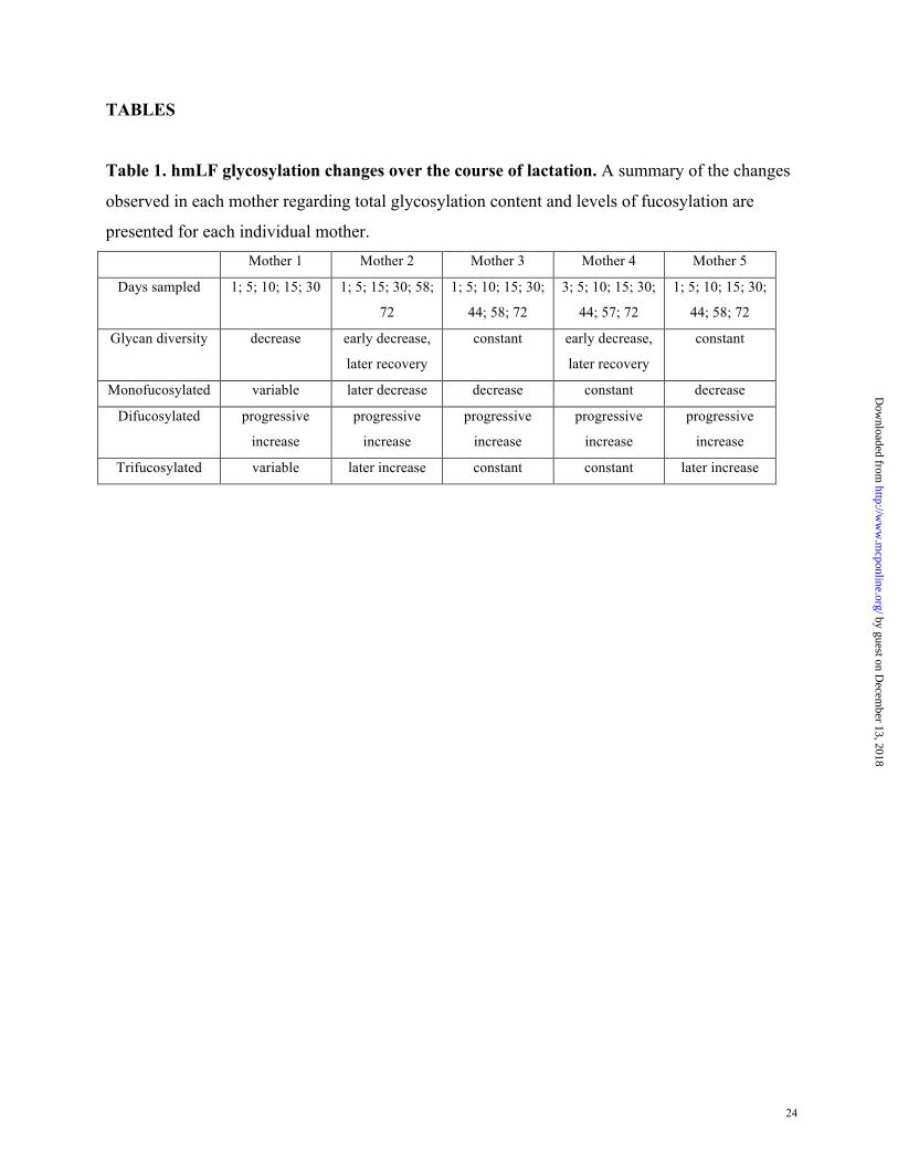
TABLES
Table 1. hmLF glycosylation changes over the course of lactation. A summary of the changes
observed in each mother regarding total glycosylation content and levels of fucosylation are
presented for each individual mother. Mother 1 Mother 2 Mother 3 Mother 4 Mother 5
Days sampled 1; 5; 10; 15; 30 1; 5; 15; 30; 58;
72
1; 5; 10; 15; 30;
44; 58; 72
3; 5; 10; 15; 30;
44; 57; 72
1; 5; 10; 15; 30;
44; 58; 72
Glycan diversity decrease early decrease,
later recovery
constant early decrease,
later recovery
constant
Monofucosylated variable later decrease decrease constant decrease
Difucosylated progressive
increase
progressive
increase
progressive
increase
progressive
increase
progressive
increase
Trifucosylated variable later increase constant constant later increase
by guest on Decem
ber 13, 2018http://w
ww
.mcponline.org/
Dow
nloaded from

25
Table 2. Gene expression of the genes associated with glycosylation at different time points
during lactation. Values are given in ‘reads per kilo base per million mapped reads’ (RPKM).
Gene
Annotation
Day 1
Day
15
Day
30
Day
40
STT3A Oligosaccharyltransferase complex subunit A 101.9 44.84 35.94 36.53
STT3B Oligosaccharyltransferase complex subunit B 26.34 21.76 32.38 28.23
DDOST
dolichyl-diphosphooligosaccharide
glycosyltransferase 17.08 94.68 70.31 88.97
RPN1 Ribophorin I 35.55 105.4 80.91 102.1
RPN2 Ribophorin II 9.33 91.35 72.28 81.54
DDA1 Cross-immunization reaction protein 2.11 6.44 5.27 6.73
OSTC oligosaccharyltransferase complex subunit 28.60 47.55 43.01 44.64
KRTCAP2 keratinocyte associated protein 2 17.00 106.7 94.4 83.35
OST4 oligosaccharyltransferase 4 361.1 175.3 139.9 169.2
FUT1 Fucα2Galβ4GlcNAc-R 0 0 0.06 0.01
FUT2 Fucα2Galβ3GlcNAc-R 3.85 12.48 8.56 6.37
FUT3
Galβ4[Fucα3]GlcNAc-R
Siaα3Galβ4[Fucα3]GlcNAc-R
Fucα2Galβ4[Fucα3]GlcNAc-R
Galβ3[Fucα4]GlcNAc-R
Siaα3Galβ3[Fucα4]GlcNAc-R
Fucα2Galβ3[Fucα4]GlcNAc-R
4.01 8.99 9.79 6.08
FUT4
Galβ4[Fucα3]GlcNAcβ3Galβ4GlcNAc-R
Galβ4]GlcNAcβ3Galβ4[Fucα3GlcNAc-R
Galβ4[Fucα3]GlcNAcβ3Galβ4[Fucα3]GlcNAc-R
SiααGalβ4GlcNAcβ3Galβ4[Fucα3]GlcNAc-R
Siαα3Galβ4[Fucα3]GlcNAc-R
0.38 0.28 0.65 1.74
FUT5 Galβ4[Fucα3]GlcNAc-R
Siaα3Galβ4[Fucα3]GlcNAc-R 0 0 0 0
FUT6 Galβ4[Fucα3]GlcNAc-R 0.11 11.1 9.77 7.29
by guest on Decem
ber 13, 2018http://w
ww
.mcponline.org/
Dow
nloaded from

26
Siaα3Galβ4[Fucα3]GlcNAc-R
FUT7 Siaα3Galβ4[Fucα3]GlcNAc-R 0.02 0.04 0.02 0.02
FUT8 GNGNManβ4GlcNAcβ4[Fucα6]GlcNAc-Asn 0.48 0.11 0.22 0.22
FUT9 Galβ4[Fucα3]GlcNAc-R 0.15 0.08 0.17 0.05
FUT10 Unknown 1.03 0.71 1.34 0.85
FUT11 Unknown 0.39 3.20 3.16 5.06
by guest on Decem
ber 13, 2018http://w
ww
.mcponline.org/
Dow
nloaded from

27
FIGURES
Figure 1. N-Glycoprofile of commercial human milk Lactoferrin. A) Positive ion mode
MALDI-FTICR mass spectrum of N-glycans recovered in 20% AcN:water fraction B) Positive
ion mode MALDI-FTICR mass spectrum of N-glycans recovered in 40% AcN:water
fraction,and C) Negative ion mode MALDI-FTICR mass spectrum of N-glycans recovered in
40% AcN:water.
!"#$%&'$()*$
!"#$+&'$()*$
!,#$+&'$()*$
by guest on Decem
ber 13, 2018http://w
ww
.mcponline.org/
Dow
nloaded from

28
Figure 2. Degree of glycosylation of human milk LF from individual donors across lactation.
All glycan signals were summed and the abudances normalized relative to Day 1, which was
regarded as 100%. Unfortunately, mother 4 did not produce milk on Day 1 and therefore was
normalized to Day 5. A paired t-test was performed to determine statistically significant
differences between Day 1 and other days.
.
by guest on Decem
ber 13, 2018http://w
ww
.mcponline.org/
Dow
nloaded from

29
Figure 3. Changes in bacterial adhesion with hmLF addition and the purified glycan from
hmLF. Bars below the x-axis indicate the p-value between the respective treatments. Control is
adhesion of the respective bacterium without glycan addition. Bacterial isolates used were ST =
Salmonella enterica ssp enterica typhimurium; SE = Salmonella enterica ssp enterica enteritidis;
Salmonella enterica ssp enterica heidelberg; LM = Listeria monocytogenes; EC = Escherichia
coli O157:H7.
by guest on Decem
ber 13, 2018http://w
ww
.mcponline.org/
Dow
nloaded from

30
Figure 4. Bacterial association changes with modification of the hmLF glycosylation.
Bacteria were incubated with CaCO-2 cells in the presence of hmLF (hLF), hmLF treated with
sialidase (Sialic acid free), hmLF treated with fucosidase (Fucose free), hmLF treated with
sialidase and fucosidase (Sia & Fucose free) hmLF treated with galactosidase (Galactose free)
and N-glycans released from hmLF (N-glycans). Baseline infection (infection) was determined
without any additives. Grey bars above the horizontal line represent the number of bacteria
adhered to the epithelial surface during 60 min of incubation, while black bars below the
horizontal line represent the amount of bacteria invaded into the epithelial cell during the same
incubation period.
by guest on Decem
ber 13, 2018http://w
ww
.mcponline.org/
Dow
nloaded from