Glycogen Synthase Kinase-3 Is Activated in Neuronal … · and G 13 by Rho-Independent and...
13
Glycogen Synthase Kinase-3 Is Activated in Neuronal Cells by G 12 and G 13 by Rho-Independent and Rho-Dependent Mechanisms C. Laura Sayas, Jesu ´ s Avila, and Francisco Wandosell Centro de Biologı ´a Molecular “Severo Ochoa”, Consejo Superior de Investigaciones Cientı ´ficas, Universidad Auto ´ noma de Madrid, Cantoblanco, Madrid 28049, Spain Glycogen synthase kinase-3 (GSK-3) was generally considered a constitutively active enzyme, only regulated by inhibition. Here we describe that GSK-3 is activated by lysophosphatidic acid (LPA) during neurite retraction in rat cerebellar granule neurons. GSK-3 activation correlates with an increase in GSK-3 tyrosine phosphorylation. In addition, LPA induces a GSK-3- mediated hyperphosphorylation of the microtubule-associated protein tau. Inhibition of GSK-3 by lithium partially blocks neu- rite retraction, indicating that GSK-3 activation is important but not essential for the neurite retraction progress. GSK-3 activa- tion by LPA in cerebellar granule neurons is neither downstream of G i nor downstream of G q /phospholipase C, suggesting that it is downstream of G 12/13. Overexpression of constitu- tively active G 12 (G 12 QL) and G 13 (G 13 QL) in Neuro2a cells induces upregulation of GSK-3 activity. Furthermore, overex- pression of constitutively active RhoA (RhoAV14) also activates GSK-3 However, the activation of GSK-3 by G 13 is blocked by coexpression with C3 transferase, whereas C3 does not block GSK-3 activation by G 12 . Thus, we demonstrate that GSK-3 is activated by both G 12 and G 13 in neuronal cells. However, GSK-3 activation by G 13 is Rho-mediated, whereas GSK-3 activation by G 12 is Rho-independent. The results presented here imply the existence of a previously unknown mechanism of GSK-3 activation by G 12/13 subunits. Key words: G 12/13 ; GSK-3 activation; lysophosphatidic acid; neurite retraction; tau hyperphosphorylation; RhoA Glycogen synthase kinase-3 (GSK-3) is a key regulator in several physiological processes, such as cell cycle, oncogenesis, apoptosis, and development (Ferkey and Kimelman, 2000). GSK-3 was originally identified as one of the serine–threonine protein ki- nases that phosphorylate and inhibit glycogen synthase (Rylatt et al., 1980), but it has a number of cellular targets (Woodgett et al., 1993). Until recently, it was believed that GSK-3 is a constitu- tively active enzyme, whose activity is decreased in response to cell stimulation. The best known signaling pathways that, when active, inhibit GSK-3, are the insulin and the Wnt pathways (Welsh and Proud, 1993; Cook et al., 1996). However, activation of kinases such as PKA and integrin-linked kinase also induce GSK-3 inhibition (Delcommenne et al., 1998; Fang et al., 2000). However, there are some recent reports that indicate that GSK-3 can be activated, in response to some cellular stimuli. GSK-3 activity is increased in some neuronal cell culture models of apoptosis, neurodegeneration, and in an in vivo model of focal ischemia (Bhat et al., 2000). Additionally, different stimuli pro- duce a transient activation of GSK-3, accompanied by an increase of tau phosphorylation in neuroblastoma cells (Hartigan and Johnson, 1999; Lesort et al., 1999). According to this line of evidence, our own recent results indicate that GSK-3 is activated during lysophosphatidic acid (LPA)-induced neurite retraction, in the SH-SY5Y human neuroblastoma cell line. This is accom- panied by the hyperphosphorylation of tau protein blocked by the tyrosine kinase inhibitor genistein, suggesting that GSK-3 activa- tion could be downstream of G 12/13 , in the Rho pathway (Sayas et al., 1999). GSK-3 is highly expressed in central nervous system (Taka- hashi et al., 1994) and directly phosphorylates several neuronal microtubule-associated proteins (MAPs), involved in microtu- bule (MT) stabilization (Goold et al., 1999; Sanchez et al., 2000; Sperber et al., 1995). A member of the Wnt family, Wnt-7a, has been recently implicated in the regulation of axonal remodeling by inhibiting GSK-3 (Hall et al., 2000). GSK-3 inhibition by Wnt 7-a induces a decrease in the phosphorylated forms of neuronal MAPs, with a concomitant reorganization of MT (Sali- nas, 1999). This suggests that the direct cell shape reorganization induced by Wnt signaling could be mediated by GSK-3 inhibi- tion and its effects on MAPs phosphorylation and MT rearrange- ment. In the present study, we have tested, first, whether GSK-3 is activated by LPA in primary neurons, and second, where GSK-3 is located in the signaling pathways downstream of LPA binding to its endothelial differentiating gene (EDG) receptor (Contos et al., 2000). Our results indicate that LPA induces neurite retrac- tion of cerebellar granule cells, accompanied by hyperphospho- rylation of tau. Both processes are at least partially prevented by lithium, a specific GSK-3 inhibitor. Direct measurement of GSK-3 activity confirms that L PA activates GSK-3 in these neurons. This activation correlates with an increase in GSK-3 tyrosine phos- phorylation. Our data show that GSK-3 activation in these neu- rons is neither downstream of G i nor downstream of G q . To investigate further whether the increase of GSK-3 activity is downstream of G 12/13 , we used the LPA-responsive neuroblas- toma Neuro2A. We show that both constitutively active G 12 and G 13 activate GSK-3. The C3 exoenzyme partially inhibits GSK-3 Received Nov. 28, 2001; revised May 22, 2002; accepted May 10, 2002. This research was supported by grants from Spanish Comisio ´n Interministerial de C iencia y Tecnologı ´a and an institutional grant from Ramon Areces Foundation. We thank Drs. A. Hall and A. C. C arreras for generously providing us with RhoV14-HA and C3-transferase cDNAs, respectively, and Drs. P. Davies, C. Mourton-Gilles, A. C. Carrera, and S. Offermans for kind gifts of antibodies. We also thank Dr. J. Dı ´az-Nido for helpful comments. Correspondence should be addressed to Francisco Wandosell, C entro de Biologı ´a Molecular “Severo Ochoa”, C SIC -Universidad Auto ´noma de Madrid, C antoblanco- Madrid 28049, Spain. E-mail: [email protected]. Copyright © 2002 Society for Neuroscience 0270-6474/02/226863-13$15.00/0 The Journal of Neuroscience, August 15, 2002, 22(16):6863–6875
Transcript of Glycogen Synthase Kinase-3 Is Activated in Neuronal … · and G 13 by Rho-Independent and...
Glycogen Synthase Kinase-3 Is Activated in Neuronal Cells by G�12and G�13 by Rho-Independent and Rho-Dependent Mechanisms
C. Laura Sayas, Jesus Avila, and Francisco Wandosell
Centro de Biologıa Molecular “Severo Ochoa”, Consejo Superior de Investigaciones Cientıficas, Universidad Autonomade Madrid, Cantoblanco, Madrid 28049, Spain
Glycogen synthase kinase-3 (GSK-3) was generally considereda constitutively active enzyme, only regulated by inhibition.Here we describe that GSK-3 is activated by lysophosphatidicacid (LPA) during neurite retraction in rat cerebellar granuleneurons. GSK-3 activation correlates with an increase in GSK-3tyrosine phosphorylation. In addition, LPA induces a GSK-3-mediated hyperphosphorylation of the microtubule-associatedprotein tau. Inhibition of GSK-3 by lithium partially blocks neu-rite retraction, indicating that GSK-3 activation is important butnot essential for the neurite retraction progress. GSK-3 activa-tion by LPA in cerebellar granule neurons is neither downstreamof G�i nor downstream of G�q/phospholipase C, suggestingthat it is downstream of G�12/13. Overexpression of constitu-
tively active G�12 (G�12QL) and G�13 (G�13QL) in Neuro2a cellsinduces upregulation of GSK-3 activity. Furthermore, overex-pression of constitutively active RhoA (RhoAV14) also activatesGSK-3 However, the activation of GSK-3 by G�13 is blocked bycoexpression with C3 transferase, whereas C3 does not blockGSK-3 activation by G�12. Thus, we demonstrate that GSK-3 isactivated by both G�12 and G�13 in neuronal cells. However,GSK-3 activation by G�13 is Rho-mediated, whereas GSK-3activation by G�12 is Rho-independent. The results presentedhere imply the existence of a previously unknown mechanism ofGSK-3 activation by G�12/13 subunits.
Key words: G�12/13 ; GSK-3 activation; lysophosphatidicacid; neurite retraction; tau hyperphosphorylation; RhoA
Glycogen synthase kinase-3 (GSK-3) is a key regulator in severalphysiological processes, such as cell cycle, oncogenesis, apoptosis,and development (Ferkey and Kimelman, 2000). GSK-3 wasoriginally identified as one of the serine–threonine protein ki-nases that phosphorylate and inhibit glycogen synthase (Rylatt etal., 1980), but it has a number of cellular targets (Woodgett et al.,1993). Until recently, it was believed that GSK-3 is a constitu-tively active enzyme, whose activity is decreased in response tocell stimulation. The best known signaling pathways that, whenactive, inhibit GSK-3, are the insulin and the Wnt pathways(Welsh and Proud, 1993; Cook et al., 1996). However, activationof kinases such as PKA and integrin-linked kinase also induceGSK-3 inhibition (Delcommenne et al., 1998; Fang et al., 2000).
However, there are some recent reports that indicate thatGSK-3 can be activated, in response to some cellular stimuli.GSK-3 activity is increased in some neuronal cell culture modelsof apoptosis, neurodegeneration, and in an in vivo model of focalischemia (Bhat et al., 2000). Additionally, different stimuli pro-duce a transient activation of GSK-3, accompanied by an increaseof tau phosphorylation in neuroblastoma cells (Hartigan andJohnson, 1999; Lesort et al., 1999). According to this line ofevidence, our own recent results indicate that GSK-3 is activatedduring lysophosphatidic acid (LPA)-induced neurite retraction,in the SH-SY5Y human neuroblastoma cell line. This is accom-
panied by the hyperphosphorylation of tau protein blocked by thetyrosine kinase inhibitor genistein, suggesting that GSK-3 activa-tion could be downstream of G�12/13, in the Rho pathway (Sayaset al., 1999).
GSK-3� is highly expressed in central nervous system (Taka-hashi et al., 1994) and directly phosphorylates several neuronalmicrotubule-associated proteins (MAPs), involved in microtu-bule (MT) stabilization (Goold et al., 1999; Sanchez et al., 2000;Sperber et al., 1995). A member of the Wnt family, Wnt-7a, hasbeen recently implicated in the regulation of axonal remodelingby inhibiting GSK-3� (Hall et al., 2000). GSK-3� inhibition byWnt 7-a induces a decrease in the phosphorylated forms ofneuronal MAPs, with a concomitant reorganization of MT (Sali-nas, 1999). This suggests that the direct cell shape reorganizationinduced by Wnt signaling could be mediated by GSK-3� inhibi-tion and its effects on MAPs phosphorylation and MT rearrange-ment.
In the present study, we have tested, first, whether GSK-3 isactivated by LPA in primary neurons, and second, where GSK-3is located in the signaling pathways downstream of LPA bindingto its endothelial differentiating gene (EDG) receptor (Contos etal., 2000). Our results indicate that LPA induces neurite retrac-tion of cerebellar granule cells, accompanied by hyperphospho-rylation of tau. Both processes are at least partially prevented bylithium, a specific GSK-3 inhibitor. Direct measurement of GSK-3activity confirms that LPA activates GSK-3 in these neurons. Thisactivation correlates with an increase in GSK-3 tyrosine phos-phorylation. Our data show that GSK-3 activation in these neu-rons is neither downstream of Gi nor downstream of Gq. Toinvestigate further whether the increase of GSK-3 activity isdownstream of G�12/13, we used the LPA-responsive neuroblas-toma Neuro2A. We show that both constitutively active G�12 andG�13 activate GSK-3. The C3 exoenzyme partially inhibits GSK-3
Received Nov. 28, 2001; revised May 22, 2002; accepted May 10, 2002.This research was supported by grants from Spanish Comision Interministerial de
Ciencia y Tecnologıa and an institutional grant from Ramon Areces Foundation. Wethank Drs. A. Hall and A. C. Carreras for generously providing us with RhoV14-HAand C3-transferase cDNAs, respectively, and Drs. P. Davies, C. Mourton-Gilles,A. C. Carrera, and S. Offermans for kind gifts of antibodies. We also thank Dr. J.Dıaz-Nido for helpful comments.
Correspondence should be addressed to Francisco Wandosell, Centro de BiologıaMolecular “Severo Ochoa”, CSIC-Universidad Autonoma de Madrid, Cantoblanco-Madrid 28049, Spain. E-mail: [email protected] © 2002 Society for Neuroscience 0270-6474/02/226863-13$15.00/0
The Journal of Neuroscience, August 15, 2002, 22(16):6863–6875

activation by G�13, indicating that GSK-3 activation by G�13 ismediated by the small GTPase RhoA. These findings demon-strate a novel mechanism of activation of GSK-3.
MATERIALS AND METHODSCell cultures and LPA treatment. Neuro2a mouse neuroblastoma cellswere routinely grown in DMEM with 10% FCS containing glutamine (2mM) and antibiotics (penicillin and streptomycin). To obtain a neuronal-like phenotype, cells were maintained for 24 hr in serum-free Neurobasalmedium supplemented with B-27. To initiate neurite retraction, 10 �Mlysophosphatidic acid [1-oleyl-9-(cis)-2-lyso-sn-glycero-3 phosphate](LPA) (Sigma, St. Louis, MO) was added to differentiated cells. Controlswithout LPA were examined simultaneously.
Cerebellar granule neurons were isolated from postnatal day 7 (P7)rats, as previously described (Levi. et al., 1989), plated onto poly-L-lysine-coated dishes, and grown in serum-free Neurobasal medium, sup-plemented with B-27 and 12.5 mM KCl, for 24 hr. LPA was added tothese neurons at a final concentration of 10 �M. GSK-3 involvement inneurite retraction was tested using lithium, a GSK-3 inhibitor (Klein andMelton, 1996). Because lithium is also an inositol monophosphataseinhibitor, we performed all of our experiments with lithium in thepresence of an excess of myo-inositol, to avoid an effect due to inositoldepletion. Cells were pretreated with lithium (10 mM) and myo-inositol(5 mM) for 4 hr before LPA addition.
Northern blot analysis of EDG-2 receptor. Total RNA was purified fromprimary cultures of P7 cerebellar granule neurons and from NIH 3T3.For Northern Blot analysis, RNA (5 �g/ lane) was electrophoresed informaldehyde–agarose gels and blotted onto nylon membranes by apositive-pressure system (Stratagene, La Jolla, CA). The RNA wascross-linked to the nylon filter by UV light (Strata-linker). Hybridizationwas performed with 32P-labeled EDG-2 cDNA, in a solution containing7.5� SSC, 50% formamide in phosphate buffer (50 mM), pH 7, withtRNA and salmon sperm DNA as carriers. The filters were incubatedwith the DNA probes in this solution at 42°C. To remove 32P-labeledprobe, filters were washed successively with 2� SSC, 0.1% SDS for 30min and 0.1� SSC, 0.5% SDS for 30 min and then exposed.
Antibodies and Western blot analysis. Antibodies used were anti-�-tyrosinated-tubulin monoclonal antibody (mAb) (Sigma); anti-�-tubulinmAb (Sigma); anti-GSK3� mAb (Transduction Laboratories, Lexington,KY); rabbit antiphospho Y 279/216 GSK-3�/� (Biosource International,Camarillo, CA); rabbit anti-G�13 (Santa Cruz Biotechnology, SantaCruz, CA); phalloidin-fluorescein (Sigma); PHF-1, which is an antiphos-pho Ser 396/404 tau mAb (kindly supplied by Dr. P. Davies, AlbertEinstein College, Bronx, NY); AD-2, which is an antiphospho Ser 396 taumAb (kind gift of Dr. C. Mourton-Gilles, Institut de Biotechnologie enInmunoanalyse et Pharmacologie, Centre National de la RechercheScientifique, Montpellier, France); anti-hemagglutinin (HA) mAb [kindgift of Dr. A. C. Carreras, Centro Nacional de Biotecnologıa, ConsejoSuperior de Investigaciones Cientıficas (CSIC)]; and rabbit anti-G�12 (agenerous gift of Dr. S. Offermans, Pharmakologisches Institut, Universityof Heidelberg, Heidelberg, Germany).
Cell extracts were prepared as follows: cells were washed with 1� PBSand then resuspended in a buffer containing 20 mM HEPES, pH 7.4, 100mM NaCl, 100 mM NaF, 1 mM sodium ortho-vanadate, 5 mM EDTA, 1%Triton X-100, and protease inhibitors (Complete; Roche Products, Hert-fordshire, UK). The soluble fraction was obtained by centrifugation at14,000 � g for 10 min at 4°C. Proteins (25–50 �g) were separated inSDS-PAGE gels and electrotransferred to a nitrocellulose filter. Filterswere blocked with 5% nonfat milk in PBS, 0.1% Tween 20 (PBS-T), andthen incubated with primary antibodies overnight at 4°C. Filters wererinsed and then incubated with the corresponding peroxidase-conjugatedsecondary antibody (Promega, Madison, WI), for 1 hr at room temper-ature. Immunoreactivity was visualized by the use of an enhancedchemiluminescence detection system (Amersham Biosciences, ArlingtonHeights, IL).
Densitometric analysis was performed on different Western blot lanes(3–5 replicates), and the data was processed with an imaging densitom-eter (GS-710 model; Bio-Rad, Hercules, CA). Data were analyzed withthe Quantity One software (Bio-Rad). The densitometric values werenormalized with respect to the values obtained for a control antibody tocorrect for any variations in the amounts of total loaded protein.
Quantitative analysis of GSK-3 activity. GSK-3 assays were performedessentially as previously described (Cross, 2001). Cells were collectedwith a cell scraper and homogenized in a buffer of 20 mM HEPES, pH
7.4, containing 100 mM NaCl, 100 mM NaF, 1 mM sodium ortho-vanadate,5 mM EDTA, 100 nM okadaic acid, 1% Triton X-100, and proteaseinhibitors (Complete; Roche). The soluble fraction was obtained aftercentrifugation at 14,000 � g for 15 min at 4°C.
Samples of 7 �g of protein were incubated in a buffer containing 25 mMTris, pH 7.5, 1 mM DTT, 10 mM MgCl2, with the specific substratepeptide 2B-SP (Welsh et al., 1997) at a final concentration of 0.75 mg/ml,in the presence of �[ 32P]ATP. After 1 hr, the reaction was stopped, andthe activity was quantified by spotting aliquots on P81 phosphocellulosepaper. The difference between the kinase activity in the presence orabsence of 20 mM LiCl was considered a measure of GSK-3 proteinkinase activity. The average range of incorporated counts per minute ina standard assay was 10 � 10 3 to 50 � 10 3 cpm. The activity values werenormalized with respect to the expression levels of GSK-3 andG�12QL-HA or G�13QL-HA in each case, to correct for any variationsin the amounts of total proteins.
Indirect immunofluorescence. After treatments, cell cultures were fixedwith PBS containing paraformaldehyde (4%) for 30 min. After severalwashes with PBS, cells were preincubated in PBS–0.1% TritonX-100–1% FCS, for 30 min. After a brief wash with PBS, they wereincubated overnight at 4°C with primary antibodies diluted in PBS–0.1%Triton X–100–1% FCS. After incubation with the primary antibody,cultures were extensively washed and then incubated for 45 min with theappropriate secondary antibody, conjugated either with fluorescein orTexas Red (The Jackson Laboratory, Bar Harbor, ME). After washing,they were immediately mounted with Fluoromount and examined undera Zeiss microscope coupled to a CCD camera, that directly captureddigital micrographs. Photographs were analyzed, and neurite length wasmeasured using the Spot software (Diagnostic Instruments, SterlingHeights, MI).
Plasmids and transfections. Plasmids used were: expression vectors forthe G�12Q229 (QL) and G�13Q226L (QL) (ATCC reference numbers63450 and 63451; supplied from Dr. H. Bourne, University of CaliforniaSan Francisco, San Francisco, CA). Both cDNAs were tagged with anHA epitope. The RhoV14-HA expression vector was kindly provided byDr. A. Hall (Medical Research Council, University College, London,UK); the one for Clostridium botulinum C3 exoenzyme cDNA was a giftfrom Dr. A. C. Carreras (CNB, CSIC, Madrid, Spain), and the expres-sion vector for green fluorescent protein (GFP) pEGFP was obtainedfrom Clontech (Cambridge, UK).
Neuro2A cells were transiently transfected using the method of trans-ferrin receptor endocytosis as described (DuoFect 80, Quantum; Appli-gene, Heidelberg, Germany). This transfection method yielded �50%transfection efficiency under optimal conditions. In the cotransfectionexperiments, total amounts of DNAs were kept constant (10 �g), and 7�g of G�12/13/Rho/C3 or plasmid without cDNA insert were mixed with3 �g of pEGFP. Twenty-four hours after transfection, cells were pro-cessed differently, depending of the kind of experiment to be done. Aproportion of the cells were fixed for immunofluorescence analysis, andthe remainders were lysed for use in Western blot analysis or for in vitroGSK-3 kinase assays.
RESULTSLPA induces neurite retraction in cerebellargranule cellsOur previous results indicated that GSK-3 is activated duringLPA-induced neurite retraction in the SH-SY5Y human neuro-blastoma cell line (Sayas et al., 1999). In this study, we have testedwhether GSK-3 activation by LPA is a more general process, notonly occurring in a particular neuroblastoma cell line, but also inprimary neurons. To determine this, we selected a cerebellargranule cell culture, as a well described and homogenous primaryneuron culture, which can be maintained in a serum-free medium(LPA is a component of serum).
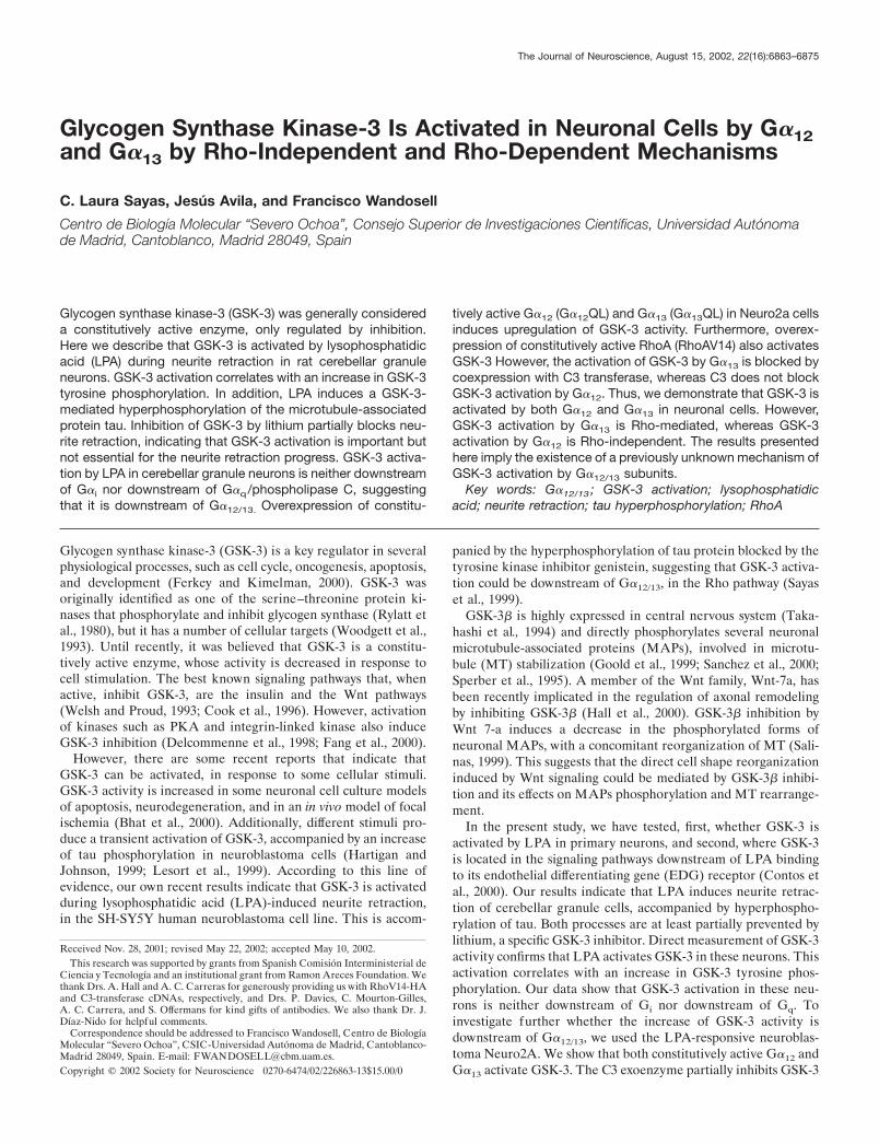
Before examining the response of cerebellar granule neurons toLPA, we analyzed by Northern blot, the expression of EDG-2, aspecific LPA receptor, in these cells. These neurons expressed a3.8 kb mRNA that corresponds to EDG-2 (Fig. 1A).
Subsequently, we examined whether cerebellar granule cellsresponded to LPA. Neurons were incubated in the presence of 10�M LPA, and cell morphology was analyzed at different times
6864 J. Neurosci., August 15, 2002, 22(16):6863–6875 Sayas et al. • G�12 and G�13 Activate GSK-3
after lipid addition (60 and 120 min). Before the LPA treatment,almost 85% of the cells bore neurites. LPA addition induced atime-dependent neurite retraction. After 60 min, 55–65% of cellswere rounded or had very short neurites, whereas 120 min afterLPA treatment, �70% of cells showed no cytoplasmic extensions(Fig. 1B).
We also analyzed the tubulin network during the neurite re-traction process, showing that 60 min after LPA addition, tubulinwas no longer present in the axon-like extension but had accu-mulated in the cell body (Fig. 1B,C), clearly correlating with themorphological retraction (Fig. 1C).
These results show that cerebellar granule cells express EDG-2,a specific LPA receptor, and respond to LPA in a time-dependentmanner, reorganizing their microtubular network.
LPA activates GSK-3 in cerebellar granule neuronsPhosphorylation of GSK-3 in a tyrosine residue (Y216 in GSK-3�,and Y279 in GSK-3�), is necessary for its activity. Recently, anincrease in the level of tyrosine phosphorylation has been corre-lated to GSK-3 activation by different stimuli (Lesort et al., 1999;Hartigan and Johnson, 1999; Bhat et al., 2000). Therefore, weexamined whether LPA addition induced an increase in GSK-3tyrosine phosphorylation in cerebellar granule neurons. For thispurpose, we performed Western blot analysis of lysates of controland LPA-treated cells. The antibody used recognizes both GSK-3� and � isoforms, phosphorylated in tyrosine. LPA addition
induced a time-dependent increase on the level of tyrosine-phosphorylated GSK-3 (� and �), whereas no differences weredetected in the total amount of the GSK-3� protein (Fig. 2A). Inaddition, the GSK3 pool phosphorylated in serine did not changealong the LPA treatment (data not shown). These results suggestthat GSK-3 is activated by LPA in these neurons.
To test this hypothesis directly, GSK-3 activity was mea-sured in extracts of control and LPA-treated cells, by using aspecific peptide as a GSK-3 substrate. Results from four dif-ferent experiments showed that the GSK-3-specific activityincreased to three times that of control levels, 60 min afterLPA addition (Fig. 2 B).
We then investigated whether GSK-3 activation modifies thephosphorylation level of the MAP tau, during neurite retraction.To test whether tau is hyperphosphorylated after LPA treatment,we analyzed the phosphorylation level of tau protein by Westernblot in lysates of control and LPA-treated neurons. Antibodiesthat recognized specifically phosphorylated (PHF-1 and AD-2)and nonphosphorylated tau epitopes (7.51), were used. The LPAtreatment results in a site-specific phosphorylation of tau, asconfirmed by the time-dependent increase of PHF-1 and AD-2immunoreactivity (Fig. 2C). No changes were detected in thetotal amount of tau or �-tubulin proteins with the treatment (Fig.2C). Results from five independent experiments showed a two-fold increase of PHF-1 immunoreactivity 60 min after LPA
Figure 1. LPA induces neurite retraction in cerebellar granule neurons. A, Northern blot analysis of EDG-2. Total RNA from NIH-3T3 (positivecontrol) and cerebellar granule neurons (CGN ) were hybridized with a 32P-labeled cDNA probe of EDG-2. A 3.8 kb mRNA is detected in both cell types.B, Neurons were cultured for 24 hr and then treated with 10 �M LPA for 60 and 120 min. Neurite lengths were measured in control and LPA-exposedcells. Error bars represent the SD of the mean values (�400 cells per data point). C, Immunofluorescence staining of control and LPA-treated neuronswith an anti-�-tyrosinated tubulin antibody. Scale bar, 20 �m.
Sayas et al. • G�12 and G�13 Activate GSK-3 J. Neurosci., August 15, 2002, 22(16):6863–6875 6865

addition, when densitometric data were related to the totalamount of tau protein (Fig. 2D).
The phosphorylation of tau in PHF-1 or AD-2 epitopes hasbeen attributed to three main kinases (GSK-3, MAP kinases,and stress kinases) (Paudel et al.,. 1993). To confirm thatGSK-3 is the kinase responsible for tau phosphorylation byLPA in cerebellar granule cells, we used lithium, an inhibitorof this kinase (Klein and Melton, 1996; Stambolic et al., 1996).Treatment of these neurons with LiCl (10 mM) 4 hr beforeLPA addition almost completely prevented the reaction withPHF-1 and AD-2 antibodies (Fig. 2C,D). As seen in Figure 2,C and D, lithium also totally inhibited the hyperphosphoryla-tion of tau induced by LPA, without changing the total amountof tau protein (Fig. 2C).
To determine the importance of GSK-3 activation for theprogress of the LPA-induced neurite retraction, we investigatedwhether its inhibition with lithium could prevent the process. Wecompared the morphology of cerebellar granule cells treated onlywith lithium and cells pretreated with lithium and subsequently
treated with LPA for 1 hr. Lithium induced neurite shorteningand thickening, accompanied by microtubule unbundling, as pre-viously described (Lucas et al., 1998) (Fig. 3A,B). However,inhibition of GSK-3 by lithium partially prevented the neuriteretraction process initiated by LPA (Fig. 3A,B).
Next we examined which of the general signal transductionpathways triggered by LPA, GSK-3 activation was located. LPAreceptors can differentially couple to three distinct G-proteins:Gi, which produces Ras-GTP accumulation and MAPK activa-tion; G�q, which causes a rise in intracellular phosphoinositidesand calcium levels, mediated by phospholipase C (PLC) signaling;and G�12/13, which induces morphological changes through theactivation of the small GTPase RhoA. We thus analyzed theeffect of pharmacological inhibitors of the different pathwaysmentioned above on LPA-induced GSK-3 activation. Neitherpretreatment of neurons with pertussis toxin (PTX), which ADP-ribosylates and inhibits G�i, nor with the PLC inhibitor U-73122,blocked GSK-3 tyrosine phosphorylation and LPA-induced acti-vation (data not shown). These results indicate that LPA-induced
Figure 2. LPA activates GSK-3 in cerebellar granule neurons. A, Western blot of soluble cell extracts obtained 30 and 60 min after LPA treatment. 0represents cell extracts from control cells. Identical samples were incubated with antiphospho Y 279/216 GSK-3 �/� (top blot) and with anti-GSK-3� (bottomblot) antibodies. B, Quantitative analysis of the GSK-3 kinase activity in control cells (0) and cells exposed to LPA (30 and 60 min). Each time pointwas normalized with respect to total GSK-3� protein present in each cell extract. Data are expressed as the mean of four different experiments. Datafrom control cells were considered 100 relative units (r.u.). Error bars represents SDs of the mean values. C, Western blots of phospho tau (PHF-1 andAD-2 antibodies), total tau (7–51 antibody), and �-tubulin. LPA induces an increase in tau phosphorylation (PHF-1 and AD-2), with no changes in theprotein amount (7.51 and �-tubulin). Pretreatment of the cells with 10 mM LiCl, 4 hr before LPA addition, blocks LPA-induced tau hyperphosphory-lation. D, The phosphorylation degree of tau protein in the PHF-1 epitope was determined for each culture by densitometry, normalized with respectto the values corresponding to total tau (7.51), and graphically represented. Diagram shows the mean normalized densitometry values and thecorresponding SDs (n � 5).
6866 J. Neurosci., August 15, 2002, 22(16):6863–6875 Sayas et al. • G�12 and G�13 Activate GSK-3

GSK-3 activation is neither downstream of Gi nor downstream ofGq. Furthermore, our results indicate that pretreatment of cere-bellar neurons with the tyrosine kinase inhibitor genistein blockstyrosine phosphorylation of GSK-3 � and � and significantlyreduces kinase activation induced by LPA (data not shown).According to this, it has been postulated that one or moretyrosine kinases, which can be inhibited by genistein, exist in theG�12/13 pathway, upstream or downstream of RhoA (Kranenburget al., 1999). These data suggest that GSK-3 activation could bedownstream of G�12/13 in cerebellar granule cells, in agreementwith our previous results that suggested that GSK-3 activation byLPA was downstream of G�12/13 in the SH-SY5Y human neuro-blastoma cell line (Sayas et al., 1999).
le;&.2qThese results demonstrate that GSK-3 is activated dur-ing LPA-induced neurite retraction in cerebellar granule cells.GSK-3 activation correlates with an increase in its tyrosine phos-phorylation level and induces hyperphosphorylation of tau pro-tein in certain epitopes (PHF-1 and AD-2). Activation of GSK-3is important but not essential for the course of the neuriteretraction process in these neurons. LPA-induced GSK-3 activa-tion in neurons is neither downstream of Gi nor downstream ofGq, suggesting that it could be downstream of G�12/13.
LPA induces cell rounding and GSK-3 activation inNeuro2-A cellsTo confirm whether GSK-3 activation by LPA was downstream ofG�12/13, we selected a murine neuroblastoma cell line, Neuro2acells, which can be transfected highly efficiently.
Initially, we tested whether differentiated Neuro2a cells re-sponded to LPA. Under the differentiation conditions used, 10%of the cells were rounded, whereas 55% presented a flat morphol-ogy, and 35% bore short neurites (data not shown). Five minutesafter LPA addition, 98% of cells were rounded with a cortical ringof actin cytoskeleton and exhibited obvious membrane blebbing(Fig. 4A). Thus, Neuro2A cells undergo rapid cell rounding inresponse to LPA treatment.
We then tested whether Neuro2a cells expressed G�12 and/orG�13, the � subunits of the heterotrimeric G-proteins, which arehypothesized as being upstream of GSK-3 activation, in the LPAactivated signaling pathways. As confirmed by Western blot anal-ysis, Neuro2a cells expressed both G�12 and G�13. Cerebellargranule neurons also expressed both � subunits (Fig. 4B).
Because in primary neurons, GSK-3 activation by LPA corre-lated with an increase in tyrosine phosphorylation of GSK-3, weinvestigated whether this also occurred in Neuro2a treated withthis lipid. For that purpose, we performed Western blot analysis,
Figure 3. GSK-3 inhibition partially prevents LPA-induced neurite retraction. A, Immunostaining of cerebellar granule neurons with an antibodyagainst �-tyrosinated tubulin antibody. Neurons were treated with 10 mM lithium for 4 hr (lef t picture) or pretreated with lithium and then treated with10 �M LPA for 60 min (right picture). Scale bar, 20 �m. B, Bar graphs represent the measurement of neurite length of control neurons and neurons treatedwith lithium, exposed or not to 10 �M LPA for 60 min.
Sayas et al. • G�12 and G�13 Activate GSK-3 J. Neurosci., August 15, 2002, 22(16):6863–6875 6867
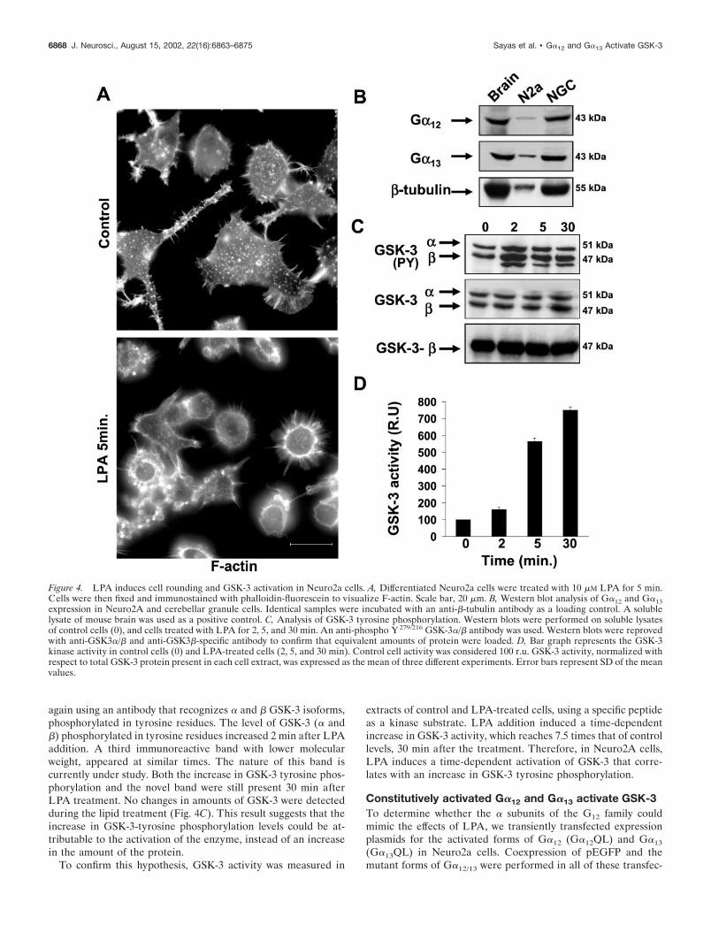
again using an antibody that recognizes � and � GSK-3 isoforms,phosphorylated in tyrosine residues. The level of GSK-3 (� and�) phosphorylated in tyrosine residues increased 2 min after LPAaddition. A third immunoreactive band with lower molecularweight, appeared at similar times. The nature of this band iscurrently under study. Both the increase in GSK-3 tyrosine phos-phorylation and the novel band were still present 30 min afterLPA treatment. No changes in amounts of GSK-3 were detectedduring the lipid treatment (Fig. 4C). This result suggests that theincrease in GSK-3-tyrosine phosphorylation levels could be at-tributable to the activation of the enzyme, instead of an increasein the amount of the protein.
To confirm this hypothesis, GSK-3 activity was measured in
extracts of control and LPA-treated cells, using a specific peptideas a kinase substrate. LPA addition induced a time-dependentincrease in GSK-3 activity, which reaches 7.5 times that of controllevels, 30 min after the treatment. Therefore, in Neuro2A cells,LPA induces a time-dependent activation of GSK-3 that corre-lates with an increase in GSK-3 tyrosine phosphorylation.
Constitutively activated G�12 and G�13 activate GSK-3To determine whether the � subunits of the G12 family couldmimic the effects of LPA, we transiently transfected expressionplasmids for the activated forms of G�12 (G�12QL) and G�13
(G�13QL) in Neuro2a cells. Coexpression of pEGFP and themutant forms of G�12/13 were performed in all of these transfec-
Figure 4. LPA induces cell rounding and GSK-3 activation in Neuro2a cells. A, Differentiated Neuro2a cells were treated with 10 �M LPA for 5 min.Cells were then fixed and immunostained with phalloidin-fluorescein to visualize F-actin. Scale bar, 20 �m. B, Western blot analysis of G�12 and G�13expression in Neuro2A and cerebellar granule cells. Identical samples were incubated with an anti-�-tubulin antibody as a loading control. A solublelysate of mouse brain was used as a positive control. C, Analysis of GSK-3 tyrosine phosphorylation. Western blots were performed on soluble lysatesof control cells (0), and cells treated with LPA for 2, 5, and 30 min. An anti-phospho Y 279/216 GSK-3�/� antibody was used. Western blots were reprovedwith anti-GSK3�/� and anti-GSK3�-specific antibody to confirm that equivalent amounts of protein were loaded. D, Bar graph represents the GSK-3kinase activity in control cells (0) and LPA-treated cells (2, 5, and 30 min). Control cell activity was considered 100 r.u. GSK-3 activity, normalized withrespect to total GSK-3 protein present in each cell extract, was expressed as the mean of three different experiments. Error bars represent SD of the meanvalues.
6868 J. Neurosci., August 15, 2002, 22(16):6863–6875 Sayas et al. • G�12 and G�13 Activate GSK-3

tion experiments. Approximately 90% of the cells transfectedwith the control plasmid (pEGFP) had flat or process-bearingshape like that seen in untransfected cells (Fig. 5A). Overexpres-sion of constitutively activated G�12 or G�13 induced a loss ofneurites and produced a rounded morphology in 60 (G�12--QL)to 95% (G�13-QL) of the transfected cells (Fig. 5A,C). Theexpression of these proteins was confirmed using and anti-HAantibody, because the expression plasmids used were tagged withan HA epitope (Fig. 5A,B). Both activated G�12 and G�13
showed a punctuate pattern of expression in the transfected cells,G�13 being localized preferentially on the plasma membrane,whereas G�12 had a more diffuse localization, probably circum-scribed to membranes of cytoplasmic organelles (Fig. 5A). Al-though the same amount of cDNA from both plasmids was usedin every transfection experiment, the expression level of G�13
protein was always 2.5–3-times that obtained for G�12 (Fig. 5B),probably because of differences in the purity of the two plasmids.
We next examined whether the overexpression of constitutively
Figure 5. Constitutively activated G�12 and G�13 activate GSK-3 in Neuro2a cells. A, Overexpression of constitutively active G�12 and G�13 in Neuro2acells induce cell rounding. G�12QL-HA and G�13QL-HA were coexpressed along with EGFP. G�12QL-HA and G�13QL-HA were detected using ananti-HA antibody. Control cells expressed only EGFP. Scale bar, 10 �m. B, Western blots of transfected cells using anti-HA (top blot) and anti-GSK-3�(bottom blot) antibodies. C, Percentage of rounded cells overexpressing EGFP (control) or EGFP along with G�12QL-HA or G�13QL-HA. Diagramshows the mean values obtained from three different experiments (�300 cells per data point and experiment). Error bars represent the SD of the meanvalues. D, Quantification of GSK-3 kinase activity in control cells (EGFP) and cells overexpressing G�12QL-HA (lef t diagram) or G�13QL-HA (rightdiagram) (along with EGFP). Data are expressed in r.u., and we considered 100 r.u. as the specific GSK-3 activity of control cells. GSK-3 activity wasnormalized with respect to total GSK-3� protein present in each cell extract. n � 3 experiments, and error bars represent SDs of the mean values. *p �0.001 is statistically significant.
Sayas et al. • G�12 and G�13 Activate GSK-3 J. Neurosci., August 15, 2002, 22(16):6863–6875 6869
active G�12 and G�13 could activate GSK-3. GSK-3 activity wasmeasured in extracts of control cells (transfected only withpEGFP) and of G�12- or G�13-overexpressing cells, using aspecific peptide as a GSK-3 substrate. Both active G�12 and G�13
increased GSK-3 activity to a similar level. However, when GSK-3activity was normalized with respect to the expression of eachprotein, G�13QL induced a twofold (Fig. 5D, right diagram)increase of GSK-3 activity, whereas G�12QL produced a sixfoldincrease of GSK-3 activity over that of controls (Fig. 5D, lef tdiagram). This GSK-3 activity increase was not attributable tochanges in GSK-3 protein levels (Fig. 5B).
Taken together these results indicate that constitutively active� subunits of the G12/13 family not only induce cell rounding ofNeuro2a cells, but also activate GSK-3.
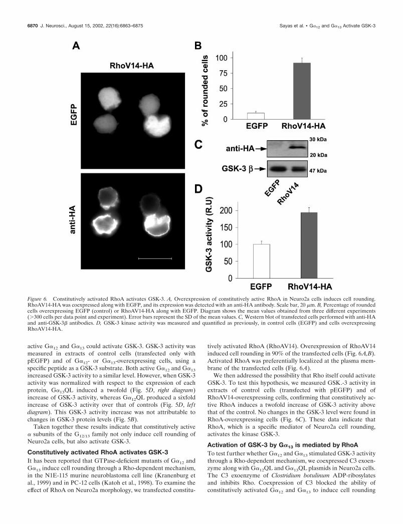
Constitutively activated RhoA activates GSK-3It has been reported that GTPase-deficient mutants of G�12 andG�13 induce cell rounding through a Rho-dependent mechanism,in the N1E-115 murine neuroblastoma cell line (Kranenburg etal., 1999) and in PC-12 cells (Katoh et al., 1998). To examine theeffect of RhoA on Neuro2a morphology, we transfected constitu-
tively activated RhoA (RhoAV14). Overexpression of RhoAV14induced cell rounding in 90% of the transfected cells (Fig. 6A,B).Activated RhoA was preferentially localized at the plasma mem-brane of the transfected cells (Fig. 6A).
We then addressed the possibility that Rho itself could activateGSK-3. To test this hypothesis, we measured GSK.-3 activity inextracts of control cells (transfected with pEGFP) and ofRhoAV14-overexpressing cells, confirming that constitutively ac-tive RhoA induces a twofold increase of GSK-3 activity abovethat of the control. No changes in the GSK-3 level were found inRhoA-overexpressing cells (Fig. 6C). These data indicate thatRhoA, which is a specific mediator of Neuro2a cell rounding,activates the kinase GSK-3.
Activation of GSK-3 by G�13 is mediated by RhoATo test further whether G�12 and G�13 stimulated GSK-3 activitythrough a Rho-dependent mechanism, we coexpressed C3 exoen-zyme along with G�12QL and G�13QL plasmids in Neuro2a cells.The C3 exoenzyme of Clostridium botulinum ADP-ribosylatesand inhibits Rho. Coexpression of C3 blocked the ability ofconstitutively activated G�12 and G�13 to induce cell rounding
Figure 6. Constitutively activated RhoA activates GSK-3. A, Overexpression of constitutively active RhoA in Neuro2a cells induces cell rounding.RhoAV14-HA was coexpressed along with EGFP, and its expression was detected with an anti-HA antibody. Scale bar, 20 �m. B, Percentage of roundedcells overexpressing EGFP (control) or RhoAV14-HA along with EGFP. Diagram shows the mean values obtained from three different experiments(�300 cells per data point and experiment). Error bars represent the SD of the mean values. C, Western blot of transfected cells performed with anti-HAand anti-GSK-3� antibodies. D, GSK-3 kinase activity was measured and quantified as previously, in control cells (EGFP) and cells overexpressingRhoAV14-HA.
6870 J. Neurosci., August 15, 2002, 22(16):6863–6875 Sayas et al. • G�12 and G�13 Activate GSK-3

(Fig. 7A). Only a small percentage of G�12 or G�13 overexpress-ing cells bore neurites (17 and 0.1%, respectively), whereas thenumber of neurite-bearing cells dramatically increased whenG�12 or G�13 were coexpressed with C3 (95 and 89%, respec-tively) (Fig. 7B). Coexpression of C3 significantly reduced theexpression level of G�12 without affecting G�13 level (Fig. 7C,center). Measurements of GSK-3 activity in extracts of controlcells and cells cotransfected with G�12 or G�13 and C3 indicatedthat C3 significantly reduced GSK-3 activation induced by G�13
(Fig. 7C, right diagram) but not that induced by G�12 (Fig. 7C , lef tdiagram). These data indicate that, although both G�12 and G�13
induce Rho-mediated cell rounding, these G� subunits activateGSK-3 through different mechanisms. GSK-3 activation by G�13
is mediated at least in part by RhoA, whereas the kinase activa-tion by G�12 does not involve RhoA function.
DISCUSSION
GSK-3 is activated by LPA during neurite retraction inprimary neuronal cellsInitially, we investigated whether cerebellar granule cells from7-d-old rat pups express an LPA receptor and respond to LPA.LPA is an intercellular lipid mediator that induces diverse bio-logical responses in many types of cells and tissues (Moolenaar,1999). LPA signals through its binding to specific G-protein-coupled receptors. Our data indicate that these neurons expressat least EDG-2, which is the most widely expressed LPA receptorin brain during development (Hecht et al., 1996). We do not ruleout the possibility that these neurons express other LPA receptors(EDG-4 and/or EDG-7) that are also expressed in mouse brain atpostnatal day 7 (Contos and Chun, 2001).
Figure 7. Activation of GSK-3 by G�13 is mediated by RhoA. A, Coexpression of C3 along with constitutively active G�12 or G�13 blocks cell roundinginduced by G�12 and G�13 in Neuro2a cells. Cells coexpress EGFP along with C3 and G�12QL-HA or G�13QL-HA. These cells were immunostainedusing an anti-HA antibody. Control cells coexpress EGFP and C3. Scale bar, 20 �m. B, Diagram showing the percentage of neurite-bearing cellsoverexpressing EGFP, G�12QL-HA, or G�13QL-HA, along with C3 transferase or not. Mean values obtained from three different experiments arerepresented (�300 cells per data point and experiment). Error bars represent the SD of the mean values. C, Western blot of transfected cells performedwith anti-HA and anti-GSK3� antibodies (center). Quantification of GSK-3 kinase activity in lysates of transfected cells, as previously described, showsthat GSK-3 activation induced by G�13QL-HA is inhibited by C3 (right diagram), whereas GSK-3 activation by G�12QL-HA is not (lef t diagram). *p �0.001 is statistically significant.
Sayas et al. • G�12 and G�13 Activate GSK-3 J. Neurosci., August 15, 2002, 22(16):6863–6875 6871

LPA induces morphological changes in neuronal cells: neuriteretraction and cell rounding in neuroblastoma and PC-12 cells(Jalink et al., 1993; Tigyi et al., 1996), growth cone collapse inprimary chick neurons (Saito, 1997), and cell rounding, mem-brane retraction, and cellular and nuclear migration in corticalneuroblasts (Fukushima et al., 2000). We show here that cerebel-lar granule neurons undergo a time-dependent neurite retractionin response to LPA.
Our previous results indicate that GSK-3 is activated duringLPA-induced neurite retraction in the SH-SY5Y human neuro-blastoma cell line (Sayas et al.,. 1999). In this study, we demon-strate that LPA induces a time-dependent activation of GSK-3 incerebellar granule cells that correlates to the neurite retractionprocess. We also show here that GSK-3 activity is increasedduring LPA-induced cell rounding in the mouse neuroblastomaNeuro2a. Therefore, the present study confirms that GSK-3 acti-vation by LPA is not a peculiarity of the cell line used as it occursin neuronal cells from different species, suggesting that it could bea widespread physiological process in neuronal cells
Until recently, it was believed that GSK-3 could be regulatedonly by inhibition, but some recent reports have indicated thatGSK-3 is activated in neuronal cells in response to diverse extra-cellular signals such as apoptotic stimuli, ischemia, a transient risein intracellular calcium, and insulin (Hartigan and Johnson, 1999;Lesort et al., 1999; Bhat et al., 2000). This activation is accom-panied by increased GSK-3 tyrosine phosphorylation. Accord-ingly, our results indicate that GSK-3 activation by LPA in cer-ebellar granule neurons and in Neuro2a cells correlates with anincrease in tyrosine phosphorylation of both GSK-3 isoforms, �and �. In addition, pretreatment of cerebellar neurons with thetyrosine kinase inhibitor genistein blocks tyrosine phosphoryla-tion of GSK-3�/� and significantly reduces kinase activation (datanot shown).
A new tyrosine kinase has recently been cloned in Dictyoste-lium discoideum, ZAK-1, that directly phosphorylates and acti-vates GSK-3 (Kim et al., 1999). In addition, it has been suggestedthat the tyrosine kinases Fyn and Pyk2 phosphorylate GSK-3(Lesort et al., 1999; Hartigan et al., 2001). Therefore, the tyrosinekinase, genistein-sensitive, responsible for LPA-induced GSK-3activation could be either a putative mammalian ZAK-1 homol-ogous or a member of Fyn or Pyk families. However, the identi-fication of this tyrosine kinase requires further investigation.Taken together, these results indicate that: (1) GSK-3 is activatedby LPA in neuronal cells, and (2) an unidentified tyrosine kinase,which can be inhibited by genistein, may be involved in GSK-3activation by LPA in cerebellar granule neurons.
GSK-3 activation contributes to the reorganization ofthe microtubular network during LPA-induced neuriteretractionWe tested how GSK-3 activation by LPA could be contributing toneurite retraction. Among GSK-3 targets are the main neuronalMAPs (Sperber et al., 1995; Lucas et al., 1998; Sanchez et al.,2000). Tau, a prominently axonal MAP, promotes microtubuleassembly and stabilizes the structure of MTs (Mandelkow et al.,1995). Abnormally hyperphosphorylated tau loses its binding toMTs, causing their disruption (Sontag et al., 1996).
Our previous results indicated that tau is hyperphosphorylatedby GSK-3 during LPA-induced neurite retraction in SH-SY5Ycells (Sayas et al., 1999). In this study, we show that tau ishyperphosphorylated in two analyzed epitopes (PHF-1 andAD-2) during LPA-induced neurite retraction in cerebellar gran-
ule neurons. This increase in tau phosphorylation is blocked bythe GSK-3 inhibitor lithium, confirming that GSK-3 is the serine–threonine kinase responsible for this hyperphosphorylation. Fur-thermore, pretreatment of neurons with lithium partially blocksneurite retraction induced by LPA. Considering that the lithiumtreatments contain an excess of myo-inositol (5 mM) to avoid thehypothetical inositol depletion, because of inhibition of myo-inositol monophosphatase This result indicates that GSK-3 acti-vation contributes to the execution of the neurite retractionprocess, possibly by tau and other MAP phosphorylation, thusfacilitating MT reorganization. The fact that GSK-3 inhibitiondoes not completely block neurite retraction implies that GSK-3activation is important but not essential for the process, in whichthe participation of other factors is needed.
Physiological and pathophysiological implications ofGSK-3 activation by LPA in neuronal cellsLPA induces proliferation as well as changes in cell morphologyin ventricular zone cortical neuroblasts during neurogenesis(Hecht et al., 1996). MTs play a key role in the mitotic spindleformation during mitosis and in the maintenance of cell morphol-ogy. Because MAPs are phosphorylated by GSK-3, GSK-3 acti-vation could promote MT reorganization, contributing to LPA-induced mitosis progression and cell rounding in ventricular zoneneuroblasts. In addition, GSK-3 phosphorylates several transcrip-tion factors, which regulation could be involved in mitosis pro-gression of neuroblasts. Thus, GSK-3 activation by LPA could beinvolved in mitosis of neuroblasts through its phosphorylation ofdifferent targets such as MAPs and transcription factors. Hyper-phosphorylation of MAPs by GSK-3 could also contribute to MTrearrangement during LPA-induced neurite retraction of differ-entiating neurons.
It has been reported that postmitotic hippocampal neuronsundergo apoptosis and necrosis in response to LPA, by an un-known mechanism (Steiner et al., 2000). On the other hand,GSK-3 activation has also been related to apoptosis in neuronalcells (Crowder and Freeman, 2000). Therefore, activation ofGSK-3 by LPA could be, at least in part, mediating the apoptoticresponse induced by LPA in mature neurons.
Neurite retraction is a significant process not only duringdevelopment (neurogenesis and neuritogenesis), but also in somepathological circumstances such as neurodegeneration. In someneurodegenerative diseases, such as Alzheimer’s disease (AD),the affected neurons bear dystrophic neurites (Onorato et al.,1989). One of the hallmarks of Alzheimer’s disease is the accu-mulation of paired helical filaments (PHFs) in the neurofibrillarytangles (Wischik et al., 1985). Hyperphosphorylated tau is themain constituent of PHFs (Goedert, 1993), and GSK-3 is one ofthe kinases responsible for tau hyperphosphorylation in Alzhei-mer’s PHFs (Hanger et al., 1992). Thus, GSK-3 activation mightbe one of the causes of PHF formation and, in part, of neurode-generation. In this sense, our group has recently reported thatconditional transgenic mice overexpressing GSK-3� show hyper-phosphorylation of tau in hippocampal neurons, resulting inpretangle-like somatodendritic localization of tau, and neuronalstress and death (Lucas et al., 2001). As we have shown thatGSK-3 is activated by LPA in neuronal cells, LPA might be oneof the factors that play an important role in accumulation ofhighly phosphorylated tau and PHF formation in AD brains.Thus, it would be of interest to determine whether LPA levels areelevated in AD brains when compared with age-matched controlbrains. Moreover, during impairment of the blood–brain barrier,
6872 J. Neurosci., August 15, 2002, 22(16):6863–6875 Sayas et al. • G�12 and G�13 Activate GSK-3

LPA could leak into the CNS, elevating its levels in injured brain.Brain injury, which constitutes one of the major risk factors ofAD, is frequently accompanied by blood–brain barrier impair-ment. Accordingly, LPA receptors might be overexpressed in ADneurons, leading to the upregulation of LPA neuronal responses,such as GSK-3 activation, during AD.
In conclusion, GSK-3 activation by LPA may have different andimportant roles during nervous system development and in thecourse of some neurodegenerative diseases, such as AD.
GSK-3 is activated by G�12 and G�13: a newmechanism for GSK-3 activationWe investigated in which of the signaling pathways triggered byLPA GSK-3 is located. LPA receptors can differentially couple tothree distinct G-proteins: Gi; G�q, and G�12/13 (Moolenaar,1999). Our data indicate that pharmacological inhibition of thesignaling pathways downstream of Gi and G�q does not blocktyrosine phosphorylation and activation of GSK-3 by LPA incerebellar granule neurons, whereas genistein treatments do(data not shown). These results indicate that GSK-3 activation isneither downstream of Gi, nor downstream of G�q in theseneurons, and suggest that GSK-3 activation is downstream G�12
or G�13.Here we demonstrate that overexpression of constitutively ac-
tive G�12 or G�13 induces GSK-3 activation, which correlateswith cell rounding. G�12 is a more potent GSK-3 activator thanG�13. This could be caused by the use of different signalingpathways by G�12 and G�13 to converge on GSK-3 activation.This possibility has been demonstrated in several studies (Wads-worth et al., 1997; Katoh et al., 1998; Gohla et al., 1999)
Constitutively active G�12 and G�13 induce stress fiber forma-tion in fibroblasts (Gohla et al., 1999) and cell rounding inneuroblastoma cell lines (Kranenburg et al., 1999) through acti-vation of the small GTPase RhoA. In this study, we show thatoverexpression of constitutively active RhoA (RhoAV14) inducesupregulation of GSK-3 activity accompanied by cell rounding inNeuro2a cells. Furthermore, coexpression of the bacterial toxinC3, which ADP-ribosylates and inhibits Rho, along with each ofthe G� subunits, completely blocks cell rounding induced by G�12
and G�13, whereas it only inhibits GSK-3 activation promoted byG�13. These results indicate that GSK-3 activation promoted byG�13 is Rho-mediated, whereas the G�12 promoted seems to beRho-independent. Additionally, these data indicate that cellrounding and GSK-3 activation induced by G�12 are mediated bydifferent signaling pathways. By contrast, G�13 causes cell round-ing and upregulation of GSK-3 activity by use of the samesignaling mechanisms. This confirms our postulated hypothesisconcerning the existence of different mechanisms of GSK-3 acti-vation by G�12 and G�13.
As mentioned above, upregulation of GSK-3 activity has beencorrelated with a rise in GSK-3 tyrosine phosphorylation(Hughes et al., 1993). Furthermore, in this study we show thatLPA induces an increase in GSK-3 � and � tyrosine phosphory-lation in cerebellar neurons and mouse neuroblastoma cells,which is apparently not downstream of Gi or Gq. This suggeststhat activation of a tyrosine kinase by G�12 or G�13 couldmediate GSK-3 activation. In addition, a number of evidencesuggests that tyrosine kinases may be mediating Rho activation byG�12 and/or G�13. Among the proposed tyrosine kinases in-volved in this process are some of the Bruton’s tyrosine kinasefamily (Tec and Bmx) (Mao et al., 1998), Pyk family (Pyk2 andFAK) (Needham and Rozengurt, 1998; Shi et al., 2000), and the
EGF-receptor tyrosine kinase (Gohla et al., 1998). Thus, GSK-3activation could be mediated by a tyrosine kinase activated byG�12 and/or G�13 in the Rho pathway. However, in Neuro2acells, the neuroblastoma used in this study, none of the tyrosinekinase inhibitors used (tyrphostin A25 and genistein) blocked cellrounding induced by constitutively active G�12 and G�13 (datanot shown). This result indicates that if a tyrosine kinase ispresent in the Rho signaling pathway downstream from G�12
and/or G�13, in Neuro2a cells, it cannot be inhibited by tyrphostinA25 or genistein.
On the other hand, GSK-3 can be inhibited by phosphorylationin a serine residue. This phosphorylation is achieved by differentserine–threonine kinases depending of the cellular stimulus:PKB in the insulin-PI3-K pathway (Cross et al., 1995), PKC in theWnt pathway (Cook et al., 1996), integrin-linked kinase down-stream of integrin binding or PI3-K pathway (Delcommenne etal., 1998), or PKA, when the signal is extracellular cAMP (Fanget al., 2000). Thus, downregulation of any of these inhibitorypathways, and/or upregulation of the activity of a phosphatase,which dephosphorylates the serine residue, are other possiblemechanisms for GSK-3 activation by G�12 and G�13 that cannotbe ruled out.
We demonstrate here that GSK-3 can be activated by G�12 andG�13. Both proteins are ubiquitously expressed. G�12/13 are notonly coupled to LPA receptors, but also to a number of otherseven transmembrane domain receptors including: sphingosine1-phosphate, thrombin, thromboxane A2, endothelin, angiotensin
Figure 8. Schematic model of GSK-3 activation by G�12 and G�13. LPAactivates GSK-3 in neuronal cells, and this activation contributes toneurite retraction. The stimulation of seven transmembrane domain re-ceptors that couple to G�12/13 may potentially activate GSK-3. AlthoughG�12 may activate RhoA, GSK-3 activation by G�12 does not involveRhoA activity. However, G�13 activates GSK-3 through a Rho-dependentmechanism.
Sayas et al. • G�12 and G�13 Activate GSK-3 J. Neurosci., August 15, 2002, 22(16):6863–6875 6873

II, bradykinin B2, vasopressin V1A, neurokinin-1, and serotonin5-HT2C (Djellas et al., 1999; Gohla et al., 1999; Windh et al.,1999) As with LPA, sphingosine 1-phosphate and thrombin in-duce neurite retraction and cell rounding in neuronal cells(Suidan et al., 1992; Postma et al., 1996). Thus, different signalsthat promote neurite retraction could converge on GSK-3 activa-tion in these cells. Further research is needed to establish whetherGSK-3 can be activated by any of the aforementioned molecules,in neuronal and/or non-neuronal cells, and what the biologicalimplications of GSK-3 activation are.
In summary, the data presented here show that GSK-3 isactivated by extracellular LPA in primary cerebellar granuleneurons. LPA-induced GSK-3 activation correlates with tau hy-perphosphorylation and neurite retraction. It is not downstreamof Gi or G�q pathways, indicating that it could be downstream ofG�12 or G�13. We show that constitutively active G�12 or G�13
induce an increase in GSK-3 activity in Neuro2a cells. GSK-3activation by G�13 is RhoA-mediated, whereas its activation byG�12 is Rho-independent (Fig. 8). These results taken togethersuggest a physiological role of GSK-3 activation during neuriteretraction, which is an important process during development andneurodegeneration. Additionally, our results point to the exis-tence of a hitherto undescribed mechanism of GSK-3 activationby G�12 and G�13 proteins.
REFERENCESBhat RV, Shanley J, Correll MP, Fieles WE, Keith RA, Scott CW, Lee
CM (2000) Regulation and localization of tyrosine216 phosphoryla-tion of glycogen synthase kinase-3beta in cellular and animal models ofneuronal degeneration. Proc Natl Acad Sci USA 97:11074–11079.
Contos JJ, Chun J (2001) The mouse lp(A3)/Edg7 lysophosphatidic acidreceptor gene: genomic structure, chromosomal localization, and ex-pression pattern. Gene 267:243–253.
Contos JJ, Ishii I, Chun J (2000) Lysophosphatidic acid receptors. MolPharmacol 58:1188–1196.
Cook D, Fry MJ, Hughes K, Sumathipala R, Woodgett JR, Dale TC(1996) Wingless inactivates glycogen synthase kinase-3 via an intracel-lular signalling pathway which involves a PKC. EMBO J 15:4526–4536.
Cross D (2001) Assays for glycogen synthase kinase-3 (GSK-3). MethodsMol Biol 124:147–159.
Cross DA, Alessi DR, Cohen P, Andjelkovich M, Hemmings BA (1995)Inhibition of glycogen synthase kinase-3 by insulin mediated by proteinkinase B. Nature 378:785–789.
Crowder RJ, Freeman RS (2000) Glycogen synthase kinase-3 beta activ-ity is critical for neuronal death caused by inhibiting phosphatidylino-sitol 3-kinase or Akt but not for death caused by nerve growth factorwithdrawal. J Biol Chem 275:34266–34271.
Delcommenne M, Tan C, Gray V, Rue L, Woodgett J, Dedhar S (1998)Phosphoinositide-3-OH kinase-dependent regulation of glycogen syn-thase kinase 3 and protein kinase B/AKT by the integrin-linked kinase.Proc Natl Acad Sci USA 95:11211–11216.
Djellas Y, Manganello JM, Antonakis K, Le Breton GC (1999) Identi-fication of G�13 as one of the G-proteins that couple to human plateletthromboxane A2 receptors. J Biol Chem 274:14325–14330.
Fang X, Yu SX, Lu Y, Bast Jr RC, Woodgett JR, Mills GB (2000)Phosphorylation and inactivation of glycogen synthase kinase 3 byprotein kinase A. Proc Natl Acad Sci USA 97:11960–11965.
Ferkey DM, Kimelman D (2000) GSK-3: new thoughts on an old en-zyme. Dev Biol 225:471–479.
Fukushima N, Weiner JA, Chun J (2000) Lysophosphatidic acid (LPA)is a novel extracellular regulator of cortical neuroblast morphology.Dev Biol 228:6–18.
Goedert M (1993) tau protein and the neurofibrillary pathology of Alz-heimer’s disease. Trends Neurosci 16:460–465.
Gohla A, Harhammer R, Schultz G (1998) The G-protein G13 but notG12 mediates signaling from lysophosphatidic acid receptor via epider-mal growth factor receptor to Rho. J Biol Chem 273:4653–4659.
Gohla A, Offermanns S, Wilkie TM, Schultz G (1999) Differential in-volvement of G�12 and G�13 in receptor-mediated stress fiber forma-tion. J Biol Chem 274:17901–17907.
Goold RG, Owen R, Gordon-Weeks PR (1999) Glycogen synthase ki-nase 3beta phosphorylation of microtubule-associated protein 1B reg-ulates the stability of microtubules in growth cones. J Cell Sci112:3373–3384.
Hall AC, Lucas FR, Salinas PC (2000) Axonal remodeling and synaptic
differentiation in the cerebellum is regulated by WNT-7a signaling.Cell 100:525–535.
Hanger DP, Hughes K, Woodgett JR, Brion JP, Anderton BH (1992)Glycogen synthase kinase-3 induces Alzheimer’s disease-like phos-phorylation of tau: generation of paired helical filament epitopes andneuronal localisation of the kinase. Neurosci Lett 147:58–62.
Hartigan JA, Johnson GV (1999) Transient increases in intracellularcalcium result in prolonged site- selective increases in tau phosphory-lation through a glycogen synthase kinase 3beta-dependent pathway.J Biol Chem 274:21395–21401.
Hartigan JA, Xiong WC, Johnson GV (2001) Glycogen synthase kinase3beta is tyrosine phosphorylated by PYK2. Biochem Biophys ResCommun 284:485–489.
Hecht JH, Weiner JA, Post SR, Chun J (1996) Ventricular zone gene-1(vzg-1) encodes a lysophosphatidic acid receptor expressed in neuro-genic regions of the developing cerebral cortex. J Cell Biol135:1071–1083.
Hughes K, Nikolakaki E, Plyte SE, Totty NF, Woodgett JR (1993)Modulation of the glycogen synthase kinase-3 family by tyrosine phos-phorylation. EMBO J 12:803–808.
Jalink K, Eichholtz T, Postma FR, van Corven EJ, Moolenaar WH(1993) Lysophosphatidic acid induces neuronal shape changes via anovel, receptor-mediated signaling pathway: similarity to thrombinaction. Cell Growth Differ 4:247–255.
Katoh H, Aoki J, Yamaguchi Y, Kitano Y, Ichikawa A, Negishi M (1998)Constitutively active G�12, G�13, and G�q induce Rho-dependentneurite retraction through different signaling pathways. J Biol Chem273:28700–28707.
Kim L, Liu J, Kimmel AR (1999) The novel tyrosine kinase ZAK1activates GSK3 to direct cell fate specification. Cell 99:399–408.
Klein PS, Melton DA (1996) A molecular mechanism for the effect oflithium on development. Proc Natl Acad Sci USA 93:8455–8459.
Kranenburg O, Poland M, van Horck FP, Drechsel D, Hall A, MoolenaarWH (1999) Activation of RhoA by lysophosphatidic acid and G�12/13subunits in neuronal cells: induction of neurite retraction. Mol Biol Cell10:1851–1857.
Lesort M, Jope RS, Johnson G V (1999) Insulin transiently increases tauphosphorylation: involvement of glycogen synthase kinase-3beta andFyn tyrosine kinase. J Neurochem 72:576–584.
Levi G, Aloisi F, Ciotti MT, Thangnipon W (1989) Preparation of 98%pure cerebellar granule cell cultures. In: A dissection and tissue culturemanual of the nervous system (Shahar A, de Vellis J, Vernadakis A,Harber B, eds), pp 211–214. New York: Liss.
Lucas FR, Goold RG, Gordon-Weeks PR, Salinas PC (1998) Inhibitionof GSK-3beta leading to the loss of phosphorylated MAP-1B is an earlyevent in axonal remodelling induced by WNT-7a or lithium. J Cell Sci111:1351–1361.
Lucas JJ, Hernandez F, Gomez-Ramos P, Moran MA, Hen R, Avila J(2001) Decreased nuclear beta-catenin, tau hyperphosphorylation andneurodegeneration in GSK-3beta conditional transgenic mice. EMBO J20:27–39.
Mandelkow EM, Biernat J, Drewes G, Gustke N, Trinczek B, MandelkowE (1995) Tau domains, phosphorylation, and interactions with micro-tubules. SAAS Bull Biochem Biotechnol 16:355–362.
Mao J, Xie W, Yuan H, Simon MI, Mano H, Wu D (1998) Tec/Bmxnon-receptor tyrosine kinases are involved in regulation of Rho andserum response factor by G�12/13. EMBO J 17:5638–5646.
Moolenaar WH (1999) Bioactive lysophospholipids and their G protein-coupled receptors. Exp Cell Res 253:230–238.
Needham LK, Rozengurt E (1998) G�12 and G�13 stimulate Rho-dependent tyrosine phosphorylation of focal adhesion kinase, paxillin,and p130 Crk-associated substrate. J Biol Chem 273:14626–14632.
Onorato M, Mulvihill P, Connolly J, Galloway P, Whitehouse P, Perry G(1989) Alteration of neuritic cytoarchitecture in Alzheimer’s disease.Prog Clin Biol Res 317:781–789.
Paudel HK, Lew J, Ali Z, Wang JH (1993) Brain proline-directed pro-tein kinase phosphorylates tau on sites that are abnormally phosphor-ylated in tau associated with Alzheimer’s paired helical filaments. J BiolChem 268:23512–23518.
Postma FR, Jalink K, Hengeveld T, Moolenaar WH (1996) Sphingosine-1-phosphate rapidly induces Rho-dependent neurite retraction: actionthrough a specific cell surface receptor. EMBO J 15:2388–2392.
Rylatt DB, Aitken A, Bilham T, Condon GD, Embi N, Cohen P (1980)Glycogen synthase from rabbit skeletal muscle. Amino acid sequence atthe sites phosphorylated by glycogen synthase kinase-3, and extensionof the N-terminal sequence containing the site phosphorylated byphosphorylase kinase. Eur J Biochem 107:529–537.
Saito S (1997) Effects of lysophosphatidic acid on primary cultured chickneurons. Neurosci Lett 229:73–76.
Salinas PC (1999) Wnt factors in axonal remodelling and synaptogen-esis. Biochem Soc Symp 65:101–109.
Sanchez C, Perez M, Avila J (2000) GSK3beta-mediated phosphoryla-tion of the microtubule-associated protein 2C (MAP2C) prevents mi-crotubule bundling. Eur J Cell Biol 79:252–260.
6874 J. Neurosci., August 15, 2002, 22(16):6863–6875 Sayas et al. • G�12 and G�13 Activate GSK-3

Sayas CL, Moreno-Flores MT, Avila J, Wandosell F (1999) The neuriteretraction induced by lysophosphatidic acid increases Alzheimer’sdisease-like tau phosphorylation. J Biol Chem 274:37046–37052.
Shi CS, Sinnarajah S, Cho H, Kozasa T, Kehrl JH (2000) G13�-mediated PYK2 activation. PYK2 is a mediator of G13�-inducedserum response element-dependent transcription. J Biol Chem275:24470–24476.
Sontag E, Nunbhakdi-Craig V, Lee G, Bloom GS, Mumby MC (1996)Regulation of the phosphorylation state and microtubule-binding ac-tivity of tau by protein phosphatase 2A. Neuron 17:1201–1207.
Sperber BR, Leight S, Goedert M, Lee VM (1995) Glycogen synthasekinase-3 beta phosphorylates tau protein at multiple sites in intact cells.Neurosci Lett 197:149–153.
Stambolic V, Ruel L, Woodgett JR (1996) Lithium inhibits glycogensynthase kinase-3 activity and mimics wingless signalling in intact cells.Curr Biol 6:1664–1668.
Steiner MR, Holtsberg FW, Keller JN, Mattson MP, Steiner SM (2000)Lysophosphatidic acid induction of neuronal apoptosis and necrosis.Ann NY Acad Sci 905:132–141.
Suidan HS, Stone SR, Hemmings BA, Monard D (1992) Thrombincauses neurite retraction in neuronal cells through activation of cellsurface receptors. Neuron 8:363–375.
Takahashi M, Tomizawa K, Kato R, Sato K, Uchida T, Fujita SC,Imahori K (1994) Localization and developmental changes of tau
protein kinase I/glycogen synthase kinase-3 beta in rat brain. J Neuro-chem 63:245–255.
Tigyi G, Fischer DJ, Sebok A, Yang C, Dyer DL, Miledi R (1996)Lysophosphatidic acid-induced neurite retraction in PC12 cells: controlby phosphoinositide-Ca 2� signaling and Rho. J Neurochem66:537–548.
Wadsworth SJ, Gebauer G, van Rossum GD, Dhanasekaran N (1997)Ras-dependent signaling by the GTPase-deficient mutant of G�12.J Biol Chem 272:28829–28832.
Welsh GI, Proud CG (1993) Glycogen synthase kinase-3 is rapidly inac-tivated in response to insulin and phosphorylates eukaryotic initiationfactor eIF-2B. Biochem J 294:625–629.
Welsh GI, Patel JC, Proud CG (1997) Peptide substrates suitable forassaying glycogen synthase kinase-3 in crude cell extracts. Anal Bio-chem 244:16–21.
Windh RT, Lee MJ, Hla T, An S, Barr AJ, Manning DR (1999) Differ-ential coupling of the sphingosine 1-phosphate receptors Edg-1, Edg-3,and H218/Edg-5 to the G(i), G(q), and G(12) families of heterotrimericG proteins. J Biol Chem 274:27351–27358.
Wischik CM, Crowther RA, Stewart M, Roth M (1985) Subunit struc-ture of paired helical filaments in Alzheimer’s disease. J Cell Biol100:1905–1912.
Woodgett JR, Plyte SE, Pulverer BJ, Mitchell JA, Hughes K (1993)Roles of glycogen synthase kinase-3 in signal transduction. BiochemSoc Trans 21:905–907.
Sayas et al. • G�12 and G�13 Activate GSK-3 J. Neurosci., August 15, 2002, 22(16):6863–6875 6875