Glucocorticoid Receptors and Regulation of ... · PNMT was demonstrated by Wurtman and Axelrod...
8
0270.6474/85/0508-2161$0200/0 The Journal of Neuroscience Copyright0 Society for Neuromence Vol. 5, No. 8, pp. 2161-2168 Printed in US A August1985 Glucocorticoid Receptors and Regulation of Phenylethanolamine-N- methyltransferase Activity in Cultured Chromaffin Cells’ KATRINA L. KELNER* AND HARVEY B. POLLARD Laboratory of Cell Biology and Genetics, National Institute of Arthritis, Diabetes and Digestive and Kidney Diseases, National Institutes of Health, Bethesda, Maryland 20205 Abstract Glucocorticoids are known to regulate the enzyme phen- ylethanolamine-N-methyltransferase (PNMT) in the adrenal medulla of the rat and are thereby thought to control the synthesis of epinephrine. We have examined the details of this relationship in a simplified system, chromaffin cell pri- mary cultures derived from bovine adrenal medulla. Cultured chromaffin cells were found to have a cytosolic, high affinity, saturable glucocorticoid-binding protein with the steroid specificity of a classical glucocorticoid receptor and a Kd of -1 nM. Treatment of cultured cells with dexamethasone or hydrocortisone at any time up to 21 days in culture increased PNMT activity in the soluble fraction of the cell. The concen- tration of hormone required to produce a half-maximal re- sponse was 10 nM dexamethasone when cells were cultured in the presence of 5% fetal calf serum, or 1 nM in a defined serum-free medium. These dose-response relationships are consistent with mediation of this effect by the glucocotticoid receptor. Unexpectedly, however, the glucocorticoid-induced increment in PNMT activity was not inhibited by cyclohexi- mide at concentrations up to 50 WM, and an acceleration of protein synthesis by insulin treatment did not augment the glucocorticoid effect on PNMT. Treatment of the cells with dexamethasone (100 PM) prevented the decline in the epi- nephrine-to-norepinephrine ratio seen over time in culture, an effect consistent with increased PNMT activity. However, there was no effect of dexamethasone on the ability of the cells to secrete catecholamines in response to stimulation with high KCI or 30 PM nicotine. We interpret these data to suggest that PNMT activity and, thus, epinephrine biosyn- thesis can be controlled in cultured chromaffin cells by a mechanism which is probably tied to the glucocotticoid re- ceptor detected in these cultures. Furthermore, these primary cultures provide a model for detailed biochemical analysis of steroid regulation of catecholamine metabolism. Phenylethanolamine-N-methyltransferase (E.C. 2.1 .1.28; PNMT), the terminal enzyme of the epinephrine biosynthetic pathway, is an ideal marker for the differentiated adrenal chromaffin cell, since it is Received September 14, 1984; Revised December 6, 1984; Accepted December 7, 1984 ’ This work was performed while K. L. K. was supported by a Pharma- cology Research Associate Training fellowship from the Natlonal lnstltute of General MedIcal Sctences, National lnstltutes of Health. We would like to acknowledge the invaluable help and advice received from Dr. Kyo]~ Morita In performlng the dopamlne p-hydroxylase assays. 2 To whom correspondence should be addressed. central to one of the cell’s primary functions-epinephrine biosyn- thesis and secretion. In the rat, PNMT is subjct to dual regulation, by both glucocorticoid hormones and direct innervation via the splanchnic nerve (Ciaranello, 1977). Glucocorticoid regulation of PNMT was demonstrated by Wurtman and Axelrod (1965, 1966) when they reported that withdrawal of endogenous glucocorticoids by hypophysectomy in the rat caused a decrease in adrenal PNMT activity. This could be reversed only by administration of high doses of replacement dexamethasone, a synthetic glucocorticoid, or by administration of ACTH. The importance of splanchnic innervation is illustrated by the fact that activation of neural input to the adrenal with 6-hydroxydopamine (Ciaranello and Black, 1971) or reserpine (Thoenen et al., 1970; Ciaranello, 1977) increases PNMT activity in rat adrenal. Berenbeim et al. (1979) have proposed that these two types of regulation, hormonal and neural, occur via distinct biochemical pathways. They suggest that glucocorticoids decrease the rate of PNMT degradation and, therefore, cause an increase in PNMT activity, whereas splanchnic nerve stimulation increases PNMT syn- thesis directly. Alternatively, Bohn (1983) has suggested that, al- though PNMT appearance during development does not depend on glucocorticoids, per se, the continued expression of the enzyme depends on maintenance of mRNA levels by glucocorticoids. It seemed possible to us that we could resolve these apparently conflicting ideas by analysis of bovine adrenal medullary chromaffin cell primary cultures. In such an in vitro system of neural crest- derived cells which are less heterogeneous than those of the central nervous system, the external milieu can be controlled and biochem- ical processes can be easily analyzed. In this paper, we report our studies on the influence of glucocor- ticoids on PNMT activity in cultures of bovine chromaffin cells. Not only were we able to replicate the in vivo finding of PNMT regulation by glucocorticoids in these cultures, but we were also able to detect an endogenous glucocorticoid receptor with an affinity in the range of physiological action of glucocorticoids on PNMT activity. These studies also suggest the potential utility of these primary cultures for investigating steroid regulation of other aspects of catecholamine metabolism. Materials and Methods Cell culture. Adrenal medullary ceils were isolated from 10 to 20 bovine adrenal glands that were obtained from the local slaughterhouse and kept on Ice for 1 to 2 hr. To remove blood, the glands were perfused five times by retrograde injection into the adrenal vein, using a sterile isotonic salt solution kept at 37°C. This salt solution contained 118 mM NaCI, 4.7 mM KCI, 1.2 mM MgSO,, 2.2 mM CaClz, 10 mM glucose, and 25 mM HEPES (pH 7.35). The glands were then perfused three times with a sterile solution of 0.095% collagenase (Sigma) which was dissolved in isotonic salt solution and kept at 37°C. The glands were incubated at 37°C for 15 min between perfusions. The medullae were dissected from the surrounding cortex, drained in 180. pm nylon mesh to remove red blood cells, and minced with scissors in 10 ml of the 0.095% collagenase solutron. The minced tissue was added to 80 2161
Transcript of Glucocorticoid Receptors and Regulation of ... · PNMT was demonstrated by Wurtman and Axelrod...
0270.6474/85/0508-2161$0200/0 The Journal of Neuroscience Copyright0 Society for Neuromence Vol. 5, No. 8, pp. 2161-2168 Printed in US A August1985
Glucocorticoid Receptors and Regulation of Phenylethanolamine-N- methyltransferase Activity in Cultured Chromaffin Cells’
KATRINA L. KELNER* AND HARVEY B. POLLARD
Laboratory of Cell Biology and Genetics, National Institute of Arthritis, Diabetes and Digestive and Kidney Diseases, National Institutes of Health, Bethesda, Maryland 20205
Abstract
Glucocorticoids are known to regulate the enzyme phen- ylethanolamine-N-methyltransferase (PNMT) in the adrenal medulla of the rat and are thereby thought to control the synthesis of epinephrine. We have examined the details of this relationship in a simplified system, chromaffin cell pri- mary cultures derived from bovine adrenal medulla. Cultured chromaffin cells were found to have a cytosolic, high affinity, saturable glucocorticoid-binding protein with the steroid specificity of a classical glucocorticoid receptor and a Kd of -1 nM. Treatment of cultured cells with dexamethasone or hydrocortisone at any time up to 21 days in culture increased PNMT activity in the soluble fraction of the cell. The concen- tration of hormone required to produce a half-maximal re- sponse was 10 nM dexamethasone when cells were cultured in the presence of 5% fetal calf serum, or 1 nM in a defined serum-free medium. These dose-response relationships are consistent with mediation of this effect by the glucocotticoid receptor. Unexpectedly, however, the glucocorticoid-induced increment in PNMT activity was not inhibited by cyclohexi- mide at concentrations up to 50 WM, and an acceleration of protein synthesis by insulin treatment did not augment the glucocorticoid effect on PNMT. Treatment of the cells with dexamethasone (100 PM) prevented the decline in the epi- nephrine-to-norepinephrine ratio seen over time in culture, an effect consistent with increased PNMT activity. However, there was no effect of dexamethasone on the ability of the cells to secrete catecholamines in response to stimulation with high KCI or 30 PM nicotine. We interpret these data to suggest that PNMT activity and, thus, epinephrine biosyn- thesis can be controlled in cultured chromaffin cells by a mechanism which is probably tied to the glucocotticoid re- ceptor detected in these cultures. Furthermore, these primary cultures provide a model for detailed biochemical analysis of steroid regulation of catecholamine metabolism.
Phenylethanolamine-N-methyltransferase (E.C. 2.1 .1.28; PNMT), the terminal enzyme of the epinephrine biosynthetic pathway, is an ideal marker for the differentiated adrenal chromaffin cell, since it is
Received September 14, 1984; Revised December 6, 1984;
Accepted December 7, 1984
’ This work was performed while K. L. K. was supported by a Pharma-
cology Research Associate Training fellowship from the Natlonal lnstltute of General MedIcal Sctences, National lnstltutes of Health. We would like to acknowledge the invaluable help and advice received from Dr. Kyo]~ Morita In performlng the dopamlne p-hydroxylase assays.
2 To whom correspondence should be addressed.
central to one of the cell’s primary functions-epinephrine biosyn- thesis and secretion. In the rat, PNMT is subjct to dual regulation, by both glucocorticoid hormones and direct innervation via the splanchnic nerve (Ciaranello, 1977). Glucocorticoid regulation of PNMT was demonstrated by Wurtman and Axelrod (1965, 1966) when they reported that withdrawal of endogenous glucocorticoids by hypophysectomy in the rat caused a decrease in adrenal PNMT activity. This could be reversed only by administration of high doses of replacement dexamethasone, a synthetic glucocorticoid, or by administration of ACTH. The importance of splanchnic innervation is illustrated by the fact that activation of neural input to the adrenal with 6-hydroxydopamine (Ciaranello and Black, 1971) or reserpine (Thoenen et al., 1970; Ciaranello, 1977) increases PNMT activity in rat adrenal.
Berenbeim et al. (1979) have proposed that these two types of regulation, hormonal and neural, occur via distinct biochemical pathways. They suggest that glucocorticoids decrease the rate of PNMT degradation and, therefore, cause an increase in PNMT activity, whereas splanchnic nerve stimulation increases PNMT syn- thesis directly. Alternatively, Bohn (1983) has suggested that, al- though PNMT appearance during development does not depend on glucocorticoids, per se, the continued expression of the enzyme depends on maintenance of mRNA levels by glucocorticoids. It seemed possible to us that we could resolve these apparently conflicting ideas by analysis of bovine adrenal medullary chromaffin cell primary cultures. In such an in vitro system of neural crest- derived cells which are less heterogeneous than those of the central nervous system, the external milieu can be controlled and biochem- ical processes can be easily analyzed.
In this paper, we report our studies on the influence of glucocor- ticoids on PNMT activity in cultures of bovine chromaffin cells. Not only were we able to replicate the in vivo finding of PNMT regulation by glucocorticoids in these cultures, but we were also able to detect an endogenous glucocorticoid receptor with an affinity in the range of physiological action of glucocorticoids on PNMT activity. These studies also suggest the potential utility of these primary cultures for investigating steroid regulation of other aspects of catecholamine metabolism.
Materials and Methods
Cell culture. Adrenal medullary ceils were isolated from 10 to 20 bovine adrenal glands that were obtained from the local slaughterhouse and kept on Ice for 1 to 2 hr. To remove blood, the glands were perfused five times by retrograde injection into the adrenal vein, using a sterile isotonic salt solution kept at 37°C. This salt solution contained 118 mM NaCI, 4.7 mM KCI, 1.2 mM MgSO,, 2.2 mM CaClz, 10 mM glucose, and 25 mM HEPES (pH 7.35). The glands were then perfused three times with a sterile solution of 0.095% collagenase (Sigma) which was dissolved in isotonic salt solution and kept at 37°C. The glands were incubated at 37°C for 15 min between perfusions. The medullae were dissected from the surrounding cortex, drained in 180. pm nylon mesh to remove red blood cells, and minced with scissors in 10 ml of the 0.095% collagenase solutron. The minced tissue was added to 80
2161

2162 Kelner and Pollard Vol. 5, No. 8, Aug. 1985
I D
jj ::f-+-•
5 I l11111~1111111111111111!11
5 10 15 20 25 TIME IN CULTURE (days)
Figure I.Time course of PNMT activrty, cell number, and soluble protern in cultured chromaffrn cells Chromaffrn cells were marntained In culture for 25 days. At various trmes, PNMT actrvity, soluble protein levels, and cell number per well were determined as descrrbed under “Materials and Meth- ods” Each point represents the mean + SD of the values obtained from three wells. Where no error bars are shown, the standard deviation IS smaller than the symbol. Within the following groups of data, there were no significant differences (p < 0.01): A, days 1 to 5, days 5 and 6, days 11 to 25 8, days 2 to 5, days 11 to 25; C, days 1 to 5, days 7 to 25; D, days 2 to 5, days 12 to 25.
ml of 0.095% collagenase and was shaken for 30 min at 37°C. Single ceils and cell clusters were separated from the undigested tissue by filtration through 180.pm nylon mesh and pelleted at 800 x g. The collagenase was removed from the cells by three resuspension and centrifugation cycles in the same sterile isotonic salt solution. After a frnal frltration through 180.Grn nylon mesh, the cells were resuspended in 10 to 20 ml of sterile isotonic salt containing 1% bovine serum albumin. Cells were plated in 24.well cluster tissue culture plates which had been previously coated with 15 gg of vitrogen (Grand Island Biological Co. (GIBCO))/well in Dulbecco’s modified Eagle’s minimal essential medium (DMEM) (GIBCO) supplemented with 5% fetal calf serum stripped of endogenous steroids. Serum was stripped by heating serum at 56°C for 30 min with 2 gm/lOO ml of activated charcoal (Sigma). The medium also contained 100 pg/ml of penicillin, 100 pg/ml of strepto- mycin, 5 Kg/ml of gentamicin, and 10 pg/ml of arabinosylcytosine to prevent divisron of non-chromaffin cells. The plating density was 1 x lo6 cells/well. Approximately 50% of these cells adhered to the plate after the frrst medtum change. This procedure yields a population of cells and cell clusters which are 99% viable by trypan blue exclusion and >95% positive by neutral red starning. The contamination by cortical cells, estrmated morphologically, is <5% (R. Ornberg, personal communication). Medium was changed every 3 to 4 days.
In some experiments, we used a modification of the defined medium described by Bottenstern and Sato (1979) and Wilson and Viveros (1981). This medium contained the same additions as described above, with the following exceptrons. Standard DMEM was replaced by 50% Ham’s F12 Nutrient Mix (GIBCO) and 50% DMEM in 1.5 mM HEPES (pH 7.4) containing 1.2 gm of NapCOZ/liter, and serum was omitted.
Steroid treatment. Steroids were obtained from Sigma. The appropriate aliquot of a 10 mM stock solution of each steroid in 95% ethanol was added to medium to yield the desired final concentration. The medium was sterilized
by filtration through a 0.22.pm filter and 1.5 ml were added to each culture well in place of a regular medium change. The concentration of ethanol was less than 1 .O% in all cases. Control wells contained an equivalent amount of ethanol. In experiments where the amount of ethanol varied, the control wells contained alcohol concentrations equivalent to that used for the highest concentration of steroid.
Enzyme assays. PNMT actrvrty was measured by a modification of the method of Axelrod (1961), as described by Pollard et al. (1979). Cell lysate was prepared by subjecting chromaffin ceils to water lysis, a freeze/thaw cycle, and centrifugatron (Beckman Microfuge) to remove particulate matter. Fifty-microliter alrquots of lysate were assayed for PNMT activity. A parallel set of samples containing lysate, but no substrate, was used to control for endogenous substrate.
Dopamine fi-hydroxylase (DBH) actrvity was measured by a modificatron of the method of Nagatsu and Udenfrrend (1972). The assay mixture contained 200 FM sodium acetate, pH 5.5, 10 mM disodium fumarate. 100 pg of catalase, 20 mM N-ethylmalermide, 10 mM ascorbrc acrd, 10 mM tyramrne. and 200 *I of sample rn a final volume of 500 ~1. The mixture was incubated at 37°C for 20 mm and the reaction was stopped with 500 ~1 of 1 .O N perchloric acrd. After removing catecholamrnes by alumrnum hydroxide- gel adsorption, the samples were transferred to 0.2.ml Ion-exchange columns (AG 5OWx8, 200 to 400 mesh, Bio-Pad). The columns were washed twice with water and adsorbed amines were eluted with 1 0 ml of 4 N ammonium hydroxide. Octopamrne was converted to p-hydroxybenzaldehyde by the periodate-oxidation method and measured at 330 nm.
Lactate dehydrogenase (LDH) was measured in water lysates of chro- maffin cells by monrtorrng the formation of NADH from NAD. Alrquots (100 ~1) of cell lysate were Incubated with 60 mM lactate, 100 mM Tris, pH 9.0, and 1 mM NAD at room temperature for 30 mm. The increase in absorbance at 340 nm was taken as an indication of LDH activrty. The molar quantrty of substrate converted was calculated, taking into account the temperature and the absorptron of NADH.
Protein amounts were quantitated by the method of Lowry et al. (1951) in aliquots of cell lysate which had been precrprtated with 10% trichloroacetrc acid (TCA).
Protein synthesis studies. Protein synthesis was monrtored rn cultured cells by the addition of 2 &I of 3H-amrno acid mixture (New England Nuclear) to the standard culture medium and subsequent determination of amino acrd incorporatron into acid-precrpitable protein. At selected trmes, the cells were lysed by a freeze/thaw cycle in water, and parkculate material was removed by centrifugation in a Beckman Mrcrofuge 12 at 12,000 x g for 5 min. The supernatant was acidrfied by the addrtion of 100% TCA (frnal concentratron. lo%), the acid-preciprtable material was pelleted by centrifugation, and the amount of radroactivrty in the pellet was determined by sctntillation spectrom- etry. Arabinosylcytosrne was omitted from the cell culture medium in all protein synthesis experiments. Using this method, 3H-amrno acrd rncorpora- tion into protein was lrnear for 48 hr. Therefore, 36 hr was chosen as the standard time point for examining the effects of insulin, cycloheximrde, and glucocorticoids on protein synthesis.
G/ucocort/co/d receptor brtding assay. Glucoconicoid brndrng was meas- ured in a high speed supernatant of cultured chromafftn cells by a modifr- cation of the sucrose pad/exchange assay, originally used for cytoplasmic estrogen receptor (Kelner et al., 1981). Chromaffin cells which had been maintarned in culture for 3 days were washed three trmes with isotonic salt solution and homogenized with 10 strokes of a tight pestle In a Dounce homogenizer In 4 ml of ice-cold buffer containing 10 mM Tns, 1 mM EDTA, 10 mM sodium molybdate, 1 mM dithiothrertol, and 10% glycerol (pH 7.4). The homogenate was centrifuged at 100,000 x g for 1 hr at 4°C. Various concentratrons of (6,7-3H(N)-dexamethasone (38.0 Ci/mmol) and (6,7-3H(~)- tnamcrnolone acetonide (31.3 Ci/mmol) (New England Nuclear) were rncu- bated with aliquots of the supernatant at 0°C for 18 to 24 hr, conditrons which were found to be optimal for ligand binding to the glucocorticoid receptor. Protamine sulfate precipitation was used to separate bound from free steroid. Unlabeled steroids used as competitors were purchased from Sigma. The data were analyzed by a modification of the Rosenthal resolution as described by Kelner et al. (1981).
Catecho/am/ne measurement. Epinephrine and norepinephrine levels were determined by one of two methods. The first was a modification of the tnhydroxyindole fluorescence method of Anton and Sayre (1962). Oxidation of alrquots of each sample at pH 7.0, a condition under which both norepi- nephnne and epinephrine are converted to their trrhydroxyrndole products, or at pH 2.0, a pH at which only eprnephnne IS significantly oxidized, allowed determination of the relative levels of these two catecholamines. In some experiments, catecholamines were measured using a high performance liquid chromatography reverse phase separation (3 pm, 100 x 4.6 mm, ODS column, Chromanetics) coupled with electrochemrcal detection (ESA model
The Journal of Neuroscience Glucocorticoid Receptors/PNMT in Chromaffin Cells 2163
175
r
5% FETAL CALF SERUM
T
.-
r 5
DEXAMETHASONE
5100 A dual electrode detector). The moblle phase (10 mM phosphate, 10 mM TCA, 0.02% sodium dodecvl sulfate, DH 2.80) was DumDed at 1 .O ml/ mln using a Gllson model 303 p&p. The area unde; the peaks was integrate; with a Gilson Datamaster and a model 620 Apple II E. This assay has been completely described and compared to the trihydroxylndole oxldatlon method previously (Kelner et al., 1985).
Statistical analyses. In experiments with two groups, the statisttcal signlf- icance of the data was calculated by the Student’s t test. In multlple group experiments with one variable, the presence of significant differences among groups was tested by analysis of variance and, when Indicated, the slgnlfi- cance of differences between each possible pair of points was determined by Tukey’s t test using the honestly significant difference
Results
PNMTactivity in acute and cultured cells. PNMT activity in lysates of acutely isolated chromaffin cells was 22.6 + 1.6 (SEM) nmol/30 min/mg of protein (n = 3) or 1.04 + 0.02 nmol/30 min/106 cells (n = 3). After 2 days in culture, the activity declined to 10.8 f 0.7 nmol/30 min/mg of protein (47.9 f 8.6% of acute cell level, n = 4) or 0.58 f 0.03 nmol/30 min/106 cells (55.5 f 2.0% of acute cell level, n = 4). This drop was not due to selective plating of norepi- nephrine-containing cells which did not contain PNMT since the epinephrine-to-norepinephrine ratio was the same in both acute and day 1 to 3 cultured cells.
With time in culture, the specific activity of PNMT continued to fall over the course of 25 days. This is shown in figure 1, which indicates PNMT sepcific acitivity expressed as both nanomoles per 30 min per milligram of protein (Fig. 1A) and as nanomoles per 30 min per 1 O6 cells (Fig. 1 i3), and the corresponding cell number (Fig. IC) and soluble protein content (Fig. ID). For the first 5 days after plating, PNMT levels expressed per milligram of protein remained constant. However, when expressed per cell, they showed a signifi- cant increase on days 2 to 5 over the levels seen on day 1 (p < 0.01). Cell number per well was also quite stable on days 1 to 5, but there was a significant rise in soluble protein on day 2 which paralleled the increase in PNMT activity per cell (p < O.Ol), perhaps reflecting cell repair after isolation. After this initial period, there was a fairly rapid decline of PNMT activity to a low, but measurable value by day 25. This was accompanied by a decrease of approximately 25% in the cell number and soluble protein levels. Nonetheless, the
DEFINED MEDIUM
-----
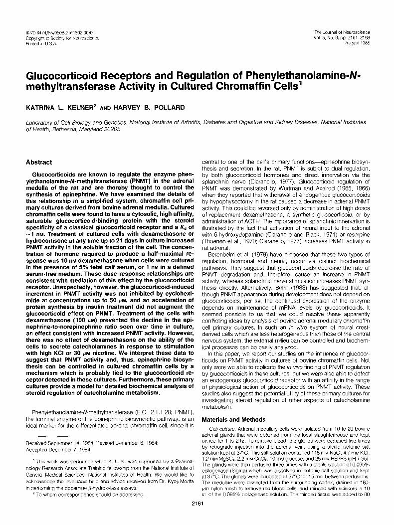
figure 2. Dose response relationships be- tween dexamethasone and PNMT activity in medium containing 5% fetal calf serum (left) and In serum-free defined medium (right). On day 7 of culture, plated chromaffln cells were treated with various concentrations of dexa- methasone for 3 days in medium containing 5% fetal calf serum or in defined serum-free medium, described in detail under “Materials and Methods.” PNMT activity and soluble pro- tein levels were measured in water lysates of these cells, and PNMT activity per milligram of soluble protein was calculated. Data were expressed as percentage of untreated con- trol. Each bar represents the mean f SD of the values obtained from four wells.
total soluble protein per cell remained constant over the 25 days in culture (6.13 + 0.08 (SEM) fig of soluble protein/IO5 cells, n = 5).
Since cell density has been shown to affect the levels of cate- cholamine biosynthetic enzymes in plated chromaffin cells (Acheson and Thoenen, 1983), we examined the effect of cell number per well on PNMT levels in our system. As the plating density was increased, the PNMT activity per cell also increased dramatically, reaching a plateau at approximately 150,000 cells/cm* (300,000 cells/well) (data not shown). All experiments reported in this paper were performed on cells plated above a density of 150,000 cells/cm’.
Glucocorticoids and PNMT activity. We tested the effect of glucocorticoids on PNMT activity in chromaffin cells cultured for various times. Addition of dexamethasone (100 PM) to the culture medium at 3, 7, or 21 days after initial plating caused an increase in PNMT specific activity, measured in the soluble fraction, of 28%, 46%, and 66% over control values, respectively. These data indicate that the cells retained responsiveness to glucocorticoids in vitro for at least 3 weeks in culture (data not shown).
Using cells cultured for 3 days, dose-response curves (1 .O nM to 100 PM) were generated with the endogenous bovine glucocorticoid, hydrocortisone, and the synthetic glucocorticoid, dexamethasone. Two types of culture media were used: standard culture medium containing 5% stripped fetal calf serum and a defined medium without serum (see “Materials and Methods”). Figures 2 and 3 illustrate the dose-response curves obtained after 2 days of incu- bation. In the presence of serum, hydrocortisone and dexametha- sone produced broad dose-response patterns of PNMT activation, with maxima at high concentrations of hormone (100 FM for hydro- cortisone and 10 FM for dexamethasone). However, in a serum-free medium, a narrower dose-response curve was seen, with increases of PNMT activity occurring at lower concentrations of hormone, approximately 1 nM, and reaching maxima at 10 to 100 nM.
The activation of PNMT by glucocorticoids required 2 days for appearance, but additional time in culture did not increase the size of the effect further (see Fig. 4). The magnitude of the effect was constant regardless of whether treatment was initiated on day 3 or on day 8 and remained the same at least until day 14 of culture (11 days or 6 days of treatment, respectively). The falling control value

5% FETAL
CALF SERUM
Kelner and Pollard Vol. 5, No. 8, Aug. 1985
HYDROCORTISONE .
DEFINED
MEDIUM
-c- Control
-+- Hydrocortisone
I I I I I I I I I I I I I 5 10
TIME IN CULTURE, DAYS
figure 3. Dose-response relationships between hydrocortlsone and PNMT activity in medium containing 5% fetal calf serum (left) and In serum-free defined medium (right). Plated chromaffin cells were treated exactly as described for Figure 2, except that hydrocotiisone was substituted for dex- amethasone. Data were calculated and ex- pressed as for Figure 2.
Figure 4. Time course of the hydrocort- sane-induced increase In PNMT activity. Plated chromaffin cells were maintained in culture for 14 days. On days 3 and 8. some wells were treated with 10 PM hydrocortlsone (arrows). At time potnts indicated, the cells were lysed with water and the levels of PNMT activity and soluble protein were measured. Data were expressed as nanomoles per 30 mln per milligram of protein. Each point rep- resents the mean -t SD of the values obtained from three wells. Where no error bars are indicated, the standard deviation IS smaller than the symbol. Statistical analyses indicated that there were no significant differences among points in the following groups (p < 0.05): control, days 2 and 3, days 3 to 6, days 6 and 7; treated, days 3 to 6, day 10, day 14. Treated points were different from controls (p < 0.05) on day 6 and day 10.
caused an apparent increase in the percentage of stimulation over time.
To test the specificity of the effect of glucocorticoids on PNMT, the lysate from cells incubated for 3 days with dexamethasone was assayed for increases in the specific activity of two other enzymes,
DBH and LDH. Under condltlons in which PNMT activity increased significantly, neither enzyme showed any change in activity (Table I,
p < 0.01). The role of protein synthesis in mediating the increase in PNMT
activity seen with glucocorticoids was investigated using the protein
synthesis inhibitor, cycloheximide. At concentrations up to 50 pM, there was no effect of protein synthesis inhibition by cycloheximide
on dexamethasone’s ability to stimulate PNMT activity (see Fig. 5, top). Dexamethasone treatment alone did not affect the rate of
protein synthesis (data not shown). In addition, acceleration of
protein synthesis by 37% by treatment of the cells with 5 wg/ml of insulin did not affect the magnitude of the increase in PNMT activity
produced by dexamethasone (data not shown). Figure 5, bottom, documents that, under the conditions used for this study, cyclohex- imide inhibited amino acid incorporation into acid-precipitable protein
in chromaffin cells. At 50 FM cycloheximide, the highest concentra- tion tested, amino acid incorporation was reduced to 2.3% of the control level.
Epinephrine synthesis in glucocorticoid-treated cells. If PNMT activity in cell lysates reflected epinephrine synthesis in the cell, then
under conditions in which PNMT activity is altered one would expect to see changes in the levels of cellular norepinephrine and epineph-
rine, the substrate and product of the enzyme, respectively. To test this, we measured the effect of constant exposure of chromaffin
cells to dexamethasone on the cellular content of epinephrine and

The Journal of Neuroscience Glucocorticoid Receptors/PNMT in Chromaffin Cells 2165
TABLE I Dexamethasone effects on chromaffin cell enzymes
PNMT, DBH. and LDH activities were measured In water lysates of 3-day
chromaffln cells that had been malntained In the presence or absence of 100 JLM dexamethasone for 3 days as described under “Materials and Methods.” The slgniflcance of the difference between groups was tested by the Student’s t test.
Control Dexamethasone
treated
PNMT DBH LDH (nmol/30 mln/mg (fimo1/30 mln/mg (nmol/30 mln/106
of protein of protein) cells
3.16 k 0.13 3.54 + 0.11 102.9 + 7.6 39.4 + 0.04” 3.21 + 0.28b 101.0 k 15.2b
“p < 0.01.
b Not slgnrflcantly different from control.
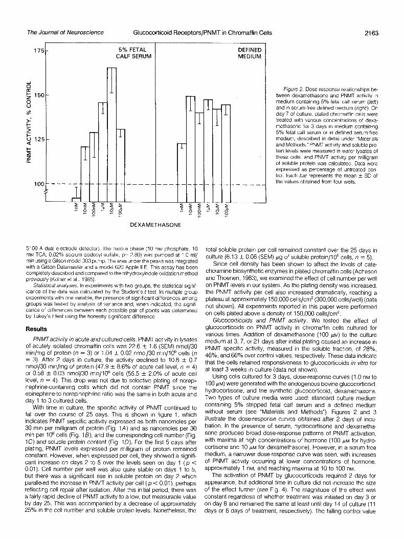
norepinephrine. The levels of the two catecholamines in acutely isolated cells were 30.9 f 2.2 nmol (SEM) of norepinephrine/106 cells (n = 6) and 63.4 ? 4.0 nmol of epinephrine/106 cells (n = 6). On day 1 of culture, these levels dropped to 56% and 48% of these values, respectively. However, during the first few days in culture, the ratio of the two catecholamines remained the same as in the acutely isolated cells (29.8% + 0.72 (SEM) norepinephrine and 70.2% + 3.08 epinephrine) (see Fig. 6). At these times, dexameth- asone had no effect on either the absolute amount of epinephrine and norepinephrine In the cells (cf. Fig. 6, left and right upper pane/s) or on the relative amounts of these catecholamines (cf. Fig. 6, left and right lower panels). With longer times in culture (more than 7 days), there was a significant drop in epinephrine levels per well, whereas norepinephrine levels remained constant (p < O.Oi), resulting in a ratio of norepinephrine to epinephrine of 52% + 3.0 (SEM):48% f 1.8 at day 17 (Fig. 6, left panels). When the cells were maintalned in the presence of dexamethasone (Fig. 6, right panels), there was a smaller but slgnlficant drop in epinephrine levels, as well as a drop in norepinephnne levels (p < 0.01). The ratlo of epinephrine to norepinephrine remained constant (29.5% + 0.5 (SEM) norepinephrine:70.5% + 0.5 epinephrine on day 17). Thus, dexamethasone treatment allowed the cells to maintain their initial proportions of norepinephrine and epinephnne.
We tested whether pretreatment with dexamethasone had any effect on the ability of cultured chromaffin cells to secrete catechol- amines in response to secretogogues. Cells were treated with 10 PM dexamethasone acutely or for 1, 3, or 6 days. Subsequently, 30 FM nicotine or 65 mM KCI was applied to the cells for 15 min, and the norepinephrine and epinephrine released into the medium during that time period was measured by the trihydroxyindole method. For both catecholamines, basal release was less than 10% and was not affected by any dexamethasone pretreatment. Similarly, dexameth- asone pretreatment of any duration had no consistent effect on the fraction of either catecholamine released into the medium with nicotine or KCI (data not shown).
Glucocorticoid receptor in chromaffin cells. Since the cultured chromaffin cells were able to respond to glucocorticoids, we exam- ined them for the presence of a glucocorticoid receptor. Saturation analyses of high speed supernatants of cultured chromaffin cells revealed the presence of a specific saturable binding site when either 3H-dexamethasone or 3H-triamcinolone acetonide was used as ligand. The saturation plots and their corresponding Scatchard analyses, shown in Figures 7 and 8, illustrate that, with both ligands, the dissociation constant of ligand binding was approximately 1 x
lo-’ M. The number of binding sites ranged from 200 to 350 fmol/ mg of soluble protein in all experiments.
A comparison of the ability of various steroids to compete for 3H- dexamethasone binding showed an order of potency of: testoster- one = estradiol < progesterone << corticosterone < triamcinolone acetonide = hydrocortisone z dexamethasone (data not shown).
o--o Dexamethasone
t +--WC----+’
-t+\t
YY ; ’ I 1
10
CYCLOHEXIMIDE, /AM
CYCLOHEXIMIDE, PM Figure 5. The effect of cycloheximide on the dexamethasone-induced
increase in PNMT activity (upper pane/) and protein synthesis (lower panel). Upper pane/, On day 5 of culture, plated chromaffin cells were treated with various concentrations of cycloheximide in the presence or absence of 100 PM dexamethasone. After 3 days, the cells were lysed and the PNMT activity and soluble protein levels were determined as described in the text. Each point represents the mean + SEM of the values obtalned from 12 wells In two separate experiments. There was no significant difference among the points withIn the control group or the treated group. The treated value was significantly different from control at 0, 1, and 50 PM cycloheximlde. The difference at 10 pM was not significant (In all cases p < 0.01). Lower pane/, On day 5 of culture, plated chromaffin cells were treated with various concentrattons of cycloheximide In the presence of 2 @Cl of ‘H-amino acids/ well. After 36 hr, the amount of radioacWty in acid-preclpltable material was determined, as described In the text. Each point IS the mean of the values obtained from three wells
Discussion
Glucocorticoid regulation of PNMT activity in vitro. Maintenance of the levels of adrenal PNMT, the methyltransferase responsible for epinephrine synthesis, is known to depend on glucocorticoids in the rat. It was therefore reasonable to suspect that some or all of the drop in PNMT levels seen over time in cultured chromaffin cells (see Fig. 1) might be due to withdrawal of the cells from the endogenous source of this hormone and that replacement of glucocorticoids might restore activity. In fact, treatment of the cultured cells with the synthetic glucocorticoid, dexamethasone, or with the endogenous bovine glucocorticoid, hydrocortisone, caused a dose-dependent increase in PNMT activity when treatment was initiated at any time
Kelner and Pollard Vol. 5, No. 8, Aug. 1985
Control Dexamethasone
E
1:
E
N 2l N
I I I
Figure 6. The effect of dexamethasone treatment on maintenance of epinephrine and norepinephrine levels in cultured chromaffin cells. Plated chromaffrn cells were cultured for 17 days In the absence (left panels) or pres- ence (fjgight panels) of 100 PM dexametha- sone. At regular Intervals, wells were lysed In 10% acetic acid and the levels of norepineph- rine and eprnephrine were analyzed by high performance liquid chromatography, as de- scrrbed under “Materials and Methods.” The data are expressed as nanomoles per well (upper panels) or as percentage of total cate- cholamine (lower panels), where total cate- cholamine equals the sum of the epinephrine and noreprnephnne values. Each point repre- sents the mean + SD of the values obtained from four wells. Where no error bar is present, the error was smaller than the symbol. Statis- tical analysrs indicates that the following val- ues are signrfrcantly different from the day 3 values (p < 0.01): control, none (N); days 12 and 17 (E); dexamethasone, days 12 and 17 (N); days 12 and 17 (E).
5 10 15 5 TIME IN CULTURE (days)
10 15
from day 3 to day 21 in culture. The specificity of this effect is illustrated by the fact that the levels of two other enzymes involved in catecholamrne metabolism, DBH and tyrosine hydroxylase, have
been shown to be stable over time in culture (Waymrre, 1977) and that glucocorticoids have no effect on the levels of DBH or LDH (Table I).
In viva, the glucoconicoid-induced increase in PNMT activity
requires unusually high concentrations of dexamethasone (Wurtman and Axelrod, 1966). In the cultured cells, there is also a requirement for high concentrations of dexamethasone to increase PNMT activity in the presence of serum, but this requirement is notably absent in
the absence of serum (Hersey and Distefano, 1979; Figs. 2 and 3). These serum-dependent differences seen in the dose requirements for the glucocorticoid-induced increase in PNMT may reflect the presence of glucocorticoid binding activity derived from the serum
which could buffer the added glucocorticoid and, thus, lower the amount of steroid available to the cell. It is not known whether a similar buffering effect of glucocorticoid-binding proteins contributes
to the high steroid requirement in viva or, alternatively, whether the properties of the receptor are altered in cultured cells.
The increase in PNMT activity produced by glucocorticoid treat- ment was characterized by a slow time course, requiring 2 days to
become evident. When protein synthesis was inhibited to 2.3% of normal rates, the response was not blunted. Conversely, accelera- tion of protein synthesis with insulin did not enhance the magnitude
of the response. These latter results are consistent with the notion that the glucocorticoid-induced increase in PNMT does not require synthesis of new protein. This is in contrast to the situation in the
adult and developing rat, where PNMT increases are blocked by protein synthesis inhibitors (Wurtman and Axelrod, 1966; Bohn,
1983). However, we have not ruled out the possibility that complete inhibition (i.e., more than 97.7%) of protein synthesis would prevent the increase in PNMT in chromaffin cells. Use of higher concentra-
tions of cycloheximide to accomplish this, however, produced toxic effects in the cells such that the results were uninterpretable.
The PNMT response to glucocorticoids in chromaffin cells is unlike the induction of many other steroid-regulated enzymes in that there IS no massive increase in enzyme activity. After reaching a
maximum value of about 3 nmol/30 min/mg of protein by 2 days,
the PNMT elevation above the control level was constant over time, although the control activity continued to fall. In fact, the glucoconi- cord-treated cells simply showed a slower rate of PNMT loss. The
levels of the enzyme were never increased by glucocorticoid treat- ment to those found in acutely isolated cells or even to the lower levels found in the cultures on day 1. This mimics the situation in viva, where restoration of PNMT levels in hypophysectomized rats
treated with replacement dexamethasone is never complete (Poh- erecky and Wurtman, 1968; Ciaranello, 1977). The drop in PNMT activity in culture that occurs regardless of the presence of gluco-
corticoids may be due to the absence of other required regulatory influences, such as splanchnic nerve innervation, which is known to be required in the rat for maintenance of PNMT levels. The time- dependent loss of cells in culture would also be expected to cause a drop in PNMT activity per cell which could account for at least
part of the drop in PNMT specific activity (see “Results”). In addition, the non-optimal long-term culture of these cells may require reorien- tation of the cell protein synthetic machinery toward proteins involved
in cell maintenance and viability. G/ucocorticoid receptor in chromaffin cells. The soluble gluco-
corticoid-binding site which we have demonstrated to be present in chromaffin cell cultures has the characteristics of a classical gluco-
corticoid type II receptor. The dissociation constant (approximately 1 X IO-’ M), the receptor concentration (200 to 300 fmol/mg of
soluble protein), and the steroid specificity are consistent with the notion that both the 3H-dexamethasone binding and the 3H-triamcin- olone acetonide binding in cultured chromaffin cells represent bind-
ing to a true glucocorticoid receptor Feldman et al., 1978; Do et al., 1979). The small variations in the steroid specificity and dissociation constant seen between this receptor and others (eg., the 1 nM Kd reported here versus 3 to 15 nM in the literature) are probably due to differing tissue compositions and assay methods and do not necessarily reflect unique properties of the chromaffin cell receptor.
Although Do et al. (1979) have shown that some bovine tissues (i.e.,
thymus, kidney, liver, adipose tissue, and adrenal cortex) contain another glucocorticoid-binding site which is not the classical recep- tor, this other binding site does not appear to be present in chro-

The Journal of Neuroscience Glucocorticoid Receptors/PNMT in Chromaffin Cells 2167
\
1 5 10 VI-Dex Free, nM
Kd = 1.28 nM
8 max = 307 fmol/mg protein
r = 0.934
jH-Dexamethasone Bound, fmol
F/gure 7. Saturation analysis of Wdexamethasone blnding to chromaffln cell cytosollc extracts. Blndtng Isotherms of %dexamethasone (?--Dex) bIndIng to chromaffln cell cytosollc extracts were constructed using concen- tratlons of %dexamethasone ranging from 0.1 to IO nM with the procedure described under “Materials and Methods.” The inset shows the saturation analysis, the upper line Indicating total binding and the lower line representing the amount of binding obtalned tn the presence of 300.fold molar excess unlabeled dexamethasone, which was assumed to represent nonspecific binding. The ma/n panel shows a Scatchard analysis of the data from the same experiment analyzed as described in the text. Each point represents a single determination The reproducibility of the method can be seen by the replication Illustrated in the inset.
maffin cell cultures in significant quantities. This is illustrated by the similarity of the saturation analyses obtained using ?--dexametha- sone and %triamcinolone acetonide, since the second non-receptor binding site does not have an appreciable affinity for triamcinolone acetonide (Do et al., 1979).
The mechanism of the glucocorticoid-induced increase in PNMT activity. There are several pieces of evidence that suggest that the glucocorticoid receptor mediates the glucocorticoid-induced in- crease in PNMT activity. Half-maximal activation of the PNMT re- sponse occurs at approximately 1 nM dexamethasone in the ab- sence of serum. Half-maximal saturation of the receptor also occurs at this concentration of steroid, the Kd being approximately 1 nM. Both saturation of the receptor and saturation of the response occur over a dose range of two orders of magnitude. In addition, the steroid specificity of this response is identical to the steroid specificity of the receptor (Hersey and Distefano, 1979; K. L. Kelner, unpub- lished observation).
The sequence of events occurring between the binding of the glucocotticoids to the receptor and the increase in PNMT activity in cultured chromaffin cells is not known, but our data may allow us to differentiate between potential mechanisms. In many systems, glu- cocorticoids have been shown to alter the levels of enzymes and proteins by a receptor-mediated process in which changes in spe- cific gene transcription are caused by action of the receptor-steroid
.05
.&I
?
$ .03 5
mo
.02
.Ol
0
0
\
0
l l
\
‘H-TA Free. nM
Kd=1.28 nM
Bmax=246 fmol/mg protein
FO.906
;:A : , 10 20 30
3H-Triamcinolone Acetonide Bound, fmol
Figure 8. Saturation analyses of ‘H-triamclnolone acetonlde bindlng to chromaffln cell cytosolic extracts. Bindlng isotherms of ‘H-triamclnolone acetonlde (%TA) bindIng to chromaffln cell cytosok extracts were con- structed as described for Figure 7. The data were analyzed and are displayed as for Figure 7.
complex on regulatory sites on the DNA. The altered levels of mRNA lead to changes in the rate of synthesis of the regulated protein. This is the mechanism proposed by Bohn (1983) for PNMT regulation by glucocorticoids. Protein synthesis inhibitors will block this type of steroid effect. For example, the hydrocortisone-induced increase in the rate of synthesis of glycerol phosphate dehydrogenase in C6 glioma cells is inhibited by 35 PM cycloheximide, as well as by RNA synthesis inhibitors (McGinnis and de Vellis, 1978). Thus, the insen- sitivity of the PNMT response to 50 FM cycloheximide reported here is inconsistent with this proposed mechanism, suggesting that in- creased PNMT synthesis is not the cause of the elevation in activity.
Glucocorticoids are also known to increase protein degradation in muscle (Goldberg, 1969; Tomas et al., 1979). In this regard, Ciaranello (1977) has suggested that, in the rat, the glucocorticoid- induced increase in PNMT activity is due to a slower rate of enzyme degradation. He suggests that this is caused by the protective action of increased levels of the PNMT cofactor, S-adenosylmethionine, which are a result of glucocorticoid treatment. Our data are consist- ent with this proposal. The amount of PNMT activity declines with time in culture, even in the presence of glucocorticoids, indicating that the rate of degradation is higher than the synthetic rate. Since protein synthesis inhibitors do not alter the glucocorticoid effect and also do not decrease the PNMT pool size (K. L. Kelner, unpublished observation), the PNMT synthetic rate must be small. Thus, the rate of degradation is the primary regulator of PNMT pool size, and a glucocorticoidinduced decrease in the rate of degradation of the enzyme would yield the results we have observed, an increase in PNMT activity. Although our data are consistent with the notion that altered protein degradation rates, rather than increased transcnption and/or translation, may mediate the glucocorticoid effect on PNMT in cultured chromaffin cells, it is also possible that it occurs by an
2168 Kelner and Pollard Vol. 5, No. 8, Aug. 1985
alternative process. For example, glucocorticoids may activate a pre-existing pool of PNMT molecules. There is also Increasing
evidence that some effects of steroids may occur by an unknown mechanism which is operative at the level of the plasma membrane (McEwen et al., 1979).
Functional consequences of glucocorticoid treatment. The ex- posure of chromaffin cells to glucocorticoids caused the levels of the substrate and the product of the PNMT reaction to be altered but had no effect on the release process itself. Without steroid
treatment, the cells were unable to maintain their original proportions of epinephrine and norepinephrine over extended time (i.e., weeks) in culture (see Fig. 6). The steady loss of epinephrine, perhaps due
to a low level of basal secretion, was prevented by maintaining the cells in the presence of dexamethasone. Addition of glucocorticoids to the medium in vitro may mimic the in vivo situation where the adrenal medullary cells are exposed to high concentrations of glu-
cocorticoid hormones which are released into the adrenal circulation by the adrenal cortex. This glucocotiicoid-induced ability to maintain catecholamine levels is probably due to the increase in PNMT activity seen in the soluble lysates of these cells. However, these data do not rule out the possibility that other factors could contribute to these
changes, such as increases in substrate or cofactor availability to PNMT.
In summary, these studies have demonstrated the presence of a classical glucocorticoid receptor in primary cultures of chromaffin cells isolated from bovine adrenal medulla. This receptor seems to mediate a slow dose-dependent increase in the activity of PNMT over control levels by a mechanism that is not easily inhibited by cycloheximide. This increase in PNMT activity may allow the cells to retain the initial content of eplnephnne over extended time in culture. Thus, primary cultures of chromaffin cells provide an excellent
system for further biochemical investigations of glucocorticoid reg- ulation of catecholamine metabolism.
References
Acheson, A. L., and H. Thoenen (1983) Cell contact-mediated regulation of tyroslne hydroxylase synthesis in cultured bovine adrenal chromaffin cells. J. Cell Btol. 97: 925-928.
Anton, A. H., and D. F. Sayre (1962) A study on the factors affecting the alumtnum oxide-trlhydroxyindole procedure for the analysis of catechol- amlnes. J. Pharmacol. Exp. Ther. 138: 360-375.
Axelrod, J. (1961) Purification and properties of phenylethanolamrne-N-methyl transferase. J. BIOI Chem. 237 1657-1660
Berenbeim, D. M., D. L. Wong, S. J. Masover, and f? Claranello (1979) Regulation of synthesis and degradation of rat adrenal phenylethanolamlne N-methyl transferase. Ill. Stabilization of PNMT against thermal and tryptic degradation by S-adenosylmethtonlne. Mol. Pharmacol. 16: 482-490.
Bohn, M. C (1983) Role of glucocotiicoids in expression and development of phenylethanolamlne~N~methy1 transferase (PNMT) in cells derived from neural crest. A revelw. Psychoneuroendocnnology 8: 381-390.
Bottensteln, J. E., and G. H. Sato (1979) Growth of a rat neuroblastoma cell line In serum-free supplemented medium. Proc. Natl. Acad. SCI. U S. A. 76: 514-517.
Ciaranello. R. D. (1977) Regulation of phenylethanolamine-N-methyl transfer- ase synthesis and degradation. In Modem Pharmacology-Tox/co/ogy. Vol.
10: Structure and function of Monoamine Enzymes. E. Usdin, N. Weiner, and M. B. H. Youdim, eds., pp. 497-525, Marcel Dekker, Inc., New York.
Claranello, R. D., and I. B. Black (1971) Kinetics of the glucocotiicoid mediated Induction of phenylethanolamlne N-methyl transferase in the hypophysectomized rat. Blochem. Pharmacol. 20: 3529-3532.
Do. Y. S., D. S. Loose, and D. Feldman (1979) Heteroaeneitv of alucocorticoid binders: A unique and a classical dkxamkthasone binding site in bovine tissues. Endocrinology 705: 1055-1063.
Feldman, D., J. Funder, and D. S. Loose (1978) Is the glucocorticoid receptor identical in various target organs? J. Steroid Biochem. 9: 141-I 45.
Goldberg, A. L. (1969) Protein turnover in skeletal muscle. II. Effects of denervation and cortisone on protein catabolism in skeletal muscle. J Biol. Chem. 244: 3223-3229.
Hersey, R. M., and V. Distefano (1979) Control of phenylethanolamine-N- methyl transferase by glucocorticoids in cultured bovine adrenal medullary cells. J. Pharmacol. Exp. Ther. 209: 147-152.
Kelner, K. L., A. L. Miller, and E. J. Peck, Jr. (1981) Estrogens and the hypothalamus: Receptors and RNA polymerase activation. J. Recept. Res. 1. 215-237.
Kelner, K. L., R. A. Levine, K. Morita, and H. B. Pollard (1985) A comparison of tnhydroxylndole and HPLC/electrochemical methods for norepinephrine and epinephrine measurement in adrenal chromaffin cells. Neurochem. Int., in press.
Lowry, 0. H., N. J. Rosebrough, A. L. Farr, and R. J. Randall (1951) Protein measurement with the Folin phenol reagent. J. Biol. Chem. 793: 265-275.
McEwen, B. S., P. G. Davis, B. Parsons, and D. W. Pfaff (1979) The brain as a target for steroid hormone actjon. Annu. Rev. Neurosci. 2: 65-112.
McGinnis, J. F., and J. de Vellis (1978) Glucocorticoid regulation in rat brain brain ceil cultures. J. Biol. Chem. 253: 8483-8492.
Nagatsu, T., and S. Udenfnend (1972) PhotometrIc assay of dopamine B- hydroxylase in human blood. Clin. Chem. 78: 980-983.
Pohorecky, L. A., and R. J. W&man (1968) Induction of epinephnne-formlng enzyme by glucocortlcolds: Steroid hydroxylatlon and induction effect. Nature 219: 392-394.
Pollard, H. B., S. S. Stopak, C. J. Pazoles, and C. E. Creutz (1979) A simplified, one-step method for radiometric analysis of phenylethanola- mine-N-methyl transferase In adrenal chromaffin cells. Anal. Biochem. 99: 281-282.
Thoenen, H., R. A. Mueller, and J. Axelrod (1970) Neuronally dependent induction of phenylethanolamlne-N-methyl transferase by 6-hydroxydopa- mine. Biochem. Pharmacol. 19: 669-673.
Tomas, F. M., H. N. Munro, and V. R. Young (1979) Effect of glucocorticoid admlnistratlon on the rate of muscle protein breakdown /n VIVO in rats, as measured by urinary excretion of N-methylhlstidine. Biochem. J. 178: 139. 146.
Waymire, J. C. K. G. Waymire, R. Boehme, D. Noritake, and J. Wardell (1977) Regulation of tyrosine hydroxylase by cyclic 3,5-adenosine monophos- phate In cultured neuroblastoma and cultured dissociated bovine adrenal chromaffin cells. In Modern Pharmacology-Toxicology. Vol. 10, Structure and function of Monoamine Enzymes. E. Usdin, N. Weiner, and M. B. H. Youdlm, eds., pp. 327-363, Marcel Dekker, Inc., New York.
Wilson, S P , and 0. H. Viveros (1981) Primary culture of adrenal medullary chromaffin cells rn a chemically defined medium. Exp. Cell Res. 133: 159- 169.
Wurtman, R. J.. and J. Axelrod (1965) Adrenaline synthesis: Control by the pituitary gland and adrenal glucocorticoids. Science 150: 1464-1465.
Wurtman. R. J., and J Axelrod (1966) Control of enzymatic synthesis of adrenaline In the adrenal medulla by adrenal cortical steroids. J Biol. Chem. 24 I. 2301-2305