Glucagon-Like Peptide (GLP)-1(9-36)Amide-Mediated...
12
Glucagon-Like Peptide (GLP)-1(9-36)Amide-Mediated Cytoprotection Is Blocked by Exendin(9-39) Yet Does Not Require the Known GLP-1 Receptor Kiwon Ban, Kyoung-Han Kim, Chan-Kyung Cho, Meghan Sauve ´, Eleftherios P. Diamandis, Peter H. Backx, Daniel J. Drucker,* and Mansoor Husain* Departments of Physiology (K.B., K.-H.K., P.H.B., M.H.), Laboratory Medicine and Pathobiology (C.-K.C., E.P.D., D.J.D., M.H.), and Medicine (P.H.B., D.J.D., M.H.), Samuel Lunenfeld Research Institute (M.S., D.J.D.), Toronto General Hospital Research Institute (M.H.), Banting and Best Diabetes Centre (M.S., D.J.D.), and Heart and Stroke Richard Lewar Centre of Excellence in Cardiovascular Research (K.B., K.- H.K., P.H.B., M.H.), University of Toronto, Toronto, Ontario, Canada M5G 2C4 The widely expressed dipeptidyl peptidase-4 enzyme rapidly cleaves the gut hormone glucagon- like peptide-1 [GLP-1(7-36)amide] at the N terminus to generate GLP-1(9-36)amide. Both intact GLP-1(7-36)amide and GLP-1(9-36)amide exert cardioprotective actions in rodent hearts; however, the mechanisms underlying the actions of GLP-1(9-36)amide remain poorly understood. We used mass spectrometry of coronary effluents to demonstrate that isolated mouse hearts rapidly convert infused GLP-1(7-36)amide to GLP-1(9-36)amide. After ischemia-reperfusion (I/R) injury of isolated mouse hearts, administration of GLP-1(9-36)amide or exendin-4 improved functional recovery and reduced infarct size. The direct actions of these peptides were studied in cultured neonatal mouse cardiomyocytes. Both GLP-1(9-36)amide and exendin-4 increased levels of cAMP and phosphory- lation of ERK1/2 and the phosphoinositide 3-kinase target protein kinase B/Akt. In I/R injury models in vitro, both peptides improved mouse cardiomyocyte viability and reduced lactate dehydroge- nase release and caspase-3 activation. These effects were attenuated by inhibitors of ERK1/2 and phosphoinositide 3-kinase. Unexpectedly, the cardioprotective actions of GLP-1(9-36)amide were blocked by exendin(9-39) yet preserved in Glp1r / cardiomyocytes. Furthermore, GLP-1(9- 36)amide, but not exendin-4, improved the survival of human aortic endothelial cells undergoing I/R injury, actions sensitive to the nitric oxide synthase inhibitor, N(G)-nitro-L-arginine methyl ester (L-NAME). In summary, our findings demonstrate separate actions for GLP-1(9-36)amide vs. the GLP-1R agonist exendin-4 and reveal the existence of a GLP-1(9-36)amide-responsive, exendin(9- 39)-sensitive, cardioprotective signaling pathway distinct from that associated with the classical GLP-1 receptor. (Endocrinology 151: 1520 –1531, 2010) G lucagon like peptide-1 [GLP-1(7-36)amide], also known as GLP-1, is released from gut endocrine cells in response to nutrient ingestion, stimulating glucose-de- pendent insulin secretion by activating a G protein-cou- pled GLP-1 receptor (GLP-1R) expressed on pancreatic islet cells (1–3). However, the half-life of intact GLP-1 is extremely short (2 min), in part due to renal clearance. GLP-1 is also rapidly metabolized to GLP-1(9-36)amide by the enzyme dipeptidyl peptidase-4 (DPP-4), which is abundantly expressed in many cell types with primary ex- ISSN Print 0013-7227 ISSN Online 1945-7170 Printed in U.S.A. Copyright © 2010 by The Endocrine Society doi: 10.1210/en.2009-1197 Received October 6, 2009. Accepted January 4, 2010. First Published Online February 19, 2010 * D.J.D. and M.H. contributed equally to this work. Abbreviations: AU, Arbitrary unit; CCF, constant coronary flow; CCPP, constant coronary perfusion pressure; CM, cardiomyocyte; CREB, cAMP response element binding; DPP-4, dipeptidyl peptidase-4; EC, endothelial cell; Ex-4, exendin-4; Ex(9-39), exendin(9-39); GLP, glucagon like peptide; GLP-1R, GLP-1 receptor; HAEC, human aortic endothelial cell; H/R, hypoxia-reoxygenation; I/R, ischemia-reperfusion; LDH, lactate dehydrogenase; L-NAME, N(G)-nitro-L-arginine methyl ester; LVDP, left ventricular developed pressure; MS, mass spectrometry; MTT, 3-(4,5-dimethylthizol-2-yl)-2,5-diphenyltetrazodium bromide; NOS, nitric oxide synthase; PI3K, phosphoinositide 3-kinase; PKB, protein kinase B; TTC, 2,3,5- triphenyl tetrazolium chloride; WT, wild type. DIABETES-INSULIN-GLUCAGON-GASTROINTESTINAL 1520 endo.endojournals.org Endocrinology, April 2010, 151(4):1520 –1531 at University of Toronto Library on April 29, 2010 endo.endojournals.org Downloaded from
Transcript of Glucagon-Like Peptide (GLP)-1(9-36)Amide-Mediated...

Glucagon-Like Peptide (GLP)-1(9-36)Amide-MediatedCytoprotection Is Blocked by Exendin(9-39) Yet DoesNot Require the Known GLP-1 Receptor
Kiwon Ban, Kyoung-Han Kim, Chan-Kyung Cho, Meghan Sauve,Eleftherios P. Diamandis, Peter H. Backx, Daniel J. Drucker,*and Mansoor Husain*
Departments of Physiology (K.B., K.-H.K., P.H.B., M.H.), Laboratory Medicine and Pathobiology (C.-K.C.,E.P.D., D.J.D., M.H.), and Medicine (P.H.B., D.J.D., M.H.), Samuel Lunenfeld Research Institute (M.S.,D.J.D.), Toronto General Hospital Research Institute (M.H.), Banting and Best Diabetes Centre (M.S.,D.J.D.), and Heart and Stroke Richard Lewar Centre of Excellence in Cardiovascular Research (K.B., K.-H.K., P.H.B., M.H.), University of Toronto, Toronto, Ontario, Canada M5G 2C4
The widely expressed dipeptidyl peptidase-4 enzyme rapidly cleaves the gut hormone glucagon-like peptide-1 [GLP-1(7-36)amide] at the N terminus to generate GLP-1(9-36)amide. Both intactGLP-1(7-36)amide and GLP-1(9-36)amide exert cardioprotective actions in rodent hearts; however,the mechanisms underlying the actions of GLP-1(9-36)amide remain poorly understood. We usedmass spectrometry of coronary effluents to demonstrate that isolated mouse hearts rapidly convertinfused GLP-1(7-36)amide to GLP-1(9-36)amide. After ischemia-reperfusion (I/R) injury of isolatedmouse hearts, administration of GLP-1(9-36)amide or exendin-4 improved functional recovery andreduced infarct size. The direct actions of these peptides were studied in cultured neonatal mousecardiomyocytes. Both GLP-1(9-36)amide and exendin-4 increased levels of cAMP and phosphory-lation of ERK1/2 and the phosphoinositide 3-kinase target protein kinase B/Akt. In I/R injury modelsin vitro, both peptides improved mouse cardiomyocyte viability and reduced lactate dehydroge-nase release and caspase-3 activation. These effects were attenuated by inhibitors of ERK1/2 andphosphoinositide 3-kinase. Unexpectedly, the cardioprotective actions of GLP-1(9-36)amide wereblocked by exendin(9-39) yet preserved in Glp1r�/� cardiomyocytes. Furthermore, GLP-1(9-36)amide, but not exendin-4, improved the survival of human aortic endothelial cells undergoingI/R injury, actions sensitive to the nitric oxide synthase inhibitor, N(G)-nitro-L-arginine methyl ester(L-NAME). In summary, our findings demonstrate separate actions for GLP-1(9-36)amide vs. theGLP-1R agonist exendin-4 and reveal the existence of a GLP-1(9-36)amide-responsive, exendin(9-39)-sensitive, cardioprotective signaling pathway distinct from that associated with the classicalGLP-1 receptor. (Endocrinology 151: 1520–1531, 2010)
Glucagon like peptide-1 [GLP-1(7-36)amide], alsoknown as GLP-1, is released from gut endocrine cells
in response to nutrient ingestion, stimulating glucose-de-pendent insulin secretion by activating a G protein-cou-pled GLP-1 receptor (GLP-1R) expressed on pancreatic
islet cells (1–3). However, the half-life of intact GLP-1 isextremely short (�2 min), in part due to renal clearance.GLP-1 is also rapidly metabolized to GLP-1(9-36)amideby the enzyme dipeptidyl peptidase-4 (DPP-4), which isabundantly expressed in many cell types with primary ex-
ISSN Print 0013-7227 ISSN Online 1945-7170Printed in U.S.A.Copyright © 2010 by The Endocrine Societydoi: 10.1210/en.2009-1197 Received October 6, 2009. Accepted January 4, 2010.First Published Online February 19, 2010* D.J.D. and M.H. contributed equally to this work.
Abbreviations: AU, Arbitrary unit; CCF, constant coronary flow; CCPP, constant coronaryperfusion pressure; CM, cardiomyocyte; CREB, cAMP response element binding; DPP-4,dipeptidyl peptidase-4; EC, endothelial cell; Ex-4, exendin-4; Ex(9-39), exendin(9-39); GLP,glucagon like peptide; GLP-1R, GLP-1 receptor; HAEC, human aortic endothelial cell; H/R,hypoxia-reoxygenation; I/R, ischemia-reperfusion; LDH, lactate dehydrogenase; L-NAME,N(G)-nitro-L-arginine methyl ester; LVDP, left ventricular developed pressure; MS, massspectrometry; MTT, 3-(4,5-dimethylthizol-2-yl)-2,5-diphenyltetrazodium bromide; NOS,nitric oxide synthase; PI3K, phosphoinositide 3-kinase; PKB, protein kinase B; TTC, 2,3,5-triphenyl tetrazolium chloride; WT, wild type.
D I A B E T E S - I N S U L I N - G L U C A G O N - G A S T R O I N T E S T I N A L
1520 endo.endojournals.org Endocrinology, April 2010, 151(4):1520–1531
at University of Toronto Library on April 29, 2010 endo.endojournals.orgDownloaded from

pression in endothelial cells (ECs) (4-6). The DPP-4-gen-erated metabolite of GLP-1, GLP-1(9-36)amide, was ini-tially believed to be either biologically inactive or a weakantagonist of the canonical GLP-1R (7, 8). Although astudy in pigs suggested that an iv infusion of GLP-1(9-36)amide enhanced glucose disposal independent of in-cretin action (9), GLP-1(9-36)amide administration tohealthy humans had no effect on glucose clearance or in-sulin secretion (10). Notably, levels of GLP-1(9-36)amideafter GLP-1 infusion did not correlate with changes inplasma glucose in diabetic subjects (11).
In contrast, multiple lines of evidence support biolog-ical actions of GLP-1(9-36)amide in the cardiovascularsystem. GLP-1(9-36)amide increased myocardial glucoseuptake and improved left ventricular performance in dogswith dilated cardiomyopathy (12), and administration ofGLP-1(9-36)amide after global ischemia in rats signifi-cantly improved left ventricular pressure, although itfailed to reduce infarct size (13). We recently showed thatGLP-1(9-36)amide was a cGMP-producing vasodilatorymolecule capable of cardioprotective effects in hearts iso-lated from GLP-1R knockout mice (Glp1r�/�) (14). Fur-thermore, suppression of the cardioprotective and vaso-dilatory effects of GLP-1 by the DPP-4 inhibitor sitagliptinsuggested that GLP-1(9-36)amide may function as a keyintermediary in a subset of the cardiovascular effects ofGLP-1 (14). A vasodilatory effect of GLP-1(9-36)amideconcentrations that cover the physiological range in vivowas recently demonstrated in rat femoral artery prepara-tions (15, 16).
Although DPP-4-resistant GLP-1R agonists (ex-endin-4, liraglutide) and DPP-4 inhibitors (sitagliptin,vildagliptin, saxagliptin) (17) are likely to produce differ-ent effects on circulating levels of GLP-1(9-36)amide invivo, the mechanisms mediating the cardiovascular ac-tions of GLP-1(9-36)amide are poorly understood. Wenow show that the isolated mouse heart rapidly convertsGLP-1 to GLP-1(9-36)amide, and that the latter peptide iscapable of reducing infarct size in an ex vivo model ofischemia-reperfusion (I/R).Usingaconstant flow modelofcoronary perfusion, we show that the protection againstI/R injuryaffordedbyGLP-1(9-36)amidedoesnot entirelydepend on its vasodilatory effects. In addition, we dem-onstrate that GLP-1(9-36)amide exerts direct cytoprotec-tive actions on isolated mouse cardiomyocytes (CMs) viaphosphoinositide 3-kinase (PI3K)-protein kinase B (PKB)/Akt- and ERK1/2-dependent mechanisms. Finally, wefound that GLP-1(9-36)amide, but not the classicalGLP-1R agonist exendin-4, protects cardiomyocytesfrom Glp1r�/� mice and human aortic endothelial cells(HAECs) against ischemic- and oxidative stress-in-duced injury models. Surprisingly, the actions of GLP-
1(9-36)amide are blocked by exendin(9-39), a widely usedGLP-1R antagonist. Together these novel data further de-fine the selectivity of GLP-1R-dependent and -indepen-dent cardioprotective pathways and implicate the exis-tence of an alternative receptor for GLP-1(9-36)amide inboth CMs and ECs.
Materials and Methods
AnimalsAll protocols were approved by Toronto General Hospital
and conformed to guidelines of the Canadian Council on AnimalCare. Experiments were performed in 12-wk-old male C57BL/6wild-type and Glp1r�/� mice. C57BL/6 mice were purchasedfrom Charles River (Montreal, Canada) and housed for at least2 wk before experiments. Creation, characterization, and geno-typing of Glp1r�/� mice are described elsewhere (18).
ReagentsGLP-1 [GLP-1(7-36)amide] and GLP-1(9-36)amide were
synthesized by Bachem (Torrance, CA) and exendin-4 (Ex-4) andthe GLP-1R antagonist exendin(9-39) [Ex(9-39)] were pur-chased from California Peptide (Napa, CA). Stock solutionswere made initially in water and then diluted in either Krebs-Hensleit or PBS solutions to achieve the final concentrationsemployed. All other chemicals used were of reagent-grade(Sigma, Mississauga, Ontario, Canada).
Isolated heart preparationsIsolated mouse hearts were prepared as described (14, 19,
20). The glucose concentration in perfusion buffers was 11 mM.Only data derived from hearts demonstrating intrinsic heartrates greater than 350 beats/min were used.
Mass spectrometry (MS) analysisCoronary effluent samples were serially collected from iso-
lated mouse hearts infused on Langendorff preparations withGLP-1 or GLP-1(9-36)amide. Samples were analyzed with aLTQ ion trap mass spectrometer (Thermo Scientific, Waltham,MA) with a nanoelectrospray ionization source to identify spe-cific signatures of GLP-1 and GLP-1(9-36)amide. Detailed meth-odology for this MS approach is provided in the SupplementalMaterials published on The Endocrine Society’s Journals Onlineweb site at http://endo.endojournals.org.
Myocardial I/R injury ex vivo
Constant coronary perfusion pressure modelIsolated mouse hearts were mounted on a Langendorff ap-
paratus and perfused with Krebs-Hensleit buffer under a con-stant coronary perfusion pressure (CCPP) of 80 mm Hg. After a20-min equilibration phase, hearts were subjected to 30 min ofglobal (no flow) ischemia and 120 min of reperfusion. EitherGLP-1(9-36)amide (0.3 nM) or Ex-4 (3 nM) was added to theperfusion buffer for the first 60 min of the reperfusion period.The heart rate (beats per minute) and left ventricular developedpressure (LVDP; mm of mercury) were continuously monitored.Recovery of LVDP was measured at the end of reperfusion and
Endocrinology, April 2010, 151(4):1520–1531 endo.endojournals.org 1521
at University of Toronto Library on April 29, 2010 endo.endojournals.orgDownloaded from

expressedasapercentageof theLVDPat the endofperfusion (i.e.before ischemia). Infarct size was measured by staining heartswith 2,3,5-triphenyl tetrazolium chloride (TTC) after I/R as pre-
viously described (20). Digital images of TTC-stained sections were acquired on a DMLBmicroscope (Leica Microsystems, Wetzlar, Ger-many) and analyzed with image J software(NIH Image; National Institutes of Health,Bethesda, MD).
Constant coronary flow (CCF) modelBy modifying our Langendorff apparatus to
deliver CCF rather than CCPP, we attempted tolimit putative vasodilatory effects of GLP-1(9-36)amide or Ex-4 on LVDP and infarct size. Dur-ing the initial stabilization period, the CCF rate(milliliters per minute) was fixed at the valueachieved with a resting CCPP of 80 mm Hg andmaintained throughout subsequent reperfusion.All other experimental steps and analyses wereidentical with those under the CCPP mode.
Cellular I/R injury in vitroFlow-independent cytoprotective effects of
our GLP-1-like peptides were also examined onfreshly isolated neonatal mouse ventricular CMsor HAECs undergoing simulated I/R injury mod-els in culture. CMs were isolated from 1-d-oldwild-type (WT) or Glp1r�/� mice as previouslydescribed (21, 22), with details provided in theSupplemental Materials. After serum deprivationfor 24 h, CMs were exposed to either hypoxia-reoxygenation (H/R) (23–27) or H2O2 (100 �M)(28–32). In the H/R injury model, CMs weretransferred to a sealed hypoxia chamber (Billup-Rothenberg, Inc., Del Mar, CA) in which 5% CO2
and 95% N2 gas were continuously infused for48 h. Subsequently reoxygenation was initiatedby transferring the CMs to an incubator undernormal gas conditions (5% CO2-95% air) andmaintained for 7 h (23–27). Peptides of interestwere added at the onset of reoxygenation. Samevolume (1 �l) of PBS was added into controlgroups. In the H2O2 model, designed to mimic theoxidative stress of I/R, CMs were exposed toH2O2 (100 �M) for 7 h after preincubation withGLP-1(9-36)amide (0.3 nM) or Ex-4 (3 nM) for 20min (28–32). HAECs were purchased (CascadeBiologics, Portland, OR) and cultured as per thevendor’s instructions. Briefly, HAECs wereplated in 24-well plates and cultured in EGM2media (Invitrogen Canada Inc., Burlington, On-tario, Canada) containing 4 mmol/liter glucoseand 2% fetal bovine serum plus growth factoradditives. Cells were then serum deprived (0.2%fetal bovine serum) for 24 h. To simulate I/R in-jury, cells were transferred to an ischemic buffer(mmol/liter: 137, NaCl; 3.8 KCl; 0.49 MgCl2; 0.9CaCl2.2 H2O; 4 HEPES; 10 deoxyglucose; 0.75sodium dithionate; 12 KCl; 20 lactate, pH 6.5)and incubated for 4 h (33–35). Reperfusion was
achieved by replacing the buffer with normal EGM2 media for7 h. At the onset of reperfusion, either GLP-1(9-36)amide (0.3nM) or Ex-4 (3 nM) was added to the media. In the H2O2 model,
FIG. 1. GLP-1 is rapidly degraded to GLP-1(9-36)amide in the isolated mouse heart. A,Specific peaks generated by pure GLP-1 [GLP-1(7-36)amide (upper panel)] and pure GLP-1(9-36)amide (lower panel) peptides. B, Both GLP-1 (upper panel) and GLP-1(9-36)amide(lower panel) were detected in coronary effluent samples collected 15 min after initiation ofa GLP-1 (0.3 nM) infusion. C, Little GLP-1 (upper panel) but abundant GLP-1(9-36)amide(lower panel) was detected in coronary effluent samples collected 30 min after initiation ofthe GLP-1 infusion. D, GLP-1(9-36)amide was found in coronary effluents collected either15 min, or 30 min (E) after initiation of a GLP-1(9-36)amide (0.3 nM) infusion. The panels shownare representative of data obtained from four individual hearts in each experimental group.
1522 Ban et al. Cytoprotective Effects of GLP-1(9-36)Amide Endocrinology, April 2010, 151(4):1520–1531
at University of Toronto Library on April 29, 2010 endo.endojournals.orgDownloaded from

FIG. 2. Treatment with GLP-1(9-36)amide or Ex-4 improved functional recovery and limited infarct size after I/R of mouse hearts ex vivo. A, Experimentalprotocol demonstrating sequence and duration of peptide infusions, ischemia, and reperfusion. B, Functional recovery of control (untreated; n � 12),GLP-1(9-36)amide (0.3 nM; n � 8), and Ex-4 (3 nM; n � 7) treatments on isolated hearts undergoing I/R in CCPP mode (P � 0.001). C, Infarct size ofcontrol (untreated; n � 6), GLP-1(9-36)amide (0.3 nM; n � 6), and Ex-4 (3 nM; n � 6) treatments as determined by TTC staining on isolated heartsundergoing I/R in the CCPP mode (P � 0.001). D, Representative LVDP and coronary perfusion pressure (CPP) recordings from normoxic hearts after GLP-1(9-36)amide or Ex-4 treatment in CCF mode. E, Functional recovery (left panel; P � 0.003) and CPP (right panel; P � 0.002) of control (untreated; n �10), GLP-1(9-36)amide (0.3 nM; n � 6), and Ex-4 (3 nM; n � 6) treatments on isolated hearts undergoing normoxia or I/R in CCF mode. Each data pointrepresents mean � SE. *, P � 0.05 compared with untreated control group by one-way ANOVA at the 130-min time point.
Endocrinology, April 2010, 151(4):1520–1531 endo.endojournals.org 1523
at University of Toronto Library on April 29, 2010 endo.endojournals.orgDownloaded from

HAECs were exposed to H2O2 (700 �M) for 7 h after preincu-bation with GLP-1(9-36)amide or Ex-4 for 20 min (28–32, 35).
Pharmacological inhibitorsTo explore mechanisms activated by GLP-1(9-36)amide or
Ex-4, antagonists of the GLP-1R [Ex(9-39); 5–50 nM], PI3K(LY294002; 10 �M), ERK1/2 (PD98059; 10 �M), and nitric ox-ide synthase (NOS) [SN (G)-nitro-L-arginine methyl ester (L-NAME); 100 �M] were examined in CMs and HAECs under-going simulated I/R injury. Each agent was used for 30 minbefore the addition of GLP-1(9-36)amide or Ex-4.
Cytoprotection assaysThe 3-(4,5-dimethylthizol-2-yl)-2,5-diphenyltetrazodium bro-
mide (MTT), extracellular lactate dehydrogenase (LDH) release,and caspase-3 activation assays used have been previously de-scribed (36–41). All experiments were repeated at least threetimes, and each condition tested was performed in quadruplicate ineach experiment.
Western blotProtein extracts from CMs were prepared as described (19,
21). Western blot was performed with rabbit monoclonal anti-bodies for PKB (Akt), phospho-PKB/Akt (Ser473), ERK1/2,phospho-ERK1/2 (Ser9), and cAMP response element binding(CREB) and phospho-CREB as per the manufacturers’ instruc-tions. All antibodies were purchased from Cell Signaling (Dan-vers, MA). Briefly, equal amounts of protein loaded on 12%percentages of sodium dodecyl sulfate-polyacrylamide gels weretransferred electrophoretically to polyvinyl difluoride mem-branes. The membrane were blocked with 5% nonfat milk, in-cubated in various concentrations of primary specific antibodyovernight at 4 C, and then incubated in horseradish peroxidase-conjugated secondary antibody. Finally, the immunoreactivebands were visualized by a chemiluminescence reagent (ECLPlus; GE Healthcare, Indianapolis, IN) and quantified by den-sitometry (Bio-Rad, Hercules, CA).
cAMP assayAfter 24 h of serum deprivation, CMs were preincubated with
3-isobutyl-1-methylxanthine (250 �M; Sigma) for 30 min. ThenCMs were treated with varying doses (0.03–30 nM) of either GLP-1(9-36)amide or Ex-4 for 20 min. Samples were then collected andanalyzed using a cAMP RIA kit (Amersham, Little Chalfont, UK).All experiments were performed in quadruplicate.
Statistical analysesAll data are presented as mean � SE. A one-way ANOVA was
used to evaluate the differences among groups. If the ANOVAwas significant, the Student-Newman-Keuls (Student-Nweman-Keuls) post hoc test was used for pair-wise multiple comparisons.Significance was defined as P � 0.05.
Results
GLP-1(9-36)amide is generated in the coronarycirculation of hearts infused with GLP-1
To determine whether GLP-1(9-36)amide can be gen-erated from the native GLP-1 (GLP-1(7-36)amide) pep-
tide by the coronary circulation, we used MS to analyzecoronary effluents of isolated mouse hearts infused withGLP-1 or GLP-1(9-36)amide. MS/MS spectra were ob-tained by injecting pure GLP-1 or GLP-1(9-36)amide pep-tides, and their identity was verified by matching experi-mental with theoretical spectra (Fig. 1A). Based on thisanalysis, parent ions of m/z 1100.37 (3�) and m/z1030.98 (3�) were chosen to detect GLP-1 and GLP-1(9-36)amide, respectively, in the coronary effluent samples.Next, we generated extracted ion chromatograms (Fig. 1,B–E) of each peptide from coronary effluent samples usingprominent charge states that were empirically determinedfor the peptides ([M�3H]�3 for both GLP-1 and GLP-1(9-36)amide). The presence of GLP-1 and GLP-1(9-36)amide in the coronary effluent was verified based onboth the retention time values for peptide ions and theMS/MS spectra result. In this manner, we demonstratedsignificant amounts of both GLP-1 and GLP-1(9-36)amidein coronary effluent samples collected 15 min after initi-ation of a GLP-1 infusion, indicating that GLP-1 is rapidlyconverted to GLP-1(9-36)amide in the isolated heart exvivo (Fig. 1B). Of interest, after 30 min of a continuousGLP-1 infusion, the vast majority of peptide collected wasGLP-1(9-36)amide, with only minimal amounts of GLP-1being detected (Fig. 1C). In contrast, GLP-1(9-36)amide lev-els were stable in coronary effluents samples collected 15 or30 min after initiation of a continuous infusion of GLP-1(9-36)amide (Fig. 1, D and E).
GLP-1(9-36)amide and Ex-4 reduce infarct size inisolated mouse hearts ex vivo
Because both GLP-1(9-36)amide and Ex-4 improvedLVDP after I/R in rodent hearts (13, 14), we tested theability of either agent to reduce infarct size because GLP-1(9-36)amide did not reduce infarct size in a rat model ofI/R (13). As GLP-1(9-36)amide also exhibits vasodilatoryproperties (14), we treated isolated mouse hearts withGLP-1(9-36)amide (0.3 nM) or Ex-4 (3 nM) for the first 60min of a 120-min reperfusion period after 30 min of globalischemia (Fig. 2A) and compared results obtained from aCCPP model with those from a CCF model. Both GLP-1(9-36)amide and Ex-4 improved recovery of LVDP afterI/R injury in the CCPP model (GLP-1(9-36)amide: 58.1 �4.6%, n � 8; Ex-4: 63.1 � 4.7%, n � 7 vs. untreated con-trols: 38.4 � 2.7%, n � 12; P � 0.05; Fig. 2B). Treatmentwith GLP-1(9-36)amide or Ex-4 also reduced infarct sizeafter I/R, as determined by TTC staining in the CCPP model[GLP-1(9-36)amide: 42.8 � 7.2%, n � 6; Ex-4: 29.8 �4.8%, n � 6 vs. untreated controls: 63.4 � 5.1%, n � 6; P �0.05; Fig. 2C]. In CCF mode, GLP-1(9-36)amide, but notEx-4, reduced coronary perfusion pressure during normoxia(Fig. 2D). GLP-1(9-36)amide again increased recovery of
1524 Ban et al. Cytoprotective Effects of GLP-1(9-36)Amide Endocrinology, April 2010, 151(4):1520–1531
at University of Toronto Library on April 29, 2010 endo.endojournals.orgDownloaded from
LVDP (39.7 � 3.5%, n � 6) vs. untreated controls (21.2 �2.1%, n � 10, P � 0.05) and did so with a lower meancoronaryperfusionpressureduringreperfusion (102.1�4.4vs. 121.4 � 5.6 mm Hg, P � 0.05). Ex-4 provided a similardegree of LVDP recovery (42.5.2 � 4.8%, n � 6, P � 0.05vs. untreated controls) but did so without lowering coronaryperfusion pressure (118.4 � 5.6 mm Hg, P � NS vs. un-treated controls) (Fig. 2E). However, under the admittedlynonphysiologicalconditionsofCCFmode,neitherGLP-1(9-36)amide nor Ex-4 reduced infarct size compared with un-treated controls (P � NS).
GLP-1(9-36)amide and Ex-4 protect cardiomyocytesfrom I/R injury models via PI3K and ERK1/2
We next examined whether the actions of GLP-1(9-36)amide or Ex-4 were direct or indirect by examining theeffects of these peptides on isolated mouse neonatal ventric-ular CMs. In CMs exposed to simulated I/R injury models invitro, administration of GLP-1(9-36)amide (0.3 nM) or Ex-4(3 nM) significantly improved CM viability as determined bytheMTTassay.These findingswereconsistentwhetherCMswere exposed to H/R [GLP-1(9-36)amide: 83.7 � 9.3%;Ex-4:87.8�5.1%;vs.PBScontrols:66.1�9.4%;P�0.05;Fig. 3A] or oxidative stress induced by H2O2 [GLP-1(9-36)amide:69.1�7.5%;Ex-4:73.7�7.9%vs.PBScontrols:54.5 � 6.7%; P � 0.05; Fig. 3B]. In CMs exposed to H/R,both peptides lowered LDH release [GLP-1(9-36)amide:
145.7 � 12.8 arbitrary units (AUs); Ex-4: 137.7 � 13.3 AUvs. PBS controls: 221.8 � 17.4 AU; P � 0.05; Fig. 3C] andcaspase-3 activation [GLP-1(9-36)amide: 146.8 � 26.1 AU;Ex-4: 138.9 � 27.5 AU vs. PBS control: 177.2 � 31.9 AU;P � 0.05; Fig. 3D].
To dissect molecular mechanism(s) underlying these ef-fects, we examined the effects of GLP-1(9-36)amide andEx-4 on known prosurvival proteins, including PKB/Akt,ERK1/2, and CREB. In normoxic CM cultures, both Ex-4and GLP-1(9-36)amide increased phosphorylation of PKB/Akt, ERK1/2, and CREB (Fig. 4A) and increased cAMP for-mation in CMs in a dose-dependent manner (Fig. 4B). Theprosurvival effects of both agents were significantly attenu-ated after cotreatment with inhibitors of PI3K (LY294002),ERK1/2 (PD98059), but not NOS (L-NAME) (Fig. 4C).To compare the minimum concentrations of GLP-1(9-36)amide and Ex-4 capable of cytoprotective action, we un-dertook a dose-response study covering six log doses of eachagent (0.003–300 nM) in CM cultures undergoing simulatedI/R injury with H2O2. A minimum cytoprotective concen-tration of 0.3 nM was identified for both peptides (Fig. 4D).
The actions of GLP-1(9-36)amide are attenuated bythe GLP-1R antagonist Ex(9-39) but preserved incardiomyocytes from Glp1r�/� mice
To investigate whether the CM actions of the structur-ally distinct peptides GLP-1(9-36)amide and Ex-4 were
A B100
120
100
120
* *
40
60
80
ell v
iabi
lity
(%)
40
60
80
ell v
iabi
lity
(%)
* **
0
20
PBS GLP-1(9-36)A Ex-4
C
0
20
PBS GLP-1(9-36)A Ex-4
C
H/R H2O2 (100uM)
PBS PBS
C D300
2 2 ( )
150
200
250
300
DH
(A.U
)
150
200
250
pase
-3 (A
.U)
* ** *
0
50
100
150
LD
0
50
100
Cle
aved
cas
pPBS GLP-1(9-36)A Ex-4
0
PBS GLP-1(9-36)A Ex-4
H/R H/R
PBS PBS
FIG. 3. Direct cytoprotective effects of GLP-1(9-36)amide and Ex-4 on CMs undergoing simulated I/R injury. GLP-1(9-36)amide and Ex-4 increasedsurvival of cultured mouse neonatal CMs after either H/R (P � 0.002) (A) or H2O2 treatment (P � 0.001) (B), as determined by the MTT assay. Cellviability was also measured by assaying extracellular release of LDH (P � 0.001) (C) and activation of caspase-3 (P � 0.048) (D). Bar graphs showmean � SE. *, P � 0.05 compared with PBS controls.
Endocrinology, April 2010, 151(4):1520–1531 endo.endojournals.org 1525
at University of Toronto Library on April 29, 2010 endo.endojournals.orgDownloaded from

FIG. 4. GLP-1(9-36)amide and Ex-4 activate cytoprotective signaling pathways in CMs. A, Both GLP-1(9-36)amide and Ex-4 treatment of culturedmouse neonatal CMs maintained in normoxic condition increased phosphorylation of prosurvival kinases, PKB/Akt (P � 0.001), ERK1/2 (P �0.001), and CREB (P � 0.031), as indicated by representative Western blots and corresponding densitometric quantification (n � 4 or 5/group). B,Treatments with GLP-1(9-36)amide and Ex-4 increased cAMP formation in a dose-dependent manner in cultured mouse neonatal CMs (n �4/dose; P � 0.001). C, Cytoprotective effects of GLP-1(9-36)amide and Ex-4 were attenuated by pretreatment with LY294002 (10 �M), orPD98059 (10 �M) but not L-NAME (100 �M) (P � 0.002). Bar graphs show mean � SE. *, P � 0.05 compared with PBS controls in each group. D,Treatment with GLP-1(9-36) amide and Ex-4 increased survival in a dose-dependent manner in cultured mouse neonatal CMs after H2O2 treatmentas determined by the MTT assay (P � 0.001). Bar graphs show mean � SE. *, P � 0.05 compared with PBS controls.
1526 Ban et al. Cytoprotective Effects of GLP-1(9-36)Amide Endocrinology, April 2010, 151(4):1520–1531
at University of Toronto Library on April 29, 2010 endo.endojournals.orgDownloaded from

mediated via GLP-1R signaling, we used the GLP-1R an-tagonist Ex(9-39) and CMs isolated from mice with ge-netic disruption of a functional GLP-1R (Glp1r�/�). Theincrease in cAMP in WT CMs after treatment with Ex-4was completely abolished by cotreatment with the classi-cal GLP-1R antagonist Ex(9-39; 50 nM) (Fig. 5A). Unex-pectedly, Ex(9-39; 5 nM) also blocked the stimulatory ac-tions of GLP-1(9-36)amide on cAMP formation (Fig. 5A).Consistent with these observations, the salutary effects ofboth peptides on CMs exposed to either I/R or H2O2 werediminished when coincubated with Ex(9-39) (5–50 nM)(Fig. 5, B and C). In contrast to the data obtained using thepeptide antagonist Ex(9-39), the cAMP stimulation and
prosurvival effects of GLP-1(9-36)amide,but not Ex-4, remained evident inGlp1r�/� CMs (Fig. 6, A and B). Alsonoteworthy was the finding that Ex(9-39)(5 nM) attenuated the effects of GLP-1(9-36)amide in CMs lacking a functionalGLP-1R (Fig. 6C), suggesting that Ex(9-39) is also a functional GLP-1(9-36)amide antagonist.
GLP-1(9-36)amide protects humanendothelial cells from I/R injurymodels
Given the ability of GLP-1(9-36)amideto vasodilate coronary, femoral, and mes-enteric arteries (14, 16), we sought to ex-amine whether GLP-1(9-36)amide exertedcytoprotective effects in ECs. GLP-1(9-36)amide but not Ex-4 improved survivalof HAECs after exposure to H/R or H2O2
(Fig. 7, A and B). These salutary effectsof GLP-1(9-36)amide were abrogatedby cotreatment with the NOS inhibitorL-NAME (Fig. 7C).
Discussion
Consistent with previous observations ofGLP-1 degradation in the systemic circu-lation, we now demonstrate that the iso-lated mouse heart rapidly converts GLP-1to GLP-1(9-36)amide. We also show thatthe metabolite GLP-1(9-36)amide exertscardioprotective actions in the mouseheart ex vivo, limiting infarct size after I/Rinjury. Consistent with coronary flow-in-dependent cytoprotection, GLP-1(9-36)amide directly activates PKB/Akt,ERK1/2, and CREB and directly protects
CMs from simulated I/R injury via PI3K- and ERK1/2-dependent mechanisms. Our finding that GLP-1(9-36)amide but not Ex-4 also promotes cell survival in CMsfrom Glp1r�/� mice and in HAECs strongly supports theexistence of an alternative receptor for GLP-1(9-36)amide,distinct from the classical GLP-1R, in both CMs and ECs.Together these results extend our understanding of the cellphysiology and molecular mechanisms of action of GLP-1(9-36)amide and shed new insights on the potential car-diovascular effects of incretin-targeted therapeutics.
In a previous report, we suggested a novel two-pathwayschema for cardiovascular actions of GLP-1: one depend-
A
50
60
70
ml) *
**
10
20
30
40
cAM
P (p
mol
/m
120B
0
GLP-1(9-36)A Ex-4 Ex(9-39) GLP-1(9-36)A+ Ex(9-39)
Ex-4+ Ex(9-39)
Forskolin
*
PBS
40
60
80
100
ells
via
bilit
y (%
) * *
0
20
PBS GLP-1(9-36)A Ex-4 Ex(9-39) GLP-1(9-36)A + Ex(9-39)
Ex-4+ Ex(9-39)
Ce
PBS
80
100
120
y (%
)
CH/R
* *
20
40
60
80
Cel
l via
bilit
y
0
PBS GLP-1(9-36)A Ex-4 Ex(9-39) GLP-1(9-36)A +Ex(9-39)
Ex-4+ Ex(9-39)
H2O2 (100uM)
PBS
FIG. 5. Actions of GLP-1(9-36)amide and Ex-4 in CMs were attenuated by pretreatmentwith Ex(9-39). A, Pretreatment with the GLP-1R antagonist Ex(9-39) (5 or 50 nM) blockedthe ability of GLP-1(9-36)amide (0.5 nM) and Ex-4 (3 nM) to increase cAMP formation incultured mouse neonatal CMs (P � 0.001). B and C, Cytoprotective effects of both GLP-1and GLP-1(9-36)amide in CMs undergoing H/R (P � 0.033) or H2O2 (100 �M) (P � 0.009)injury were abolished by pretreatment with Ex(9-39) (5 or 50 nM). Bar graphs showmean � SE. *, P � 0.05 compared with PBS controls in each group.
Endocrinology, April 2010, 151(4):1520–1531 endo.endojournals.org 1527
at University of Toronto Library on April 29, 2010 endo.endojournals.orgDownloaded from

ing on the canonical GLP-1R found on ECs, vascularsmooth muscle cells, and CMs (14) and a second depend-ing on rapid metabolism of GLP-1 to GLP-1(9-36)amide,the latter having GLP-1R-independent effects. In this re-gard, we developed a MS approach to qualitatively detectthe presence of GLP-1 and GLP-1(9-36)amide in coronaryeffluents. MS has emerged as a powerful analytical tech-nique for protein identification and can also be applied topeptide metabolism due to its high sensitivity and resolu-tion (42). We were able to detect GLP-1(9-36)amidewithin 15 min of the initiation of a GLP-1 infusion toisolated mouse hearts ex vivo. The fact that only trace
amounts of intact GLP-1 remained in cor-onary effluents after 30 min of continu-ous GLP-1 infusion supports the notionthat GLP-1(9-36)amide may function asa critical intermediary in GLP-1-inducedcardiovascular biology.
Consistent with previous studies, ad-ministration of GLP-1(9-36)amide or thedegradation resistant GLP-1R agonistEx-4 significantly improved functionalrecovery and reduced infarct size. How-ever, unlike the study by Sonne et al.(13) in which Ex-4 but not GLP-1(9-36)amide reduced infarct size in a ratmodel of I/R, the current study found thatboth Ex-4 and GLP-1(9-36)amide are ca-pable of limiting infarct size in a mousemodel of I/R using the CCPP mode ofmyocardial reperfusion. Although iso-lated hearts in CCF mode allowed us toexamine vascular effects, this prepara-tion has the disadvantage of not resem-bling in vivo conditions. Autoregula-tion of the myocardial vasculature islost in this model, and the results obtainedwith respect to infarct size differed consid-erably from those obtained in CCPP mode.Whereas the absence of an infarct-sparingeffect of GLP-1(9-36)amide in CCF modesuggests that vasodilatory actions are es-sential for cardioprotection, data from ourin vitro studies examining isolated celltypes support the notion that vasodilatoryeffects of GLP-1(9-36)amide are not theonly mechanism underlying its cardiopro-tective action.
Whereas the magnitude of the cardio-protective effects of Ex-4 appeared to begreater than that of GLP-1(9-36)amide, itis important to highlight that the dose ofEx-4 used (3 nM) was an order of magni-
tude greater than that of GLP-1(9-36)amide (0.3 nM). In-deed, in isolated CMs undergoing simulated I/R injury, thedose-response relationship for cytoprotective effects ofboth agents was essentially identical.
Our finding that GLP-1(9-36)amide but not Ex-4 wascapable of lowering coronary perfusion pressure duringthe reperfusion phase provides further support for the no-tion that GLP-1(9-36)amide has vasodilatory effects (evenwhen coronary flow was kept constant) beyond its directcardioprotective actions (14). We have extended these ob-servations by now demonstrating that GLP-1(9-36)amide
A
60
70
80
l/ml) *
10
20
30
40
50
cAM
P (p
mol
100
120 **
B
0PBS GLP-1(9-36)A Ex-4 EX(9-39) GLP-1(9-36)A
+ EX(9-39)Ex-4
+ Ex(9-39)Forskolin
40
60
80
100
Cel
l via
bilit
y (%
) *
0
20
PBS GLP-1(9-36)A Ex-4 PBS GLP-1(9-36)A Ex-4
C
H/R H2O2 (100 µµM)
PBS
H/R H2O2 (100 µµM)
80
100
120
y (%
)
**C
20
40
60
Cel
l via
bilit
y
0PBS GLP-1(9-36)A Ex-4 Ex(9-39) GLP-1(9-36)A
+ Ex(9-39)Ex-4
+ Ex(9-39)
H/R
PBS
FIG. 6. Actions of GLP-1(9-36)amide were maintained in CMs from Glp1r�/� mice but stillabolished by Ex(9-39). A, GLP-1(9-36)amide (0.3 nM) but not Ex-4 (3 nM) augmented cAMPformation in cultured neonatal CMs from Glp1r�/� mice, an effect blocked by pretreatmentwith Ex(9-39) (5 nM) (P � 0.001). B and C, Cytoprotective actions of Ex-4, but not GLP-1(9-36)amide, were lost in CMs from Glp1r�/� mice (P � 0.001). The cytoprotective effects ofGLP-1(9-36)amide in Glp1r�/� CMs were abolished by pretreatment with Ex(9-39) (5 nM)(P � 0.001). Bar graphs show mean � SE. *, P � 0.05 compared with PBS control group.
1528 Ban et al. Cytoprotective Effects of GLP-1(9-36)Amide Endocrinology, April 2010, 151(4):1520–1531
at University of Toronto Library on April 29, 2010 endo.endojournals.orgDownloaded from
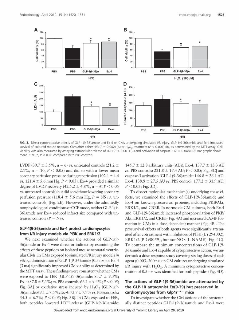
exerts direct effects on two cardiovascular cell types,namely CMs and ECs. In cultured neonatal CMs from WTmice, I/R injury was induced by either H/R or H2O2 treat-ment protocols, both well-recognized models for simu-lating I/R injury in vitro (23–27). Treatment of CMswith GLP-1(9-36)amide or Ex-4 resulted in improvedcell viability in response to these I/R injury models, asdetermined by the MTT assay, LDH release, andcaspase-3 activation.
Previous studies demonstrated that multiple down-stream signaling pathways are involved in mediating theprotective effects of GLP-1 (43). Similar to findings madewith whole hearts studied ex vivo, we observed that bothGLP-1(9-36)amide and Ex-4 significantly increased
cAMP levels and phosphorylation ofPI3K targets PKB/Akt and ERK1/2 andCREB in CMs maintained under nor-moxic conditions in vitro. It is importantto note that each of these signaling path-ways has been reported to play a criticalrole in cardioprotection against I/R in-jury (44–49). Of these key candidates,the cytoprotective effects of both GLP-1(9-36)amide and Ex-4 were attenu-ated by inhibitors of PI3K andERK1/2. Together these data providestrong evidence that both GLP-1(9-36)amide and Ex-4 exert cytoprotec-tive effects through PI3K-PKB- andMAPK-ERK1/2-dependent signalingpathways.
Consistent with our hypothesis that aGLP-1R-independent (i.e. alternate re-ceptor) signaling mechanism exists forGLP-1(9-36)amide, the actions of GLP-1(9-36)amide, but not Ex-4, were main-tained in CMs isolated from Glp1r�/�
mice. Moreover, only GLP-1(9-36)amide,and not Ex-4, improved the survival ofHAECs undergoing simulated I/R injury.Unexpectedly, the actions of GLP-1(9-36)amide in CMs isolated from both WTand Glp1r�/� mice were blunted by co-treatment with Ex(9-39). This intriguingfinding suggests that Ex(9-39) is capableof blocking not only GLP-1R-dependentmechanisms but also GLP-1R-independentactionsmediatedbyGLP-1(9-36)amide.Todate, it has been widely assumed that theactions of Ex(9-39) are relatively specificfor the classical GLP-1R. However, ourcurrent results strongly suggest Ex(9-39)also functions as a GLP-1(9-36)amide an-
tagonist. In retrospect, there have been many previous re-ports on nonspecific effects of Ex(9-39). For example,Ex(9-39) antagonized cAMP production and insulin releaseby glucose-dependent insulinotropic polypeptide (50, 51),and Ex(9-39) alone can cause concentration-dependent va-sorelaxation (52). Hence, our current findings shouldprompt a reassessment of the specificity of Ex(9-39) whenused as a putative GLP-1R antagonist.
Finally, it is noteworthy that although the concentra-tions of GLP-1(9-36)amide capable of eliciting vasodila-tory responses in the rat femoral artery (0.001–10 nM) (16)overlap the range of GLP-1(9-36)amide concentrationsmeasured in man (0.025–0.1 nM) (15), our study identi-
A
80
100
120
y (%
) * *
0
20
40
60
80
Cel
l via
bilit
y
B100
120
%) * *
H/R
0
PBS Insulin GLP-1(9-36)A Ex-4PBS
20
40
60
80
Cel
l via
bilit
y (% *
120C H2O2 (700 µµM)
0
20
PBS Insulin GLP-1(9-36)A Ex-4PBS
60
80
100
viab
ility
(%) * *
0
20
40
Cel
lv
PBS Insulin GLP-1(9-36)A Ex-4 PBS Insulin GLP-1(9-36)A Ex-4
+ L-NAME
H2O2 (700 µM)
PBS
FIG. 7. GLP-1(9-36)amide but not Ex-4 exerts cytoprotective effects in human ECs. A, GLP-1(9-36)amide but not Ex-4 improved survival of HAECs undergoing simulated I/R injury (H/R:P � 0.033; H2O2: P � 0.014). B, Salutary effects of GLP-1(9-36)amide in HAECs wereabolished by pretreatment with L-NAME (150 �M) (P � 0.009). Bar graphs show mean � SE.*, P � 0.05 compared with PBS controls.
Endocrinology, April 2010, 151(4):1520–1531 endo.endojournals.org 1529
at University of Toronto Library on April 29, 2010 endo.endojournals.orgDownloaded from

fied the minimum cytoprotective concentration of GLP-1(9-36)amide (or Ex-4) in isolated CMs to be 0.3 nM, avalue admittedly more pharmacological than physiologi-cal. Nevertheless, local tissue concentrations of GLP-1(9-36)amide have not been well studied and the minimumconcentration of GLP-1(9-36)amide capable of elicitingcardioprotective actions in vivo has yet to be established.
In conclusion, we demonstrate that GLP-1(9-36)amideexhibits many of the beneficial cardiovascular effects attrib-uted to GLP-1 but mediates these effects through aGLP-1 receptor-independent, Ex(9-39)-sensitive mecha-nism. Identification and characterization of the putativeGLP-1(9-36)amide receptor and further elucidation of therelevance of GLP-1(9-36)amide action in humans may leadto enhanced understanding of the cardiovascular biology ofGLP-1 and related peptides.
Acknowledgments
Address all correspondence and requests for reprints to: MansoorHusain, TMDT 3-904, 200 Elizabeth Street, Toronto, Ontario,Canada M5G 2C4. E-mail: [email protected].
This work was supported in part by operating Grants IRO-80668 from the Canadian Institutes of Health Research (toD.J.D.) and NA-5926 from the HSFO (to M.H. and D.J.D.). K.B.and K.-H.K. were supported in part by graduate studentshipsfrom the Heart and Stroke Richard Lewar Centre of Excellencein Cardiovascular Research, University of Toronto. D.J.D. issupported by a Chair in Regulatory Peptides from the CanadaResearch Chairs Program. M.H. and P.H.B. are supported byCareer Investigator Awards (CI-5503 and I-6891) of theHeart and Stroke Foundation of Ontario (HSFO).
Disclosure Summary: D.J.D. has served as an adviser or con-sultant within the past 12 months to Amgen Inc., Amylin Phar-maceuticals, Arisaph Pharmaceuticals Inc., Chugai Inc., Eli LillyInc., Glaxo Smith Kline, Glenmark Pharmaceuticals, Isis Phar-maceuticals Inc., Johnson & Johnson, Merck Research Labora-tories, Novartis Pharmaceuticals, Novo Nordisk Inc., NPS Phar-maceuticals Inc., Phenomix Inc., Takeda, and TransitionPharmaceuticals Inc. Neither D.J.D. nor M.H. or their familieshold stock directly or indirectly in any of these companies. Noother authors have any disclosures.
References
1. Elrick H, Stimmler L, Hlad Jr CJ, Arai Y 1964 Plasma insulin re-sponses to oral and intravenous glucose administration. J Clin En-docrinol Metab 24:1076–1082
2. McIntyre N, Holdsworth CD, Turner DS 1965 Intestinal factors in thecontrol of insulin secretion. J Clin Endocrinol Metab 25:1317–1324
3. Baggio LL, Drucker DJ 2007 Biology of incretins: GLP-1 and GIP.Gastroenterology 132:2131–2157
4. Kieffer TJ, McIntosh CH, Pederson RA 1995 Degradation of glu-cose-dependent insulinotropic polypeptide and truncated glucagon-
like peptide 1 in vitro and in vivo by dipeptidyl peptidase IV. En-docrinology 136:3585–3596
5. Mentlein R, Gallwitz B, Schmidt WE 1993 Dipeptidyl-peptidase IVhydrolyses gastric inhibitory polypeptide, glucagon-like peptide-1(7-36)amide, peptide histidine methionine and is responsible fortheir degradation in human serum. Eur J Biochem 214:829–835
6. Vilsbøll T, Agersø H, Krarup T, Holst JJ 2003 Similar eliminationrates of glucagon-like peptide-1 in obese type 2 diabetic patients andhealthy subjects. J Clin Endocrinol Metab 88:220–224
7. Deacon CF, Johnsen AH, Holst JJ 1995 Degradation of glucagon-like peptide-1 by human plasma in vitro yields an N-terminally trun-cated peptide that is a major endogenous metabolite in vivo. J ClinEndocrinol Metab 80:952–957
8. Knudsen LB, Pridal L 1996 Glucagon-like peptide-1-(9-36) amide isa major metabolite of glucagon-like peptide-1-(7-36) amide after invivo administration to dogs, and it acts as an antagonist on thepancreatic receptor. Eur J Pharmacol 318:429–435
9. Deacon CF, Plamboeck A, Møller S, Holst JJ 2002 GLP-1-(9-36)amide reduces blood glucose in anesthetized pigs by a mechanismthat does not involve insulin secretion. Am J Physiol 282:E873–E879
10. Vahl TP, Paty BW, Fuller BD, Prigeon RL, D’Alessio DA 2003Effects of GLP-1-(7-36)NH2, GLP-1-(7-37), and GLP-1-(9-36)NH2 on intravenous glucose tolerance and glucose-induced in-sulin secretion in healthy humans. J Clin Endocrinol Metab 88:1772–1779
11. Zander M, Madsbad S, Deacon CF, Holst JJ 2006 The metabolitegenerated by dipeptidyl-peptidase 4 metabolism of glucagon-likepeptide-1 has no influence on plasma glucose levels in patients withtype 2 diabetes. Diabetologia 49:369–374
12. Nikolaidis LA, Elahi D, Shen YT, Shannon RP 2005 Active metab-olite of GLP-1 mediates myocardial glucose uptake and improvesleft ventricular performance in conscious dogs with dilated cardio-myopathy. Am J Physiol Heart Circ Physiol 289:H2401–H2408
13. Sonne DP, Engstrøm T, Treiman M 2008 Protective effects of GLP-1analogues exendin-4 and GLP-1(9-36) amide against ischemia-reperfusion injury in rat heart. Regul Pept 146:243–249
14. Ban K, Noyan-Ashraf MH, Hoefer J, Bolz SS, Drucker DJ, HusainM 2008 Cardioprotective and vasodilatory actions of glucagon-likepeptide 1 receptor are mediated through both glucagon-like peptide1 receptor-dependent and -independent pathways. Circulation 117:2340–2350
15. Meier JJ, Nauck MA, Kranz D, Holst JJ, Deacon CF, Gaeckler D,Schmidt WE, Gallwitz B 2004 Secretion, degradation, and elimina-tion of glucagon-like peptide 1 and gastric inhibitory polypeptide inpatients with chronic renal insufficiency and healthy control sub-jects. Diabetes 53:654–662
16. Nathanson D, Erdogdu O, Pernow J, Zhang Q, Nystrom T 2009Endothelial dysfunction induced by triglycerides is not restored byexenatide in rat conduit arteries ex vivo. Regul Peptides 157:8–13
17. Drucker DJ, Nauck MA 2006 The incretin system: glucagon-likepeptide-1 receptor agonists and dipeptidyl peptidase-4 inhibitors intype 2 diabetes. Lancet 368:1696–1705
18. Scrocchi LA, Brown TJ, MaClusky N, Brubaker PL, Auerbach AB,Joyner AL, Drucker DJ 1996 Glucose intolerance but normal satietyin mice with a null mutation in the glucagon-like peptide receptorgene. Nat Med 2:1254–1258
19. Ban K, Cooper AJ, Samuel S, Bhatti A, Patel M, Izumo S, PenningerJM, Backx PH, Oudit GY, Tsushima RG 2008 Phosphatidylinositol3-kinase � is a critical mediator of myocardial ischemic and ade-nosine-mediated preconditioning. Circ Res 103:643–653
20. Noyan-Ashraf MH, Momen MA, Ban K, Sadi AM, Zhou YQ, RiaziAM, Baggio LL, Henkelman RM, Husain M, Drucker DJ 2009GLP-1R agonist liraglutide activates cytoprotective pathways andimproves outcomes after experimental myocardial infarction inmice. Diabetes 58:975–983
21. Kim KH, Oudit GY, Backx PH 2008 Erythropoietin protects againstdoxorubicin-induced cardiomyopathy via a phosphatidylinositol3-kinase-dependent pathway. J Pharmacol Exp Ther 324:160–169
1530 Ban et al. Cytoprotective Effects of GLP-1(9-36)Amide Endocrinology, April 2010, 151(4):1520–1531
at University of Toronto Library on April 29, 2010 endo.endojournals.orgDownloaded from

22. Costantini DL, Arruda EP, Agarwal P, Kim KH, Zhu Y, Zhu W,Lebel M, Cheng CW, Park CY, Pierce SA, Guerchicoff A, PollevickGD, Chan TY, Kabir MG, Cheng SH, Husain M, Antzelevitch C,Srivastava D, Gross GJ, Hui CC, Backx PH, Bruneau BG 2005 Thehomeodomain transcription factor Irx5 establishes the mouse car-diac ventricular repolarization gradient. Cell 123:347–358
23. Fujio Y, Nguyen T, Wencker D, Kitsis RN, Walsh K 2000 Akt pro-motes survival of cardiomyocytes in vitro and protects against isch-emia-reperfusion injury in mouse heart. Circulation 101:660–667
24. Gao F, Gao E, Yue TL, Ohlstein EH, Lopez BL, Christopher TA, MaXL 2002 Nitric oxide mediates the antiapoptotic effect of insulin inmyocardial ischemia-reperfusion: the roles of PI3-kinase, Akt, andendothelial nitric oxide synthase phosphorylation. Circulation 105:1497–1502
25. Morisco C, Marrone C, Trimarco V, Crispo S, Monti MG,Sadoshima J, Trimarco B 2007 Insulin resistance affects the cy-toprotective effect of insulin in cardiomyocytes through an im-pairment of MAPK phosphatase-1 expression. Cardiovasc Res76:453– 464
26. Tao L, Gao E, Jiao X, Yuan Y, Li S, Christopher TA, Lopez BL, KochW, Chan L, Goldstein BJ, Ma XL 2007 Adiponectin cardioprotec-tion after myocardial ischemia/reperfusion involves the reduction ofoxidative/nitrative stress. Circulation 115:1408–1416
27. Oshima Y, Ouchi N, Sato K, Izumiya Y, Pimentel DR, Walsh K 2008Follistatin-like 1 is an Akt-regulated cardioprotective factor that issecreted by the heart. Circulation 117:3099–3108
28. Ohori K, Miura T, Tanno M, Miki T, Sato T, Ishikawa S, Horio Y,Shimamoto K 2008 Ser9 phosphorylation of mitochondrial GSK-3�is a primary mechanism of cardiomyocyte protection by erythro-poietin against oxidant-induced apoptosis. Am J Physiol Heart CircPhysiol 295:H2079–H2086
29. Eguchi M, Liu Y, Shin EJ, Sweeney G 2008 Leptin protects H9c2rat cardiomyocytes from H2O2-induced apoptosis. FEBS J 275:3136 –3144
30. Han H, Long H, Wang H, Wang J, Zhang Y, Wang Z 2004 Pro-gressive apoptotic cell death triggered by transient oxidative insultin H9c2 rat ventricular cells: a novel pattern of apoptosis and themechanisms. Am J Physiol Heart Circ Physiol 286:H2169–H2182
31. Murata H, Ihara Y, Nakamura H, Yodoi J, Sumikawa K, Kondo T2003 Glutaredoxin exerts an antiapoptotic effect by regulating theredox state of Akt. J Biol Chem 278:50226–50233
32. Yasuoka C, Ihara Y, Ikeda S, Miyahara Y, Kondo T, Kohno S 2004Antiapoptotic activity of Akt is down-regulated by Ca2� in myo-cardiac H9c2 cells: evidence of Ca2�-dependent regulation of pro-tein phosphatase 2Ac. J Biol Chem 279:51182–51192
33. Emanueli C, Bonaria Salis M, Stacca T, Pintus G, Kirchmair R, IsnerJM, Pinna A, Gaspa L, Regoli D, Cayla C, Pesquero JB, Bader M,Madeddu P 2002 Targeting kinin B1 receptor for therapeutic neo-vascularization. Circulation 105:360–366
34. Esumi K, Nishida M, Shaw D, Smith TW, Marsh JD 1991 NADHmeasurements in adult rat myocytes during simulated ischemia.Am J Physiol Heart Circ Physiol 260:H1743–H1752
35. Ma H, Zhang HF, Yu L, Zhang QJ, Li J, Huo JH, Li X, Guo WY,Wang HC, Gao F 2006 Vasculoprotective effect of insulin in theischemic/reperfused canine heart: role of Akt-stimulated NO pro-duction. Cardiovasc Res 69:57–65
36. Bergmann MW, Rechner C, Freund C, Baurand A, El Jamali A,Dietz R 2004 Statins inhibit reoxygenation-induced cardiomyocyteapoptosis: role for glycogen synthase kinase 3� and transcriptionfactor �-catenin. J Mol Cell Cardiol 37:681–690
37. Dhanasekaran A, Gruenloh SK, Buonaccorsi JN, Zhang R, GrossGJ, Falck JR, Patel PK, Jacobs ER, Medhora M 2008 Multiple an-
tiapoptotic targets of the PI3K/Akt survival pathway are activatedby epoxyeicosatrienoic acids to protect cardiomyocytes from hyp-oxia/anoxia. Am J Physiol Heart Circ Physiol 294:H724–H735
38. Germack R, Dickenson JM 2005 Adenosine triggers precondition-ing through MEK/ERK1/2 signalling pathway during hypoxia/reoxygenation in neonatal rat cardiomyocytes. J Mol Cell Cardiol39:429–442
39. Gomez LA, Alekseev AE, Aleksandrova LA, Brady PA, Terzic A1997 Use of the MTT assay in adult ventricular cardiomyocytes toassess viability: effects of adenosine and potassium on cellular sur-vival. J Mol Cell Cardiol 29:1255–1266
40. Thuerauf DJ, Marcinko M, Gude N, Rubio M, Sussman MA, Glem-botski CC 2006 Activation of the unfolded protein response in in-farcted mouse heart and hypoxic cultured cardiac myocytes. CircRes 99:275–282
41. Woo AY, Cheng CH, Waye MM 2005 Baicalein protects rat car-diomyocytes from hypoxia/reoxygenation damage via a prooxidantmechanism. Cardiovasc Res 65:244–253
42. KulasingamV,SmithCR,Batruch I,BucklerA, JefferyDA,DiamandisEP 2008 “Product ion monitoring” assay for prostate-specific antigenin serum using a linear ion-trap. J Proteome Res 7:640–647
43. Bose AK, Mocanu MM, Carr RD, Brand CL, Yellon DM 2005Glucagon-like peptide-1 (GLP-1) can directly protect the heartagainst ischemia/reperfusion injury. Diabetes 54:146–151
44. Das S, Tosaki A, Bagchi D, Maulik N, Das DK 2005 Resveratrol-mediated activation of cAMP response element-binding proteinthrough adenosine A3 receptor by Akt-dependent and -independentpathways. J Pharmacol Exp Ther 314:762–769
45. Marais E, Genade S, Lochner A 2008 CREB activation and isch-aemic preconditioning. Cardiovasc Drugs Ther 22:3–17
46. Markou T, Hadzopoulou-Cladaras M, Lazou A 2004 Phenyleph-rine induces activation of CREB in adult rat cardiac myocytesthrough MSK1 and PKA signaling pathways. J Mol Cell Cardiol37:1001–1011
47. Matus M, Lewin G, Stumpel F, Buchwalow IB, Schneider MD,Schutz G, Schmitz W, Muller FU 2007 Cardiomyocyte-specific in-activation of transcription factor CREB in mice. FASEB J 21:1884–1892
48. Mehrhof FB, Muller FU, Bergmann MW, Li P, Wang Y, Schmitz W,Dietz R, von Harsdorf R 2001 In cardiomyocyte hypoxia, insulin-likegrowthfactor-I-inducedantiapoptoticsignalingrequiresphosphatidyl-inositol-3-OH-kinase-dependent and mitogen-activated protein ki-nase-dependent activation of the transcription factor cAMP responseelement-binding protein. Circulation 104:2088–2094
49. Nagy N, Shiroto K, Malik G, Huang CK, Gaestel M, Abdellatif M,Tosaki A, Maulik N, Das DK 2007 Ischemic preconditioning in-volves dual cardio-protective axes with p38MAPK as upstream tar-get. J Mol Cell Cardiol 42:981–990
50. Gault VA, O’Harte FP, Harriott P, Mooney MH, Green BD, Flatt PR2003 Effects of the novel (Pro3)GIP antagonist and exendin(9-39)amide on GIP- and GLP-1-induced cyclic AMP generation, in-sulin secretion and postprandial insulin release in obese diabetic(ob/ob) mice: evidence that GIP is the major physiological incretin.Diabetologia 46:222–230
51. Wheeler MB, Gelling RW, McIntosh CH, Georgiou J, Brown JC,Pederson RA 1995 Functional expression of the rat pancreatic isletglucose-dependent insulinotropic polypeptide receptor: ligand bindingand intracellular signaling properties. Endocrinology 136:4629–4639
52. Green BD, Hand KV, Dougan JE, McDonnell BM, Cassidy RS,Grieve DJ 2008 GLP-1 and related peptides cause concentration-dependent relaxation of rat aorta through a pathway involvingKATP and cAMP. Arch Biochem Biophys 478:136–142
Endocrinology, April 2010, 151(4):1520–1531 endo.endojournals.org 1531
at University of Toronto Library on April 29, 2010 endo.endojournals.orgDownloaded from


![Hannah v Peel (1945) - [1945] K.B. 509](https://static.fdocuments.us/doc/165x107/544f9c22b1af9f11098b460a/hannah-v-peel-1945-1945-kb-509.jpg)