GLOBAL ATLAS OF ALLERGY 1 DIAGNOSIS - SKIN TESTS€¦ · - Allergy diagnosis SKIN TESTS FOR FOOD...
27
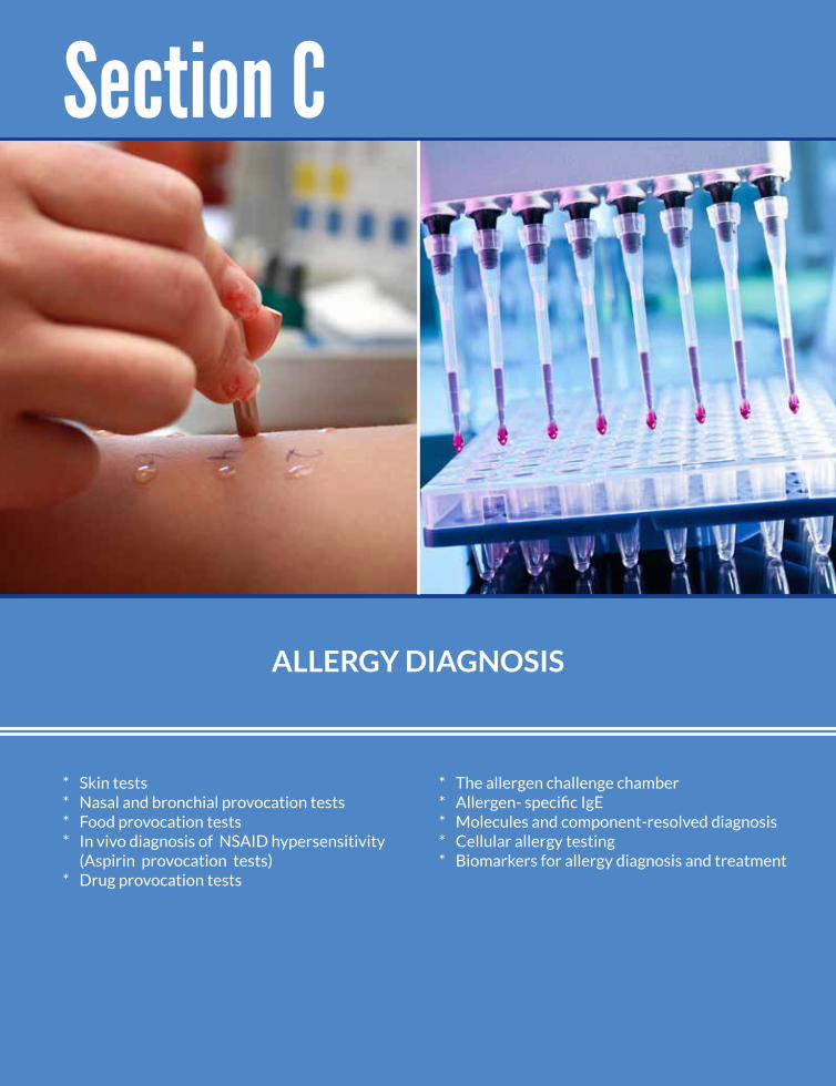
Section C ALLERGY DIAGNOSIS * Skin tests * Nasal and bronchial provocation tests * Food provocation tests * In vivo diagnosis of NSAID hypersensitivity (Aspirin provocation tests) * Drug provocation tests * The allergen challenge chamber * Allergen- specific IgE * Molecules and component-resolved diagnosis * Cellular allergy testing * Biomarkers for allergy diagnosis and treatment
Transcript of GLOBAL ATLAS OF ALLERGY 1 DIAGNOSIS - SKIN TESTS€¦ · - Allergy diagnosis SKIN TESTS FOR FOOD...
Section C
ALLERGY DIAGNOSIS
* Skin tests* Nasal and bronchial provocation tests* Food provocation tests * In vivo diagnosis of NSAID hypersensitivity
(Aspirin provocation tests)* Drug provocation tests
* The allergen challenge chamber * Allergen- specific IgE * Molecules and component-resolved diagnosis * Cellular allergy testing* Biomarkers for allergy diagnosis and treatment
150
Global atlas oF allerGyse
ct
ion
c -
Alle
rgy
dia
gno
sis • Skin tests are a cornerstone in the diagnosis of allergic diseases
• Performing skin tests needs a specific training• Skin Prick Tests are an excellent tool to detect sensitizations to
inhalant and food allergens• Intradermal tests are of paramount importance in venom and
drug hypersensitivity
Skin tests are an important diag-nostic tool in the work up of many allergic diseases (Table 1). Skin tests are widely used for the diag-nosis of inhalant allergies, but also allergies to foods, venom, occupa-tional agents and drugs can be in-vestigated by skin tests.
TYPES OF SKIN TESTS AND REQUIREMENTSSkin prick tests (SPTs) and intra-dermal tests are still the corner-stone of the diagnosis of IgE-me-diated (type I) allergy. Especially SPTs are widely used (Figure 1). As they are usually easy to perform, cheap and allow a fast reading, they are an ideal tool for bedside diagnosis. Intradermal also named intracutaneous testing (Figure 2) is more demanding, but allows by titration of various allergen con-centrations a very accurate rating of the sensitization level.
Epicutaneous or patch tests are used in patients with suspected type IV allergies for contact der-matitis or delayed type of drug hypersensitivity. Due to its po-tentially scar-inducing effect skin testing by scratching is limited to some special aspects of drug or mostly occupational allergens. The atopy patch tests (APT) may help to find out causes of exacerbation
in atopic dermatitis or Eosinophil-ic Esophagitis, but its value is still controversial. Performing skin tests needs a specific training, es-pecially for intradermal and epicu-taneous tests with nonstandard-ized allergen material.
Contraindications such as the use of some drugs, pregnancy or other conditions precluding some types of allergy testing should be evalu-ated. Also the skin condition of the patient must allow testing in the skin. Patients have to abstain from medications such as antihistamin-ics that could mask a positive type I reactions; on the other hand con-ditions such as dermographism/pressure urticaria able to provoke false positive skin reactions must be considered. Using controls such as histamine for positive and saline as negative controls are cru-cial for SPT/intradermal testing.
SKIN TESTS FOR INHALANT ALLERGENS SPT are the classical diagnostic tool get prompt information about sensitizations against inhalant al-lergens such as pollen, house dust mites, pets and, to a lesser extent moulds. As they are usually easy to perform, cheap and allow a fast reading, thus an ideal tool for bed-side diagnosis. Recommendations for standard series of SPT solu-tions are available for many geo-graphic and climatic regions.
Skin prick tests can also be used in tropical areas and in countries with limited resources, where al-lergy is booming. However, local allergens may not necessarily be identified or available and can therefore be missed for skin test-ing. For example, it is crucial to include allergens such as Blomia tropicalis in the skin test battery of tropical countries.
IN VIVO ALLERGY DIAGNOSIS - SKIN TESTS1
Ke y m e ssag e s
In vivo allergy diagnosis - skin tests
Julia Katharina Genser Peter Schmid-GrendelmeierUniversity Hospital of Zürich
Zürich, Switzerland

151
Global atlas oF allerGyse
ct
ion
c - A
llergy diagn
osis
In vivo allergy diagnosis - skin tests
TABLE 1
Difference between basophils and mast cells
Skin Prick Test
Intradermal Tests
Epicutaneous (Patch) Tests
Atopy Patch Test
Inhalant allergies+++
StandardRarely needed, few available allergens
n.a.Some forms of atopic
dermatitis1
Food allergy
++ With commercially
available extracts and fresh food
n.a. n.a.
Eosinophilic esophagitis? Some
forms of atopic dermatitis2
Venom allergy+
(reduced sensitivity) +++
(Titration)n.a. n.a.
Drug hypersenstivity + to +++++
(with all soluble drugs; Titration)
(++) In delayed drug
hypersensitivity3
n.a.
Contact allergens(+)
( in case of protein contact dermatitis)
n.a. +++
1 If Atopic Dermatitis seems to exacerbated by the presence of inhalant allergens 2 If Atopic Dermatitis seems to exacerbated by the presence of food allergens3 Especially if cutaneous manifestations of delayed type (eczema,maculo-papular rash) occur
a
bFigure 1 Skin prick test with a)
inhalant allergens and b) fresh food.
152
Global atlas oF allerGyse
ct
ion
c -
Alle
rgy
dia
gno
sis
SKIN TESTS FOR FOOD ALLERGY For food allergy mostly SPT is used, done with commercially available food allergen extracts but also with fresh food (Prick-to Prick technique, Figure 1b). Test-ing with fresh food enables a fast detection of sensitization also to foods individually provided by the patient or only locally available. As skin testing does not discriminate between sensitization and clinical relevant food allergy, additional tools such as food allergen oral provocation tests, elimination diets and more recently compo-nent-resolved in vitro diagnosis are indicated. Intradermal testing with food allergens is not recom-mended.
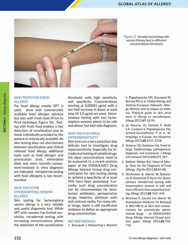
SKIN TESTS FOR HYMENOPTERA VENOM ALLERGYSkin testing for hymenoptera venom allergy is a very reliable and useful diagnostic tool. While SPT with venoms has limited sen-sitivity, intradermal testing with increasing concentrations allows the detection of the sensitization
threshold, with high sensitivity and specificity. Concentrations starting at 0.00001 μg/ml with a ten-fold increase in doses at each step till 1.0 μg/ml are used. Simul-taneous testing with two hyme-noptera venoms seems to be safe and allows fast bed-side diagnosis.
SKIN TESTS IN DRUG HYPERSENSITIVITYSkin tests are a very useful but also delicate tool to investigate drug hypersensitivity. Especially for in-tradermal testing of soluble drugs, the ideal concentrations need to be evaluated. In a recent position paper of the ENDA/EAACI Drug Allergy Interest Group drug con-centration for skin testing aiming to achieve a specificity of at least 95% have been postulated. Cur-rently such drug concentration can be recommended for beta-lactam antibiotics, perioperative drugs, heparins, platinum salts and contrast media. For many oth-er drugs, there is still insufficient evidence to define an appropriate drug concentration.
KEY REFERENCES1. Bousquet J, Heinzerling L, Bachert
C, Papadopoulos NG, Bousquet PJ, Burney PG et al. Global Allergy and Asthma European Network; Aller-gic Rhinitis and its Impact on Asth-ma. Practical guide to skin prick tests in allergy to aeroallergens. Allergy 2012;67:18-24.
2. de Monchy JG, Demoly P, Akdis CA, Cardona V, Papadopoulos NG, Schmid-Grendelmeier P et al. Al-lergology in Europe, the blueprint. Allergy 2013;68:1211-1218.
3. Sicherer SH, Sampson HA. Food al-lergy: Epidemiology, pathogenesis, diagnosis, and treatment. J Allergy Clin Immunol 2014;133:291-307.
4. Ballmer-Weber B.K. Value of Aller-gy Tests for the Diagnosis of Food Allergy. Dig Dis 2014;32:84-88.
5. Strohmeier B, Aberer W, Bokano-vic D, Komericki P, Sturm GJ. Simul-taneous intradermal testing with hymenoptera venoms is safe and more efficient than sequential test-ing. Allergy 2013;68: 542–544.
6. Brockow K, Garvey LH, Aberer W, Atanaskovic-Markovic M, Barbaud A, Bilo MB et al. Skin test concen-trations for systemically admin-istered drugs -- an ENDA/EAACI Drug Allergy Interest Group posi-tion paper. Allergy 2013;68:702-712.
Figure 2 Intradermal testing with venom (Honey bee) in different
concentrations (titration).
In vivo allergy diagnosis - skin tests
153
Global atlas oF allerGyse
ct
ion
c - A
llergy diagn
osis
• Nasal and bronchial provocation tests are of additional value in the understanding and diagnosis of allergic rhinitis and asthma
• These tests need to be performed in a clinical setting by dedicated staff
• It is strongly recommended to follow a validated and standardized protocol
• In experienced hands, the safety risks of challenge tests are low
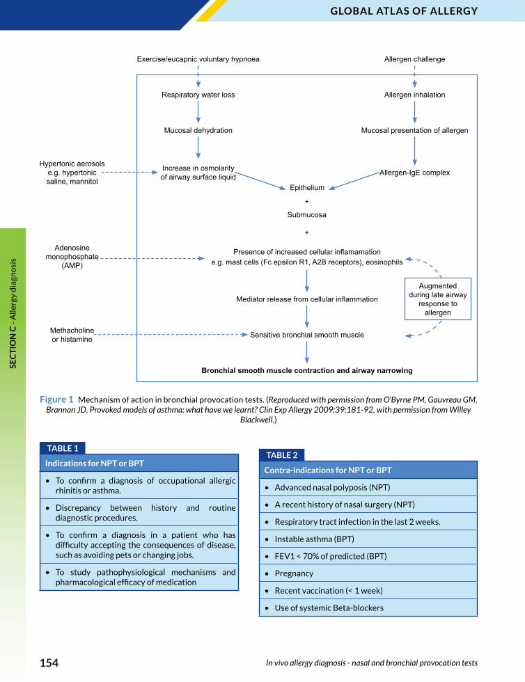
INTRODUCTIONEpidemiological studies have shown that inhaled allergens are an important cause of allergic rhi-nitis (AR) and asthma. Allergen inhalation challenge mimics the natural situation and is useful for understanding the mechanisms of allergic airway inflammation and airway hyperresponsiveness (AHR). Moreover, provocation tests play an important role in the diagnosis of asthma, and to a less-er extent also in AR. Via a bron-chial provocation test (BPT), AHR can be measured by challenging the airways with a variety of phys-ical and inhaled chemical stimuli resulting in airway constriction (Figure 1). The airway narrowing is measured by changes in FEV1 after gradually increasing the dose of provoking agent.
A nasal provocation test (NPT) is used in case of a typical history of AR and a negative allergy test (Figure 2). It is of importance to differentiate AR from non-allergic rhinitis since management may be different. A positive test result is defined by the presence of symp-toms typical for AR plus objective parameters, such as decreased upper airway patency (measured with acoustic rhinometry or an-
IN VIVO ALLERGY DIAGNOSIS- NASAL AND BRONCHIAL
PROVOCATION TESTS2a
Ke y m e ssag e s
In vivo allergy diagnosis - nasal and bronchial provocation tests
terior rhinomanometry) or nasal secretion of inflammatory media-tors. Patients can have immediate or dual responses.
METHODSDifferent methods for NPT and measurement of nasal respons-es are used. Medication that may interfere with the results should be stopped prior to the test and contra-indications should be ruled out. If available, it is advised to fol-low a validated and standardized protocol.
Inhaled allergen challenge starts early in the morning in order to measure a late asthmatic response. Ideally, patients are observed for a longer period. It is strongly recom-mended to perform BPT in a clini-cal setting. In daily clinical practice for AHR diagnosis, the preferred agents are histamine, methacho-
line and mannitol. These agents give a bronchoconstrive response that is less prolonged and quickly reversible with bronchodilators and inhaled corticosteroids. Diag-nosis of occupational asthma and rhinitis may require specific inha-lation tests.
LIMITATIONSInhaled allergen provocation tests are safe in a dedicated setting with experienced staff. Several objec-tive and validated parameters to measure response are available for BPT, but not for NPT. Allergen application may produce nonspe-cific reactions. A limited number of standardized allergen extracts are commercially available. Aller-gen challenge can result in a tran-sient increase in symptoms and medication use.
Gert Jan Braunstahl St. Franciscus Gasthuis
Rotterdam, the Netherlands
154
Global atlas oF allerGyse
ct
ion
c -
Alle
rgy
dia
gno
sis
In vivo allergy diagnosis - nasal and bronchial provocation tests
TABLE 2
Contra-indications for NPT or BPT
• Advanced nasal polyposis (NPT)
• A recent history of nasal surgery (NPT)
• Respiratory tract infection in the last 2 weeks.
• Instable asthma (BPT)
• FEV1 < 70% of predicted (BPT)
• Pregnancy
• Recent vaccination (< 1 week)
• Use of systemic Beta-blockers
Figure 1 Mechanism of action in bronchial provocation tests. (Reproduced with permission from O’Byrne PM, Gauvreau GM, Brannan JD. Provoked models of asthma: what have we learnt? Clin Exp Allergy 2009;39:181-92, with permission from Willey
Blackwell.)
TABLE 1
Indications for NPT or BPT
• To confirm a diagnosis of occupational allergic rhinitis or asthma.
• Discrepancy between history and routine diagnostic procedures.
• To confirm a diagnosis in a patient who has difficulty accepting the consequences of disease, such as avoiding pets or changing jobs.
• To study pathophysiological mechanisms and pharmacological efficacy of medication
Exercise/eucapnic voluntary hypnoea
Respiratory water loss
Mucosal dehydration
Increase in osmolarity of airway surface liquid
Allergen inhalation
Mucosal presentation of allergen
Allergen-IgE complexHypertonic aerosols
e.g. hypertonic saline, mannitol
Adenosine monophosphate
(AMP)
Methacholine or histamine
Epithelium
Submucosa
Presence of increased cellular inflamamatione.g. mast cells (Fc epsilon R1, A2B receptors), eosinophils
Mediator release from cellular inflammation
Sensitive bronchial smooth muscle
Bronchial smooth muscle contraction and airway narrowing
Allergen challenge
+
+
Augmented during late airway
response to allergen

155
Global atlas oF allerGyse
ct
ion
c - A
llergy diagn
osis
In vivo allergy diagnosis - nasal and bronchial provocation tests
SAFETY AND SIDE EFFECTSNPT and BPT are safe procedures. Generalized reactions are rare after BPT, but asthma exacerba-tions may occur. After allergen challenge the magnitude of the bronchoconstriction may be more difficult to control than with direct challenges, such as histamine or methacholine. Moreover, the in-duced bronchoconstriction is usu-ally prolonged due to the release of proinflammatory mediators.
Therefore, it is necessary that during an inhaled allergen provo-cation a doctor is present, the res-cue medication is ready to use and a resuscitation set is available on the ward.
KEY REFERENCES1. Bousquet J, Van Cauwenberge P,
Khaltaev N. Allergic rhinitis and its impact on asthma. J Allergy Clin Im-munol 2001;108:S147-334.
2. O'Byrne PM, Gauvreau GM, Bran-nan JD. Provoked models of asth-
ma: what have we learnt? Clin Exp Allergy 2009;39:181-192.
3. Diamant Z, Gauvreau GM, Cock-croft DW, Boulet LP, Sterk PJ, de Jongh FH, et al. Inhaled allergen bronchoprovocation tests. J Allergy Clin Immunol 2013;132:1045-1055 e6.
4. Rondon C, Campo P, Togias A, Fok-kens WJ, Durham SR, Powe DG, et al. Local allergic rhinitis: con-cept, pathophysiology, and man-agement. J Allergy Clin Immunol 2012;129:1460-1467.
Figure 2 Diagnostic approach to the patient with allergic rhinitis. (Reprinted from J Allergy Clin Immunol, 129/6, Rondon C, Campo P, Togias A, et al. Local allergic rhinitis: concept, pathophysiology, and management, 1460-1467, Copyright 2012, with
permission from Elsevier.)
Clinical History suggestive of rhinitis symptoms
Skin Prick Test with aeroallergens
Positive Negative
Concordant with clinical history
Disconcordant with clinical history Serum specific-IgE
Allergological target organ evaluation:NAPT and/or nasal specific IgE
Local Allergic Rhinitis Non - Allergic Rhinitis
Positive & concordant with clinical history
Positive & disconcordant with clinical history Negative
Positive Negative
Allergic Rhinitis
156
Global atlas oF allerGyse
ct
ion
c -
Alle
rgy
dia
gno
sis • Food challenge is the diagnostic gold standard for food allergy
since the presence of atopy in an individual might lead to false positive interpretation of IgE testing
• Patient safety is key to the design of a food challenge • Interpreting a food provocation test can be challenging, the
experience of the staff is decisive• Food challenges are essential procedures in the follow-up of
food allergy
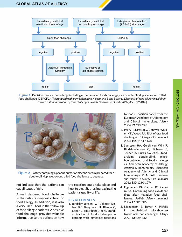
WHY DO WE DO FOOD CHALLENGES?Diagnosis of food allergy can be obvious, for example in the case of a child reacting with urticar-ia 10 minutes after having eaten peanuts. In this case caused by an IgE-mediated reaction, skin prick tests and in-vitro diagnosis, by measuring specific IgE in the se-rum, are most often positive and clearly relate to the symptoms. Food challenge might be done in patients with vague symptoms possibly related to food allergy, with a significant proportion of patients having positive IgE tests not related to the symptoms but to an atopic predisposition (false positive tests indicating only sen-sitization). In this context, a food challenge will be the gold standard for diagnosis (Figure 1).
HOW DO WE DO FOOD CHALLENGES?Several variables have to be taken into account when organizing a food challenge. Most important-ly, the food challenge must be designed in a safe way. The food challenge must be performed in a location, where emergency medi-cation can be administered. When doing high risk challenges, it is im-portant to have rapid access to in-
Philippe Eigenmann University Hospitals of Geneva
Geneva, Switzerland
IN-VIVO ALLERGY DIAGNOSIS- FOOD PROVOCATION TESTS 2b
Ke y m e ssag e s
In-vivo allergy diagnosis – food provocation tests
tensive care facilities. In all cases, the food challenge must be super-vised by nurses trained to recog-nize early signs of reactions. Also, the supervising physician must be trained in order to have sufficient experience in interpreting the various clinical signs possibly ob-served during a food challenge.
The food is provided to the patient in increasing doses. The starting dose, as well as the time interval between the doses, will be deter-mined by the initial history of the patient and by the purpose of the challenge. A food challenge aiming to determine the threshold level of a reaction will start with a very low dose.
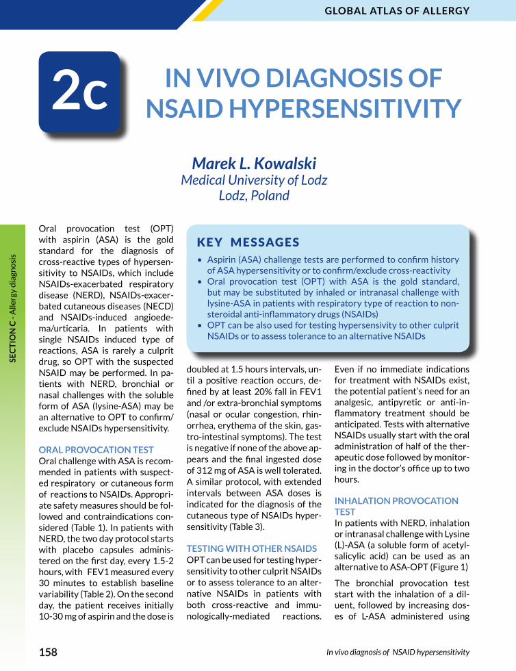
The food can be administered openly, but also in a double-blind placebo controlled manner. Dur-
ing this procedure, the challenge is separated into two parts, the food being hidden in a vehicle in order for blind both the patient and the examiner (Figure 2).
LIMITATIONS OF A FOOD CHALLENGE A well designed food challenge will provide definite information about the presence or the absence of a food allergy. Nevertheless, one needs to be aware of some caveats. A food challenge is valid only for the type of preparation of the food. As an example, patients may react to raw egg, but not to cooked egg, and a patient with a negative challenge to cooked egg might afterwards react to a prepa-ration with raw or not completely cooked egg. Similarly, a negative challenge to a specific fish does
157
Global atlas oF allerGyse
ct
ion
c - A
llergy diagn
osis
Figure 1 Decision tree for food allergy including either an open food challenge, or a double-blind, placebo-controlled food challenge (DBPCFC). (Reproduced with permission from Niggemann B and Beyer K. Diagnosis of food allergy in children:
toward a standardization of food challengeJ Pediatr Gastroenterol Nutr 2007, 45, 399-404.)
not indicate that the patient can eat all types of fish.
A well designed food challenge is the definite diagnostic tool for food allergy. In addition, it is also a very useful tool in the follow-up of food allergic patients. A positive food challenge provides valuable information to the patient on how
the reaction could take place and how to treat it, thus increasing the patient’s quality of life.
KEY REFERENCES1. Bindslev-Jensen C, Ballmer-We-
ber BK, Bengtsson U, Blanco C, Ebner C, Hourihane J et al. Stand-ardization of food challenges in patients with immediate reactions
to foods – position paper from the European Academy of Allergology and Clinical Immunology. Allergy 2004;59:690-697.
2. Perry TT, Matsui EC, Conover-Walk-er MK, Wood RA. Risk of oral food challenges. J Allergy Clin Immunol 2004;114:1164-1168.
3. Sampson HA, Gerth van Wijk R, Bindslev-Jensen C, Sicherer S, Teuber SS, Burks AW et al. Stand-ardizing double-blind, place-bo-controlled oral food challeng-es: American Academy of Allergy, Asthma & Immunology–European Academy of Allergy and Clinical Immunology PRACTALL consen-sus report. J Allergy Clin Immunol 2012;130:1260-1274.
4. Eigenmann PA, Caubet JC, Zamo-ra SA. Continuing food-avoidance diets after negative food chal-lenges. Pediatr Allergy Immunol 2006;17:601-605.
5. Niggemann B, Beyer K. Pitfalls in double-blind, placebo-con-trolled oral food challenges. Allergy 2007;62:729-732.
Figure 2 Pastry containing a peanut butter or placebo cream prepared for a double-blind, placebo-controlled food challenge to peanuts.
In-vivo allergy diagnosis – food provocation tests
Immediate type clinical reaction < 1 year of age
Immediate type clinical reaction 1+ year of age
Late phase clinic reaction (AE & GI) at any age
Open food challenge DBPCFC
negative positive
Objective, immediate symptom
Subjective or late phase reaction
no diet diet
negative positive
no diet
158
Global atlas oF allerGyse
ct
ion
c -
Alle
rgy
dia
gno
sis • Aspirin (ASA) challenge tests are performed to confirm history
of ASA hypersensitivity or to confirm/exclude cross-reactivity • Oral provocation test (OPT) with ASA is the gold standard,
but may be substituted by inhaled or intranasal challenge with lysine-ASA in patients with respiratory type of reaction to non-steroidal anti-inflammatory drugs (NSAIDs)
• OPT can be also used for testing hypersensivity to other culprit NSAIDs or to assess tolerance to an alternative NSAIDs
Oral provocation test (OPT) with aspirin (ASA) is the gold standard for the diagnosis of cross-reactive types of hypersen-sitivity to NSAIDs, which include NSAIDs-exacerbated respiratory disease (NERD), NSAIDs-exacer-bated cutaneous diseases (NECD) and NSAIDs-induced angioede-ma/urticaria. In patients with single NSAIDs induced type of reactions, ASA is rarely a culprit drug, so OPT with the suspected NSAID may be performed. In pa-tients with NERD, bronchial or nasal challenges with the soluble form of ASA (lysine-ASA) may be an alternative to OPT to confirm/exclude NSAIDs hypersensitivity.
ORAL PROVOCATION TEST Oral challenge with ASA is recom-mended in patients with suspect-ed respiratory or cutaneous form of reactions to NSAIDs. Appropri-ate safety measures should be fol-lowed and contraindications con-sidered (Table 1). In patients with NERD, the two day protocol starts with placebo capsules adminis-tered on the first day, every 1.5-2 hours, with FEV1 measured every 30 minutes to establish baseline variability (Table 2). On the second day, the patient receives initially 10-30 mg of aspirin and the dose is
Marek L. Kowalski Medical University of Lodz
Lodz, Poland
IN VIVO DIAGNOSIS OF NSAID HYPERSENSITIVITY2c
Ke y m e ssag e s
In vivo diagnosis of NSAID hypersensitivity
doubled at 1.5 hours intervals, un-til a positive reaction occurs, de-fined by at least 20% fall in FEV1 and /or extra-bronchial symptoms (nasal or ocular congestion, rhin-orrhea, erythema of the skin, gas-tro-intestinal symptoms). The test is negative if none of the above ap-pears and the final ingested dose of 312 mg of ASA is well tolerated. A similar protocol, with extended intervals between ASA doses is indicated for the diagnosis of the cutaneous type of NSAIDs hyper-sensitivity (Table 3).
TESTING WITH OTHER NSAIDS OPT can be used for testing hyper-sensitivity to other culprit NSAIDs or to assess tolerance to an alter-native NSAIDs in patients with both cross-reactive and immu-nologically-mediated reactions.
Even if no immediate indications for treatment with NSAIDs exist, the potential patient’s need for an analgesic, antipyretic or anti-in-flammatory treatment should be anticipated. Tests with alternative NSAIDs usually start with the oral administration of half of the ther-apeutic dose followed by monitor-ing in the doctor’s office up to two hours.
INHALATION PROVOCATION TEST In patients with NERD, inhalation or intranasal challenge with Lysine (L)-ASA (a soluble form of acetyl-salicylic acid) can be used as an alternative to ASA-OPT (Figure 1)
The bronchial provocation test start with the inhalation of a dil-uent, followed by increasing dos-es of L-ASA administered using
159
Global atlas oF allerGyse
ct
ion
c - A
llergy diagn
osis
In vivo diagnosis of NSAID hypersensitivity
TABLE 1
Indications and contra indications to oral aspirin challenge in patients with NSAIDs hypersensitivity
Indications
• Ambiguous or not clear history of response to aspirin/NSAIDs
• Confirmation/exclusion of cross-sensitivity if aspirin is not a culprit NSAID
• Beginning of desensitization
Contraindications
• History of severe anaphylactic reactions induced by aspirin or other NSAIDs
• Not well controlled underlying disease (asthma/urticaria)
• Low baseline FEV1 (below 70% of the predicted value )
• Severe delayed type reactions
• Concomitant disorders potentially aggravated by the challenge
TABLE 2
Protocol for the oral aspirin challenge in a patient with suspected NSAIDs exacerbated respiratory diseases
8:00 Placebo 10 mga
9:30 Placebo 27 mg
11:00 Placebo 44 mg
12:30 Placebo 117 mg
14:00 312 mg
a Optional in patients with a history of severe reaction to NSAIDs
TABLE 3
Protocol for oral aspirin challenge in a patient with suspected NSAIDs exacerbated cutaneous disease
Time Day 1 Day 2
8:00 Placebo 71 mg
10:00 Placebo 117 mg
12:00 Placebo 312 mg
14:00 Placebo 500 mg
a dosimeter at 30 minutes inter-vals, allowing to complete the procedure in an outpatient set-ting in less than 5 hours. The test is considered positive, if at least 20% drop in FEV1 is recorded at 10, 20 or 30 minutes after inha-
lation. If the positive reaction is observed PD20 ASA is calculated from the dose-response curve. Inhalation challenge may induce extrabronchial symptoms and may also involve a late phase reaction. Both oral and inhaled tests have
similar sensitivity and specificity
but, as compared to the oral chal-
lenge, the inhalation test, is faster
and safer to perform (the reaction
is usually easily reversible with
nebulized beta2 agonists).

160
Global atlas oF allerGyse
ct
ion
c -
Alle
rgy
dia
gno
sis
Advantages Limitations
Oral challenge
Gold standard (reliable ) Risk of severe reaction
Useful in patients with extra bronchial symptoms
Time-consuming
Bronchial challenge
Short duration (< 4 h) Only in patients with
bronchial/nasal symptoms
Reaction easily reversibleRequires special equipment
(dosimeter)
Nasal challenge
Safe in patients with severe/ unstable asthma
Only in patients with bronchial/nasal symptoms
Not possible in patients with nasal obstruction
The nasal provocation test with L-ASA is particularly recommend-ed in asthmatic patients with low pulmonary function not suitable for bronchial provocation. The challenge starts with instillation of saline followed by L-ASA solution increasing concentrations applied into each nostril every 30 minutes. All parameters (objective and subjective) are recorded every 10 minutes. Alternatively, ketorolac nasal spray solution can be used. For the assessment of the chal-lenge, clinical symptoms are com-bined with the objective measures of nasal airflow patency (acoustic rhinometry, rhinomanometry or peak nasal inspiratory flow).
The reaction is considered posi-tive, if clinical symptoms appear and/or a significant nasal obstruc-
tion is objectively documented (a 25% decrease in total nasal flow or 40% drop of the inspiratory nasal flow, as compared to baseline).
In experienced hands the sensi-tivity of intranasal aspirin prov-ocation may rise above 80% and specificity approaches 95%, thus reaching the performance of the bronchial challenge.
The nasal challenge route is not recommended in patients with significant nasal obstruction, tur-bulent nasal flow or with unspecif-ic nasal responsiveness.
KEY REFERENCES 1. Kowalski ML, Asero R, Bavbek S,
Blanca M, Blanca-Lopez N, Bo-chenek G et al. Classification and practical approach to the diagnosis and management of hypersensitiv-
ity to nonsteroidal anti-inflamma-tory drugs. Allergy 2013;68:1219-1232.
2. Nizankowska-Mogilnicka E, Bo-chenek G, Mastalerz L, Swierczyńs-ka M, Picado C, Scadding G et al. EAACI/GA2LEN guideline: aspirin provocation tests for diagnosis of aspirin hypersensitivity. Allergy 2007;62 :1111-1118.
3. Melillo G, Balzano G, Bianco S, Dahlen B, Godard P, Kowalski ML, et al. Oral and inhalation provoca-tion tests for the diagnosis of aspi-rin-induced asthma. Allergy 2001; 56:899-911.
4. Lee RU, White AA, Ding D, Dursun AB, Woessner KM, Simon RA, et al. Use of intranasal ketorolac and modified oral aspirin challenge for desensitization of aspirin-exac-erbated respiratory disease. Ann Allergy Asthma Immunol 2010;105: 130-135.
Figure 1 Advantages and limitations of oral, bronchial and intranasal route of aspirin challenge in patients with NSAIDs- exacerbated respiratory disease.
In vivo diagnosis of NSAID hypersensitivity
161
Global atlas oF allerGyse
ct
ion
c - A
llergy diagn
osis
• For many drug hypersensitivity reactions, no sensitization can be demonstrated
• A drug provocation test is the controlled administration of a drug for diagnostic purposes
• It is the golden standard to confirm or exclude drug hypersensitivity
• Specific protocols are available for several drugs• A drug provocation test is often the only reliable way to establish
a diagnosis and should be considered after risk-benefit analysis
BACKGROUND AND INDICATIONSA drug hypersensitivity (DH) re-action is particularly difficult to prove, as the history may be un-reliable and for many drugs and reactions no sensitization can be demonstrated by skin tests or in-vitro tests. A drug provoca-tion test (DPT) is performed: a) to exclude DH in a non-suggestive history (e.g. unspecific symptoms arising after intake of a penicillin), b) if a cross-reactivity in proven hypersensitivity has to be exclud-ed (e.g. to other pain medications in acetylsalicylic acid hypersen-sitivity), c) if a firm diagnosis in suggestive history of DH with neg-ative, non-conclusive or non-avail-able allergological tests shall be established (Table1). It is consid-ered to be the gold standard for DH diagnosis, required when oth-er allergy tests do not allow rele-vant conclusions.
PROCEDUREA drug provocation test (DPT) is the controlled administration of a drug for diagnostic purposes, performed under close medical surveillance with emergency med-ication available and always after an individual risk-benefit analysis. Requirements for a DPT include
Knut Brockow Technische Universität München
München, Germany
IN VIVO ALLERGY DIAGNOSIS - DRUG
PROVOCATION TEST 2d
Ke y m e ssag e s
In vivo allergy diagnosis - drug provocation test
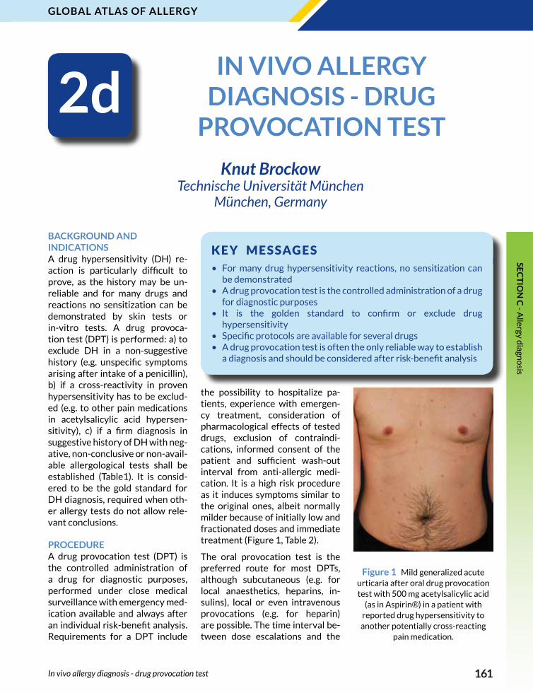
the possibility to hospitalize pa-tients, experience with emergen-cy treatment, consideration of pharmacological effects of tested drugs, exclusion of contraindi-cations, informed consent of the patient and sufficient wash-out interval from anti-allergic medi-cation. It is a high risk procedure as it induces symptoms similar to the original ones, albeit normally milder because of initially low and fractionated doses and immediate treatment (Figure 1, Table 2).
The oral provocation test is the preferred route for most DPTs, although subcutaneous (e.g. for local anaesthetics, heparins, in-sulins), local or even intravenous provocations (e.g. for heparin) are possible. The time interval be-tween dose escalations and the
Figure 1 Mild generalized acute urticaria after oral drug provocation test with 500 mg acetylsalicylic acid
(as in Aspirin®) in a patient with reported drug hypersensitivity to another potentially cross-reacting
pain medication.

162
Global atlas oF allerGyse
ct
ion
c -
Alle
rgy
dia
gno
sis
duration of provocation have to be determined considering the origi-nal reaction and suspected mech-anism.
General guidelines for perform-ing DPT have been published and specific protocols are available for some drugs, such as betalactam drugs, nonsteroidal anti-inflam-matory drugs or radiocontrast media.
INTERPRETATIONIn the majority of patients, DH can be excluded by DPT. In some pa-tients, the reappearance of reac-tions confirms the diagnosis. False
positive and false negative results to DPTs are possible. In medicine, a DPT can never guarantee toler-ance in 100% of patients, but helps to decide, if lifelong avoidance is justified. Studies evaluating the validity of the DPT show that the majority of patients (>95%) toler-ated the drug in real life, if there were no reaction in the DPT and conclude that this test is beneficial for the patient.
KEY REFERENCES1. Aberer W, Bircher A, Romano A,
Blanca M, Campi P, Fernandez J et al. Drug provocation testing in the diagnosis of drug hypersensitivity
reactions: general considerations. Allergy 2003;58:854-863.
2. Messaad D, Sahla H, Benahmed S, Godard P, Bousquet J, Demoly P. Drug provocation tests in pa-tients with a history suggesting an immediate drug hypersensi-tivity reaction. Ann Intern Med 2004;140:1001-1006.
3. Joint Task Force on Practice Pa-rameters; American Academy of Allergy, Asthma and Immunology; American College of Allergy, Asth-ma and Immunology; Joint Council of Allergy, Asthma and Immunolo-gy: Drug allergy: an updated prac-tice parameter. Ann Allergy Asthma Immunol 2010;105:259-273.
TABLE 1
Indications for a drug provocation test
• To exclude hypersensitivity in non-suggestive history of drug hypersensitivity
• To exclude cross-reactivity of related drugs in proven hypersensitivity
• To establish a firm diagnosis in suggestive history of drug hypersensitivity with negative, non-conclusive or non-available allergological tests
• Validation of the history and other allergological tests (reference standard)
Modified from Aberer W, Bircher A, Romano A, et al. Drug provocation testing in the diagnosis of drug hypersensitivity reactions: general considerations. Allergy 2003;58: 854-863.
TABLE 2
Clinical reactions to drug provocation tests in 241 patients with confirmed drug hypersensitivity
Manifestation Number (Percentage)
Urticaria 160 (66.4%)
Maculopapular exanthema 22 (9.1%)
Bronchospasm 9 (7.9%)
Laryngeal edema 10 (4.1%)
Anaphylaxis without shock 17 (7%)
Anaphylatic shock 13 (5.4%)
In vivo allergy diagnosis - drug provocation test
163
Global atlas oF allerGyse
ct
ion
c - A
llergy diagn
osis
• A challenge in the allergen challenge chamber (ACC) affects several target organs of type I allergy
• A stable and reproducible allergen load in the ACC is the basis for consistent subjective symptom scores
• Challenge sessions with comparable meaningful results are possible all over the year
• ACC enables to obtain objective parameters supporting subjective symptom scores
• ACC can monitor specific immunotherapy and pharmacotherapy• ACC can reduce the cost of drug development
In contrast to several other prov-ocation tests, allergen challenge chamber (ACC) tests are not fo-cused on single organs, but on several targets of type I allergic reactions like eyes, upper and lower respiratory tract and even skin. In contrast to other allergy tests, ACCs are usually not ap-plied for individual diagnosis, but for assessing allergic treatments, studies in occupational allergy and basic research.
At present, there are twelve ACCs active in Europe, USA, Canada and Japan (Figure 1). There is a broad conformity between the systems, although individual differences are to be mentioned. All ACCs are validated for pollen challenges like grass/ birch/ragweed or Japanese cedar pollen; some are also vali-dated for house dust mite or even for cat (Figure 2).
The main common feature of all ACCs is a pre-defined and stable allergen concentration in the air, which has to be high enough to induce symptoms in sensitized subjects. In addition, the allergen concentration climatic conditions like temperature, humidity and CO2 concentration are kept sta-ble over several hours. All param-eters are constantly controlled to
Friedrich Horak Research Consult GmbH
Vienna, Austria
THE ALLERGEN CHALLENGE CHAMBER 2e
Ke y m e ssag e s
The allergen challenge chamber
ensure high reproducibility of the ACC model. During a challenge session patients record their sub-jective symptoms several times an hour with good compliance. There is usually no application of rescue medication, which could possibly complicate the interpretation of the symptom score. Usually nasal, ocular and/or bronchial symptoms are scored using a 4-point scale (0 to 3).
A challenge session lasts at least two hours, because by then a pla-teau is normally reached in all of the patients’ symptoms, which will then not vary, if the allergen load is kept stable. This plateau-reaction is usually monitored for two to six hours (Figure 3).
In contrast to field or park studies, it is possible to additionally monitor and report objective assessments during the challenge sessions. These objective parameters offer the opportunity to check the proper-ness and accuracy of the subjective scoring (Table 1). Furthermore, it is possible to take blood samples at several time points of the challenge session to follow the ongoing aller-gic reaction.
The ACC model is approved by the FDA and EMA, at least for phase II trials and further supporting phase III trials. EMA suggests ACC trials for conducting dose finding trials of immunotherapeutic prod-ucts. Potential fields of application of ACC trials, however, can be more extensive (Table 2).

164
Global atlas oF allerGyse
ct
ion
c -
Alle
rgy
dia
gno
sis
ACC can monitor specific immu-notherapy and reduce the cost of drug development substantially, not only by proof of concept stud-ies. Requiring significantly less time than field studies and small-er numbers of individuals per trial are the essential arguments for application of ACCs. Besides sev-eral advantages some disadvan-
tages are to be mentioned (Table 3). The standardization of several ACCs worldwide and the “be-tween-unit” reproducibility are still unmet targets.
KEY REFERENCES1. Day JH, Horak F, Briscoe MP, Ca-
nonica GW, Fineman SM et al. The role of allergen challenge chambers in the evaluation of anti-allergic
TABLE 1
Objective assessments performed in an ACC
Rhinomanometry
Spirometry
Peak Expiratory Flow (PEF)
Nasal secretion weight
Nasal scraping
Nasal lavage
Nasal Peak Inspiratory Flow (NPIF)
Slit lamp investigation
Conjunctival imaging
Digital Endoscopy
Acoustic rhinometry
Blood samples (drug level, mediators,…)
Figure 1 Distribution of Allergen Challenge Chambers in the World.
Figure 2 The Vienna Challenge Chamber (VCC)
The allergen challenge chamber

165
Global atlas oF allerGyse
ct
ion
c - A
llergy diagn
osis
TABLE 2
Documented clinical trials in an ACC
Proof of concept
Dose finding
Onset of action
Magnitude and duration of action
Additive treatments
Pharmacodynamics
Pharmacokinetics
Competitive trials
TABLE 3
Possible environmental factors influencing development of allergic diseases
Advantages Disadvantages and unmet needs
Exposure to a defined, controlled level of allergen
Potential effects of claustrophobia or the influence of other subjects on subjective symptom scores
Out-of-season use for seasonal inhalant allergens and the ability to save up to 12 months in the development programme
Short exposure times (no more than several hours)
Rescue medication use can be prohibited or suspended during the challenge
A potentially abrupt challenge
Ability to collect biological samples from participants
Lack of seasonal priming in out-of-season challenges
Investigation of dose-ranging and onset of action
Low number of operational ACCs worldwide
Overall cost and ethical benefits due to use of lower number of subjects than in natural-exposure trials
Lack of standardization between the different ACCs in the World
medication: an international con-sensus paper. Clin Exp Allergy Re-views 2006;6:31-59.
2. Ellis AK, North ML, Walker T, Stea-cy LM. Environmental exposure unit: a sensitive, specific, and re-producible methodology for aller-gen challenge. Ann Allergy Asthma Immunol 2013;111:323-328.
3. Hohlfeld JM1, Holland-Letz T, Lar-big M, Lavae-Mokhtari M, Wieren-ga E, Kapsenberg M et al. Diag-
nostic value of outcome measures following allergen exposure in an environmental challenge chamber compared with natural conditions. Clin Exp Allergy 2010;40:998-1006.
4. Devillier P, Le Gall M, Horak F. The allergen challenge chamber: a valu-able tool for optimizing the clinical development of pollen immuno-therapy. Allergy 2011;66:163-169.
5. Yuki A, Terada T, Ichihara T, Fujii K, Hyo S, Kawata R, et al. Evaluat-
ing the effects of testing period on pollinosis symptoms using an aller-gen challenge chamber. Allergol Int 2011;60:533-539.
6. Bernstein JA. Correlation between a pollen challenge chamber and a natural allergen exposure study design for elicting ocular and nasal symptoms: early evidence support-ing a paradigm shift in drug investi-gation? J Allergy Clin Immunol 2012; 130:128-129.
Figure 3 Total Nasal Symptom Score (TNSS) of > 500 subjects with placebo
treatment.
The allergen challenge chamber
166
Global atlas oF allerGyse
ct
ion
c -
Alle
rgy
dia
gno
sis • Elevated serum levels of allergen-specific IgE demonstrate
the atopic status of a patient and are indicative for an eventual clinically significant allergy
• Determination of allergen-specific IgE in serum allows rapid screening of the sensitization spectrum of a patient without risks for adverse reactions
• Multiplex specific IgE measurements to pure natural or recombinant allergens allow a component-resolved diagnostics, which can be useful to design a patient-tailored immunotherapeutic intervention
• For allergy diagnosis elevated allergen-specific serum IgE levels need to be interpreted in the light of clinical history. In some cases, in vivo provocation tests performed with the suspected allergen are needed to confirm the diagnosis
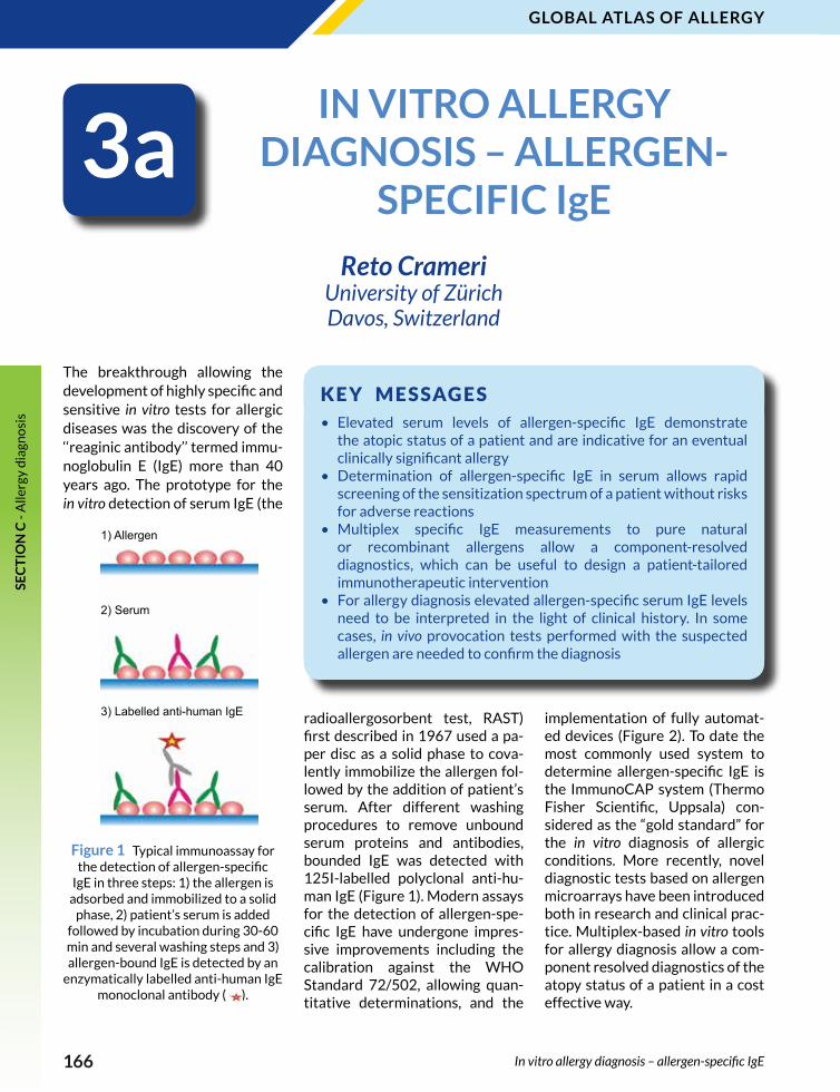
The breakthrough allowing the development of highly specific and sensitive in vitro tests for allergic diseases was the discovery of the ‘‘reaginic antibody’’ termed immu-noglobulin E (IgE) more than 40 years ago. The prototype for the in vitro detection of serum IgE (the
Reto Crameri University of Zürich Davos, Switzerland
IN VITRO ALLERGY DIAGNOSIS – ALLERGEN-
SPECIFIC IgE 3a
Ke y m e ssag e s
In vitro allergy diagnosis – allergen-specific IgE
radioallergosorbent test, RAST) first described in 1967 used a pa-per disc as a solid phase to cova-lently immobilize the allergen fol-lowed by the addition of patient’s serum. After different washing procedures to remove unbound serum proteins and antibodies, bounded IgE was detected with 125I-labelled polyclonal anti-hu-man IgE (Figure 1). Modern assays for the detection of allergen-spe-cific IgE have undergone impres-sive improvements including the calibration against the WHO Standard 72/502, allowing quan-titative determinations, and the
implementation of fully automat-ed devices (Figure 2). To date the most commonly used system to determine allergen-specific IgE is the ImmunoCAP system (Thermo Fisher Scientific, Uppsala) con-sidered as the “gold standard” for the in vitro diagnosis of allergic conditions. More recently, novel diagnostic tests based on allergen microarrays have been introduced both in research and clinical prac-tice. Multiplex-based in vitro tools for allergy diagnosis allow a com-ponent resolved diagnostics of the atopy status of a patient in a cost effective way.
Figure 1 Typical immunoassay for the detection of allergen-specific
IgE in three steps: 1) the allergen is adsorbed and immobilized to a solid
phase, 2) patient’s serum is added followed by incubation during 30-60 min and several washing steps and 3) allergen-bound IgE is detected by an
enzymatically labelled anti-human IgE monoclonal antibody ( ).
1) Allergen
2) Serum
3) Labelled anti-human IgE
167
Global atlas oF allerGyse
ct
ion
c - A
llergy diagn
osis
Despite the relevant technological improvement, several problems related to the in vitro diagnosis of
allergy remain unsolved (Figure 3). Allergen-specific IgE in serum is a marker for sensitization and a
Figure 2 Most commonly used techniques for the detection of allergen-specific IgE used in research and clinics. All techniques are based on the
immunoassay principle described in Figure 1.
Allergen- Specific
IgE
Basophil Activation
tests
Allergen Challenge
tests
Allergic Disease
ChronicAllergic Disease
Sensitization
Cell activation / mediator release
In vivo cell activation / mediator release
Inflammation / hypersensitivity
Tissue remodelling
Figure 3 Steps involved in the development of allergic diseases. A switch to the production of allergen-specific IgE detectable in serum leads to the
sensitization of a patient, reflecting its atopic status. However, not all sensitized individuals suffer from allergic symptoms highlighting the limitation of in
vitro allergy diagnoses. Allergen challenge tests are needed to confirm the clinical relevance of an allergy suspected from the clinical history and from the
presence of allergen-specific IgE in serum. (Reproduced with permission from Bousquet J, Anto JM, Bachert C,et al. Factors responsible for differences between
asymptomatic subjects and patients presenting and IgE sensitization to allergens: a GA2LEN project. Allergy 2006:61;671-680, with permission from Willey Blackwell.)
prerequisite for the development of an IgE-mediated allergy, but is not sufficient for the induction of symptoms. In fact, more than 20% of the individuals with aller-gen-specific serum IgE are asymp-tomatic. We should be aware that allergen-specific serum IgE is bi-ologically irrelevant as long as it doesn’t bind to the high affinity receptor for IgE on effector cells. Therefore, the presence or the absence of allergen-specific IgE in serum is not sufficient to confirm or exclude an allergy in all cases.
At the current state of the art, it is worthwhile to maintain a healthy scepticism about allergy diagnos-tic test results solely based on the determination of allergen-specif-ic serum IgE levels and to let the clinical history, corroborated by adequate provocation tests, which drive the diagnosis before consid-ering immunotherapeutic inter-ventions.
KEY REFERENCES1. Ishizaka K, Ishizaka T. Identification
of gamma-E antibodies as a carri-er of reaginic activity. J Immunol 1967;99:1187–1198.
2. Wide L, Bennich H, Johansson SG. Diagnosis of allergy by an in-vitro test for allergen antibodies. Lancet 1967;2:1105-1107.
3. Shreffler WG. Microarrayed re-combinant allergens for diagnos-tic testing. J Allergy Clin Immunol 2011;127:843-849.
4. Bousquet J, Anto J, Bachert C, Bousquet PJ, Colombo P, Crameri R et al. Factors responsible for differ-ences between asymptomatic sub-jects and patients presenting and IgE sensitization to allergens: a GA-2LEN project. Allergy 2006:61;671-680.
5. Crameri R. The crux with a reliable in vitro and in vivo diagnosis of al-lergy. Allergy 2013:68;393-394.
In vitro allergy diagnosis – allergen-specific IgE
ELISA-Plates Research
Western blots Research
Strips Commercial
ImmunoCAPs Commercial
Microarrays Commercial
168
Global atlas oF allerGyse
ct
ion
c -
Alle
rgy
dia
gno
sis • The advent of molecular allergen technologies has created a shift
of paradigm in diagnosing IgE-mediated allergies• Component-resolved analysis of the specific IgE response provides
a more individualized and stratified diagnosis of allergic patients• Component-resolved diagnosis (CRD) of the specific IgE response
improves the sensitivity, specificity and clinical performance of the laboratory assay
• CRD has the potential to better select patients for immunotherapy, to predict the risk for severe allergic reactions and to monitor patients for immunotherapy outcome
The history of in vitro (laboratory) tests for allergy diagnosis started in the early seventies, soon after the discovery of IgE in 1967. The first generation assays, starting from the very first RAST, allowed researchers and allergists to im-prove the diagnosis of IgE-medi-ated diseases. The first generation IgE assays were mainly based on allergenic extracts – a mixture of allergenic and non-allergenic compounds derived from natural sources (e.g. a pollen extract).
A great step forward to a broader use of in vitro diagnosis in allergy was achieved in the early nine-ties, when the more reliable sec-ond generation assays appeared in laboratories worldwide and in parallel a new kind of test reagent started to be available. In fact, during the nineties an increasing number of allergenic molecules have been identified, character-ized and, most important, progres-sively translated into the daily use in diagnosis (Figure 1A). Most of the available allergenic molecules have been obtained by using the recombinant DNA technology, allowing cloning and production of recombinant allergens in a re-producible large scale. Within the last decades, allergenic molecules
IN VITRO ALLERGY DIAGNOSIS – MOLECULES AND COMPONENT-
RESOLVED DIAGNOSIS 3b
Ke y m e ssag e s
In vitro allergy diagnosis – molecules and component-resolved diagnosis
have been increasingly used in re-search and clinical studies leading to a cumulative number of scientif-ic publications (Figure 1B).
This knowledge is now displayed in several free accessible inter-net-based resources documenting the magnitude of the worldwide allergy health problem (Table 1). The most recent keystone im-provement in this area has been the application of microtechnolo-gy to in vitro diagnosis of IgE-me-diated diseases. In the last decade, a new tool has become available to allergists allowing them the parallel diagnosis on hundreds of allergenic molecules from a huge collection of allergenic sources in a single test run (Figure 2). This multiplex microarray test is now
available worldwide and allows allergic patients to be tested with the same panel of allergens re-gardless of age, gender and kind of allergen and route of exposure.
Within the last decade, multi-ple studies from many countries around the world independently reported an added diagnostic val-ue by using molecular components for in vitro allergy diagnosis in the singleplex test systems – the more traditional approach in allergy laboratory testing. This progress became mainly evident in some of the most severe allergic diseases such as anaphylaxis due to food intake or insect stings, which can result in fatal outcome. Through the advent of molecular technolo-gies, some of the weaknesses and
Markus Ollert Technische Universität München
Munich, Germany
Adriano Mari Associated Centers for Molecular
Allergology, Rome, Italy

169
Global atlas oF allerGyse
ct
ion
c - A
llergy diagn
osis
In vitro allergy diagnosis – molecules and component-resolved diagnosis
Figure 1 A - Cumulative number of identified allergenic molecules during the last three decades. (source: www.allergome.org); B - Number of scientific papers on identification, characterization and clinical use of allergenic molecules
per year published in the last three decades.
Figure 2 The new microtechnology-based tool used to detect specific IgE. Allergenic molecules, represented by proteins available as purified natural
compounds or recombinants, are immobilized as microspots (200 μm in diameter) on a solid phase area of 7 x 10 mm and are grouped by sources and
tissues and also on the basis of their biochemical structure and their IgE epitope sharing. It is thus possible to visualize the IgE cross-reactivity, now better-
defined as IgE co-recognition, as a cluster of fluorescent spots. The extension of cluster IgE recognition can help clinical decision on whether an allergenic
source must be avoided or not.
shortcomings of the classical diag-nostic algorithm, which was solely based on allergenic extracts from nature, could be solved in terms of assay sensitivity, specificity and prediction of clinical severity and risk. Nowadays, several prominent examples exist, where the meas-urement of sIgE to molecular aller-gen components is a recommend-ed state-of-the-art procedure within the algorithm for clinical decision-making (Table 2).
From a global perspective – de-spite the immense progress that has been achieved in the field of molecular allergy in vitro testing within the last two decades – there is a clinical and research need for a better reflection of allergenic molecules based on regional dif-ferences in the world. Not all aller-gens are global, some of them are regional, but still can cause severe allergic disease. This new era of allergy diagnosis, which calls for a global definition of allergenic source and molecules, has already been entered. The analysis of such wealth of data is now possible by using advanced information and communication technologies, which will ultimately result in bet-ter care for allergic patients.

170
Global atlas oF allerGyse
ct
ion
c -
Alle
rgy
dia
gno
sis
TABLE 2
Molecular components or allergens with a demonstrated utility for clinical decision making in various allergic conditions
Allergic DiseaseMolecular Component / Allergen
Diagnostic Problem SolvedCountries of Major
Studies
Insect venom anaphylaxis
Api m 1, Ves v 1, Ves v 5, Pol d 5
Better differentiation of honey bee and vespid venom double-sensitized patients
GER, SUI, AUT, SLO, USA, ITA, POL, ESP
Insect venom anaphylaxis
Ves v 5Increased sIgE assay sensitivity through spiking of vespid venom with rVes v 5
GER, SUI, AUT
F/WDEIA Tri a 19 Marker allergen for food-/wheat-dependent exercise-induced anaphylaxis (F/WDEIA)
JAP, FIN, DAN, GER, SUI
Peanut allergy Ara h 2, Ara h 6Predictive marker allergens for severe reactions to ingested peanut
USA, GBR, GER, AUS
Allergy to red meat α-GalMarker allergen for delayed anaphylactic reactions to red meat and offal
USA, SWE, GER, AUT
Pollen Allergy; Insect venom anaphylaxis
CCDMarker for broad IgE-based cross-reactivity without clinical relevance
ITA, NED, GER, AUT
Insect venom anaphylaxis
Api m 10, Api m 3Prediction of patients at risk for therapeutic failures in honey bee venom immunotherapy
GER, SUI
Abbreviations and explanations:Tri a 19, wheat allergen ω-5 gliadin; α-Gal, mammalian carbohydrate determinant (galactose-α-1,3-galactose); CCD, carbohy-drate cross-reactive determinants (β-1,2-xylose and α-1,3-fucose); Api m 1, 3, 10, honey bee venom allergens Phospholipase A2, Acid Phosphatase, Icarapin; Ves v 1, 5, vespid venom allergens Phospholipase A1, Antigen 5; Pol d 5, Polistes venom allergen Antigen 5; Ara h 2, 6, major peanut allergens storage protein allergens.
TABLE 1
Comprehensive allergen databases providing free access.
Database URL Target/Function
WHO/IUIS Allergen Nomenclature Sub-Committee
http://www.allergen.org/Responsible for maintaining and developing a unique, unambiguous and systematic nomenclature for allergenic proteins
ALLERGOME http://www.allergome.org/A web site designed to supply information on allergenic molecules (allergens) for clinicians and researchers
AllergenOnlinehttp://www.allergenonline.org/
Provides access to a peer reviewed allergen list and sequence searchable database
AllFamhttp://www.meduniwien.ac.at/allergens/allfam/
The AllFam database is a resource for classifying allergens into protein families (based on the Allergome website)
Abbreviations and explanations:WHO, World Health Organization; IUIS, International Union of Immunological Societies.
KEY REFERENCES1. Mari A. When does a protein be-
come an allergen? Searching for a dynamic definition based on most advanced technology tools. Clin Exp Allergy 2008;38:1089-1094.
2. Scala E, Alessandri C, Bernardi ML, Ferrara R, Palazzo P, Pomponi D et al. Cross-sectional survey
on immunoglobulin E reactivity in 23 077 subjects using an aller-genic molecule-based microarray detection system. Clin Exp Allergy 2010;40:911-921.
3. Koid AE, Chapman MD, Hamilton RG, Van Ree R, Versteeg SA, Dre-skin SC et al. Ara h 6 complements Ara h 2 as an important marker for IgE reactivity to peanut. J Ag-
ric Food Chem 2013 Dec 11. [Epub ahead of print].
4. Grunwald T, Bockisch B, Spillner E, Ring J, Bredehorst R, Ollert MW. Molecular cloning and expres-sion in insect cells of honey bee venom allergen acid phosphatase (Api m 3). J Allergy Clin Immunol 2007;117:848-854.
In vitro allergy diagnosis – molecules and component-resolved diagnosis
171
Global atlas oF allerGyse
ct
ion
c - A
llergy diagn
osis
• Cellular allergy tests expand the tools of the allergist to diagnose and monitor allergic disease
• The basophil activation test (BAT) documents type I sensitisation to an allergen, as fraction of blood basophils activated by soluble allergen
• Measuring basophil sensitivity as activation at graded dilutions of allergen expands the options since sensitivity to allergen is a more reproducible metric of allergic disease than provocation testing. It can be used to identify sufficiently sensitised individuals and to monitor natural and therapeutic resolution of allergy
• Identification of culprit drugs in T cell mediated Type IV drug allergy by Elispot or flow cytometry is only available at very few laboratories
Cellular allergy testing is essen-tially induction of the allergic response in a test tube. As any in vitro test, it has the advantage of quantifying the allergic response without any risk to the patient.
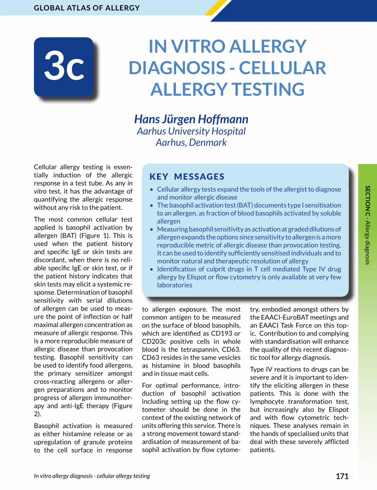
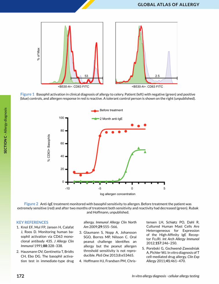
The most common cellular test applied is basophil activation by allergen (BAT) (Figure 1). This is used when the patient history and specific IgE or skin tests are discordant, when there is no reli-able specific IgE or skin test, or if the patient history indicates that skin tests may elicit a systemic re-sponse. Determination of basophil sensitivity with serial dilutions of allergen can be used to meas-ure the point of inflection or half maximal allergen concentration as measure of allergic response. This is a more reproducible measure of allergic disease than provocation testing. Basophil sensitivity can be used to identify food allergens, the primary sensitizer amongst cross-reacting allergens or aller-gen preparations and to monitor progress of allergen immunother-apy and anti-IgE therapy (Figure 2).
Basophil activation is measured as either histamine release or as upregulation of granule proteins to the cell surface in response
Hans Jürgen Hoffmann Aarhus University Hospital
Aarhus, Denmark
IN VITRO ALLERGY DIAGNOSIS - CELLULAR
ALLERGY TESTING3c
Ke y m e ssag e s
In vitro allergy diagnosis - cellular allergy testing
to allergen exposure. The most common antigen to be measured on the surface of blood basophils, which are identified as CD193 or CD203c positive cells in whole blood is the tetraspannin, CD63. CD63 resides in the same vesicles as histamine in blood basophils and in tissue mast cells.
For optimal performance, intro-duction of basophil activation including setting up the flow cy-tometer should be done in the context of the existing network of units offering this service. There is a strong movement toward stand-ardisation of measurement of ba-sophil activation by flow cytome-
try, embodied amongst others by the EAACI-EuroBAT meetings and an EAACI Task Force on this top-ic. Contribution to and complying with standardisation will enhance the quality of this recent diagnos-tic tool for allergy diagnosis.
Type IV reactions to drugs can be severe and it is important to iden-tify the eliciting allergen in these patients. This is done with the lymphocyte transformation test, but increasingly also by Elispot and with flow cytometric tech-niques. These analyses remain in the hands of specialised units that deal with these severely afflicted patients.
172
Global atlas oF allerGyse
ct
ion
c -
Alle
rgy
dia
gno
sis
KEY REFERENCES1. Knol EF, Mul FP, Jansen H, Calafat
J, Roos D. Monitoring human ba-sophil activation via CD63 mono-clonal antibody 435. J Allergy Clin Immunol 1991;88:328–338.
2. Hausmann OV, Gentinetta T, Bridts CH, Ebo DG. The basophil activa-tion test in immediate-type drug
allergy. Immunol Allergy Clin North Am 2009;29:555–566.
3. Glaumann S, Nopp A, Johansson SGO, Borres MP, Nilsson C. Oral peanut challenge identifies an allergy but the peanut allergen threshold sensitivity is not repro-ducible. PloS One 2013;8:e53465.
4. Hoffmann HJ, Frandsen PM, Chris-
tensen LH, Schiøtz PO, Dahl R. Cultured Human Mast Cells Are Heterogeneous for Expression of the High-Affinity IgE Recep-tor FcεRI. Int Arch Allergy Immunol 2012;157:246–250.
5. Porebski G, Gschwend-Zawodniak A, Pichler WJ. In vitro diagnosis of T cell-mediated drug allergy. Clin Exp Allergy 2011;41:461–470.
Figure 1 Basophil activation in clinical diagnosis of allergy to celery. Patient (left) with negative (green) and positive (blue) controls, and allergen response in red is reactive. A tolerant control person is shown on the right (unpublished).
Figure 2 Anti-IgE treatment monitored with basophil sensitivity to allergen. Before treatment the patient was extremely sensitive (red) and after two months of treatment both sensitivity and reactivity had decreased (green). Rubak
and Hoffmann, unpublished.
In vitro allergy diagnosis - cellular allergy testing
100
80
60
40
20
0
-10 -5 0 5
log allergen concentration
% C
D63
+ B
asop
hils
Before treatment
2 Month anti-IgE
<B530-A>: CD63 FITC <B530-A>: CD63 FITC
53 2.5
% o
f Max
173
Global atlas oF allerGyse
ct
ion
c - A
llergy diagn
osis
• Novel biomarkers, as well as appropriate biomarker combinations are important to improve allergy diagnosis and treatment
• Classifying allergic diseases into several endotypes and using appropriate biomarkers to evaluate the molecular target for endotype will increase drug effectiveness
• Fractional exhaled nitric oxide is considered a surrogate biomarker for eosinophilic airway inflammation and for steroid responsiveness in asthma patients
• Periostin has emerged as a surrogate biomarker of type 2 inflammation and tissue remodeling in asthma
Biomarkers are used to diagnose diseases, estimate disease se-verity, or investigate underlying inflammation types, thus predict-ing the efficacy of therapeutics. For example, measuring a specific IgE or urinary leukotriene E4 is important for diagnosis of atopic status or aspirin-sensitive asthma, respectively. Discovering novel biomarkers, as well as finding ap-propriate combinations with ex-isting biomarkers, is an important goal in allergy research aiming to improve diagnosis and treatment.
“Companion diagnostics”, which can predict therapeutic efficacy, is now being widely recognized due the development of molecular targeted drugs against allergic dis-eases, particularly asthma. These drugs, mostly biologics, are expen-sive and are effective only for par-ticular types of patients. It is now thought that asthma is not a single disease, but a “syndrome.” Thus, it is important to classify asthma into several endotypes, based on their pathogenesis, and evaluate for which endotype, using the ap-propriate biomarkers each mo-lecular targeted drug is effective using the appropriate biomarkers.
Although inhaled corticosteroids (ICS) provide beneficial effects for
Kenji Izuhara Saga Medical School
Saga, Japan
BIOMARKERS FOR ALLERGY DIAGNOSIS AND
TREATMENT4
Ke y m e ssag e s
Biomarkers for allergy diagnosis and treatment
most asthma patients, 5-10% of patients are resistant or hypo-re-sponsive to ICS. Any biomarker in-dicating the cause of the resistance to ICS would be useful. Moreover, biomarkers that can predict asth-ma relapse upon steroid reduction would be also valuable. Eosino-philc airway inflammation is at least one factor associated with steroid responsiveness. Fractional exhaled nitric oxide (FeNO) is con-sidered a surrogate biomarker for eosinophilic airway inflammation and is regarded as a biomarker for steroid responsiveness (Figure 1). Although still controversial, the ac-cumulated evidence has suggested that FeNO is useful for assessing control with ICS, predicting relapse upon ICS reduction, and estimating
lung function.
Periostin, a downstream molecule of IL-4 and/or IL-13, has emerged as a surrogate biomarker of type 2 inflammation and tissue remod-eling in bronchial asthma. It has been shown that serum perios-tin can predict the efficacy of an-ti-IL-13 antibody (lebrikizumab) and anti-IgE antibody (omalizum-ab), thus proving a prototype bi-omarker for the companion diag-nostic of allergic diseases (Figure 2). Moreover, high serum periostin is linked to hypo-responsiveness for ICS, particularly in late-onset eosinophilic asthma (Figure 3). In addition to periostin, sputum eo-sinophils are useful for estimating the efficacy of anti-IL-5 antibody (mepolizumab).

174
Global atlas oF allerGyse
ct
ion
c -
Alle
rgy
dia
gno
sis
Genetic biomarkers, such as the
filaggrin gene for skin barrier dys-
function or the glucocorticoid-in-
duced transcript 1 gene for re-
sponsiveness to steroid therapy,
represent another avenue to be
explored in allergy research.
KEY REFERENCES1. Vijverberg SJ, Koenderman L, Ko-
ster ES, van der Ent CK, Raaijmak-ers JA, Maitland-van der Zee AH. Biomarkers of therapy respon-siveness in asthma: pitfalls and promises. Clin Exp Allergy 2011; 41:615-629.
2. Szefler SJ, Wenzel S, Brown R, Er-
zurum SC, Fahy JV, Hamilton RG, et al. Asthma outcomes: biomark-ers. J Allergy Clin Immunol 2012; 129:S9-23.
3. Lötvall J, Akdis CA, Bacharier LB, Bjermer L, Casale TB, Custovic A et al. Asthma endotypes: a new approach to classification of dis-ease entities within the asthma
FeNO <25 ppb 25-50 ppb 50< ppb
Symptoms (+)Consider
alternative diagnosis
Uncontrolled Uncontrolled
Symptoms (-)
Well controlled
Consider taper or withdrawal
Well controlledRisk of relapse
upon ICS reduction
Serum periostin High Low
Anti-IL-13 antibody
(lebrikizumab)Responsive No responsive
Anti-IgE antibody
(omalizumab)Responsive No responsive
Asthma patients
Other agents
Th2 antagonists
ICS
Low
High
Effective
Ineffective
Measurement of serum periostin levels
Figure 1 A treatment guideline for adult asthma patients based on
existing symptoms and FeNO levels (Modified from Ref. 4)
Figure 2 Serum periostin as a biomarker to select Th2 antagonists for asthma patients Upper panel: Responsiveness in asthma patients to Th2 antagonists depending on serum periostin is depicted. Patients showing high serum periostin
are responsive to anti-IL-13 antibody (lebrikizumab) and anti-IgE antibody (omalizumab), whereas those showing low serum periostin are unresponsive to these agents. (Summarized from Corren J, Lemanske RF, Hanania NA, Korenblat PE, Parsey MV, Arron JR, Harris JM. Lebrikizumab treatment in adults with asthma. N Engl J Med 2011 365; 12, 1088-1098, Hanania NA, Wenzel S, Rosen K, Hsieh HJ, Mosesova S, Choy D F. Exploring the effects of omalizumab in allergic asthma: an analysis of biomarkers in the EXTRA study. Am J Respir Crit Care Med 2013 187; 804-811) Lower panel: An algorithm
of treatment of asthma patients is shown. The first choice for asthma patients is ICS. If the patients are resistant to ICS, measurement of serum periostin is recommended. If serum periostin is high, Th2 antagonists are recommended; if low,
other agents are recommended.
Biomarkers for allergy diagnosis and treatment

175
Global atlas oF allerGyse
ct
ion
c - A
llergy diagn
osis
syndrome. J Allergy Clin Immunol 2011;127:355-360.
4. Barnes PJ, Dweik RA, Gelb AF, Gib-son PG, George SC, Grasemann H et al. Exhaled nitric oxide in pulmo-nary diseases: a comprehensive review. Chest 2010;138:682-692.
5. Izuhara K, Arima K, Ohta S, Su-zuki S, Inamitsu M, Yamamoto K. Periostin in allergic inflammation. Allergol Int 2014 Mar 25. [Epub ahead of print]
6. Corren J, Lemanske RF, Hanania NA, Korenblat PE, Parsey MV, Ar-ron JR et al. Lebrikizumab treat-ment in adults with asthma. N Engl J Med 2011;365:1088-1098.
7. Hanania NA, Wenzel S, Rosen K, Hsieh HJ, Mosesova S, Choy D F. Exploring the effects of omali-zumab in allergic asthma: an anal-ysis of biomarkers in the EXTRA study. Am J Respir Crit Care Med 2013;187:804-811.
8. Kanemitsu Y, Matsumoto H, Izu-hara K, Tohda Y, Kita H, Horiguchi T, et al. Increased periostin asso-ciates with greater airflow limita-tion in patients receiving inhaled corticosteroids. J Allergy Clin Im-munol 2013;132:305-312.
9. Nagasaki T, Matsumoto H, Kane-mitsu Y, Izuhara K, Tohda Y, Kita H et al. Integrating longitudinal on pulmonary function and in-
flammation using asthma phe-notypes. J Allergy Clin Immunol 2014;133:1474-1477.e2.
10. Agache I, Akdis C, Jutel M, Vir-chow JC. Untangling asthma phe-notypes and endotypes. Allergy 2012;67:835-846.
11. Akdis CA, Bachert C, Cingi C, Dykewicz MS, Hellings PW, Na-clerio RM et al. Endotypes and phenotypes of chronic rhinosi-nusitis: A PRACTALL document of the European Academy of Allergy and Clinical Immunology and the American Academy of Allergy, Asthma & Immunology. J Allergy Clin Immunol 2013;131:1479-1490.
Figure 3 Serum periostin as a biomarker to predict responsiveness to ICS in asthma patients. Upper left panel: Serum periostin levels in rapid and non-rapid FEV1 decliners are depicted (8). Upper right panel: Clustering of asthma patients
based on peripheral neutrophils and eosinophils is depicted. Lower left panel: Characteristics of each subtype are shown. Lower right panel: Correlation between serum periostin and responsiveness to ICS in cluster 3 is depicted. (Reprinted from
J Allergy Clin Immunol, 133/5, Nagasaki T, Matsumoto H, Kanemitsu Y et al. Integrating longitudinal information on pulmonary function and inflammation using asthma phenotypes, 1474-1477, Copyright 2014, with permission from Elsevier.)
Biomarkers for allergy diagnosis and treatment
Cluster 1
Cluster 2Cluster 3
Cluster 4
1 2 3 4 60Peripheral Eosinophils (X102/µL)
Per
iphe
ral n
eutro
phils
(X 1
02 /µL)
65
60
50
40
30
25
104.6
89.2
Rapid decliners(n=52)
Non-rapid decliners(n=172)
Ser
um p
erio
stin
leve
l (ng
/ml)
140
120
100
80
60
40
20
0
Rapid decliners: DFEV1 ≤ -30 ml/yr
Non-rapid decliners : DFEV1 > -30 ml/yr
Cluster 1 Cluster 3
Late-onset Non-atopic
Late-onset Eosinophil-dominant Highest periostin
Cluster 2 Cluster 4
Early-onset Atopic
Poor control Low FEV1 High IL-6
Serum periostin Responsiveness to ICS
High Bad
Low Good
Steroid responsive
Steroid hypo-responsive