Glaucoma Referral Refinement and OHT Enhanced …€¦ · The aim of a glaucoma referral refinement...
20
Copyright © LOC Central Support Unit 2009. All rights reserved. Glaucoma Referral Refinement and OHT Enhanced Service Pathways following updated NICE guidance (April 2009) Glaucoma Referral Refinement, Ocular Hypertension Monitoring and Ocular Hypertension Diagnosis Issued by LOC Support Unit May 2009
-
Upload
truongquynh -
Category
Documents
-
view
217 -
download
1
Transcript of Glaucoma Referral Refinement and OHT Enhanced …€¦ · The aim of a glaucoma referral refinement...

Copyright © LOC Central Support Unit 2009. All rights reserved.
Glaucoma Referral Refinement and OHT Enhanced Service Pathways following updated NICE guidance
(April 2009) Glaucoma Referral Refinement, Ocular Hypertension Monitoring
and Ocular Hypertension Diagnosis
Issued by LOC Support Unit
May 2009
Copyright © LOC Central Support Unit 2009. All rights reserved.
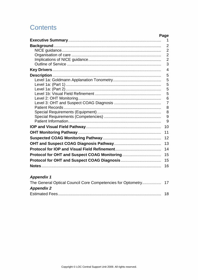
Contents Page Executive Summary ................................................................................... 1 Background ................................................................................................ 2 NICE guidance......................................................................................... 2 Organisation of care ................................................................................ 2 Implications of NICE guidance ................................................................. 2 Outline of Service .................................................................................... 3 Key Drivers ................................................................................................. 4 Description ................................................................................................. 5 Level 1a: Goldmann Applanation Tonometry ........................................... 5 Level 1a: (Part 1) ..................................................................................... 5 Level 1a: (Part 2) ..................................................................................... 5 Level 1b: Visual Field Refinement ........................................................... 5 Level 2: OHT Monitoring .......................................................................... 6 Level 3: OHT and Suspect COAG Diagnosis .......................................... 7 Patient Records ....................................................................................... 8 Special Requirements (Equipment) ......................................................... 8 Special Requirements (Competencies) ................................................... 9 Patient Information ................................................................................... 9 IOP and Visual Field Pathway ................................................................... 10 OHT Monitoring Pathway .......................................................................... 11 Suspected COAG Monitoring Pathway .................................................... 12 OHT and Suspect COAG Diagnosis Pathway .......................................... 13 Protocol for IOP and Visual Field Refinement ......................................... 14 Protocol for OHT and Suspect COAG Monitoring ................................... 15 Protocol for OHT and Suspect COAG Diagnosis .................................... 15 Notes ........................................................................................................... 16 Appendix 1 The General Optical Council Core Competencies for Optometry................. 17 Appendix 2 Estimated Fees ............................................................................................ 18
Copyright © LOC Central Support Unit 2009. All rights reserved.
1
Executive Summary The aim of a glaucoma referral refinement pathway is to reduce unnecessary referrals to the hospital eye service, reducing patient anxiety and increasing capacity within the overburdened hospital glaucoma clinics. This will provide a more cost effective service with a greater number of patients managed within the primary care setting. This has been shown in the North East of Scotland1 and in Manchester2
1 The Influence of the New General Ophthalmic Services (GOS) Contract in Optometrist Referrals for
Glaucoma in Scotland. GS Ang, WS Ng and A Azuara-Blanco. Eye (Nov 2007):1–5. 2 Community Refinement of Glaucoma Referrals. DB Henson, AF Spencer, R Harper and EJ Cadman.
Eye (2003):17;21–26 Nature Publishing Group.
where there was a reduction in false positive referrals of 40%. The referral refinement pathway would allow accredited optometrists to repeat diagnostic tests to confirm the risk of disease and thus improve the accuracy of referrals and deflect unnecessary referrals. A further service would allow diagnosis of ocular hypertension (OHT) and suspect chronic open angle glaucoma (COAG) by specialist accredited optometrists, retaining more patients in primary care. The aim of an OHT and suspect COAG monitoring pathway is to reduce the number of secondary care consultations for the cohort of patients who are diagnosed either as having OHT i.e. consistently high intra-ocular pressures (IOPs) but no glaucoma, or as being COAG suspect i.e. other suspicious signs. Currently, patients considered to have a greater chance of developing COAG due to elevated IOP or other suspicious signs are generally retained in secondary care and reviewed there on an annual basis, following referral in. The new service would provide for these patients to be discharged back into primary care for monitoring by community optometrists. Patients would only be referred back into secondary care if there was a change in clinical status.
Copyright © LOC Central Support Unit 2009. All rights reserved.
2
Background Nice Guidance NICE clinical guideline 85 (Diagnosis and management of chronic open angle glaucoma and ocular hypertension) issued 22 April 2009 sets out how best to diagnose COAG and OHT, how people with COAG, OHT or at risk of COAG should be monitored, and which treatments should be considered. Affecting an estimated 480,00 people in England, COAG is a common condition involving optic nerve damage and loss of the visual field that can lead to blindness if it’s not diagnosed early and treated promptly. Around 14% of UK blindness registrations are due to glaucoma. However many people won’t know that their eyesight is at risk – there are usually no symptoms until the later stages when their vision is already seriously damaged. OHT (raised pressure in the eye) is a major risk factor for developing COAG, although COAG can occur with or without raised eye pressure. Glaucoma is more common with increasing age, and people of African descent or with a family history of glaucoma may be at greater risk of developing the condition. With changes in population demographics the number of people affected by the condition is expected to rise. Once diagnosed, people with COAG need lifelong monitoring so that any progression of visual damage can be detected. Controlling the condition to prevent or minimise further damage is crucial to maintaining a sighted lifetime. By implementing this guideline more people will be prevented from going blind. Organisation of Care NICE states that diagnosis of OHT and suspected COAG and formulation of a management plan should be made by a suitably trained healthcare professional with relevant experience who either has a specialist qualification, or is working under the supervision of a consultant ophthalmologist. However, diagnosis of COAG should be confirmed by a consultant ophthalmologist. NICE also states that monitoring of people with a confirmed diagnosis of OHT or suspected COAG, who have an established management plan, can be carried out by a suitably trained healthcare professional with the relevant skills and ability to detect a change in clinical status. Implications of NICE guidance Currently due to the constraints of a GOS sight test, and recent NICE guidelines, optometrists are referring all cases of suspect glaucoma to secondary care for confirmation of the diagnosis and treatment where necessary. This works well when the diagnosis is positive. However, there is no simple single test for glaucoma and this, coupled with the low prevalence of the condition, makes it difficult to detect with certainty. Referral refinement by repeating visual fields and intra-ocular pressures on another occasion has been shown to reduce onward referrals by as much as 40%2 and this is our proposed level 1. OHT is defined in the NICE guidance as repeatable intra-ocular pressure over 21 mmHg as measured by Goldmann tonometry.
Copyright © LOC Central Support Unit 2009. All rights reserved.
3
Previously the threshold for OHT was set by local ophthalmologists and in most cases was around 25 mmHg. This lowering of the threshold will increase the number of referrals by community optometrists who will now be following the NICE guideline. However, most optometrists measure the pressure using an air-puff tonometer. These are considered by NICE to be less accurate, and so repeating the pressures using Goldmann tonometry will reduce the number of false positive referrals. The use of Goldmann tonometry is not a requirement of a GOS sight test, although it is a core competency of optometrists. The lowered threshold for diagnosis of OHT and the requirement for a standardised battery of clinical tests will mean an inevitable significant increase in the number of patients. This could quickly overwhelm hospital eye service departments. In order to ensure that follow up care of patients with established glaucoma does not suffer, a service to manage OHT in the community should be established. Outline of Service There are three proposed levels of service with full details on page 7: Level 1 would provide primary care referral refinement to deflect unnecessary referrals and is within the basic competency of community optometrists. Some accreditation and additional equipment would be required. This service would be below secondary care PbR tariff. Level 2 would provide primary care monitoring of patients diagnosed with OHT who will need to be seen annually. This will enable discharge into primary care both of newly diagnosed and large numbers of existing patients with OHT who can be seen closer to home at lower cost. This is within the basic competency of optometrists. Some accreditation will be required. Level 3 would provide for primary care diagnosis of ocular hypertension and glaucoma by an optometrist with additional training and accreditation as an alternative to referral to secondary care from the level 1 service.
Copyright © LOC Central Support Unit 2009. All rights reserved.
4
Key Drivers The national key drivers include: • NICE clinical guideline 85: Diagnosis and Management of Chronic Open
Angle Glaucoma and Ocular Hypertension • World Class Commissioning (2008) • Creating a Patient-led NHS: Delivering the NHS Improvement Plan (March
2005) • Commissioning Framework for 2007-8 • Implement Care Closer to Home; Convenient Quality Care for Patients
(April 2007) • Commissioning Framework for Health and Well-being (March 2007) • Trust, Assurance and Safety – the Regulation of Health Professionals
(February 2007) • Safeguarding Patients ( February 2007) • The NHS in England: Operating Framework 2007–08 (December 2006) • Practice Based Commissioning: Practical Implementation (November
2006) • Health Reform in England: Update and Commissioning Framework (July
2006) • Tackling Hospital Waiting: The 18 week Patient Pathway (May 2006) • Standards for Better Health (April 2006) • White Paper: Our Health, Our Care, Our Say (January 2006) • NHS Plan: A plan for Reform, a Plan for Investment (2000)
Copyright © LOC Central Support Unit 2009. All rights reserved.
5
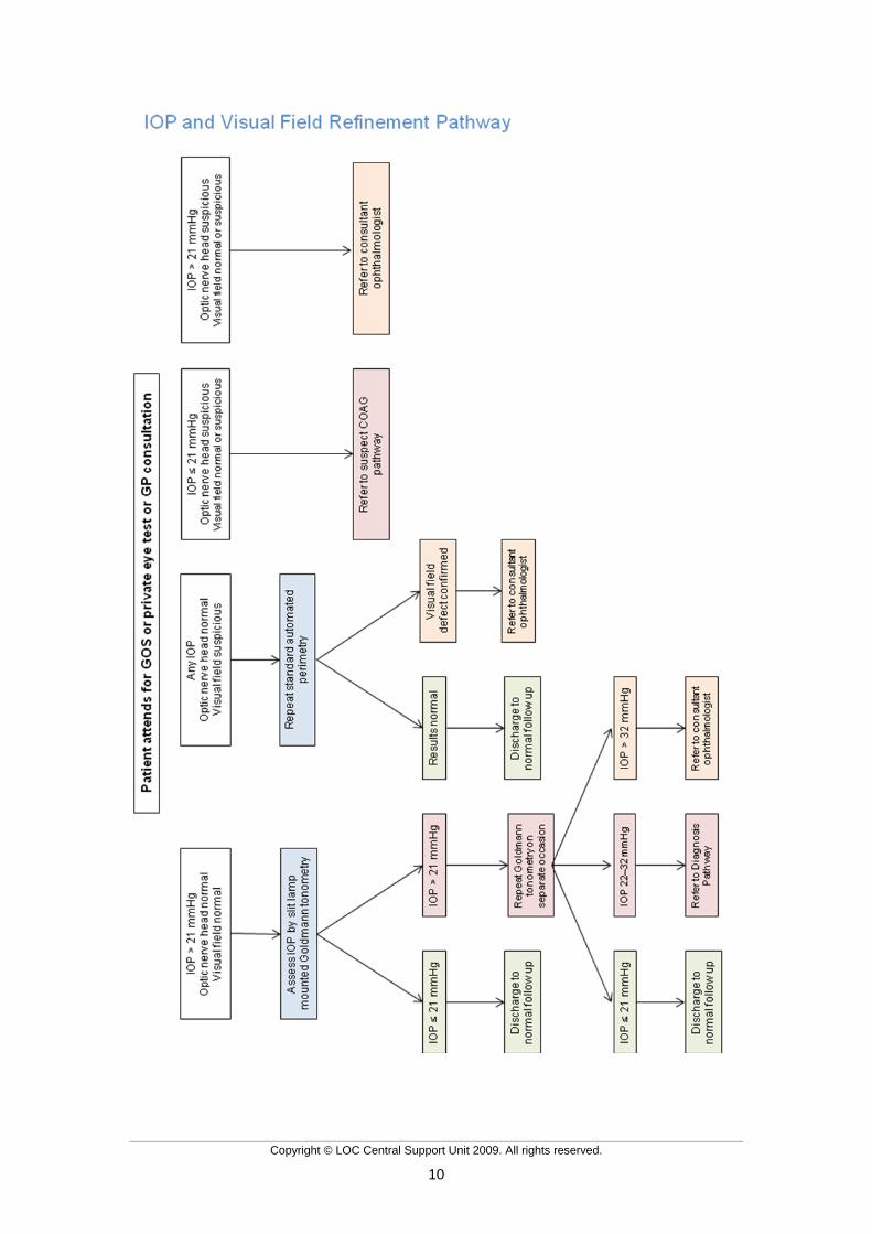
Description There are three different levels of enhanced services included in this package. Commissioners may decide to choose one service independently of the others based on local need, however, it is anticipated that all three services will be beneficial in most areas. Level 1a: Goldmann Applanation Tonometry A low level enhanced service for IOP refinement where other signs of glaucoma are not present will reduce unnecessary referrals to the hospital eye service, reducing patient anxiety and minimising capacity issues within the already overburdened hospital glaucoma clinics. The service will be cost effective with a greater number of patients managed within the primary care setting. Level 1a (Part 1) Patients who are identified as having IOP > 21 mmHg during a standard GOS or private sight test will have immediate Goldmann applanation tonometry assuming the optometrist is accredited to provide the service. This service falls within core competencies for optometrists. Set up costs of purchasing slit lamp mounted Goldmann tonometers should be considered. Outcomes There are two possible outcomes from Part 1: 1. The results are within normal limits and the patient can be discharged.
At risk groups should be monitored appropriate intervals under GOS. 2. Pressure is > 21 mmHg patient proceeds to Part 2. Level 1a (Part 2) Patient attends for repeat Goldmann applanation tonometry on a separate occasion Outcomes There are three possible outcomes from repeating this test: 1. The results are within normal limits and the patient can be discharged.
At risk groups should be monitored at appropriate intervals under GOS. 2. The pressure is confirmed as 22–32 mmHg and the patient is referred to
the OHT diagnosis pathway. 3. Pressure is > 32 mmHg is confirmed and the patient is referred to
consultant ophthalmologist. Level 1b: Visual Field Refinement A low level enhanced service for visual field refinement will reduce unnecessary referrals to the hospital eye service, reducing patient anxiety and minimising capacity issues within the already overburdened hospital glaucoma clinics. The service will be cost effective with a greater number of patients managed within the primary care setting.
Copyright © LOC Central Support Unit 2009. All rights reserved.
6
Patients who are identified as having suspicious visual fields during a standard GOS or private sight test will have visual fields repeated on a separate occasion assuming the optometrist is accredited to provide the service This service falls within core competencies for optometrists Outcomes There are three possible outcomes from these tests: 1. The results are within normal limits and the patient can be discharged. At
risk groups should be monitored annually under GOS. 2. Visual field is suspicious and requires monitoring at appropriate intervals 3. Visual field defect is confirmed and the patient is referred to consultant
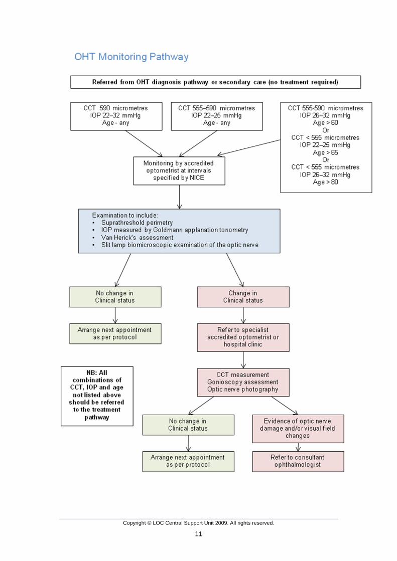
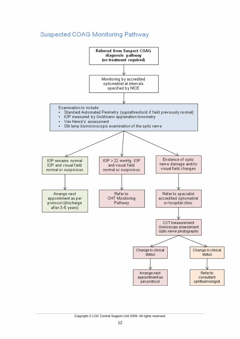
ophthalmologist. The criteria for inclusion of patients in level 1a and/or 1b may include the following: • IOP > 21 mmHg • Field defects • Difference in IOP between the two eyes of > 5 mmHg Note: If a community OHT and Suspect COAG diagnosis service (Level 3) is in place, commissioners may wish to implement Goldmann applanation tonometry (Level 1a) only and direct suspicious visual fields to the Level 3 service. If however, there is no Level 3 service in place, Level 1a and Level 1b are recommended. Level 2: OHT and suspect COAG Monitoring Patients who have a confirmed diagnosis of OHT or suspect COAG and who do not require treatment should follow the OHT or suspect COAG monitoring pathway and be monitored at regular intervals as specified by NICE. Patients will be referred to this pathway from the community OHT diagnostic clinic or secondary care. The skills required for this pathway are covered by the core competencies for optometrists. Training and accreditation for optometrists would include knowledge of the NICE guidelines, referral criteria, interpretation of results and the disease process. If a change in clinical status is found, the patient should be referred to a specialist community optometrist or hospital eye department for further investigation, depending on local protocol. This service will reduce unnecessary hospital eye service consultations and minimise capacity issues in secondary care. Set up costs of purchasing slit lamp mounted Goldmann tonometers should be considered. The accredited optometrist will carry out slit lamp mounted Goldmann tonometry, suprathreshold perimetry, Van Herick’s test, and dilated slit lamp biomicroscopic examination of the optic nerve head. Outcomes There are two possible outcomes from these tests: 1. No change in clinical status. Next appointment as per protocol. 2. Change in clinical status. Patient referred to specialist optometrist or
hospital clinic depending on local arrangement.
Copyright © LOC Central Support Unit 2009. All rights reserved.
7
The criteria for inclusion of patients may include the following: • Diagnosed ocular hypertension discharged from hospital eye service (HES)
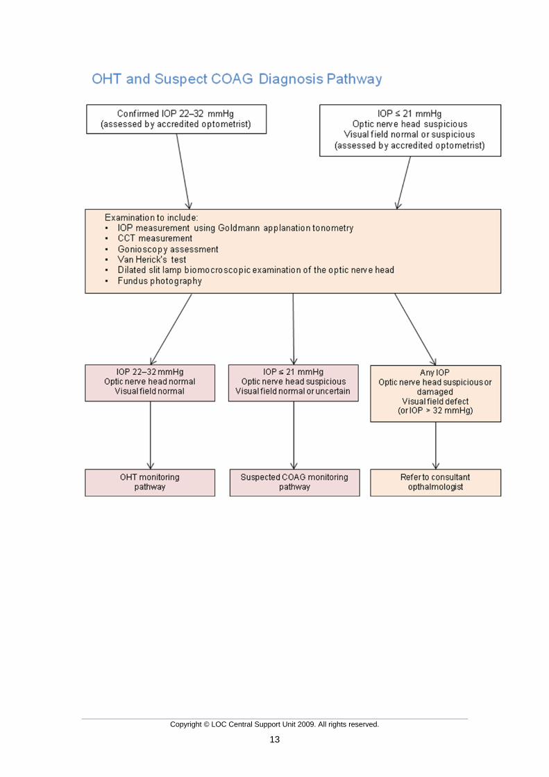
or specialist practitioner • Diagnosed suspect COAG discharged from HES or specialist practitioner Level 3: OHT and Suspect COAG Diagnosis For patients whose pressure is confirmed as above the OHT threshold of 21 mmHg, or whose pressure is ≤ 21 mmHg but the optic nerve head and/or visual fields are suspicious, the OHT and Suspect COAG Diagnosis Pathway should be followed. Patients referred into this pathway would be those who are identified as being at risk of glaucoma or OHT during a standard GOS or private sight test or GP consultation. The referring optometrist would send all the relevant clinical details with the referral. (In many cases the examining optometrist and the accredited optometrist will be the same person, or may be referring within the same practice). Patients will be assessed by accredited optometrists working to a protocol to ensure NICE compliance. Patients would then follow a specific management pathway depending on outcome. Again this service will be cost effective and reduce unnecessary referrals to secondary care. Additional equipment costs should be considered. The accredited optometrist will carry out slit lamp mounted Goldmann tonometry, central corneal thickness (CCT) measurement, gonioscopy assessment, central threshold perimetry, Van Herick’s test, dilated slit lamp biomicroscopic examination of the optic nerve head and fundus photography. Outcomes There are three possible outcomes from these tests; 1. IOP 22–32 mmHg with optic nerve and visual field normal or suspicious.
The patient should be referred to the OHT monitoring pathway where assessments will take place at intervals specified by NICE guidelines.
2. IOP < 21 mmHg, optic nerve head suspicious, visual field normal or uncertain. Patient should be referred to Suspect COAG monitoring pathway where assessments will take place at intervals specified by NICE.
3. Visual field defect, with suspicious or damaged optic nerve head and any IOP. The patient should be referred to consultant ophthalmologist.
The criteria for inclusion of patients may include the following: • GP referral • IOP > 21 mmHg • Suspicious visual fields • Suspicious cupping or asymmetry of optic discs • Difference in IOP between the two eyes of > 5 mmHg • Significant change in IOPs • Suspicious HRT/GDx or similar results

Copyright © LOC Central Support Unit 2009. All rights reserved.
8
Patient Records All advice given to the patient, and procedures undertaken should be recorded on a patient card or electronic device, and stored in a safe retrieval system. On conclusion of a referral refinement, OHT diagnosis or OHT monitoring assessment the optometrist must complete the appropriate report form, entering the information on the IT system for audit purposes (where applicable) and report to the referring GP, and to the hospital eye service, should an onward referral be necessary. NB: Glaucoma is a very slow developing disease and there is very little risk to the patient in delaying the repeat tests. The reason for repeating the tests on a different occasion is to ensure that factors that may have influenced the patient responses, particularly in the fields test, the first time around will be different. Special Requirements (Equipment) All practices joining the IOP and Visual Field refinement service and the OHT monitoring service will be expected to employ an accredited optometrist and have the following equipment available: • Access to the Internet • Slit lamp and fundus viewing lens • Goldmann applanation tonometer • Threshold fields equipment capable of producing a printed report • Distance test chart • Appropriate ophthalmic drugs
− Mydriatic − Anaesthetic − Staining agents
All practices joining the OHT diagnosis service will be expected to employ an accredited optometrist and have the following equipment available: • Access to the Internet • Slit lamp and fundus viewing lens • Goldmann applanation tonometer • Threshold fields equipment capable of producing a printed report • Gonioscope • Pachymeter • Distance test chart • Appropriate ophthalmic drugs
− Mydriatic − Anaesthetic − Staining agents
Copyright © LOC Central Support Unit 2009. All rights reserved.
9
Special Requirements - Competencies The competencies required for optometrists participating in IOP and Visual Field refinement and OHT monitoring are all included in the core competencies defined by the GOC. (See Appendix 1) Training and accreditation for optometrists participating in these pathways will include knowledge of the referral criteria and interpretation of results and the disease process. Optometrists participating in OHT diagnosis will need to demonstrate additional competence in gonioscopy. Patient Information Patient information leaflets as recommended by NICE will be available to patients.
Copyright © LOC Central Support Unit 2009. All rights reserved.
10
Copyright © LOC Central Support Unit 2009. All rights reserved.
11
Copyright © LOC Central Support Unit 2009. All rights reserved.
12
Copyright © LOC Central Support Unit 2009. All rights reserved.
13
Copyright © LOC Central Support Unit 2009. All rights reserved.
14
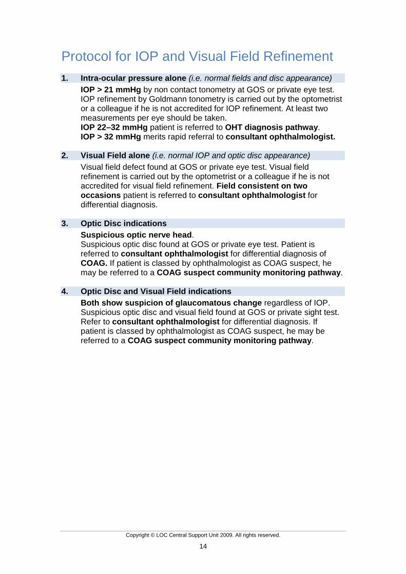
Protocol for IOP and Visual Field Refinement 1. Intra-ocular pressure alone (i.e. normal fields and disc appearance) IOP > 21 mmHg by non contact tonometry at GOS or private eye test.
IOP refinement by Goldmann tonometry is carried out by the optometrist or a colleague if he is not accredited for IOP refinement. At least two measurements per eye should be taken.
IOP 22–32 mmHg patient is referred to OHT diagnosis pathway. IOP > 32 mmHg merits rapid referral to consultant ophthalmologist. 2. Visual Field alone (i.e. normal IOP and optic disc appearance) Visual field defect found at GOS or private eye test. Visual field
refinement is carried out by the optometrist or a colleague if he is not accredited for visual field refinement. Field consistent on two occasions patient is referred to consultant ophthalmologist for differential diagnosis.
3. Optic Disc indications Suspicious optic nerve head. Suspicious optic disc found at GOS or private eye test. Patient is
referred to consultant ophthalmologist for differential diagnosis of COAG. If patient is classed by ophthalmologist as COAG suspect, he may be referred to a COAG suspect community monitoring pathway.
4. Optic Disc and Visual Field indications Both show suspicion of glaucomatous change regardless of IOP. Suspicious optic disc and visual field found at GOS or private sight test.
Refer to consultant ophthalmologist for differential diagnosis. If patient is classed by ophthalmologist as COAG suspect, he may be referred to a COAG suspect community monitoring pathway.
Copyright © LOC Central Support Unit 2009. All rights reserved.
15
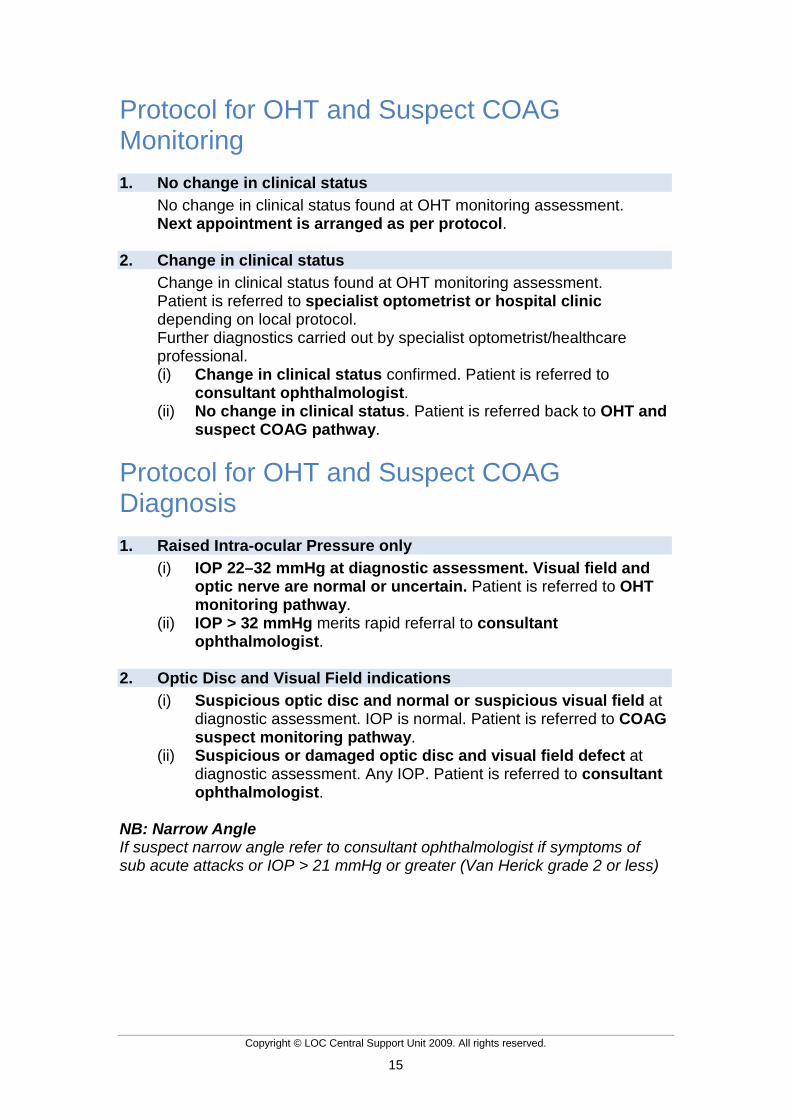
Protocol for OHT and Suspect COAG Monitoring 1. No change in clinical status No change in clinical status found at OHT monitoring assessment. Next appointment is arranged as per protocol.
2. Change in clinical status Change in clinical status found at OHT monitoring assessment. Patient is referred to specialist optometrist or hospital clinic
depending on local protocol. Further diagnostics carried out by specialist optometrist/healthcare
professional. (i) Change in clinical status confirmed. Patient is referred to
consultant ophthalmologist. (ii) No change in clinical status. Patient is referred back to OHT and
suspect COAG pathway.
Protocol for OHT and Suspect COAG Diagnosis 1. Raised Intra-ocular Pressure only
(i) IOP 22–32 mmHg at diagnostic assessment. Visual field and optic nerve are normal or uncertain. Patient is referred to OHT monitoring pathway.
(ii) IOP > 32 mmHg merits rapid referral to consultant ophthalmologist.
2. Optic Disc and Visual Field indications
(i) Suspicious optic disc and normal or suspicious visual field at diagnostic assessment. IOP is normal. Patient is referred to COAG suspect monitoring pathway.
(ii) Suspicious or damaged optic disc and visual field defect at diagnostic assessment. Any IOP. Patient is referred to consultant ophthalmologist.
NB: Narrow Angle If suspect narrow angle refer to consultant ophthalmologist if symptoms of sub acute attacks or IOP > 21 mmHg or greater (Van Herick grade 2 or less)
Copyright © LOC Central Support Unit 2009. All rights reserved.
16
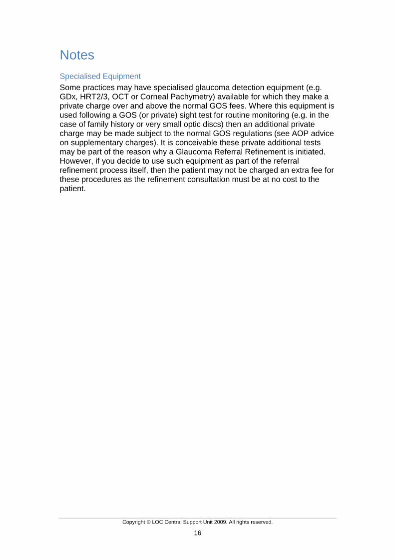
Notes Specialised Equipment Some practices may have specialised glaucoma detection equipment (e.g. GDx, HRT2/3, OCT or Corneal Pachymetry) available for which they make a private charge over and above the normal GOS fees. Where this equipment is used following a GOS (or private) sight test for routine monitoring (e.g. in the case of family history or very small optic discs) then an additional private charge may be made subject to the normal GOS regulations (see AOP advice on supplementary charges). It is conceivable these private additional tests may be part of the reason why a Glaucoma Referral Refinement is initiated. However, if you decide to use such equipment as part of the referral refinement process itself, then the patient may not be charged an extra fee for these procedures as the refinement consultation must be at no cost to the patient.
Copyright © LOC Central Support Unit 2009. All rights reserved.
17
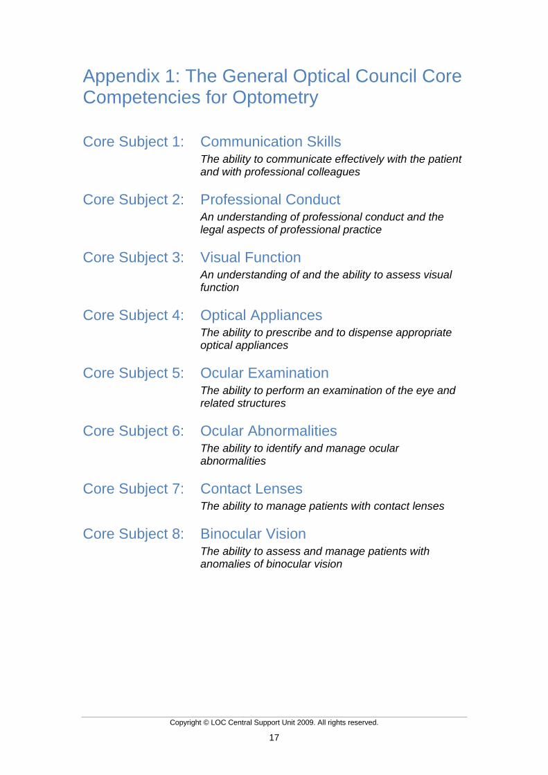
Appendix 1: The General Optical Council Core Competencies for Optometry Core Subject 1: Communication Skills The ability to communicate effectively with the patient
and with professional colleagues Core Subject 2: Professional Conduct An understanding of professional conduct and the
legal aspects of professional practice Core Subject 3: Visual Function An understanding of and the ability to assess visual
function Core Subject 4: Optical Appliances The ability to prescribe and to dispense appropriate
optical appliances Core Subject 5: Ocular Examination The ability to perform an examination of the eye and
related structures Core Subject 6: Ocular Abnormalities The ability to identify and manage ocular
abnormalities Core Subject 7: Contact Lenses The ability to manage patients with contact lenses Core Subject 8: Binocular Vision The ability to assess and manage patients with
anomalies of binocular vision
Copyright © LOC Central Support Unit 2009. All rights reserved.
18
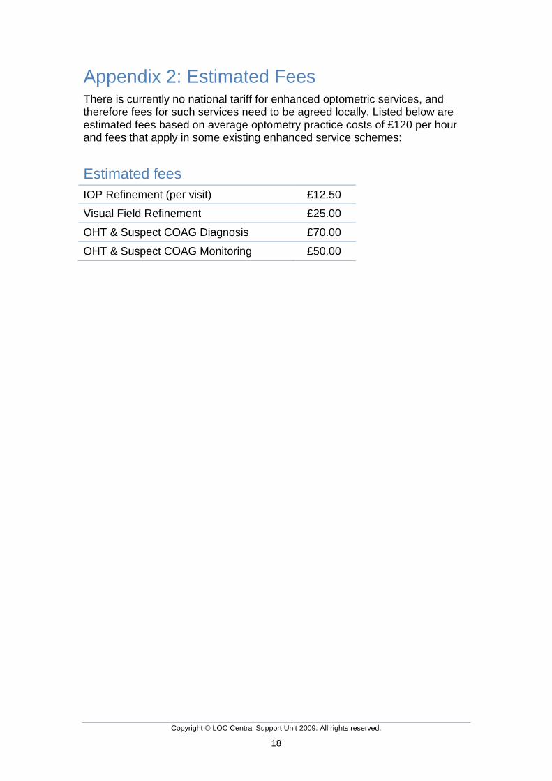
Appendix 2: Estimated Fees There is currently no national tariff for enhanced optometric services, and therefore fees for such services need to be agreed locally. Listed below are estimated fees based on average optometry practice costs of £120 per hour and fees that apply in some existing enhanced service schemes:
Estimated fees IOP Refinement (per visit) £12.50
Visual Field Refinement £25.00
OHT & Suspect COAG Diagnosis £70.00
OHT & Suspect COAG Monitoring £50.00