GISTs recent but long history - · PDF fileCarmen Ardeleanu “Victor Babes”...
33
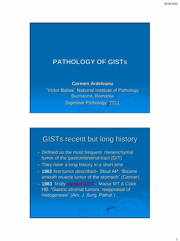
09.05.2012 1 PATHOLOGY OF GISTs Carmen Ardeleanu “Victor Babes” National Institute of Pathology Bucharest, Romania Digestive Pathology, 2011 GISTs recent but long history Defined as the most frequent mesenchymal tumor of the gastrointestinal tract (GIT) They have a long history in a short time 1962 first tumor described– Stout AP: “Bizarre smooth muscle tumor of the stomach” (Cancer) 1983 firstly named GIST – Mazur MT & Clark HB: “Gastric stromal tumors: reappraisal of histogenesis” (Am. J. Surg. Pathol.)
Transcript of GISTs recent but long history - · PDF fileCarmen Ardeleanu “Victor Babes”...
09.05.2012
1
PATHOLOGY OF GISTs
Carmen Ardeleanu
“Victor Babes” National Institute of Pathology
Bucharest, Romania
Digestive Pathology, 2011
GISTs recent but long history
Defined as the most frequent mesenchymal
tumor of the gastrointestinal tract (GIT)
They have a long history in a short time
1962 first tumor described– Stout AP: “Bizarre
smooth muscle tumor of the stomach” (Cancer)
1983 firstly named GIST – Mazur MT & Clark
HB: “Gastric stromal tumors: reappraisal of
histogenesis” (Am. J. Surg. Pathol.)
09.05.2012
2
GISTS history
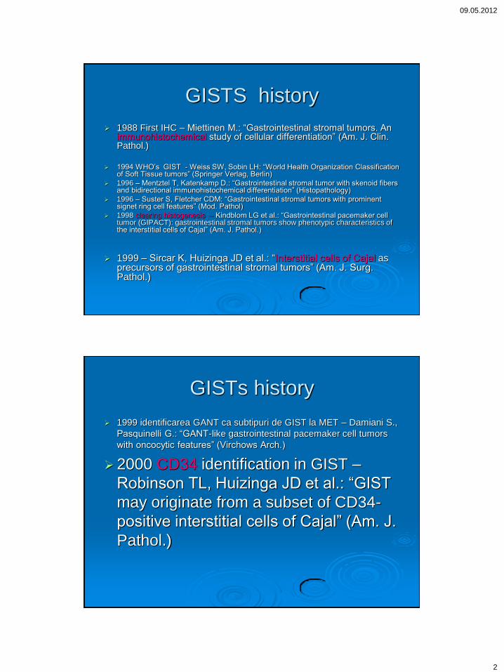
1988 First IHC – Miettinen M.: “Gastrointestinal stromal tumors. An immunohistochemical study of cellular differentiation” (Am. J. Clin. Pathol.)
1994 WHO’s GIST - Weiss SW, Sobin LH: “World Health Organization Classification
of Soft Tissue tumors” (Springer Verlag, Berlin)
1996 – Mentztel T, Katenkamp D.: “Gastrointestinal stromal tumor with skenoid fibers and bidirectional immunohistochemical differentiation” (Histopathology)
1996 – Suster S, Fletcher CDM: “Gastrointestinal stromal tumors with prominent signet ring cell features” (Mod. Pathol)
1998 clearing histogenesis – Kindblom LG et al.: “Gastrointestinal pacemaker cell tumor (GIPACT): gastrointestinal stromal tumors show phenotypic characteristics of the interstitial cells of Cajal” (Am. J. Pathol.)
1999 – Sircar K, Huizinga JD et al.: “Interstitial cells of Cajal as precursors of gastrointestinal stromal tumors” (Am. J. Surg. Pathol.)
GISTs history
1999 identificarea GANT ca subtipuri de GIST la MET – Damiani S.,
Pasquinelli G.: “GANT-like gastrointestinal pacemaker cell tumors
with oncocytic features” (Virchows Arch.)
2000 CD34 identification in GIST –
Robinson TL, Huizinga JD et al.: “GIST
may originate from a subset of CD34-
positive interstitial cells of Cajal” (Am. J.
Pathol.)
09.05.2012
3
GIST history
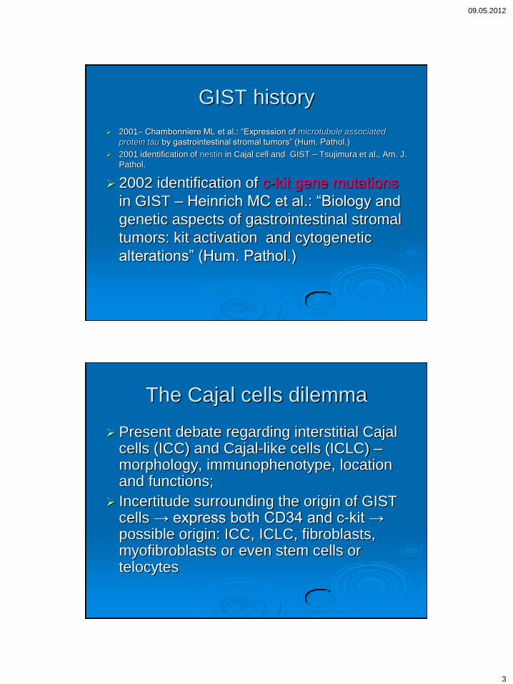
2001– Chambonniere ML et al.: “Expression of microtubule associated
protein tau by gastrointestinal stromal tumors” (Hum. Pathol.)
2001 identification of nestin in Cajal cell and GIST – Tsujimura et al., Am. J.
Pathol.
2002 identification of c-kit gene mutations
in GIST – Heinrich MC et al.: “Biology and
genetic aspects of gastrointestinal stromal
tumors: kit activation and cytogenetic
alterations” (Hum. Pathol.)
The Cajal cells dilemma
Present debate regarding interstitial Cajal cells (ICC) and Cajal-like cells (ICLC) – morphology, immunophenotype, location and functions;
Incertitude surrounding the origin of GIST cells → express both CD34 and c-kit → possible origin: ICC, ICLC, fibroblasts, myofibroblasts or even stem cells or telocytes

09.05.2012
4
GIST’ Histogenesis: ICC / ICLC
Initial gastrointestinal location, extended to many others
Gut pacemaker cells
Form intramural networks
Develop from intrinsic gut mesenchyme? Or from bone marrow CD 34 positive stem cells ?
IHC: CD34, CD117, Nestin, Tau, S-100 protein, Leu 7, DOG1,PDGFRA, PGP9.5.
ICC location
ICC described initially by Cajal as interstitial neurons in the gut and thereafter identified all over the gastrointestinal tract
The ICC network is distributed within the submucosal, intramuscular and intermuscular (myenteric) layer and deep muscular plexus
Only ICC or also ICLC? (Pieri et al 2008).
09.05.2012
5
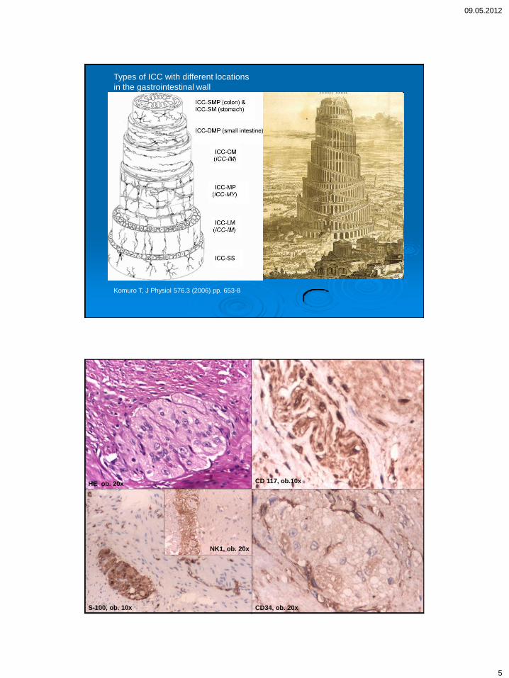
Types of ICC with different locations
in the gastrointestinal wall
Komuro T, J Physiol 576.3 (2006) pp. 653-8
CD 117, ob.10x
CD34, ob. 20x
HE, ob. 20x
S-100, ob. 10x
NK1, ob. 20x

09.05.2012
6
Small intestine
PKCӨ 20x
CD34 20x
HE 20x
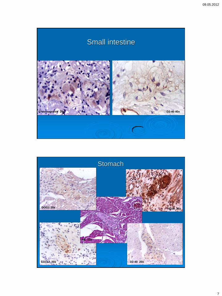
Small intestine
Nestin 20x
S-100 protein
Tau 20x
09.05.2012
7
Small intestine
Chromogranin 20x D2-40 40x
Stomach
CD117 20x
S100 20x
D2-40 20x
DOG1 20x
HE 10x

09.05.2012
8
GIST
The most frequent mesenchymal tumour
of the gastrointestinal tract
Thought to arise from the interstitial cells
of Cajal (ICC) and, more recently, from the
interstitial Cajal-like cells (ICLC)
IHC: CD117, CD 34,PDGFRA, DOG1,
PKCӨ, D2-40, Nestin, Tau
Histogenetic considerations
GISTs do not originate exclusively from ICC, since these tumors can occur in sites where ICC do not exist (omentum, peritoneum, retroperitoneum).
ICLC and other candidates, including a multipotent progenitor, can also constitute the origin of these tumors.
CD34 positivity, considered an expression of immaturity for ICC, has not the same significance in GISTs: >70% are CD34+, but only 25-30% have a biologically malignant behavior.
09.05.2012
9
Clinical features
most of the patients are elderly persons of
both genders,
sometimes GISTs can arise in children,
having particular features and evolution.
these tumors can be single or multiple
sporadic, the last type being sometimes
misinterpreted as recurrent or metastatic
disease.
Location
They may arise throughout the gut, but the commonest sites are the stomach (60-70%), the small bowel (20-30%), the colorectum (5%), the esophagus (up to 5%).
GISTs developed in the retroperitoneum, the omentum and the mesentery are considered extraGISTs.
09.05.2012
10
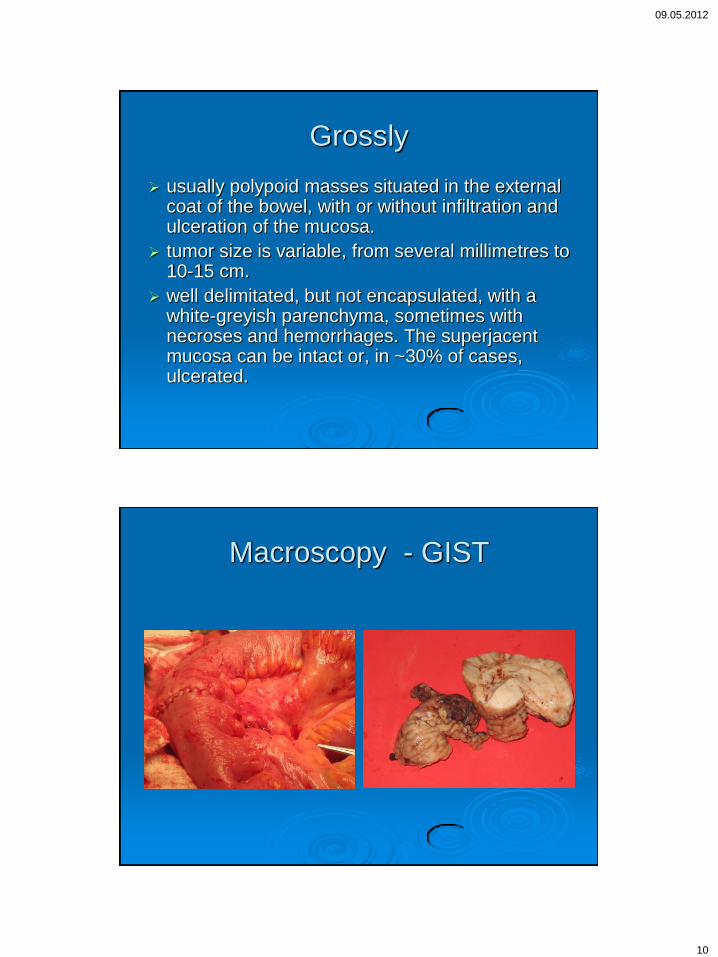
Grossly
usually polypoid masses situated in the external coat of the bowel, with or without infiltration and ulceration of the mucosa.
tumor size is variable, from several millimetres to 10-15 cm.
well delimitated, but not encapsulated, with a white-greyish parenchyma, sometimes with necroses and hemorrhages. The superjacent mucosa can be intact or, in ~30% of cases, ulcerated.
Macroscopy - GIST
09.05.2012
11
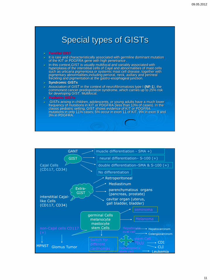
Special types of GISTs
Familial GIST
It is rare and characteristically associated with germline dominant mutation of the KIT or PDGFRA gene with high penetrance .
In this context,GIST is usually multifocal and variably associated with hyperplasia of the interstitial cells of Cajal and abnormalities of mast cells such as urticaria pigmentosa or systemic mast cell disease, together with pigmentary abnormalities,including perioral, neck, axillary and perineal freckling and pigmentation at the gastro-esophageal junction.
Syndromic GISTs
Association of GIST in the context of neurofibromatosis type I (NF-1), the commonest cancer-predisposition syndrome, which carries up to 25% risk for developing GIST. Multifocal.
Pediatric GISTs:
GISTs arising in children, adolescents, or young adults have a much lower frequency of mutations in KIT or PDGFRA (less than 10% of cases). In the classic pediatric setting, GIST shows evidence of KIT or PDGFRA mutations in only 11% cases; 5% occur in exon 11 of KIT, 9% in exon 9 and 3% in PDGFRA.
Cajal Cells (CD117, CD34)
GIST
interstitial Cajal-like Cells (CD117, CD34)
Extra- GIST
non-Cajal cells CD117 (+)
germinal Cells melanocyte mastocyte stem Cells
seminoma
Melanoma
Switch for different carcinomas
Retroperitoneal
Mediastinum
cavitar organ (uterus, gall bladder, bladder)
parenchymatous organs (pancreas, prostate)
muscle differentiation - SMA +)
neural differentiation– S-100 (+)
double differentiation–SMA & S-100 (+)
No differentiation
Hepatocyte oval cell
stem Cell TDLU
Hepatocarcinom
Colangiocarcinom
CDI
hematopoetic stem cell
CLI
Leukemia
GANT
MPNST Glomus Tumor
09.05.2012
12
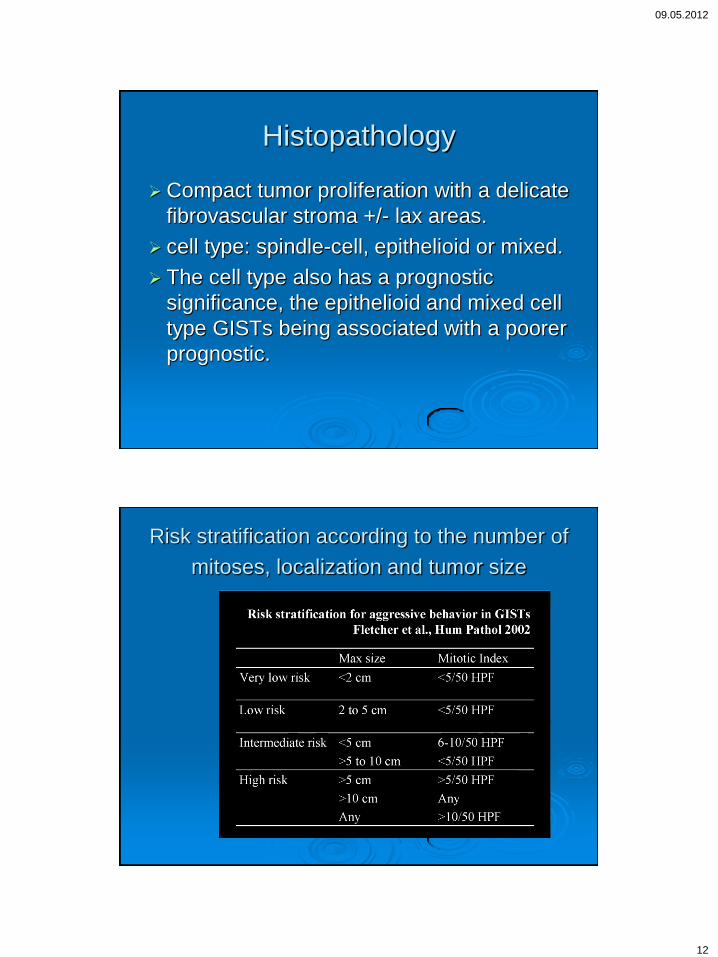
Histopathology
Compact tumor proliferation with a delicate
fibrovascular stroma +/- lax areas.
cell type: spindle-cell, epithelioid or mixed.
The cell type also has a prognostic
significance, the epithelioid and mixed cell
type GISTs being associated with a poorer
prognostic.
Risk stratification according to the number of
mitoses, localization and tumor size
09.05.2012
13
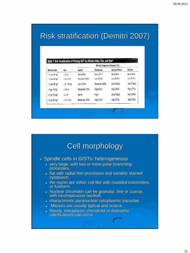
Risk stratification (Demitri 2007)
Cell morphology
Spindle cells in GISTs: heterogeneous very large, with two or more polar branching
processes,
flat with radial thin processes and variably stained cytoplasm.
the nuclei are either rod-like with rounded extremities, or fusiform.
Nuclear chromatin can be granular, fine or coarse, with inconspicuous nucleoli.
characteristic paranuclear cytoplasmic vacuolae.
Mitoses are usually typical and scarce.
Rarely, metaplastic chondroid or distrophic calcifications can occur.
09.05.2012
14
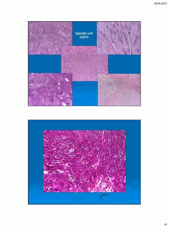
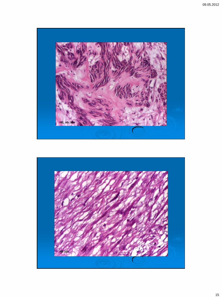
Spindle cell
GISTs
HE, ob. 10x
09.05.2012
15
HE, ob. 20x
HE 20x

09.05.2012
16
HE, ob. 40x
HE, ob. 60x

09.05.2012
17
HE, ob. 40x
HE, ob. 20x

09.05.2012
18
Cell morphology
Epithelioid type (5-10% of cases)
proliferate as islets or sheaths of large rounded
cells with pale eosinophilic cytoplasm,
sometimes foamy or plasmacytoid,
rounded vesiculous nuclei and visible nucleoli.
the tumor stroma can be finely fibrovascular or
more abundant, myxoid or edematous.
A significant number of mitoses can be
considered as prognostic factor.
Epithelioid type GIST

09.05.2012
19
Epithelioid pattern, clear cells
plasmocytoid pattern
Stroma hyalinization
Mixoide
Hialina

09.05.2012
20
Distrophic calcifications
Morfologic features: plasmacytoid cells;
paranuclear vacuolization
HE, 10x HE, 10x
09.05.2012
21
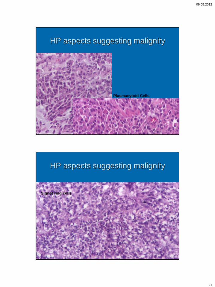
HP aspects suggesting malignity
Plasmacytoid Cells
HP aspects suggesting malignity
Signet ring cells

09.05.2012
22
Immunohistochemical profile
CD34 – the first marker used
CD117 – c-kit gene protein
PDGFRA - platelet derived growth factor
receptor A
DOG1 - discovered on GIST
D2-40 / podoplanin
Nestin, Caveolin 1, Tau, PKC teta
C-kit protein (CD117)
- it is a 145-kD glycosylated transmembranary receptor with tyrosine kinase activity
structure :
* an extracellular domain (consists of 5 Ig-like domains)
* a transmembrane region (TM)
* a juxtamembrane (JM) domain
* an intracytoplasmic tyrosine kinase domain
- its specific ligand is the stem cells growth factor (SCF)

09.05.2012
23
Heterogeneous c-kit staining
patterns
CD117 is expressed in >95% of GISTs
IHC patterns in tumor cells: Membrane
Cytoplasm: Homogenous
Coarse granular
Fine granular
Paranuclear dot
CD117 staining patterns can be associated with certain c-kit gene mutations (homozygous or heterozygous) involved in the response to imatinib (Tabone-Eglinger S et al. Clin Cancer Res, 2008, 14(8):2285-2294)
CD117 20x
GASTRIC GISTs
CD117 20x
09.05.2012
24
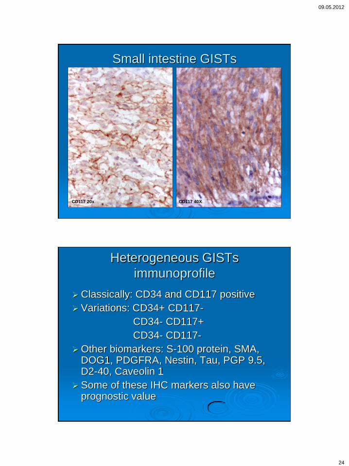
Small intestine GISTs
CD117 20x CD117 40X
Heterogeneous GISTs
immunoprofile
Classically: CD34 and CD117 positive
Variations: CD34+ CD117-
CD34- CD117+
CD34- CD117-
Other biomarkers: S-100 protein, SMA, DOG1, PDGFRA, Nestin, Tau, PGP 9.5, D2-40, Caveolin 1
Some of these IHC markers also have prognostic value
09.05.2012
25
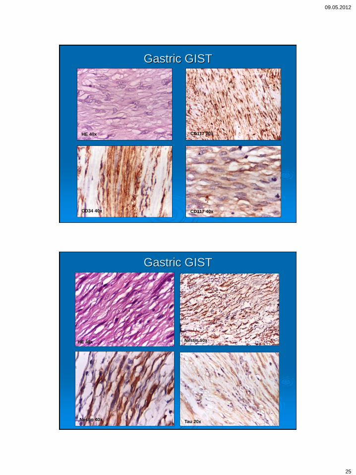
Gastric GIST
HE 40x CD117 20x
CD117 40x CD34 40x
Gastric GIST
Nestin 10x
Nestin 40x Tau 20x
HE 40x

09.05.2012
26
Gastric GIST
CD117 20x PDGFRA 20x
Caveolin-1 20x Vimentin 20x
Caveolin 10x
CD117 20x
CD117 20x
09.05.2012
27
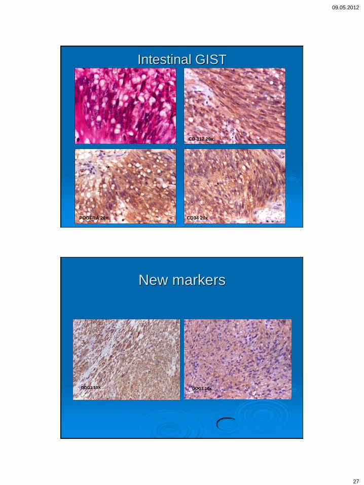
Intestinal GIST
CD 117 20x
PDGFRA 20x CD34 20x
New markers
DOG1 10X DOG1 10x
09.05.2012
28
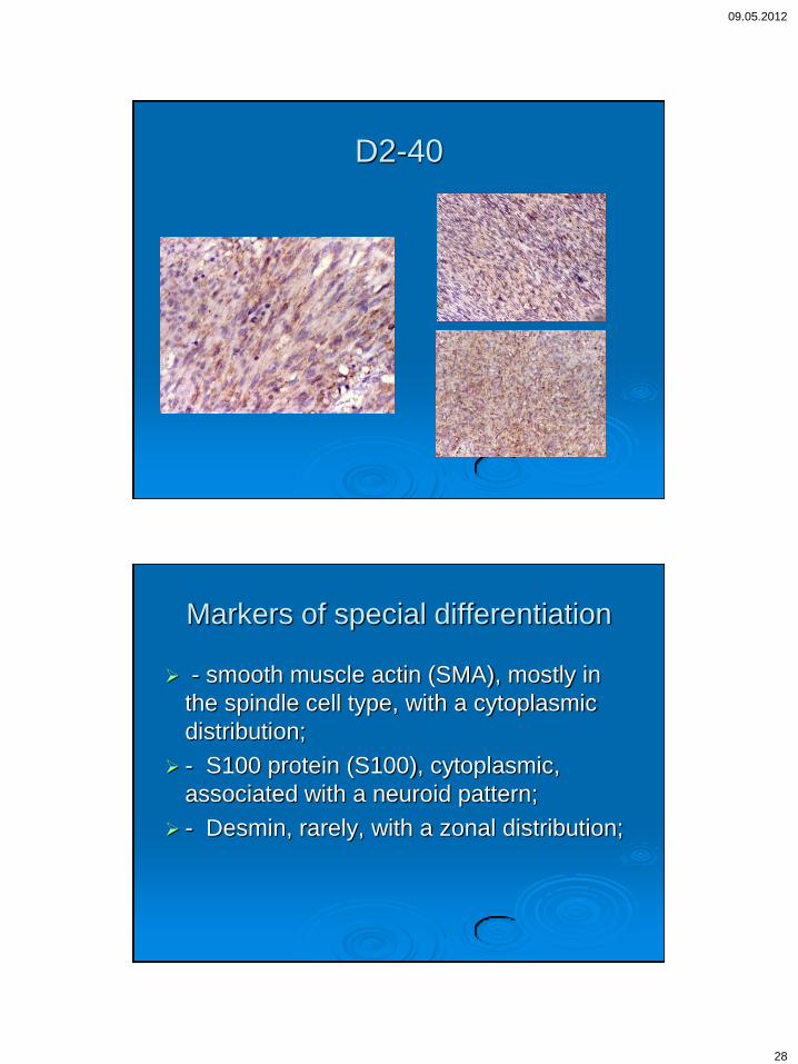
D2-40
Markers of special differentiation
- smooth muscle actin (SMA), mostly in
the spindle cell type, with a cytoplasmic
distribution;
- S100 protein (S100), cytoplasmic,
associated with a neuroid pattern;
- Desmin, rarely, with a zonal distribution;

09.05.2012
29
Special differentiation
SMA
10x/ 20x
S-100
10x/20x
Molecular features- ckit mutations
occur on exon 11 which codifies a juxtamembranary domain with regulatory function; it is the most frequently encountered, in >70% of GISTs.
Other mutations, in decreasing order of frequency, are located on exons 9, 13 and 17.
Recent studies: GISTs with mutations on exon 11 are the most sensitive to the specific treatment with Imatinib, compared with mutations on exons 9 or 13. Also, deletions compared with point mutations in exon 11 have been found to be a significant unfavorable factor in patients with gastric GISTs.

09.05.2012
30
PDGFRA mutations
located on exon 18 (over 80%), exon 12
(much less frequent - 6%) or exon 14 (the
rarest - 1%). PDGFRA mutations on exon
18 characterize the category of GISTs with
primary resistance to Imatinib, while
mutations on exon 14 are usually
associated with a less favorable
prognosis. Exon 12 mutations are the
most sensitive to specific treatment
PCR pentru c-Kit Exoni 9 si 11
1 2 3 4 5 6 7 M 1’ 2’ 3’ 4’ 5’ 6’ 7’
Verificare electroforetica pe gel de agaroza 2% a reactiilor PCR pentru gena C-KIT-exonii 9 si 11
Probe (1-7=exon 9 si 1’-7’=exon 11):
1 – ADS (control negativ)
2- 123750
3 – 126904
4 – 128048
5 – 130530
6 – 174048
7 – proba de control pozitiv 174934-2
M – 50bp DNA Ladder (Fermentas)
09.05.2012
31
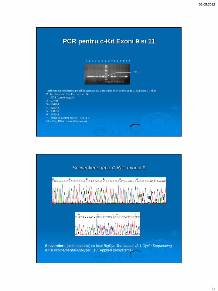
PCR pentru c-Kit Exoni 9 si 11
Verificare electroforetica pe gel de agaroza 2% a reactiilor PCR pentru gena C-KIT-exonii 9 si 11
Probe (1-7=exon 9 si 1’-7’=exon 11):
1 – ADS (control negativ)
2- 123750
3 – 126904
4 – 128048
5 – 130530
6 – 174048
7 – proba de control pozitiv 174934-2
M – 50bp DNA Ladder (Fermentas)
1 2 3 4 5 6 7 M 1’ 2’ 3’ 4’ 5’ 6’ 7’
200 pb
Secventiere gena C-KIT, exonul 9
Secventiere (bidirectionala) cu kitul BigDye Terminator v3.1 Cycle Sequencing
Kit si echipamentul Analyzer 310 (Applied Biosystems)
09.05.2012
32
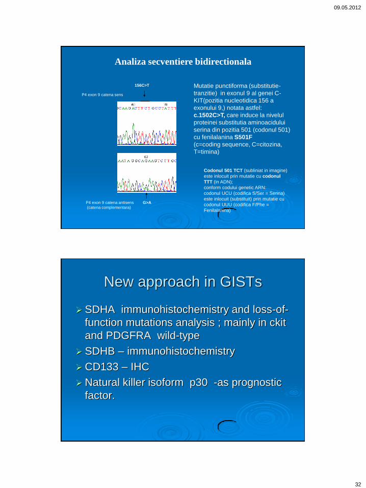
Codonul 501 TCT (subliniat in imagine)
este inlocuit prin mutatie cu codonul
TTT (in ADN);
conform codului genetic ARN:
codonul UCU (codifica S/Ser = Serina)
este inlocuit (substituit) prin mutatie cu
codonul UUU (codifica F/Phe =
Fenilalanina)
156C>T
P4 exon 9 catena sens
P4 exon 9 catena antisens G>A
(catena complementara)
Mutatie punctiforma (substitutie-
tranzitie) in exonul 9 al genei C-
KIT(pozitia nucleotidica 156 a
exonului 9,) notata astfel:
c.1502C>T, care induce la nivelul
proteinei substitutia aminoacidului
serina din pozitia 501 (codonul 501)
cu fenilalanina S501F
(c=coding sequence, C=citozina,
T=timina)
Analiza secventiere bidirectionala
New approach in GISTs
SDHA immunohistochemistry and loss-of-
function mutations analysis ; mainly in ckit
and PDGFRA wild-type
SDHB – immunohistochemistry
CD133 – IHC
Natural killer isoform p30 -as prognostic
factor.
09.05.2012
33
Victor Babes Institute GIST team
Simona Enache
Mihaela Mihai
Cristina iosif
Florina Vasilescu
Dana Terzea
Florin Andrei
Georgeta Cardos
Alina Grigore
Thank you!