Ginsenoside-Rb2 displays anti-osteoporosis effects … · 2 displays anti-osteoporosis effects...
9
Original Full Length Article Ginsenoside-Rb 2 displays anti-osteoporosis effects through reducing oxidative damage and bone-resorbing cytokines during osteogenesis Qiang Huang 1 , Bo Gao 1 , Qiang Jie 1 , Bo-Yuan Wei, Jing Fan, Hong-Yang Zhang, Jin-Kang Zhang, Xiao-Jie Li, Jun Shi, Zhuo-Jing Luo 2 , Liu Yang 2 , Jian Liu ⁎ Institute of Orthopedic Surgery, Xijing Hospital, Fourth Military Medical University, Xi'an 710032, People's Republic of China abstract article info Article history: Received 27 February 2014 Revised 5 June 2014 Accepted 6 June 2014 Available online 13 June 2014 Edited by J. Aubin Keywords: Ginsenoside-Rb 2 Oxidative damage Osteogenesis Osteoporosis Ovariectomized mouse Bone-resorbing cytokine Reactive oxygen species (ROS) are a significant pathogenic factor of osteoporosis. Ginsenoside-Rb 2 (Rb 2 ), a 20(S)- protopanaxadiol glycoside extracted from ginseng, is a potent antioxidant that generates interest regarding the bone metabolism area. We tested the potential anti-osteoporosis effects of Rb 2 and its underlying mechanism in this study. We produced an oxidative damage model induced by hydrogen peroxide (H 2 O 2 ) in osteoblastic MC3T3-E1 cells to test the essential anti-osteoporosis effects of Rb 2 in vitro. The results indicated that treatment of 0.1 to 10 μM Rb 2 promoted the proliferation of MC3T3-E1 cells, improved alkaline phosphatase (ALP) expression, elevated calcium mineralization and mRNA expressions of Alp, Col1a1, osteocalcin (Ocn) and osteopontin (Opn) against oxidative damage induced by H 2 O 2 . Importantly, Rb 2 reduced the expression levels of receptor activator of nuclear factor kappa-B ligand (RANKL) and IL-6 and inhibited the H 2 O 2 -induced production of ROS. The in vivo study indicated that the Rb 2 administered for 12 weeks partially decreased blood malondialdehyde (MDA) activity and elevated the activity of reduced glutathione (GSH) in ovariectomized (OVX) mice. Moreover, Rb 2 improved the micro-architecture of trabecular bones and increased bone mineral density (BMD) of the 4th lumbar vertebrae (L4) and the distal femur. Altogether, these results demonstrated that the potential anti-osteoporosis effects of Rb 2 were linked to a reduction of oxidative damage and bone-resorbing cytokines, which suggests that Rb 2 might be effective in preventing and alleviating osteoporosis. © 2014 The Authors. Published by Elsevier Inc. This is an open access article under the CC BY-NC-SA license (http://creativecommons.org/licenses/by-nc-sa/3.0/). Introduction Oxidative stress is described as an imbalance between an overpro- duction of reactive oxygen species (ROS) and an insufficient defense of antioxidants [1]. Oxidative stress can lead to oxidative damage that affects all of the cellular components, including proteins, lipids and nucleic acids. Osteoporosis is defined as a systemic degenerative dis- ease, which is characterized by decreased bone mass and progressive bone micro-architectural deterioration and results in increasing bone fragility and susceptibility to fractures [2]. Currently, many studies have demonstrated a relationship between oxidative damage and osteoporosis or aging. Sendur and colleagues found that the decreased BMD of postmenopausal osteoporotic women was related to higher oxidation of plasma lipid [3] and lowered superoxide dismutase (SOD), catalase and glutathione peroxidase efficacy [4–6]. Ovariectomy has been shown to induce oxidative damage and to decrease the efficacy of antioxidant defense mechanisms, thus leading to osteoporosis. The activities of lipid peroxidation and H 2 O 2 were increased, and enzymatic antioxidants, such as superoxide dismutase, glutathione peroxidase, and glutathione S transferase, were decreased in ovariectomized ani- mals [7]. Moreover, many studies have demonstrated that superoxide and H 2 O 2 were highly deleterious to cell survival and that they played a major causative role in the aging process [8]. These research findings provided a paradigm shift of osteoporosis pathogenesis from the “estrogen-centric” concept to one in which age-related mechanisms intrinsic to bone and oxidative stress were protagonists. Moreover, the age-related changes in other organs and tissues, such as the ovaries, accentuated these alterations [9]. Bone remodeling is a process that occurs throughout one's entire life and involves two types of cells: osteogenic cells and monocyte-derived osteoclasts [10–12]. The osteogenic cells participate in bone formation, and osteoclasts are responsible for bone resorption [13,14]. The balance between bone formation and bone resorption is essential for maintain- ing bone homeostasis. Bone turnover changes significantly in postmen- opausal osteoporosis: bone resorption is maintained or increases, but bone formation decreases, thereby leading to a net bone loss [15]. Emerging evidence has shown that ROS increased bone resorption by enhancing osteoclastic development and activity [16]. Indeed, in vivo bone resorption occurs preferentially in sites where ROS and Bone 66 (2014) 306–314 ⁎ Corresponding author. Fax: +86 29 84771013. E-mail address: [email protected] (J. Liu). 1 These authors contributed equally to the work. 2 Co-corresponding author. http://dx.doi.org/10.1016/j.bone.2014.06.010 8756-3282/© 2014 The Authors. Published by Elsevier Inc. This is an open access article under the CC BY-NC-SA license (http://creativecommons.org/licenses/by-nc-sa/3.0/). Contents lists available at ScienceDirect Bone journal homepage: www.elsevier.com/locate/bone
Transcript of Ginsenoside-Rb2 displays anti-osteoporosis effects … · 2 displays anti-osteoporosis effects...

Bone 66 (2014) 306–314
Contents lists available at ScienceDirect
Bone
j ourna l homepage: www.e lsev ie r .com/ locate /bone
Original Full Length Article
Ginsenoside-Rb2 displays anti-osteoporosis effects through reducingoxidative damage and bone-resorbing cytokines during osteogenesis
Qiang Huang 1, Bo Gao 1, Qiang Jie 1, Bo-Yuan Wei, Jing Fan, Hong-Yang Zhang, Jin-Kang Zhang, Xiao-Jie Li,Jun Shi, Zhuo-Jing Luo 2, Liu Yang 2, Jian Liu ⁎Institute of Orthopedic Surgery, Xijing Hospital, Fourth Military Medical University, Xi'an 710032, People's Republic of China
⁎ Corresponding author. Fax: +86 29 84771013.E-mail address: [email protected] (J. Liu).
1 These authors contributed equally to the work.2 Co-corresponding author.
http://dx.doi.org/10.1016/j.bone.2014.06.0108756-3282/© 2014 The Authors. Published by Elsevier Inc
a b s t r a c t
a r t i c l e i n f oArticle history:Received 27 February 2014Revised 5 June 2014Accepted 6 June 2014Available online 13 June 2014
Edited by J. Aubin
Keywords:Ginsenoside-Rb2Oxidative damageOsteogenesisOsteoporosisOvariectomized mouseBone-resorbing cytokine
Reactive oxygen species (ROS) are a significant pathogenic factor of osteoporosis. Ginsenoside-Rb2 (Rb2), a 20(S)-protopanaxadiol glycoside extracted from ginseng, is a potent antioxidant that generates interest regarding thebone metabolism area. We tested the potential anti-osteoporosis effects of Rb2 and its underlying mechanism inthis study. We produced an oxidative damage model induced by hydrogen peroxide (H2O2) in osteoblasticMC3T3-E1 cells to test the essential anti-osteoporosis effects of Rb2 in vitro. The results indicated that treatment of0.1 to 10 μM Rb2 promoted the proliferation of MC3T3-E1 cells, improved alkaline phosphatase (ALP) expression,elevated calcium mineralization and mRNA expressions of Alp, Col1a1, osteocalcin (Ocn) and osteopontin (Opn)against oxidative damage induced by H2O2. Importantly, Rb2 reduced the expression levels of receptor activator ofnuclear factor kappa-B ligand (RANKL) and IL-6 and inhibited the H2O2-induced production of ROS. The in vivostudy indicated that the Rb2 administered for 12 weeks partially decreased bloodmalondialdehyde (MDA) activityand elevated the activity of reduced glutathione (GSH) in ovariectomized (OVX)mice. Moreover, Rb2 improved themicro-architecture of trabecular bones and increased bonemineral density (BMD) of the 4th lumbar vertebrae (L4)and the distal femur. Altogether, these results demonstrated that the potential anti-osteoporosis effects of Rb2 werelinked to a reduction of oxidative damage and bone-resorbing cytokines, which suggests that Rb2might be effectivein preventing and alleviating osteoporosis.
© 2014 The Authors. Published by Elsevier Inc. This is an open access article under the CC BY-NC-SA license(http://creativecommons.org/licenses/by-nc-sa/3.0/).
Introduction
Oxidative stress is described as an imbalance between an overpro-duction of reactive oxygen species (ROS) and an insufficient defenseof antioxidants [1]. Oxidative stress can lead to oxidative damage thataffects all of the cellular components, including proteins, lipids andnucleic acids. Osteoporosis is defined as a systemic degenerative dis-ease, which is characterized by decreased bone mass and progressivebone micro-architectural deterioration and results in increasing bonefragility and susceptibility to fractures [2]. Currently, many studieshave demonstrated a relationship between oxidative damage andosteoporosis or aging. Sendur and colleagues found that the decreasedBMD of postmenopausal osteoporotic women was related to higheroxidation of plasma lipid [3] and lowered superoxide dismutase(SOD), catalase and glutathione peroxidase efficacy [4–6]. Ovariectomyhas been shown to induce oxidative damage and to decrease the efficacyof antioxidant defense mechanisms, thus leading to osteoporosis. The
. This is an open access article under
activities of lipid peroxidation and H2O2 were increased, and enzymaticantioxidants, such as superoxide dismutase, glutathione peroxidase,and glutathione S transferase, were decreased in ovariectomized ani-mals [7]. Moreover, many studies have demonstrated that superoxideand H2O2 were highly deleterious to cell survival and that they playeda major causative role in the aging process [8]. These research findingsprovided a paradigm shift of osteoporosis pathogenesis from the“estrogen-centric” concept to one in which age-related mechanismsintrinsic to bone and oxidative stress were protagonists. Moreover, theage-related changes in other organs and tissues, such as the ovaries,accentuated these alterations [9].
Bone remodeling is a process that occurs throughout one's entire lifeand involves two types of cells: osteogenic cells and monocyte-derivedosteoclasts [10–12]. The osteogenic cells participate in bone formation,and osteoclasts are responsible for bone resorption [13,14]. The balancebetween bone formation and bone resorption is essential for maintain-ing bone homeostasis. Bone turnover changes significantly in postmen-opausal osteoporosis: bone resorption is maintained or increases, butbone formation decreases, thereby leading to a net bone loss [15].
Emerging evidence has shown that ROS increased bone resorptionby enhancing osteoclastic development and activity [16]. Indeed,in vivo bone resorption occurs preferentially in sites where ROS and
the CC BY-NC-SA license (http://creativecommons.org/licenses/by-nc-sa/3.0/).

307Q. Huang et al. / Bone 66 (2014) 306–314
hence oxidative stress levels are high [17,18]. ROS also led to osteoblastapoptosis, as well as reducing their activity, which led to decreased os-teogenesis [19]. Oxidative stress decreases the life span of osteoblast inthe bone, as highlighted by the observation that the administration ofantioxidants abrogates osteoblast apoptosis in ovariectomized or agedmice [20,21]. Therefore, we recommend substances that contain antiox-idants because they may ameliorate the dysfunction of these two celltypes bymaintaining bone hemostasis. This suggestionmight be imple-mented by acting as a latent method for the prevention and new treat-ment for osteoporosis or other related bone metabolic diseases.
Because of fewer side-effects and longer term usage than Westernmedicine, traditional Chinese medicines with anti-oxidative propertieshave recently attracted more attention. The ginseng root, which is ahighly effective phytomedicinal remedy, is awell-recognized, tradition-al Chinesemedicine that is widely used. The primary components foundin ginseng are ginsenosides, which contribute to themost active proper-ties. Ginsenoside-Rb2 (Fig. 1) is the most quantitative saponin that iscontained in Panax ginseng [22]. It was reported that Rb2 possessedanti-diabetic, anti-adipocyte, anti-carcinogenic and anti-oxidativeproperties [23–26]. More importantly, recent studies showed thatH2O2,which is known as a cancer promoter, inhibited the gap junctionalintercellular communication (GJIC) of epithelial cells. Rb2 supplementa-tionmight inhibit the occurrence of cancer through the up-regulation ofGJIC in the cancer-accelerating phase [27]. SOD1 is considered to be oneof the antioxidant enzymes. A previous study [28] has shown thatginsenoside-Rb2 could induce the transcriptional expressions of Cu,Zn-superoxide dismutase gene (SOD1). The mutated AP2 binding site inthe promoter of SOD1 gene abrogated this effect, which suggests thatthe SOD1 gene was highly activated by ginsenoside-Rb2 through theAP2 binding site [28]. Kang et al. studied the hydroxyl radical (•OH)clearing capacity change of Rb2 through heat procedure using an elec-tron spin resonance spectrometer. Specially, ginsenoside-Rb2 was heatprocessed using the same amount of glycine, which was an aminoacid commonly employed in the Maillard response model system [26].
Whether ginsenoside-Rb2 might provide the potential anti-osteoporosis effects andwhether these effects are produced by reducingoxidative damage and bone-resorbing cytokines have not beenestablished. Therefore, in the present study, we tested the anti-osteoporosis effects of Rb2 and the underlying mechanisms in vitro
Fig. 1. Chemical structure of Rb2 (from National Center of Biotechnology Information,Pubchem CID: 5458674).
and in vivo using an H2O2-induced oxidative damage model ofosteoblastic MC3T3-E1 cells and a murine ovariectomized model.
Materials and methods
Materials
Rb2 extracted from ginseng (molecular weight, 1079; purity,N98.0%; dissolved in distilled water) was purchased from ShanghaiTauto Biotech Co., LTD (China). The culture flasks and plates wereobtained fromNunc (Denmark). The ALP activity assay kit was obtainedfrom GENMED Scientific Inc. (USA). RANKL and IL-6 ELISA assay kitswere obtained from R&D system Inc. (Minneapolis, MN, USA). Thetotal RNA kit was obtained from OMEGA. Prime Script RT reagent kitand SYBR Premix Ex Taq were obtained from TaKaRa Biotechnology(Dalian, China). The oligonucleotide primers were synthesized byTaKaRa Biotechnology. BCIP/NBT alkaline phosphatase color develop-ment kit was obtained from Gibco Life Technologies (Grand Island,USA). The reactive oxygen species assay kit, GSH and MDA assay kitswere obtained from the Beyotime Institute of Biotechnology (Shanghai,China). All other reagents were of analytical grade.
Cell cultures
Murine MC3T3-E1 cells were purchased from the Center Laboratoryfor Tissue Engineering, College of Stomatology, Fourth Military MedicalUniversity, Xi'an, China [29,30]. The cells were cultured inα-MEMusing10% heat-inactivated FBS and 100 U/ml penicillin and 100 mg/ml strep-tomycin in a condition of 5% CO2 and 37 °C. H2O2 acted as exogenousROS treatment, whereas N-acetyl-L-cysteine (NAC) acted as an ROScleaner. When the cells reached confluence, the serum-free mediumcontaining Rb2was dissolved in distilledwater and cultured for 24 h be-fore the administration of 0.3 mM H2O2 for 24 h. For each experiment,Rb2 administration continued after the pretreatment. All of the experi-ments were performed in duplicate wells and repeated three times.
Assays of cell survival
For this experiment, the murine MC3T3-E1 cells were administeredusing different consistencies of Rb2 (0 μM, 0.1 μM, 1 μM, and 10 μM) for24 h and 72 h to test the toxicity of Rb2. Subsequently, the cells were in-cubated using the serum-free regular culture medium, which containedRb2 (0 μM, 0.1 μM, 1 μM, and 10 μM) for 24 h followed by the administra-tion of 300 μMH2O2 for 24h. TheMTT assayswereused formeasuring cellsurvival. The absorbances of all of the wells were recorded using amicro-plate reader at 492 nmwavelength. The cell survival of the controlgroup,whichwas not exposed to eitherH2O2 or Rb2,was defined as 100%.
Alkaline phosphatase (ALP) staining and activity assay
After a 6-day osteogenic induction, themurineMC3T3-E1 cells wereincubated using serum-free medium, which contained Rb2 and/or H2O2
for 2 days. The cells were stained using the BCIP/NBT alkaline phospha-tase color development kit according to themanufacturer's instructions.To evaluate the ALP activity, the cell monolayer was lysed using the celllysis buffer. Subsequently, the lysate was centrifuged at 10,000 g for5 min. The clear supernatant was used to measure the ALP activity,which was determined employing the ALP activity assay kit. The totalprotein consistencies were determined using the Bradford proteinassay method. The ALP activity was normalized to total protein, whichwas measured using the Bradford protein assay method.
Calcium mineralization assay
After a 21-day osteogenic induction, the murine MC3T3-E1 cellswere incubated using a serum-free medium, which contained Rb2

308 Q. Huang et al. / Bone 66 (2014) 306–314
and/or H2O2 for 2 days. After the cells were collected, the cells werefixed with formalin for 20 min and stained with Alizarin Red S for45 min under room temperature. To measure matrix calcification, un-bound alizarin red was washed off using PBS. Subsequently, the stainwas solubilized using 10% cetylpyridinium chloride by shaking for15 min. The absorbances of the released Alizarin Red S were recordedusing a micro-plate reader at 562 nm wavelength.
Quantitative real-time PCR
After a 6-day osteogenic induction, murine MC3T3-E1 cells were in-cubated using a serum-free medium, which contained Rb2 and/or H2O2
for 2 days. The total cellular RNA was extracted from these cells usingTrizol reagent. Single strand cDNA synthesis was determined using thePrime Script RT reagent kit (TaKaRa). The RT-PCR was performedusing the CFX96 (BIO-RAD) instrument, and individual PCRs were con-ducted in 96-well optical reaction plates using SYBR Green-I (TaKaRa)according to the manufacturer's instructions. Target gene (Alp, Col1a1,Ocn and Opn) expressions were normalized to the reference geneβ-actin. The 2−ΔΔCt method was applied to calculate the relative geneexpression. These PCR products were subjected to a melting curveanalysis and a standard curve to confirm the correct amplification. Allof the PCRs were performed in triplicate, and the primers used for PCRare shown in Table 1.
RANKL and IL-6 measurements
After a 6-day osteogenic induction, the murine MC3T3-E1 cells wereincubated using serum-free medium, which contained Rb2 and/or H2O2
for 2 days. The expressions of RANKL and IL-6 in culture medium weretested using the sandwich ELISA assay kit according to themanufacturer'sinstructions. The total protein consistencies were measured using theBradford protein assay method.
Intracellular reactive oxygen species (ROS) measurement
After a 6-day osteogenic induction, themurineMC3T3-E1 cells wereincubated using serum-free medium, which contained Rb2 and/or H2O2
for 2 days. The intracellular ROS expression level was determined usingthe ROS assay kit. DCFH-DA can be oxidized by ROS in viable cells to2′,7′-dichlorofluorescein (DCF), which is highly fluorescent at 530 nm.These cells were washed three times with PBS. DCFH-DA, whichwas di-luted to a final consistency of 10 μM,was added and cultured for 30minat 37 °C in the dark. When washed three times by PBS, the relativeexpression of fluorescence was quantified using a multi-detectionmicro-plate reader (485 nm excitation and 535 nm emission).
Animals and ginsenoside-Rb2 intervention
Forty 8-week-old, BALB/c female mice, weighing 20.84 ± 1.21 g,were obtained from the Experimental Animal Center of The FourthMilitary Medical University (Xi'an, China). There was no significantdifference in the initial body weights of the mice among all 4 groupsin this experiment. The mice were allowed to adapt to the laboratoryenvironment (a well-ventilated controlled room at 20 °C on a 12-h
Table 1Real-time PCR primers for amplification of specific MC3T3-E1 mRNA.
Gene Forward (5′–3′) Reverse (5′–3′)
Alp GCAGTATGAATTGAATCGGAACAAC ATGGCCTGGTCCATCTCCACColla1 GACATGTTCAGCTTTGTGGACCTC GGGACCCTTAGGCCATTGTGTAOcn ACCATCTTTCTGCTCACTCTGCT CCTTATTGCCCTCCTGCTTGOpn TACGACCATGAGATTGGCAGTGA TATAGGATCTGGGTGCAGGCTGTAAβ-Actin CATCCGTAAAGACCTCTATGCCAAC ATGGAGCCACCGATCCACA
light/dark cycle; the animals were given free access to water andfood) for 1 week before the surgery. Subsequently, the mice experi-enced sham-operation (n = 10) or were surgically ovariectomized(OVX) (n = 30) under anesthesia using pentobarbital sodium(50 mg/kg body weight, i.p.). The ovariectomy operation was per-formed according to Steven K. Boyd's procedure [31]. A total of 30BALB/c female mice were randomly divided into three groups: 1) OVXgroup, administered intraperitoneally with distilled water (n = 10);2) OVX group, administered intraperitoneally with Rb2 (body weight,4.6 μmol/kg; n = 10) daily; and 3) OVX group, administered intra-peritoneally with Rb2 (body weight, 18.5 μmol/kg; n = 10) daily.Rb2 was dissolved in distilled water. One week after the operation,the treatments commenced and continued for 12 weeks. Bloodsamples were obtained from the hearts in anesthetized mice andserum samples were prepared by centrifugation. The left femursand 4th lumbar vertebrae (L4) of the mice were collected and theadherent tissue was discarded. All of the experimental procedureswere officially approval by the Ethics in the Animal Research Com-mittee of the Fourth Military Medical University (permission code2010C00843).
Measurements of serummalondialdehyde (MDA) and reduced glutathione(GSH)
The activity of MDA inwhole blood sampleswas determined using alipid peroxidation MDA assay kit according to the manufacturer'sinstructions. The binding of thiobarbituric acid to malondialdehyde,which was formed during lipid peroxidation, results in a chromogeniccomplex. In the spectrophotometer, the change of absorptionpeak was detected at 532 nm. Colorimetry was used to detect the
Fig. 2. Protection by Rb2 on H2O2-induced cytotoxicity in MC3T3-E1 cells. A: MC3T3-E1cells were cultured in different concentrations of Rb2. B: MC3T3-E1 cells were adminis-tered with Rb2 for 24 h before 0.3 mM H2O2 administration for 24 h. The control valuesfor cell survival were 0.61 ± 0.02 OD. ##P b 0.01 compared with untreated control cells;*P b 0.05 and **P b 0.01 in contrast to the group treated with H2O2 alone.
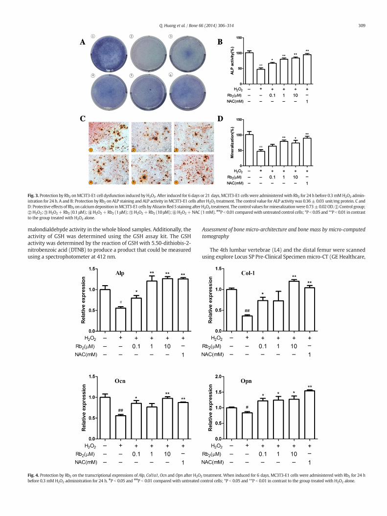
Fig. 3. Protection by Rb2 onMC3T3-E1 cell dysfunction induced by H2O2. After induced for 6 days or 21 days, MC3T3-E1 cells were administeredwith Rb2 for 24 h before 0.3 mMH2O2 admin-istration for 24 h. A and B: Protection by Rb2 on ALP staining and ALP activity inMC3T3-E1 cells after H2O2 treatment. The control value for ALP activity was 0.36± 0.03 unit/mg protein. C andD: Protective effects of Rb2 on calciumdeposition inMC3T3-E1 cells byAlizarin Red S staining afterH2O2 treatment. The control values formineralizationwere 0.73±0.02OD.①Control group;②H2O2;③H2O2+Rb2 (0.1 μM);④H2O2+Rb2 (1 μM);⑤H2O2+Rb2 (10 μM);⑥H2O2+NAC (1mM). ##P b 0.01 comparedwith untreated control cells; *P b 0.05 and **P b 0.01 in contrastto the group treated with H2O2 alone.
309Q. Huang et al. / Bone 66 (2014) 306–314
malondialdehyde activity in the whole blood samples. Additionally, theactivity of GSH was determined using the GSH assay kit. The GSHactivity was determined by the reaction of GSH with 5.50-dithiobis-2-nitrobenzoic acid (DTNB) to produce a product that could be measuredusing a spectrophotometer at 412 nm.
Fig. 4. Protection by Rb2 on the transcriptional expressions of Alp, Col1a1, Ocn and Opn after H2Obefore 0.3 mM H2O2 administration for 24 h. #P b 0.05 and ##P b 0.01 compared with untreated c
Assessment of bone micro-architecture and bone mass by micro-computedtomography
The 4th lumbar vertebrae (L4) and the distal femur were scannedusing explore Locus SP Pre-Clinical Specimen micro-CT (GE Healthcare,
2 treatment. When induced for 6 days, MC3T3-E1 cells were administered with Rb2 for 24 hontrol cells; *P b 0.05 and **P b 0.01 in contrast to the group treated with H2O2 alone.

Fig. 5. Rb2 inhibited the expressions of RANKL and IL-6 after H2O2 treatment. After osteo-genic induction for 6 days, MC3T3-E1 cells were administered with Rb2 for 24 h before0.3 mM H2O2 administration for 24 h. A: Expression of RANKL with the presence of Rb2and/or H2O2. The control values for RANKL were 2.27 ± 0.18 ng/mg. B: Expression ofIL-6 with the presence of Rb2 and/or H2O2. The control values for IL-6 were 0.72 ±0.17 ng/mg. ##P b 0.01 compared with untreated control cells; *P b 0.05 and **P b 0.01in contrast to the group treated with H2O2 alone.
310 Q. Huang et al. / Bone 66 (2014) 306–314
USA) with 8-mm resolution, 50-kV tube voltage and 0.1-mA tube cur-rent. The reconstruction and 3D quantitative analyses were determinedusing software provided by a desktop micro-CT system (GE Healthcare,USA). Similar settings for scans and analyses were used for all of thesamples. In the femur, the scanning regions were confined to the distalmetaphysis, extending proximally 2.0 mm from the proximal tip of theprimary spongiosa. The trabecular bone region from the vertebral bodywas outlined for each micro-CT slice, excluding both the cranial andcaudal endplate regions. The following 3D indices in the defined regionof interest (ROI) were analyzed: bone mineral density (BMD), connectiv-ity density (Conn.D), structure model index (SMI), trabecular number(Tb.N), trabecular thickness (Tb.Th), trabecular separation (Tb.Sp) andrelative bone volume over total volume (BV/TV, %). The operator whoconducted the scan analysis was blinded to the procedure associatedwith the specimens.
Histological examination by Van Gieson (VG) staining
The 4th lumbar vertebrae and the left femur of all of the mice werecollected and fixed in 4% paraformaldehyde for 48 h. After dehydrationand embedding, the 4th lumbar vertebrae and the distal femur wereembedded in polymethyl-methacrylate (PMMA) and processed into240-mm-thick sections in the coronal plane using a rotationmicrotome.Subsequently, all of the sections were hand-grounded to a thicknessof 20 mm for VG staining, which was used for staining collagenfiber [32].
Statistical analysis
The data represented the mean ± SD values of multiple repeats ofthe same experiment (n = 5). The data for all of the measurementswere analyzed using a one-way analysis of variance (ANOVA)with sub-sequent post hoc multiple comparison by Dunnett's test. Statisticallysignificant values were defined as P b 0.05.
Results
Rb2 inhibited H2O2-induced cytotoxicity in MC3T3-E1 cells
Before the anti-cytotoxicity effect of Rb2 was tested, the toxicity ofRb2 onMC3T3-E1was observed. The results showed that Rb2 treatmentalone did not affect cell survival at the consistencies tested in this exper-iment (Fig. 2A). Subsequently, we tested the survival of MC3T3-E1 cellsto observe the defensive effects by Rb2 on the repercussion of the cellsafter H2O2-induced oxidative damage. We implemented oxidativedamage onMC3T3-E1 cells using 0.3 mMH2O2 for 24 h.When differentconcentrations of Rb2 (0.1, 1, 10 μM) were administered to MC3T3-E1cells for 24 h before H2O2 treatment, the viability of the cells increasedcompared to the group without Rb2 treatment, which indicates thatRb2, in part, inhibited H2O2-induced cytotoxicity (Fig. 2B). N-acetyl-L-cysteine represented the positive control, which significantlysuppressed cytotoxicity induced by H2O2 at 1 mM.
Protection by Rb2 on H2O2-induced dysfunction of MC3T3-E1 cells
We tested the protective effect of Rb2 on H2O2-induced dysfunctionof MC3T3-E1 cells by evaluating osteoblast differentiation and mineral-ization with ALP staining, ALP activity and Alizarin Red S staining. Addi-tionally, we detected the expression of osteogenic genes including Alp,Col1a1, Ocn and Opn. Compared with the control group, all markers ofosteogenic differentiation, such as ALP expression, calcium depositionand osteogenic genes (Figs. 3 and 4), decreased after treatment ofH2O2. However, when we pretreated MC3T3-E1 cells with Rb2(0.1 μM, 1 μM, and 10 μM), cellular ALP expression (Fig. 3A) and activity(Fig. 3B) increased in a dose-dependent manner. As shown in Fig. 3Cand D, Rb2 (0.1 μM, 1 μM, and 10 μM) exhibited a good recovery effecton calcium deposition, which was suppressed by H2O2. Furthermore,pretreatment with Rb2 partially elevated the expression of osteogenicgenes compared with the group of H2O2 treated alone (Fig. 4). Ourdata indicated that Rb2, in part, attenuated H2O2-induced dysfunctionof MC3T3-E1 cells.
Rb2 inhibited RANKL and IL-6 expression in MC3T3-E1 cells
Osteoblasts always express and secrete several cytokines, whichbind to receptors in osteoclasts and affect osteoclast activity in bone re-modeling. RANKL and IL-6 are two essential bone-resorbing factorsexpressed in osteoblasts. After the addition of 0.3mMH2O2, the expres-sions of RANKL and IL-6 increased. However, when pretreated with Rb2of 0.1 μM, 1 μM and 10 μM, the elevated expressions of RANKL and IL-6were, in part, inhibited (Fig. 5).
Rb2 inhibited the production of reactive oxygen species induced by H2O2 inMC3T3-E1 cells
To clarify whether the cell-protective capacity of Rb2 was associatedwith its antioxidant property, we tested ROS production using a fluores-cent probe DCFH-DA. The results in Fig. 6 showed that whenMC3T3-E1cells were treatedwith 0.3mMH2O2, ROS increased. These data indicat-ed that H2O2 stimulated the generation of oxidants and resulted inoxidative stress to MC3T3-E1 cells. However, when pretreated with Rb2of different concentrations, ROS production was partially suppressed

Fig. 6. Rb2 inhibited H2O2-induced ROS expression in MC3T3-E1 cells. After osteogenic induction for 6 days, the cells were administered with Rb2 for 24 h before a 0.3 mMH2O2 admin-istration for 24 h. A: ROS detection by fluorescent probe DCFH-DA; B: quantitative analysis of ROS detection.① Control group;② H2O2;③ H2O2 + Rb2 (0.1 μM);④ H2O2 + Rb2 (1 μM);⑤ H2O2 + Rb2 (10 μM); ⑥ H2O2 + NAC (1 mM). ##P b 0.01 compared with untreated control cells; *P b 0.05 and **P b 0.01 in contrast to the group treated with H2O2 alone.
311Q. Huang et al. / Bone 66 (2014) 306–314
(Fig. 6). These results suggested that the protection by Rb2 might berelated to its antioxidant activity.
Rb2 inhibited serum oxidative damage of ovariectomized mice
To test whether Rb2 can inhibit serum oxidative damage caused byestrogen insufficiency, the activities of serum malondialdehyde (MDA)and reduced glutathione (GSH) were observed in ovariectomized micewith or without Rb2 intervention. The results in Fig. 7 showed that incontrast to the sham-operated group, the activity of MDA in serumincreased, whereas the GSH activity in the ovariectomized groupdecreased. However, Rb2 treatment partly rescued the serum activitiesof MDA and GSH.
Rb2 improved bone mass and bone structure of ovariectomized mice
To evaluate Rb2 acting on trabecular bone mass and micro-architecture, different concentrations of Rb2 were supplemented tothe ovariectomized (OVX) mice. When all of the mice were collectedat 13 weeks after operation, there were no significant differencesin body weights of all four groups. The body weights of the sham-operated group, OVX group, OVX + Rb2 (4.6 μmol/kg) group andOVX + Rb2 (18.5 μmol/kg) group were 24.26 ± 0.99 g, 24.12 ±
1.06 g, 24.03 ± 1.09 g and 24.07 ± 0.98 g, respectively. The micro-architectures of distal femurs are shown in Fig. 8A and B. The analysesof the trabecular bone of the 4th lumbar vertebrae (L4) and the distalfemur showed that ovariectomy reduced bone mass and deterioratedbone micro-architecture, which was indicated through decreases inBMD, Conn.D, Tb.N, Tb.Th and BV/TV (Fig. 8C and D) (P b 0.05). SMIand Tb.Sp exhibited increases that contributed to ovariectomy(P b 0.05), as shown in Fig. 8E and F. However, the treatment of ovari-ectomized mice with 4.6 μmol/kg or 18.5 μmol/kg Rb2, in part, rescuedthese bone parameters and improved the micro-architecture of thetrabecular bone in the 4th lumbar vertebrae and the distal femur.Moreover, we evaluated the changes of bone micro-architecture by VGstaining. As shown in Fig. 8G and H, compared with the sham-operated group, the number of trabeculae decreased and the trabecularspace became broader in the OVX group. The supplement of Rb2 re-versed these changes by an elevated number of trabecular bone and areduction of trabecular bone space (Fig. 8G and H). These results wereconsistent with the micro-CT data (Fig. 8A–F).
Discussion
The reactive oxygen species are highly reactive oxygen free radicalsor non-radical molecules, which include hydrogen peroxide (H2O2),

Fig. 7.Rb2 inhibited serumoxidative damage in the ovariectomizedmodel. A: SerumMDAconcentrations in OVX mice that were administered different concentrations of Rb2.B: Serum GSH concentrations in OVX mice that were administered different concentrationsof Rb2. Groups: Sham; OVX; OVX+Rb2 (4.6 μmol/kg); OVX+Rb2 (18.5 μmol/kg). The con-trol values forMDA and GSHwere 9.24± 0.67 nM/mg and 11.87± 1.27 nM/mg. ##P b 0.01compared with the sham group; *P b 0.05 and **P b 0.01 in contrast to the OVX group.
312 Q. Huang et al. / Bone 66 (2014) 306–314
superoxide anion (O2•−) and hydroxyl radical (•OH) [33]. Their over-
production induces oxidative stress. A considerable number of studiesindicated that increased oxidative stress was involved in the pathogen-esis of osteoporosis caused by estrogen deficiency and aging [34,35]. Be-cause of good stability and property to pass through cell membranes,hydrogen peroxide is favorable to serve as both an extra- and an inter-cellular signal [36]. Therefore, an in vitro H2O2-induced oxidativedamage model was used in this study. The study of Zhang et al. showedthat the treatment of 0.3 mM H2O2 for 24 h reduced the number ofMC3T3-E1 cells to 50% and that 0.3 mMH2O2 was suitable for gener-ating an in vitro oxidative stress model [32]. Therefore, we used0.3 mM H2O2 for 24 h to make an oxidative damage model in thepresent study. Our data showed that H2O2 toxicity was reversed toa certain degree by pretreatment with Rb2. This study demonstratedthat Rb2, in part, decreased H2O2-induced cytotoxicity on MC3T3-E1cells.
Oxidative damage not only affects osteoblast survival but also has animpact on its differentiation. H2O2-induced oxidative damage is report-ed to inhibit osteoblast differentiation in primary murine bone marrowstem cells and to reduce the expression of osteoblast markers in osteo-blasticMC3T3-E1 cells [19,37]. Our study also showed that H2O2 supple-mentwas linked to a reduction of cellular alkaline phosphatase activity,a decrease of calciummineralization and lowered expressions of osteo-genic genes, including Alp, Col1a1, Ocn and Opn, which confirms previ-ous results that cytotoxicity of H2O2, in part, results in MC3T3-E1 celldysfunction. Moreover, our data showed that H2O2-induced inhibitionof osteogenic differentiation could be reversed by Rb2. Rb2 alone couldnot significantly enhance MC3T3-E1 cell survival and osteogenic differ-entiation within experimented consistencies. Therefore, we speculatedthat the antioxidant property of Rb2 contributed to the protective effect.Because Rb2 improves osteoblastic survival and differentiation againstoxidative damage, Rb2 might become an anti-osteoporosis agent inthe bone metabolism area.
Several pathways and cytokines couple the link between osteoblastsandosteoclasts. RANKL and IL-6 are generated by osteoblasts and functionas bone-resorbing cytokines by stimulating osteoclast activity. RANKLbelongs to the superfamily of tumor necrosis factor [38]. By binding toRANK, RANKL leads to osteoclastic differentiation, prolongs osteoclasticactivity and increases bone resorption [11]. Several studies have shownthat reactive oxygen species could elevate the expression of RANKL inosteoblasts [39]. IL-6 is also generated by osteoblastswhen it ismotivat-ed by IL-1, TNF-α, and lipopolysaccharide [40]. IL-6 could affect the ex-pression of RANKL, improve osteoclast development and be a significantpathogenic factor of estrogen deficiency osteoporosis [41,42]. It wasreported that reactive oxygen species might indirectly influence os-teoclasts by increasing the expression of bone-resorptive cytokines,which are highly involved in estrogen deficiency osteoporosis [43].In our study, Rb2 was noted to inhibit, in part, the H2O2-induced gener-ations of RANKL and IL-6 in MC3T3-E1 cells. The reductions of RANKLand IL-6 might contribute to the anti-resorbing property, which Rb2exhibited.
Ginsenoside-Rb2 (Rb2), which is extracted from Panax ginseng,belongs to traditional Chinese medicine. Recently, its anti-oxidativeproperty has been reported [16]. In this study, we could reverse, inpart, H2O2-induced production of reactive oxygen species by pretreat-mentwith Rb2 for 24 h. Our data indicated that Rb2might be a useful an-tioxidant to protect MC3T3-E1 cells from cytotoxicity induced byoxidative damage. Consequently, the protection by Rb2 to MC3T3-E1cells could be mediated via its antioxidant property. The beneficial effectof Rb2 might be linked to reduced oxidative damage and bone-resorbing cytokines. However, the molecular mechanism has not beenestablished. Comparedwith other growth factors, reactive oxygen speciesresult in the retention of FoxOs in the nucleus and the activation of theirtranscription [44]. FoxO protein family is characterized by a commonwinged-helix DNA binding domain called Forkhead box [45]. Animalstudies have demonstrated that in response to oxidative damage, c-junkinase (JNK) and mammalian sterile 20-related kinase-1 (Mst1) bondand phosphorylate FoxOs in a direct manner [46–49]. FoxO-dependentoxidative defense systems provide an approach for dealing with oxygenfree radicals that are constantly produced by the aerobic metabolism ofosteoblasts and are therefore essential for bone mass balance [50].Under these circumstances, we deduced that Rb2 provided protectiveeffects against cytotoxicity and osteoblast dysfunction induced by H2O2
through the JNK,Mst1 and FoxOs signaling pathway. In additional studies,we will explore this molecular mechanism.
Themechanismbywhich estrogen deficiency activates bone loss hasnot been established. Recently, oxidative damage has been documentedto result in postmenopausal osteoporosis. Almeida et al. reported thatsimilar increases in oxidative stress and p66shc phosphorylation ob-served with age-related changes in bone of C57BL/6 mice were causedby removing gonads in the female or male mice [17,20]. Moreover, thealterations were ameliorated by administering antioxidants, such asN-acetyl-L-cysteine, ascorbate, and catalase, which is as effective as hor-mone replacement [9]. Malondialdehyde (MDA), which commonlyserved as a trustful parameter in evaluating the oxidative damage status[51], is an end product of lipid peroxidation induced by reactive oxygenspecies. Reduced glutathione (GSH) is an intracellular antioxidant,which prevents cells from oxidative stress caused by free radicals, per-oxides and toxins. It can effectively remove O2•
− and provide electronsfor glutathione peroxidase to transform H2O2 into H2O [52]. Additional-ly, GSH reduction is a marker of oxidative damage [53]. Our researchshowed that Rb2 administration for 12 weeks reversed, in part, serumMDA and GSH activities of ovariectomized mice.
Sharing similarities with postmenopausal women, ovariectomizedmice experience significant bone loss due to estrogen deficiency,which primarily results from trabecular bone loss [54]. Therefore,the trabecular bone micro-architecture is considered to be a properpredictor of OVX-induced bone loss and bone quality deterioration[55]. The micro-architecture of trabecular bone has been shown to

Fig. 8. Protection by Rb2 on the bone mass and micro-architecture of trabecular bone of OVX mice. A and B: Analysis of micro-CT in the distal metaphyseal femur region. Groups: Sham;OVX; OVX + Rb2 (4.6 μmol/kg); OVX+ Rb2 (18.5 μmol/kg). C and E: Analysis of micro-CT quantification in the distal metaphyseal femur region. D and F: Analysis of micro-CT quantifi-cation in the 4th lumbar vertebrae. The following 3D indices in the defined region of interest (ROI) were analyzed: BMD, Conn.D, SMI, Tb.N, Tb.Th, Tb.Sp and BV/TV. G and H: Van Gieson(VG) staining of the distal femur and body of the 4th lumbar vertebrae. The figure was 40× and 100× of the original section. *P b 0.05 and ##P b 0.01 compared with the sham group;*P b 0.05 and **P b 0.01 in contrast to the OVX group.
313Q. Huang et al. / Bone 66 (2014) 306–314
dramatically deteriorate after ovariectomy by both micro-CT scan-ning and VG staining [32,56]. Nevertheless, these negative effectscould be reversed, to some degree, after administering Rb2. These re-sults further confirmed that Rb2 might, in part, ameliorate trabecularmicro-architecture and bonemass of OVXmice and that Rb2 might be agood candidate for the prevention and the treatment of estrogen-deficient osteoporosis.
To conclude, the results described in this article indicated that in anin vitro analysis, ginsenoside-Rb2 protected osteoblastic MC3T3-E1 cellsfromcytotoxicity and osteoblast dysfunction induced by hydrogen perox-ide at 0.1 to 10 μM. This activity is linked to a reduction of oxidative dam-age and bone-resorbing cytokines. More importantly, ginsenoside-Rb2decreased serum reactive oxygen species to a certain degree and partlyreversed estrogen-deficient effects in vivo at doses of 4 to 18 μmol/kgbody weight. Ginsenoside-Rb2 may act as a substantive alternative inbone metabolic diseases, especially osteoporosis. Extensive research isneeded in the future to explore the intricate molecular mechanisms ofginsenoside-Rb2 on bone metabolism.
Acknowledgments
This work was supported by theMinistry of Science and Technologyof the People's Republic of China (2011CB964703), National HighTechnology Research and Development Program 863 (2012AA020502)and National Natural Science Foundation of China (30901504), and theProgram for Changjiang Scholars and Innovative Research Team inUniversity (no. IRT1053). No benefits in any form have been or will bereceived from a commercial party directly or indirectly by the authorsof this manuscript.
References
[1] Sanchez-Rodriguez MA, Ruiz-Ramos M, Correa-Munoz E, Mendoza-Nunez VM.Oxidative stress as a risk factor for osteoporosis in elderly Mexicans as characterizedby antioxidant enzymes. BMC Musculoskelet Disord 2007;8:124.
[2] Bouillon, Burckhardt R, Christiansen P, Fleisch C, Fujita HA, Gennari T, et al. Consen-sus development conference: diagnosis, prophylaxis and treatment of osteoporosis.Am J Med 1993;94:646–50.

314 Q. Huang et al. / Bone 66 (2014) 306–314
[3] Sendur OF, Turan Y, Tastaban E, Serter M. Antioxidant status in patients withosteoporosis: a controlled study. Joint Bone Spine 2009;76:514–8.
[4] Witko-Sarsat V, Friedlander M, Capeillere-Blandin C, Nguyen-Khoa T, Nguyen AT,Zingraff J, et al. Advanced oxidation protein products as a novel marker of oxidativestress in uremia. Kidney Int 1996;49:1304.
[5] Ozgocmen S, Kaya H, Fadillioglu E, Aydogan R, Yilmaz Z. Role of antioxidant systems,lipid peroxidation, and nitric oxide in postmenopausal osteoporosis. Mol CellBiochem 2007;295:45–52.
[6] Ozgocmen S, Kaya H, Fadillioglu E, Yilmaz Z. Effects of calcitonin, risedronate, andraloxifene on erythrocyte antioxidant enzyme activity, lipid peroxidation, and nitricoxide in postmenopausal osteoporosis. Arch Med Res 2007;38:196–205.
[7] Muthusami S, Ramachandran I, Muthusamy B, Vasudevan G, Prabhu V,Subramaniam V, et al. Ovariectomy induces oxidative stress and impairs bone anti-oxidant system in adult rats. Clin Chim Acta 2005;360:81–6.
[8] Linnane AW, Eastwood H. Cellular redox regulation and prooxidant signalingsystems: a new perspective on the free radical theory of aging. Ann N Y Acad Sci2006;1067:47.
[9] Manolagas SC. From estrogen-centric to aging and oxidative stress: a revisedperspective of the pathogenesis of osteoporosis. Endocr Rev 2010;31:266–300.
[10] Theill LE, Boyle WJ, Penninger JM. RANK-L and RANK: T cells, bone loss, andmammalian evolution. Annu Rev Immunol 2002;20:795–823.
[11] Boyle WJ, Simonet WS, Lacey DL. Osteoclast differentiation and activation. Nature2003;423:337–42.
[12] Karsenty G, Wagner EF. Reaching a genetic and molecular understanding of skeletaldevelopment. Dev Cell 2002;2:389–406.
[13] Soltanoff CS, Yang S, ChenW, Li YP. Signaling networks that control the lineage commit-ment and differentiation of bone cells. Crit Rev Eukaryot Gene Expr 2009;19:1–46.
[14] HuangW, Yang S, Shao J, Li YP. Signaling and transcriptional regulation in osteoblastcommitment and differentiation. Front Biosci 2007;12:3068–92.
[15] Isomura H, Fujie K, Shibata K, Inoue N, Iizuka T, Takebe G, et al. Bonemetabolism andoxidative stress in postmenopausal rats with iron overload. Toxicology 2004;197:93–100.
[16] Lee NK, Choi YG, Baik JY, Han SY, Jeong DW, Bae YS, et al. A crucial role for reactiveoxygen species in RANKL-induced osteoclast differentiation. Blood 2005;106:852–9.
[17] Lean JM, Davies JT, Fuller K, Jagger CJ, Kirstein B, et al. A crucial role for thiol antiox-idants in estrogen-deficiency bone loss. J Clin Invest 2003;112:915–23.
[18] Lean JM, Jagger CJ, Kirstein B, Fuller K, Chambers TJ. Hydrogen peroxide is essentialfor estrogen-deficiency bone loss and osteoclast formation. Endocrinology 2005;146:728–35.
[19] Arai M, Shibata Y, Pugdee K, Abiko Y, Ogata Y. Effects of reactive oxygen species(ROS) on antioxidant system and osteoblastic differentiation in MC3T3-E1 cells.IUBMB Life 2007;59:27–33.
[20] Almeida M, Han L, Martin-Millan M, Plotkin LI, Stewart SA, Roberson PK, et al.Skeletal involution by age-associated oxidative stress and its acceleration by lossof sex steroids. J Biol Chem 2007;282:27285–97.
[21] Jilka RL, Almeida M, Ambrogini E, Han L, Roberson PK, et al. Decreased oxidativestress and greater bone anabolism in the aged, as compared to the young, murineskeleton by parathyroid hormone. Aging Cell 2010;9:851–67.
[22] MaWG,Mizutani M, Malterud KE, Lu SL, Ducrey B, Tahara S. Saponins from the rootsof Panax notoginseng. Phytochemistry 1999;52:1133–9.
[23] Lee KT, Jung TW, Lee HJ, Kim SG, Shin YS, Whang WK. The antidiabetic effect ofginsenoside Rb2 via activation of AMPK. Arch Pharm Res 2011;34(7):1201–8.
[24] Kim EJ, Lee HI, Chung KJ, Noh YH, Ro Y, Koo JH. The ginsenoside-Rb2 lowers choles-terol and triacylglycerol levels in 3T3-L1 adipocytes cultured under high cholesterolor fatty acids conditions. BMB Rep 2009;42(4):194–9.
[25] Fujimoto J, Sakaguchi H, Aoki I, Toyoki H, Khatun S, Tamaya T. Inhibitory effectof ginsenoside-Rb2 on invasiveness of uterine endometrial cancer cells to the base-ment membrane. Eur J Gynaecol Oncol 2001;22(5):339–41.
[26] Kang KS, Kim HY, Baek SH, Yoo HH, Park JH, Yokozawa T. Study on the hydroxyl rad-ical scavenging activity changes of ginseng and ginsenoside-Rb2 by heat processing.Biol Pharm Bull 2007;30(4):724–8.
[27] Kang KS, Kang BC, Lee BJ, Che JH, Li GX, Trosko JE, et al. Preventive effect of epicate-chin and ginsenoside Rb2 on the inhibition of gap junctional intercellular communi-cation by TPA and H2O2. Cancer Lett 2000;152:97–106.
[28] Kim YH, Park KH, Rho HM. Transcriptional activation of the Cu, Zn-superoxide dis-mutase gene through the AP2 site by ginsenoside Rb2 extracted from a medicinalplant, Panax ginseng. J Biol Chem 1996;40:24539–43.
[29] Li Y, Tang L, Duan Y, Ding Y. Up-regulation of MMP-13 and TIMP-1 expression inresponse to mechanical strain in MC3T3-E1 osteoblastic cells. BMC Res Notes2010;3:309.
[30] Li Y, MaW, Feng Z,Wang Z, Zha N, Deng B, et al. Effects of irradiation on osteoblast-likecells on different titanium surfaces in vitro. J Biomed Mater Res B Appl Biomater2012;101B:9–17.
[31] Boyd Steven K, Davison Peter, Müller Ralph, Gasser Jürg A. Monitoring individualmorphological changes over time in ovariectomized rats by in vivo micro-computed tomography. Bone 2006;39:854–62.
[32] Zhang JK, Yang L, Meng GL, Yuan Z, Fan J, Li D, et al. Protection by salidroside againstbone loss via inhibition of oxidative stress and bone-resorbing mediators. PLoS ONE2013;8(2):e57251.
[33] Shi Y, Tang B, Yu PW, Tang B, Hao YX, Lei X, et al. Autophagy protects againstoxaliplatin-induced cell death via ER stress and ROS in Caco-2 cells. PLoS ONE2012;7:e51076.
[34] Baek KH, Oh KW, Lee WY, Lee SS, Kim MK, Kwon HS, et al. Association of oxidativestress with postmenopausal osteoporosis and the effects of hydrogen peroxide onosteoclast formation in human bone marrow cell cultures. Calcif Tissue Int2010;87:226–35.
[35] Maggio D, Barabani M, Pierandrei M, Polidori MC, Catani M, Catani M, et al. Markeddecrease in plasma antioxidants in aged osteoporotic women: results of a cross-sectional study. J Clin Endocrinol Metab 2003;88:1523–7.
[36] Denisova NA, Cantuti-Castelvetri I, Hassan WN, Paulson KE, Joseph JA. Role of mem-brane lipids in regulation of vulnerability to oxidative stress in PC12 cells: implicationfor aging. Free Radic Biol Med 2001;30:671–8.
[37] Liu AL, Zhang ZM, Zhu BF, Liao ZH, Liu Z.Metallothionein protects bonemarrow stromalcells against hydrogen peroxide-induced inhibition of osteoblastic differentiation. CellBiol Int 2004;28:905–11.
[38] Lacey DL, Timms E, Tan HL, Kelley MJ, Dunstan CR, Burgess T, et al. Osteoprotegerinligand is a cytokine that regulates osteoclast differentiation and activation. Cell1998;93:165–76.
[39] Bai XC, Lu D, Liu AL, Zhang ZM, Li XM, Zou ZP, et al. Reactive oxygen species stimu-lates receptor activator of NF-kappaB ligand expression in osteoblast. J Biol Chem2005;280:17497–506.
[40] Girasole G, Jilka RL, Passeri G, Boswell S, Boder G, Williams DC, et al. 17 beta-estradiol inhibits interleukin-6 production by bone marrow-derived stromal cellsand osteoblasts in vitro: a potential mechanism for the antiosteoporotic effect ofestrogens. J Clin Invest 1992;89:883–91.
[41] Kurihara N, Bertolini D, Suda T, Akiyama Y, Roodman GD. IL-6 stimulates osteoclast-like multinucleated cell formation in long term human marrow cultures by inducingIL-1 release. J Immunol 1990;144:4226–30.
[42] Papanicolaou DA, Vgontzas AN. Interleukin-6: the endocrine cytokine. J ClinEndocrinol Metab 2000;85:1331–3.
[43] Kitazawa R, Kimble RB, Vannice JL, Kung VT, Pacifici R. Interleukin-1 receptor antag-onist and tumor necrosis factor binding protein decrease osteoclast formation andbone resorption in ovariectomized mice. J Clin Invest 1994;94:2397–406.
[44] Van der HA, Burgering BM. Stressing the role of FoxO proteins in lifespan anddisease. Nat Rev Mol Cell Biol 2007;8:440–50.
[45] Greer EL, Brunet A. FOXO transcription factors at the interface between longevityand tumor suppression. Oncogene 2005;24:7410–25.
[46] Essers MA, Weijzen S, Vries-Smits AM, Saarloos I, de Ruiter ND, Bos JL, et al. FOXOtranscription factor activation by oxidative stress mediated by the small GTPaseRal and JNK. EMBO J 2004;23:4802–12.
[47] Lehtinen MK, Yuan Z, Boag PR, Yang Y, Villen J, Becker EB, et al. A conserved MST-FOXO signaling pathway mediates oxidative-stress responses and extends lifespan. Cell 2006;125:987–1001.
[48] WangMC, BohmannD, Jasper H. JNK extends life span and limits growth by antagoniz-ing cellular and organism-wide responses to insulin signaling. Cell 2005;121:115–25.
[49] Oh SW, Mukhopadhyay A, Svrzikapa N, Jiang F, Davis RJ, Tissenbaum HA. JNK regu-lates lifespan in Caenorhabditis elegans by modulating nuclear translocation offorkhead transcription factor/DAF-16. Proc Natl Acad Sci U S A 2005;102:4494–9.
[50] Ambrogini E, Almeida M, Martin-Millan M, Paik JH, DePinho RA, Han L, et al.FoxO-mediated defense against oxidative stress in osteoblasts is indispensable forskeletal homeostasis in mice. Cell Metab 2010;11(2):136.
[51] Cakatay U, Aydin S, Yanar K, Uzun H. Gender-dependent variations in systemicbiomarkers of oxidative protein, DNA, and lipid damage in aged rats. Aging Male2010;13:51.
[52] Estrela JM, Ortega A, Obrador E. Glutathione in cancer biology and therapy. Crit RevClin Lab Sci 2006;43:143–81.
[53] Kinov P, Leithner A, Radl R, Bodo K, Khoschsorur GA, Schauenstein K, et al. Role offree radicals in aseptic loosening of hip arthroplasty. J Orthop Res 2006;24:55–62.
[54] Kim TH, Jung JW, Ha BG, Hong JM, Park EK, Kim HJ, et al. The effects of luteolin onosteoclast differentiation, function in vitro and ovariectomy-induced bone loss. JNutr Biochem 2011;22:8–15.
[55] Chappard D, Basle MF, Legrand E, Audran M. Trabecular bone microarchitecture: areview. Morphologie 2008;92:162–70.
[56] Campbell GM, Buie HR, Boyd SK. Signs of irreversible architectural changes occurearly in the development of experimental osteoporosis as assessed by in vivomicro-CT. Osteoporos Int 2008;19:1409–19.