
GIK794 .zenobiosic ..enmes. w. (10 lig/ml), and phenylmethylsul-fonyl fluoride (44 pg/ml), the cell...
5
i - * * & i Expression of Xenobiotic-Metabolizing Enzymes in Cultured Rat Tracheal Epithelial Cells 1 2 2 Andre Castonguay, Lila Overby, Paul Nettesheim, George C. Clark, and Richard M. Philpot2 'School of Pharmacy, Laval University, Quebec City, GIK 794 Canada; 2National Institute of Environmental Health Sciences, Research Triangle Park, NC 27606 USA Rat qtrachealepitheial (RTE) celki were cul- tured on membrane support with and with- out retinoic acid (RA). In early (6-day-old) cultures, the epithelium is a monolayer or bilayer of unddif t cels a secretes little mucu .ike product either in the absence or.resence of A. n`l.ate :(1i:- to 15-day-old) cutures, the epithelim.differ- entiates as a mucociiary epithelium in the presence of RA and as a epitheli- um in the absence of RA. The purpose of our study wa so. detrmine whetea num- ber of zenobiosic enmesare...os:d in . . ........ . .. . ... w. .. ... ..... ..... . these c ises whether ei.p...r..... .e..sir on .depends o t. sta:te of die tio. .Enzyme epr.es.sonwas ch a.rterzedt. electrophoreuis and immunobosttig. as a function of time in culture and phenotypic differentiatin C re P450 lAl was *::not expresedinfreshly harestedTE. ..el .. .h..i.n...e ....s by .. age wth:.tiS Z,,-ssaclrdabno--io: ...... .. ... .. in (TCDIM or by expoue eal ell ....... t. -" .-:-.. cultures to. TICDD), provided "A wA so added to the. cultures. Cytochro .me P450 *2B1 was observed in fieshly isolated RTE cl, but: n.t in earyor hte RTE cultures. n: contras:t expression o:f ADPH- ... cycochrome P50: reuOs adcesdi ..._t....N~~~~......... ........ : ::early cultures, but was i. esd l-d ferentiated cut'ures. Flavi-n contawing -monooy se was detected in lung tissue, but not in fieshly. or cultured:RTE .cells. Glutahio e St.-ransfei.es (GST) p and : were express-..:..e.d.. : arves. .: *T cels GT::it wa iexprressed u al n * tl ...... : : ~~~~ ~~ ~ ~~~~~~~~~~~~~~.... ..... . . .. .7.. ...-E. I 1. R ...- l..ate cu wheasGTp aseressed n late c e but. could not be found in early cultured RTdE ceils Levies of GST *~ ~~~~~~~~~~~~~~~~~~~~~~~~~~~~. ..... *isoenymesi. were .unfetd by BtA These .:resulsparale the. exprsion of enzms ... ... ... .............s. observd n rliraig and d&i n ig. cycli aroat ic y; c aron resultsin :RTE cels with metboli activatang ade dactivat- ing enz n situtes a tem suited for some toiclga studiesKe w r cytochrome P450; fla c tninvg mono g tone r rase; ... muci; rttchal Cells retiMStocacd . TG99.*: S. vern He*14tP e. ....l.33 TCDD* £ Hel P 13 2428 (195 The carcinogenic potential of chemicals in a given tissue may be determined by the ability of the tissue to activate and detoxify these chemicals. Most chemical carcino- gens are activated by the cytochrome P450 monooxygenase system, which comprises the hemoprotein cytochrome-450 and the flavoprotein NADPH-cytochrome 450 reductase. The cytochromes P450 are a superfamily of enzymes (1); subfamily P450 IA catalyzes the activation of many carcinogens, including benzo[a]pyrene, aflatoxin B1, and tobacco-specific nitrosamines (2). A second monooxygenase system, the flavin-containing monooxyge- nase (FMO), is involved in the activation of aromatic amines and in the detoxifica- tion of carcinogens having a pyridine ring (3,4). Glutathione S-thransferases (GSTs) are an important class of carcinogen-detox- ifying enzymes that catalyze the nucle- ophilic attack of the thiol group of glu- tathione on reactive electrophilic metabo- lites, including proximate and ultimate car- cinogens (5). Expression of these enzymes is usually down-regulated in human and animal tissues cultured in vitro. Maintenance of enzyme expression in cultured cells depends on culture condi- tions, cell type, and the enzyme consid- ered. For instance, under conventional cul- ture conditions, rat hepatocytes lose 80% of their cytochrome P450 during 24 hr of culture and, during the same period, lose 50% of their NADPH-P450 reductase activity (6). Recently, Schuetz et al. (7) developed a membrane matrix, "matrigel," which allows for the in vitro induction of some cytochrome P450 isoforms to levels observed in vivo. Under these conditions, secretions of proteins considered markers of hepatocyte differentiation increased sig- nificantly in cultures, whereas cr-fetopro- tein, an indicator of dedifferentiation, was not observed (7). These studies demon- strated that maintenance of in vivo-like cel- lular morphology correlates with the preservation of activities and expression of enzymes in cultured cells. The normal pseudostratified columnar epithelium of rat trachea consists of basal, secretory, and ciliated cells. Considerable efforts by various investigators have been directed toward maintaining the differenti- ated morphology of tracheobronchial cells in vitro (8-11). Trump et al.(12) hypothe- sized that tracheobronchial mucous cells are the major target for initiation of tumorigenesis. Recently, we used a bipha- sic culture system to obtain mucociliary differentiation of RTE cells in the presence of retinoic acid (RA). In biphasic cultures, a mucous phenotype is induced by RA. With no RA in the culture medium, a metaplastic stratified squamous epithelium develops (13). Several enzymes involved in drug metabolism can be induced in the lung by exposure to environmental chemicals. One potent inducer of several xenobiotic- metabolizing enzymes is 2,3,7,8-tetra- chlorodibenzo-p-dioxin (TCDD). The mechanistic basis for TCDD induction of these enzymes is fairly well understood, at least for P450 lAl. TCDD binds to a spe- cific high-affinity cytoplasmic receptor (the Ah receptor); the receptor-TCDD complex then translocates to the nucleus and binds to upstream regulatory ele- ments, resulting in transcriptional activa- tion of the CYPIAI gene (14,15). Other genes forming the Ah gene battery are thought to be similarly induced to high level expression in sensitive tissues (16). It is important to determine whether these induction processes are functional in cul- tures of airway cells. The aim of this study was to determine the expression of various enzymes impor- tant in chemical toxication/detoxication in RTE cells cultured under newly developed conditions. Expression of these enzymes was compared in RTE cells differentiated into stratified squamous or pseudostrati- fied mucociliary epithelium. Materials and Methods Male F344 rats were bred in the National Institute of Evironmental Health Sciences colony. A stock solution of TCDD in corn oil (50 pg/ml; lot 2715-020-016-CO; purity >98% by gas chromatography/mass spectrometry) was purchased from Radian Corporation (Austin, Texas). This solution had been prepared by diluting TCDD into an acetone:corn oil emulsion and remov- ing the acetone by rotary evaporation. Rats were treated once with 50 pg TCDD/kg (0.1 ml/100 g body weight, per os). The rats were euthanized with CO2 7 days after exposure to TCDD, and tracheal epitheli- um was isolated. Tracheas were cannulated, and tracheal Address correspondence to A. Castonguay, Laboratory of Cancer Etiology and Chemo- prevention, School of Pharmacy, Laval University, Quebec City, GI K 7P4 Canada. This work was supported by NIEHS. We appreciate the technical assistance of Scott H. Randell in morphological studies of RTE cells and Veronica B. Godfrey with cell culture. Received 8 August 1994; accepted 13 December 1994. Environmental Health Perspectives 254
Transcript of GIK794 .zenobiosic ..enmes. w. (10 lig/ml), and phenylmethylsul-fonyl fluoride (44 pg/ml), the cell...

i - * *& i
Expression of Xenobiotic-Metabolizing Enzymesin Cultured Rat Tracheal Epithelial Cells
1 2 2Andre Castonguay, Lila Overby, Paul Nettesheim, George C. Clark, andRichard M. Philpot2'School of Pharmacy, Laval University, Quebec City, GIK 794 Canada; 2National Instituteof Environmental Health Sciences, Research Triangle Park, NC 27606 USA
Rat qtrachealepitheial (RTE) celki were cul-tured on membrane support with and with-out retinoic acid (RA). In early (6-day-old)cultures, the epithelium is a monolayer orbilayer of unddif t cels a secreteslittle mucu .ike product either in theabsence or.resence of A. n`l.ate:(1i:- to15-day-old) cutures, the epithelim.differ-entiates as a mucociiary epithelium in thepresence of RA and as a epitheli-um in the absence of RA. The purpose ofour study wa so. detrmine whetea num-ber of zenobiosic enmesare...os:d in. . ........ . .. . ... w. .. ... ..... ..... .these c ises whether ei.p...r......e..siron.depends ot . sta:teofdie tio..Enzyme epr.es.sonwas cha.rterzedt.electrophoreuis and immunobosttig. as afunction of time in culture and phenotypicdifferentiatin C re P450 lAl was
*::not expresedinfreshly harestedTE...el ...h..i.n...e ....sby ..
agewth:.tiSZ,,-ssaclrdabno--io:...... .. .....
in (TCDIM or by expoue eal ell....... t.-".-:-..
cultures to. TICDD), provided"A wA soadded to the. cultures. Cytochro.me P450*2B1 was observed in fieshly isolated RTEcl, but:n.t in earyor hte RTE cultures.n: contras:t expression o:f ADPH-
...
cycochrome P50:reuOs adcesdi..._t....N~~~~......... ........ :::early cultures, but wasi. esd l-dferentiated cut'ures. Flavi-n contawing-monooy se was detected in lung tissue,but not in fieshly. or cultured:RTE.cells. Glutahio e St.-ransfei.es (GST) pand : were express-..:..e.d..: arves. .:
*T cels GT::it wa iexprressed u al n* tl ...... : : ~~~~ ~ ~ ~~~~~~~~~~~~~~~.......... . .. .7.. ...-E. I 1. R ...-
l..ate cu wheasGTp aseressedn late c e but. could not be found inearly culturedRTdE ceils Levies of GST
*~ ~~~~~~~~~~~~~~~~~~~~~~~~~~~~. .....
*isoenymesi.were .unfetd by BtA These.:resulsparale the. exprsion of enzms
... ... ... .............s.observd n rliraigand d&i n ig.
cycli aroat icy;caron resultsin:RTEcels with metboli activatangadedactivat-ing enz n situtes a tem suitedfor some toiclga studiesKew rcytochrome P450; fla c tninvgmono g tone r rase;
...muci; rttchal Cells retiMStocacd.TG99.*: S. vern He*14tP e.....l.33TCDD* £ Hel P 132428 (195
The carcinogenic potential of chemicals ina given tissue may be determined by theability of the tissue to activate and detoxifythese chemicals. Most chemical carcino-gens are activated by the cytochrome P450monooxygenase system, which comprisesthe hemoprotein cytochrome-450 and theflavoprotein NADPH-cytochrome 450reductase. The cytochromes P450 are asuperfamily of enzymes (1); subfamilyP450 IA catalyzes the activation of manycarcinogens, including benzo[a]pyrene,aflatoxin B1, and tobacco-specificnitrosamines (2). A second monooxygenasesystem, the flavin-containing monooxyge-nase (FMO), is involved in the activationof aromatic amines and in the detoxifica-tion of carcinogens having a pyridine ring(3,4). Glutathione S-thransferases (GSTs)are an important class of carcinogen-detox-ifying enzymes that catalyze the nucle-ophilic attack of the thiol group of glu-tathione on reactive electrophilic metabo-lites, including proximate and ultimate car-cinogens (5). Expression of these enzymesis usually down-regulated in human andanimal tissues cultured in vitro.
Maintenance of enzyme expression incultured cells depends on culture condi-tions, cell type, and the enzyme consid-ered. For instance, under conventional cul-ture conditions, rat hepatocytes lose 80%of their cytochrome P450 during 24 hr ofculture and, during the same period, lose50% of their NADPH-P450 reductaseactivity (6). Recently, Schuetz et al. (7)developed a membrane matrix, "matrigel,"which allows for the in vitro induction ofsome cytochrome P450 isoforms to levelsobserved in vivo. Under these conditions,secretions of proteins considered markersof hepatocyte differentiation increased sig-nificantly in cultures, whereas cr-fetopro-tein, an indicator of dedifferentiation, wasnot observed (7). These studies demon-strated that maintenance of in vivo-like cel-lular morphology correlates with thepreservation of activities and expression ofenzymes in cultured cells.
The normal pseudostratified columnarepithelium of rat trachea consists of basal,secretory, and ciliated cells. Considerableefforts by various investigators have beendirected toward maintaining the differenti-ated morphology of tracheobronchial cellsin vitro (8-11). Trump et al.(12) hypothe-sized that tracheobronchial mucous cells
are the major target for initiation oftumorigenesis. Recently, we used a bipha-sic culture system to obtain mucociliarydifferentiation of RTE cells in the presenceof retinoic acid (RA). In biphasic cultures,a mucous phenotype is induced by RA.With no RA in the culture medium, ametaplastic stratified squamous epitheliumdevelops (13).
Several enzymes involved in drugmetabolism can be induced in the lung byexposure to environmental chemicals. Onepotent inducer of several xenobiotic-metabolizing enzymes is 2,3,7,8-tetra-chlorodibenzo-p-dioxin (TCDD). Themechanistic basis for TCDD induction ofthese enzymes is fairly well understood, atleast for P450 lAl. TCDD binds to a spe-cific high-affinity cytoplasmic receptor(the Ah receptor); the receptor-TCDDcomplex then translocates to the nucleusand binds to upstream regulatory ele-ments, resulting in transcriptional activa-tion of the CYPIAI gene (14,15). Othergenes forming the Ah gene battery arethought to be similarly induced to highlevel expression in sensitive tissues (16). Itis important to determine whether theseinduction processes are functional in cul-tures of airway cells.
The aim of this study was to determinethe expression of various enzymes impor-tant in chemical toxication/detoxication inRTE cells cultured under newly developedconditions. Expression of these enzymeswas compared in RTE cells differentiatedinto stratified squamous or pseudostrati-fied mucociliary epithelium.
Materials and MethodsMale F344 rats were bred in the NationalInstitute of Evironmental Health Sciencescolony. A stock solution ofTCDD in cornoil (50 pg/ml; lot 2715-020-016-CO;purity >98% by gas chromatography/massspectrometry) was purchased from RadianCorporation (Austin, Texas). This solutionhad been prepared by diluting TCDD intoan acetone:corn oil emulsion and remov-ing the acetone by rotary evaporation. Ratswere treated once with 50 pg TCDD/kg(0.1 ml/100 g body weight, per os). Therats were euthanized with CO2 7 days afterexposure to TCDD, and tracheal epitheli-um was isolated.
Tracheas were cannulated, and tracheal
Address correspondence to A. Castonguay,Laboratory of Cancer Etiology and Chemo-prevention, School of Pharmacy, Laval University,Quebec City, GIK 7P4 Canada.This work was supported by NIEHS.We appreciate the technical assistance of Scott H.Randell in morphological studies of RTE cells andVeronica B. Godfrey with cell culture.Received 8 August 1994; accepted 13 December1994.
Environmental Health Perspectives254

A .*. *iis I-S;;&SE; - ie-i&aiM S i & So;
epithelial cells were isolated by pronasedigestion as described previously (13). TheRTE cells were seeded at a density of 1.2 x10⁵ cells/insert and cultured on Transwell-Col tissue culture inserts (24.5 mm diame-ter; Costar Co., Cambridge, Massachusetts).Cells were cultured with or without 10 nMRA as described previously (13).
We dissolved TCDD (lot MLB 15091-55) purchased from Cambridge IsotopeLaboratories, Inc. (Woburn, Massachusetts)was dissolved in toluene (1 mg/ml) and seri-ally diluted it in dimethyl sulfoxide(DMSO) to a concentration of 10 pM. Justbefore medium change, aliquots of thissolution were added to the culture mediumto a final dilution of 10 nM TCDD.
We removed RTE cells from the insertsby treatment with 0.25% trypsin-EDTA(0.3 ml/insert) at 37°C for 2 min. Afteradding 0.5 ml phosphate-buffered saline(PBS) containing aprotinin (50 pg/ml), leu-peptin (10 lig/ml), and phenylmethylsul-fonyl fluoride (44 pg/ml), the cell suspen-sion was removed by suction from theinsert. We washed each insert three timeswith PBS containing the inhibitorsdescribed above. The cell suspension wascentrifuged at 1OOOg for 10 min. The pelletwas homogenized in a Broeck tissue grinderand sonicated three times at 40C. After cen-trifugation (900g, 0°C, 10 min) the super-natant was fractionated at 110,000g for 1hr at 40C. The microsomal fraction wasdispersed in 1 mM EDTA, 10 mM TrisHCl, and 20% glycerol buffer. Protein con-centrations were determined with bicin-choninic acid/copper sulfate reagents usingbovine serum albumin as standard (Pierce;Rockford, Illinois).
Culture medium (0.5 ml) present in theinserts on day 6 of culture was removed
after a period of 24 hr (day 7 of culture).Each insert was washed twice with 0.5 mlof PBS. On day 12, we washed inserts with0.5 ml PBS. Culture media and washingswere combined, and aliquots were diluted10-fold (day 6) or 200- to 2000-fold (day12) with 10 mM EDTA and 0.6 M KSCN.Aliquots were applied to nitrocellulosepaper using a slot-blot apparatus(Schleicher & Schuell, Keene, NewHampshire). The nitrocellulose was treatedsuccessively with 1) 5% nonfat milk inPBS containing 0.05% Tween 20 for 30min; 2) monoclonal antibody RTE 1 1against mucuslike secretions diluted 1:20for 30 min; 3) peroxidase conjugate donkeyanti-mouse IgG Uackson Immunoresearch,Philadelphia, Pennsylvania) diluted 1:3000;and 4) 10 mg 3,3'-diaminobenzidine, 100pl of 3% H2O2 in 100 ml of 0.05 M TrisHCl buffer, pH 7.6. Intensities of thebands were quantitated with a video densit-ometer (model 620; Bio-Rad, RockvilleCentre, New York) in the reflection mode.
Electrophoresis of microsomal andcytosolic proteins was carried out on 7.5%and 10% polyacrylamide gels, respectively, inthe presence of sodium dodecyl sulfate(SDS). Samples were applied to 5x Laemmlibuffer (0.312 M Tris, pH 6.8; 0.6% g-mer-captoethanol; 10% sucrose; 10% SDS).Cytosolic samples were heated at 95°C for 3min. Transfer to nitrocellulose and immunos-taining using rabbit anti-goat IgG (dilution1/100) and goat peroxidase-immunoperoxi-dase (Organon Teknika Corp, Westchester,Pennsylvania) have been described (17,18).The specificities of the following goat anti-bodies have been described previously: goatanti-rat P450 lAl (19); goat anti-rat P4502B (20); goat anti-rabbit NADPH P450reductase (21); goat anti-pig liver FMO IA
(18); and goat anti-rabbit GST p and goatanti-rabbit GST X (22).
ResultsRTE cells, cultured for 6 days on tran-swell inserts in the presence or absence ofRA (Fig. 1), formed a flat monolayer ofundifferentiated epithelial cells. On day12, the cultures supplemented with RAshowed a pseudostratified epithelium con-taining many alcian blue-phosphatasealkaline substrate-positive secretory cells(Fig. 1B). Ciliated cells only appearedbetween days 14 and 17 (not shown). Atday 12, the RA-deprived cultures showeda thick, keratinizing squamous epithelium(Fig. ID).
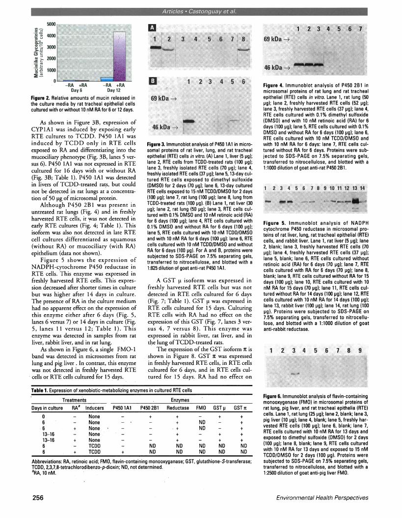
The monoclonal antibody RTE 1 1,specific for granule components of secreto-ry cells, was used to assay for the produc-tion of mucinlike glycoproteins by cul-tured RTE cells (23). As shown in Figure2, RTE cells cultured for 6 days producedonly low levels of mucinlike material. Onday 12 of culture, the amount of RTE-1 1reactive product increased. RA-treated cul-tures produced more than 10-fold greateramounts of mucinlike material than RA-deficient cultures.
Analysis of P450 lAl by immunoblot-ting showed that freshly harvested RTEcells do not express this P450 isoform (Fig.3A) and that P450 lAl could not bedetected in lungs of untreated rats.However, P450 lAI was induced in RTEcells and in lung by treatment of rats withTCDD 1 week before sacrifice. RTE cellscultured for 13 days and subsequentlyexposed to TCDD for 2 days did notexpress P450 lAI (Fig. 3A, lanes 6 versus5). P450 lAI was also detected in samplesofTCDD-induced rat liver.
Figure 1. Representative photomicrographs of rat tracheal epithelial cells cultured on Transwell inserts, cut in 2-pm-thick plastic sections, and stained with alcianblue-periodic acid-Schiff-hematoxylin. (A) Day 6, plus 10 nM retinoic acid (RA); (B) day 6 minus RA; (C) day 12 plus 10 nM RA; (D) day 12 minus RA. On day 6, bothwith and without RA, the cells appear flat and undifferentiated. On day 12 in cultures containing RA, the epithelium is pseudostratified columnar and contains numer-ous secretory cells (arrowheads). On day 12 in cultures without RA, the epithelium is stratified squamous and exhibits epidermoid differentiation. Bar = 50 pm.
Volume 103, Number 3, March 1995 255
Afi -*S3 1 *:i J &Ds S F 2 " i
So0WI-C
a - -
= co
-RA +RA -RA +RADay 6 Day 12
Figure 2. Relative amounts of mucin released inthe culture media by rat tracheal epithelial cellscultured with or without 10 nM RA for 6 or 12 days.
As shown in Figure 3B, expression ofCYPlAl was induced by exposing earlyRTE cultures to TCDD. P450 lAI wasinduced by TCDD only in RTE cellsexposed to RA and differentiating into themucociliary phenotype (Fig. 3B, lanes 5 ver-sus 6). P450 lAI was not expressed in RTEcultured for 16 days with or without RA(Pig. 3B; Table 1). P450 1A1 was detectedin livers of TCDD-treated rats, but couldnot be detected in rat lungs at a concentra-tion of 50 pg of microsomal protein.
Although P450 2B1 was present inuntreated rat lungs (Fig. 4) and in freshlyharvested RTE cells, it was not detected inearly RTE cultures (Fig. 4; Table 1). Thisisoform was also not. detected in late RTEcell cultures differentiated as squamous(without RA) or mucociliary (with RA)epithelium (data not shown).
Figure 5 shows the expression ofNADPH-cytochrome P450 reductase inRTE cells. This enzyme was expressed infreshly harvested RTE cells. This expres-sion decreased after shorter times in culturebut was higher after 14 days in culture.The presence of RA in the culture mediumhad no apparent effect on the expression ofthis enzyme either after 6 days (Fig. 5,lanes 6 versus 7) or 14 days in culture (Fig.5, lanes 11 versus 12; Table 1). Thisenzyme was detected in samples from ratliver, rabbit liver, and in rat lung.
As shown in Figure 6, a single FMO-1band was detected in microsomes from ratlung and pig liver . In contrast, this enzymewas not detected in freshly harvested RTEcells or RTE cells cultured for 15 days.
Figure 3. Immunoblot analysis of P450 lAl in micro-sormal proteins of rat liver, lung, and rat trachealepithelial (RTE) cells in vitro. (A) Lane 1, liver (5 pg);lane 2, RTE cells from TCDD-treated rats (100 pg);lane 3, freshly isolated RTE cells (70 jig); lane 4,freshly isolated RTE cells (37 pg); lane 5,13-day cul-tured RTE cells exposed to dimethyl sulfoxide(DMSO) for 2 days (70 pg); lane 6, 13-day culturedRTE cells exposed to 15 nM TCDD/DMSO for 2 days(100 pg); lane 7, rat lung (100 pg); lane 8, lung fromTCDD-treated rats (100 pg). (B) Lane 1, rat liver (30pg); lane 2, rat lung (50 pg); lane 3, RTE cells cul-tured with 0.1% DMSO and 10 nM retinoic acid (RA)for 6 days (100 pg); lane 4, RTE cells cultured with0.1% DMSO and without RA for 6 days (100 pg);lane 5, RTE cells cultured with 10 nM TCDD/DMSOand with 10 nM RA for 6 days (100 pg): lane 6, RTEcells cultured with 10 nM TCDD/DMSO and withoutRA for 6 days (100 pg). For A and B, proteins weresubjected to SDS-PAGE on 7.5% separating gels,transferred to nitrocellulose, and blotted with a1:825 dilution of goat anti-rat P450 lAl.
A GST ji isoform was expressed infreshly harvested RTE cells but was notdetected in RTE cells cultured for 6 days(Fig. 7; Table 1). GST p was expressed inRTE cells cultured for 15 days. CulturingRTE cells with RA had no effect on theexpression of this GST (Fig. 7, lanes 3 ver-sus 4, 7 versus 8). This enzyme wasexpressed in rabbit liver, rat liver, and inthe lung ofTCDD-treated rats.
The expression of the GST isoform i isshown in Figure 8. GST x was expressedin freshly harvested RTE cells, in RTE cellscultured for 6 days, and in RTE cells cul-tured for 15 days. RA had no effect on
Figure 4. Immunoblot analysis of P450 2B1 inmicrosomal proteins of rat lung and rat trachealepithelial (RTE) cells in vitro. Lane 1, rat lung (50pg); lane 2, freshly harvested RTE cells (52 pg);lane 3, freshly harvested RTE cells (37 pg); lane 4,RTE cells cultured with 0.1% dimethyl sulfoxide(DMSO) and with 10 nM retinoic acid (RA) for 6days (100 pg); lane 5, RTE cells cultured with 0.1%DMSO and without RA for 6 days (100 pg); lane 6,RTE cells cultured with 10 nM TCDD/DMSO andwith 10 nM RA for 6 days: lane 7, RTE cells cul-tured without RA for 6 days. Proteins were sub-jected to SDS-PAGE on 7.5% separating gels,transferred to nitrocellulose, and blotted with a1:1000 dilution of goat anti-rat P450 2B1.
Figure 5. Immunoblot analysis of NADPHcytochrome P450 reductase in microsomal pro-teins of rat liver, lung, rat tracheal epithelial (RTE)cells, and rabbit liver. Lane 1, rat liver (5 pg); lane2, blank; lane 3, freshly harvested RTE cells (70pg); lane 4, freshly harvested RTE cells (37 pg);lane 5, blank; lane 6, RTE cells cultured withoutretinoic acid (RA) for 6 days (70 pg); lane 7, RTEcells cultured with RA for 6 days (70 pg); lane 8,blank; lane 9, RTE cells cultured without RA for 15days (100 pg); lane 10, RTE cells cultured with 10nM RA for 15 days (70 pg); lane 11, RTE cells cul-tured without RA for 14 days (100 pg); lane 12, RTEcells cultured with 10 nM RA for 14 days (100 pg);lane 13, rabbit liver (100 pg); lane 14, rat lung (100pg). Proteins were subjected to SDS-PAGE on7.5% separating gels, transferred to nitrocellu-lose, and blotted with a 1:1000 dilution of goatanti-rabbit reductase.
Table 1. Expression of xenobiotic-metabolizing enzymes in cultured RTE cells
Treatments EnzymesDays in culture RAa Inducers P450 lAI P450 2B1 Reductase FMO GST p GST n
0 - None - + + - + +6 - None - - + ND - +
*6 + None - - ND - +13-16 - None - - + - + +13-16 + None - - + - + +6 - TCDD - ND ND ND ND ND6 + TCDD + ND ND ND ND ND
Abbreviations: RA, retinoic acid; FMO, flavin-containing monooxyganase; GST, glutathione-S-transferase;TCDD, 2,3,7,8-tetrachlorodibenzo-p-dioxin; ND, not determined.aRA, 10 nM.
Figure 6. Immunoblot analysis of flavin-containingmonooxygenase (FMO) in microsomal proteins ofrat lung, pig liver, and rat tracheal epithelia (RTE)cells. Lane 1, rat lung (25 pg); lane 2, blank; lane 3,pig liver (10 pg); lane 4, blank; lane 5, freshly har-vested RTE cells (100 pg); lane 6, blank; lane 7,RTE cells cultured with 10 nM RA for 13 days andexposed to dimethyl sulfoxide (DMSO) for 2 days(100 pg); lane 8, blank; lane 9, RTE cells culturedwith 10 nM RA for 13 days and exposed to 15 nMTCDD/DMSO for 2 days (100 pg). Proteins weresubjected to SDS-PAGE on 7.5% separating gels,transferred to nitrocellulose, and blotted with a1:2500 dilution of goat anti-pig liver FMO.
Environmental Health Perspectives256

M -I MQ9§- .
Figure 7. Immunoblot analysis of glutathione-S-transferase (GST) isoenzyme p in cytosolic pro-teins from rabbit liver, rat liver, rat lung, and rattracheal epithelial (RTE) cells. Lane 1, rabbit liver(10 pg); lane 2, blank; lane 3, RTE cells culturedwith 10 nM retinoic acid (RA) for 15 days (10 pg);lane 4, RTE cells cultured without RA for 15 days(10 pg); lane 5, freshly harvested RTE cells (10pg); lane 6, freshly harvested RTE cells (10 pg);lane 7, RTE cells cultured without RA for 6 days(10 pg); lane 8, RTE cells cultured with 10 nM RAfor 6 days (10 pg); lane 9, RTE cells cultured withRA for 16 days (10 pg); lane 10, blank; lane 11,lung from TCDD-treated rat (50 pg); lane 12,blank; lane 13, rat liver (10 pg). Proteins weresubjected to SDS-PAGE on 10% separating gels,transferred to nitrocellulose, and blotted with a1:1000 dilution of goat anti-rabbit GST p.
Figure 8. Immunoblot analysis of glutathione-S-transferase (GST) t in cytosolic proteins fromrabbit lung, rat lung, and rat tracheal epithelial(RTE) cells. Lane 1, rabbit lung (10 pg); lane 2,blank; lane 3, RTE cells cultured with retinoic acid(RA) for 15 days (10 pg); lane 4, RTE cells culturedwithout RA for 15 days (10 pg); lanes 5 and 6,freshly harvested RTE cells (10 pg); lane 7, RTEcells cultured without RA for 6 days (10 pg); lane8, RTE cells cultured with RA for 6 days (10 pg);lane 9, RTE cells cultured with RA for 16 days (10pg). Proteins were subjected to SDS-PAGE on10% separating gels, transferred to nitrocellulose,and blotted with a 1:1000 dilution of goat anti-rab-bit GST n.
expression at 16 days. A time-dependentincrease in the expression of GST it wasobserved (Fig. 8, lanes 7 versus 4, lanes 8versus 3, lanes 6 versus 8).
DiscussionIn this study, we demonstrated that tra-cheal expression of P450 lA1 can beinduced by a single treatment of rats withTCDD (Fig. 3A). In vitro exposure ofRTE to TCDD did not induce P450 lA1unless retinoic acid was included in theculture system of proliferating cells (Fig.3B). TCDD induction of P450 lA1 wasobserved in undifferentiated proliferatingRTE culture but not in growth-arrested,well-differentiated culture (Fig. 3). This isconsistent with the results of Clark et al.(24), who observed that human peripheralblood lymphocytes do not express the Ahreceptor until the cells have been stimulat-ed to proliferate.
Altered morphology, kinetics of cellregeneration, and enzyme expression fol-
lowing mechanical injuries have been doc-umented in rat and hamster tracheas. Aftermild abrasion of the rat tracheal epitheli-um, basal cells remaining at the site ofinjury undergo flattening in order to re-form a complete cell layer. This is followedby increased mitotic activity (25). In ham-sters, this single-layer epithelium changesrapidly to a multilayered epithelium. In thefinal stage of healing, regenerating cellsshowing early stages of ciliogenesis and for-mation of mucous granules are observed(26). The insert culture of RTE cellsdescribed in this paper and in a previousreport from this laboratory (13) exhibitsthe same sequence of proliferation and dif-ferentiation observed in vivo. Duringregeneration of the epithelium in hamsters,the expression of a P450 2B isozyme isreduced significantly. This isoform isstrongly expressed in secretary cells ofintact epithelium. In line with this obser-vation, we did not observe P450 2B1expression in proliferating RTE cultures(Fig. 4). Similarly, McDowell et al. (27)did not observe cytochrome P450 reduc-tase expression in regenerating trachealepithelium. In our study, the expression ofcytochrome P450 reductase was lower inproliferating RTE cells than in freshly har-vested RTE cells or well-differentiatedRTE cells (Fig. 5). Taken together, ourdata show that cultures of RTE cells onmembrane support with an air-liquidinterface are not only morphologically butalso biochemically comparable to trachealregeneration in vivo.
The GSTs are a family of enzymesinvolved in the detoxification of elec-trophilic intermediates generated duringthe metabolism of toxic agents. They cat-alyze the conjugation of electrophilic inter-mediates to the tripeptide glutathione(GSH). The rat enzymes are divided intothree classes, a, p, and x, according totheir structures and enzymatic properties(28). Their levels of expression differ fromtissue to tissue and within different cells ofthe same tissue (29). The expression of aand p transferases are low in rat lung (30).In this study, we demonstrated that GST pis expressed in freshly harvested RTE cellsand in RTE cells differentiated into squa-mous or mucociliary phenotype, but not inproliferating RTE cells (Fig. 7).
Results from this study and from previ-ous studies show that RTE cells expressvarious markers of squamous or mucocil-iary differentiation after 6 days in culture(13). Preliminary results from this labora-toil showed that RTE cells seeded at 1.2 x10 /insert were in proliferation during thefirst 6 days of culture. At this stage of cul-ture, RA has little effect on cell morpholo-gy (unpublished results). Culturing RTEcells for 12-15 days, with or without RA,
induced a mucociliary or squamous differ-entiation, respectively. GST it wasexpressed in RTE cells having either phe-notype as well as in proliferating RTE cells,demonstrating that GST X would be avail-able to detoxify carcinogen-derived elec-trophilic species that are substrates for thisenzyme, regardless of the proliferative ordifferentiated stage ofRTE cultures.
In summary, biphasic RTE cell cultureprovides a versatile system that showsexcellent potential for screening environ-mental carcinogens and chemopreventiveagents involved in the detoxification of car-cinogen derived intermediates.
REFERENCES
1. Nelson DR, Kamataki T, Waxman DJ,Guengerich FP, Estabrook RW, Feyereisen R,Gonzalez FJ, Coon MJ, Gunsalus IC, Gotoh0, Okuda K, Nebert DW. The P450 super-family: update on new sequences, gene map-ping, accession numbers, early trivial names ofenzymes, and nomenclature. DNA Cell Biol12:1-51 (1993).
2. Gonzalez FJ, Crespi CL, Gelboin HV. cDNA-expressed human cytochrome P450s: a new ageof molecular toxicology and human risk assess-ment. Mutat Res 247:113-127 (1991).
3. Nagata C, Kodama M, Kimura T, NakayamaT. Mechanism of metabolic activation of car-cinogenic aromatic amines. In: P-450 andchemical carcinogenesis (Tagashira Y, OmuraT, eds). New York:Plenum Press,1985;93-1 10.
4. Pillay NS, Castonguay A. The role of P-450,flavin containing monooxygenase and speciesdifferences in the metabolism of 4-(methylni-trosamino)-1-(3-pyridyl)-1-butanone (NNK).Proc Am Assoc Cancer Res 31:117 (1990).
5. Lotlikar PD, Graj H, Prasanna HR, Jhee EC,Ho LL, Magee, PN. Role of glutathione(GSH) and GSH S-transferases in conjugationof reactive metabolites of chemical carcinogens.Indian J Biochem Biophys 24:36-43 (1987).
6. Guzelian PS, Bisell DM, Meyer UA. Drugmetabolism in adult rat hepatocytes in primarymonolayer culture. Gastroenterology72:1232-1239 (1977).
7. Schuetz EG, Li D, Omiecinski CJ, Muller-Eberhard U, Kleinman HK, Elswick B,Guzelian PS. Regulation of gene expression inadult rat hepatocytes cultured on a basementmembrane matrix. J Cell Phys 134:309-323(1988).
8. Adler KB, Schwarz JE, Whitcutt MJ, Wu R. Anew chamber system for maintaining differen-tiated guinea pig respiratory epithelial cellsbetween air and liquid phases. Biotechniques5:462-465 (1987).
9. Lee TC, Wu R, Brody AR, Barett JC,Nettesheim P. Growth and differentiation ofhamster tracheal epithelial cells in culture. ExpLung Res 6:27-45 (1984).
10. Moller PC, Partridge LR, Cox RA, PellegriniV, Ritchie DG. The development of ciliatedand mucus cells from basal cells in hamster tra-cheal epithelial cell cultures. Tissue Cell 21:195-198 (1989).
11. Niles R, Kim KC, Hyman B, Christensen T,Wasano K, Brody J. Characterization ofextended primary and secondary cultures ofhamster tracheal epithelial cells. In Vitro Cell
Volume 103, Number 3, March 1995 257

-~~~
Dev Biol 24:457-463 (1988).12. Trump BF, McDowell EM, Harris CC.
Chemical carcinogenesis in the tracheo-bronchial epithelium. Environ Health Perspect55:77-84 (1984).
13. Kaartinen L, Nettesheim P, Adler KB, RandellSH. Rat tracheal epithelial (RTE) cell differen-tiation in vitro. In Vitro 29A:481-492 (1993).
14. Whitlock JP. Genetic and molecular aspects of2,3,7,8-tetrachlorodibenzo-p-dioxin action.Annu Rev Pharmacol Toxicol 30:251-277(1990).
15. Poland A, Knutson JC. 2,3,7,8-tetra-chlorodibenzo-p-dioxin and related halogenat-ed aromatic hydrocarbons: examination of themechanism of toxicity. Annu Rev PharmacolToxicol 22:517-554 (1982).
16. Sutter T, Guzman K, Dold K, Greenlee W.Targets for dioxin: Genes for plasminogen acti-vator inhibitor-2 and interleukin-lb. Science254:415-418 (1991).
17. Domin BA, Serabjit-Singh CJ, Philpot RM.Quantitation of rabbit cytochrome P-450,form 2, in microsomal preparations bounddirectly to nitrocellulose paper using a modi-fied peroxidase-immunostaining technique.Anal Biochem 136:390-396 (1984).
18. Tynes RE, Philpot RM. Tissue- and species-dependent expression of multiple forms ofmammalian microsomal flavin-containingmonooxygenase. J Pharmacol Exp Ther31:569-574 (1987).
19. Andries MJ, Lucier GW, Goldstein J,Thompson CL. Involvement of cytochrome P-450c in a-naphthoflavone metabolism by ratliver microsomes. Mol Pharmacol 37:990-995(1990).
20. Yeowell HN, Linko P, Hodgson E, GoldsteinJA. Induction of specific cytochrome P-450isozymes by methylenedioxyphenyl compoundsand antagonism by 3-methylcholanthrene.Arch Biochem Biophys 243:408-419 (1985).
21. Serabjit-Singh CJ, Wolf CR, Philpot RM. Therabbit pulmonary monooxygenase system:Immunochemical and biochemical characteri-zation of the enzyme components. J Biol Chem254:9901-9907 (1979).
22. Overby LH, Gardlik S, Philpot RM, Serabjit-Singh CJ. Unique distribution profiles of glu-tathione S-transferases in regions of kidney,ureter and bladder of rabbit. Lab Invest70:468-478 (1994).
23. Shimizu T, Nettesheim P, Eddy EM, RandellSH. Monoclonal antibody (Mab) markers forsubpopulations of rat tracheal epithelial (RTE)cells. Exp Lung Res 18:323-342 (1992).
24. Clark GC, Vanden Heuvel JP, Thompson C,Poland A, Glover E, Lucier GW. Ah receptorexpression in human peripheral blood lympho-cytes: effects of in vitro exposure to 2,3,7,8-tetrachlorodibenzo-p-dioxin (TCDD).Toxicologist 13:33 (1993).
25. Gordon RE, Lane BP. Regeneration of rat tra-cheal epithelium after mechanical injury. II.
Restoration of surface integrity during the earlyhours after injury. Am Rev Respir Dis113:799-807 (1976).
26. McDowell EM, Becci PJ, Schurch W, TrumpBF. The respiratory epithelium. VII. Epidermoidmetaplasia of hamster tracheal epithelium duringregeneration following mechanical injury. J NadCancer Inst 62:995-1008 (1979).
27. McDowell EM, Zhang XM, Philpot RM, DeSanti AM, Strum JM. Immunohistochemicaldemonstration of cytochrome P-450 monooxy-genase in regenerating tracheal epithelium: arecapitulation of fetal development. Virch ArchB Cell Pathol 59:243-249 (1990).
28. Mannervik B, Alin P, Guthenberg C, JenssonH, Tahir MK, Warholm M, Jorvall H.Identification of three classes of cytosolic glu-tathione transferase common to several mam-malian species: correlation between structuraldata and enzymatic properties. Proc Natd AcadSci USA 82:7202-7206 (1985).
29. Vos RME, Van Bladeren PJV. Glutathione S-transferases in relation to their role in the bio-transformation of xenobiotics. Chem-BiolInteract 75:241-265 (1990).
30. Robertson IGC, Jensson H, Mannervik B,Jernstrom B. Glutathione transferases in ratlung: the presence of transferase 7-7, highlyefficient in the conjugation of glutathione withthe carcinogenic (+)-7P,8a-dihydroxy-9a,10a-oxy-7,8,9,1 0-tetrahydrobenzo [a] pyrene.Carcinogenesis 7:295-299 (1986).
1995 Swine in Biomedical ResearchTHE INTERNATIONAL SYMPOSIUM
October 22-25, 1995The Inn and Conference Center
University of Maryland, University College, College Park, MD
SPONSORED BYTHE UNIVERSITY OF MINNESOTA &
THE UNIVERSITY OF ILLINOIS AT URBANA-CHAMPAIGN
CONFERENCE AGENDAAbstracts accepted relating to:Transplantation PharmacologyNutrition TransgenicsGenetic Models ToxicologyBehavior Infectious DiseasesImmunology PhysiologyObesity Dermatology
Other Subjects
ABSTRACT DEADLINE IS JULY 15,1995For Registration and Abstract Information contact:
SWINE IN BIOMEDICAL RESEARCHSecretariat International Symposium
College of Veterinary Medicine295 AS/VM Bldg., 1988 Fitch Avenue
St. Paul, MN 55108-6009Internet: pigmodel~gold.tc.umn.edu
258 Environmental Health Perspectives


![Facilitator: [Insert name] Date: [Insert] Venue: [Insert] Wellcome !](https://static.fdocuments.us/doc/165x107/56649dd05503460f94ac59be/facilitator-insert-name-date-insert-venue-insert-wellcome-.jpg)


![Welcome [] · 2019. 7. 31. · Shipper ID: 00000000 Insert #1 Insert #2 Shipping Method: 2ND DAY Insert #3 Insert #4 CARRIER: UPS Insert #5 Insert #6 Address: Insert #7 Insert #8](https://static.fdocuments.us/doc/165x107/606af0d80d38412add396492/welcome-2019-7-31-shipper-id-00000000-insert-1-insert-2-shipping-method.jpg)

