GHOSE - British Journal of Ophthalmology · 620 K. SEN'a'nd N. GHOSE cephalic...
10
618 K. SEN and N. GHOSE suggestion is justifiable pending microscopical or biochemical evidence. Treatment of the amblyopic patients has been undertaken by Major R. H. Girdwood, R.A.M.C. In additiorn to a full European diet supplementary vitamins are administered., Each man receives ten tablets of "lMultivite," a preparation containing thiamin 1 mg., riboflavine 1 mg., ascorbic acid 25 mg. and nicotinic acid amide 10 mg., together with aneurin 6 mgs. daily, and at the start paren- teral vitamins in the form of 8 c.c. of crtude- liver intramuscularlv for 6 days. Severe cases also receive parenteral riboflavine which is in short supply. Yeast is not locally obtainable. Amblyopic patients are still arriving at the rate of about 5 per day, and it would be serious if 1 per cent., and may be more, of the survivors from Japanese prison camps are to be permanently dis- abled by defective sight. So far there has- been insufficient time to assess improvement, though after a fortnight about half the patients consider that they can see better especially when trying to read. In some cases acuity has improved by as much as two lines of the test type and in several the density of the scotoma is reduced. Some men in the early stages recovered during captivity when better food was supplied and it is to be hoped that even at this late stage. all will improve to some degree in the course of months, Presumably those with, established optic atrophy are the least favourable. Arrangements have been made to follow up all the cases, British, Australian and Dutch, and it is hoped to publish a full report at a later date. Thanks are tendered to Lieut.-Colonel Arthur Lister, R.A.M.C., Adviser in Ophthalmology,, A.L.F.S.E.A., for permission to publish this paper andto Major John Macaskill, R.A.M.C., for his help and co-operation and especially foir performing the majority of the'visual field examinations. OCULAR GNATHOSTOMIASIS* BY K.-SEN HONORARY SURGEON, EYE INFIRMARY, MEDICAL COLLEGE HOSPITALS, CALCUTTA ajsd N.- GHOSE OFF. HOUSE SVRGEON, EYE INFIRMARY, CALCUTTA PARASITES in the human eye are rare. Onchocerca is responsible t',' ,for cases of blindness in certain regions of Africa and America. Undoubted cases of filaria bancrofti, doubtful cases of filaria loa * Received for publication, February'26, 1945. copyright. on March 12, 2020 by guest. Protected by http://bjo.bmj.com/ Br J Ophthalmol: first published as 10.1136/bjo.29.12.618 on 1 December 1945. Downloaded from
Transcript of GHOSE - British Journal of Ophthalmology · 620 K. SEN'a'nd N. GHOSE cephalic...
618 K. SEN and N. GHOSE
suggestion is justifiable pending microscopical or biochemicalevidence.
Treatment of the amblyopic patients has been undertaken byMajor R. H. Girdwood, R.A.M.C. In additiorn to a full Europeandiet supplementary vitamins are administered., Each man receivesten tablets of "lMultivite," a preparation containing thiamin 1 mg.,riboflavine 1 mg., ascorbic acid 25 mg. and nicotinic acid amide10 mg., together with aneurin 6 mgs. daily, and at the start paren-teral vitamins in the form of 8 c.c. of crtude- liver intramuscularlvfor 6 days. Severe cases also receive parenteral riboflavine whichis in short supply. Yeast is not locally obtainable.
Amblyopic patients are still arriving at the rate of about 5 per day,and it would be serious if 1 per cent., and may be more, of thesurvivors from Japanese prison camps are to be permanently dis-abled by defective sight. So far there has- been insufficient time toassess improvement, though after a fortnight about half the patientsconsider that they can see better especially when trying to read.In some cases acuity has improved by as much as two lines of thetest type and in several the density of the scotoma is reduced. Somemen in the early stages recovered during captivity when better foodwas supplied and it is to be hoped that even at this late stage. allwill improve to some degree in the course of months, Presumablythose with, established optic atrophy are the least favourable.Arrangements have been made to follow up all the cases, British,Australian and Dutch, and it is hoped to publish a full report at alater date.Thanks are tendered to Lieut.-Colonel Arthur Lister, R.A.M.C.,
Adviser in Ophthalmology,, A.L.F.S.E.A., for permission to publishthis paper andto Major John Macaskill, R.A.M.C., for his help andco-operation and especially foir performing the majority of the'visualfield examinations.
OCULAR GNATHOSTOMIASIS*BY
K.-SENHONORARY SURGEON, EYE INFIRMARY, MEDICAL
COLLEGE HOSPITALS, CALCUTTA
ajsdN.- GHOSE
OFF. HOUSE SVRGEON, EYE INFIRMARY, CALCUTTA
PARASITES in the human eye are rare. Onchocerca is responsiblet',' ,for cases of blindness in certain regions of Africa and America.
Undoubted cases of filaria bancrofti, doubtful cases of filaria loa* Received for publication, February'26, 1945.
copyright. on M
arch 12, 2020 by guest. Protected by
http://bjo.bmj.com
/B
r J Ophthalm
ol: first published as 10.1136/bjo.29.12.618 on 1 Decem
ber 1945. Dow
nloaded from

OCULAR GNATHOSTOMIASIS 619
(F. oculi), and filaria medinensis have been described.. Cysticercuscelltlosae, echinococcus, the lazia callipaeda and larvae 'offlies have been reported. This is probably the first case ofGnathostoma in the human eye to be reported. Human infectionwith Gnathostoma is very rare. According to Maplestone andBhaduri (1937) since the report of the first case of human infectionin Siam in 1890, only 24 definite and 60 clinical cases were reportedup to 1937. These cases were distributed as follows, 15 definiteand 60 clinical cases from Siam, one from Malaya State, threefrom China, one from Japan and four from Bengal. The sitesfrom which the worm had been recovered in the four cases re-corded from Bengal were two from superficial abscesses infingertips, one from a mastoid abscess and one from an abscesson the right side of the forehead. The other reported sites inwhich the worm had been found were the subcutaneous tissuesand internal organs, e.g., stomach, lungs and kidney. The fol-lowing brief description of the worm, its life cycle and habitat hasbeen taken from Faust (1939).
Gnathostoma spinigerum, Owen, 1836
It is a nematode of the superfamily Spiruroidea, Railliet andHenry (1915), and family Gnathostomatidae, R. Blanchard (1895).The synonyms are Cheiracanthus, Diesing (1836), Cheiracanthus-siamensis, Levinsen (1890), Gnathostoma siamense (Levinsen),Railliet (1893). This worm was first recovered from the stomachnodule of a tiger by Owen in 1836. Since then it had beenrecovered from the stomach nodules of domestic cats,- wild cats,leopards and dogs and oesophageal nodules in weasels in MalayaStates, China, India and Japan. The first human- infection wasreported by Levinsen in 1890, when he studied a single immatureworm obtained by Deuntzer from a breast abscess of a nativewoman from -Siam. Since then immature worms have beenrecovered from peripheral abscesses, cutaneous nodules or whilemigrating under the skin, in Malaya, Japan, China, India, andNorth Queensland. The only reference of a human being har-bouring the adult worm in the intestinal tract is that of Chandler(1927), who on two occasions found eggs of Gnathostomaspinigerum in examination' of stools presumably human. fromBurma and Eastern Bengal where the infection is very common incats. Siam is the country of greatest prevalence of this wormboth'in human and reservoirhosts.
Structure of adult worm.-The length of the adult male wormin tigers is 11-25 mm., and female worm 25-54 mm. In dogs theyare somewhat smaller and in cats they are still restricted in size.It is reddish in colour, slightly transparent with a globular
copyright. on M
arch 12, 2020 by guest. Protected by
http://bjo.bmj.com
/B
r J Ophthalm
ol: first published as 10.1136/bjo.29.12.618 on 1 Decem
ber 1945. Dow
nloaded from

620 K. SEN'a'nd N. GHOSE
cephalic sw~~~~~~~~~~~~~~~~~~~~~~~~~~~~~~~~~~~~~~~ellnseaatdfrom. the rest of the bodykby a cervicaconstriction. The. 'oral end "is- frequently curved. ventrad, -whilstthe, posterior end is strongly curved'ventrad and 'inw'ards. Theanterior half o'f the worm's cuticle is provided with leaflikie spineswhich' ar'e most common imnMediately behind the cervical region',become -less,common behind the'equatorial 'region while th'e pos-terior part of the body is entirely dev'oid of spines and the cuticleis entirely smooth. Superficial annular -creasings of the bodyare. common. The hea'd -portion of the mature worm is coveredwith eight transverse rwofsmlshpypinted recurved
hooks. Th mot -pninto the oesophagus. Four club shapedcervical secretory glands. are' arranged symmetrically round theoesophagus.
Life cycle.-From the eggs first. stage, larvae Tare hatched inwater.On inestion by ay variety ocyclops, second stage
larvae develop. The'y are like' the immature- worm but muchsmaller. In the second intermediate host, hc is ete rs
waer fish, a frog or a -snake, adolescent or imaturewomdevelop. They are encapsulated in muscles, liver, mesentery oroteisues Th"maueworm has a head, bulb encircled 'by
four roW-s of- spines, a distinct digestive tracf and two pairs ofcervical secretory glanids. 'The worm reaches its adult form inits --reservoir host which'is e the tie,ct, dog or man. The
*reservoir host afcquires the infect'ion by eatinig infecte feswaefishes, frog or snkes. It is not known whether human ifciontak(es paeithswy or, by accidental swallowing of~infectedCyclops in raw drinking wtr. It is proble'matical whethe'r-thelarvae.enter the human' body vathe skin or the mouth,,althoughthe latter route is the common one in other reservoir hosts.Pathogenicity and symptoatolodgy.-Leson roduced in
the intestinal walt,. primaLrily in t'he stomach, have been describedfrom non-hu'man hosts. They consist of iridurate'd nodules aroundone.-or more mature or maturing worms, which lie free i'n an abscess:pocket *in the centre of the niodule. The worms are' bathed in a
mik uulent' exudate.. Ther isfequentl an pening intothintestnallmen troughwhichthe eggs. laid by the adult fem~Ilestaedischarged. 'The peripheral lesi'ons, seen in.the hmanhssare dermal orsbdermal in position and conis f no-dules withabscess centres Qr tunn'els between: th-e ep'idermis arid corium withinfiltra'tion of a larg number of eosinophils and plasa cels
*The Migrating variety causes creeping" eruptions. Th!e develop-ment of immature ,worms in peripheral abscess- pockets or. their'migraton itunelled passages under the s ct n u'superficial layers ofthekinproucesextemediscomfort consisting of intermittent
pricking piand -continuied dull pa-in. Marked eosinophilia ispresent.
copyright. on M
arch 12, 2020 by guest. Protected by
http://bjo.bmj.com
/B
r J Ophthalm
ol: first published as 10.1136/bjo.29.12.618 on 1 Decem
ber 1945. Dow
nloaded from
r- ..2-L'.i' ,'
OCULAR GNATHOSTO.MIASIS 621
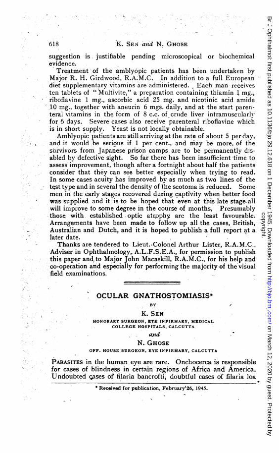
Case reportM.N.S., Hindu brahmin male, aged. 26 years, of Serajgunj,
Northern Bengal, was admitted into the Eye Infirmary for theinvestigation and treatment of orbital cellulitis with haemorrhagesin the retina and the vitreous in his left eye.Habits a-nd previous history.-He is a man of average build
and looks slightly anaemic.- He had very rarely visited EasternBengal and. that was only for -a day or two. His habits areregular. He lives on average bengali diet consisting of rice,pulses, vegetables and fish, milk irregularly and eggs. and- meatrarely. He had never eaten raw or dried fish. He does not- drinkor smoke. He does not remember having suffered from -anyserious illness, any attack of urticaria, any painful transientswelling or any creeping eruption in any part of the body.
History of present- illness.-On May 25, 1944, he felt adull aching pain on the left side of his nose extending- to thefrontal and temporal regions on the same side. On May 26, 1944,the pain increased and a swelling appeared on the left cheek-ex-tending to the lower eyelid. The swelling was very itchy. OnMay 29, 1944, the swelling had increased and spread from theangle of the 'mouth to the roots of the hair o?h the same (left) side.The eyelids were so swollen that he could not open the eye. Hewas running a low temperature till -June 5, 1944, when hereported at the outpatient's department of the Eye Infirmary.Examination.-On that day, he had no swelling of his left cheek
and forehead, but the tids were swollen and oedematous, tfthe coo-junctiva was chemosed, movements of the eyeball were restricted,the pupil was slightly dilated and reacted to light sluggishly, thetension was normaL, and the vision was 6/6. The right.eye was,normal. He was given treatment for orbital cellulitis includingsulphonamide 4 tablets stat. ,and 2 tablets every 4 hours. Twodays later on June 7, 1944, he reported. again, conplaining ofdimness of vision and increase of pain in the eye.. His vision hadfallen to hand movements only. The swelling of the lids and thechemosis of the conjunctiva had almost disappeared -and. themovements of the eyeball were almo$t normal. The pupil was -slightly more dilated and reacted sluggishly to light and the ten-sion was normal. On examination of the fundus, the vitreouswas hazy, the retinal veins were engQrged and there werehaemorrhages from the veins on the upper part of the disc- andthe upper temporal and nasal parts of the- retina. On admissionto the hospital on June 8, 1944, the swelling of the lids and-.thechemosis of the conjunctiva had completely disappeared, move-ments of the eyeball were, nornial, the pupil was still dilated andsluggish in reaction to light, tension was normal, and the
copyright. on M
arch 12, 2020 by guest. Protected by
http://bjo.bmj.com
/B
r J Ophthalm
ol: first published as 10.1136/bjo.29.12.618 on 1 Decem
ber 1945. Dow
nloaded from
K. SEN and N. GHOSE
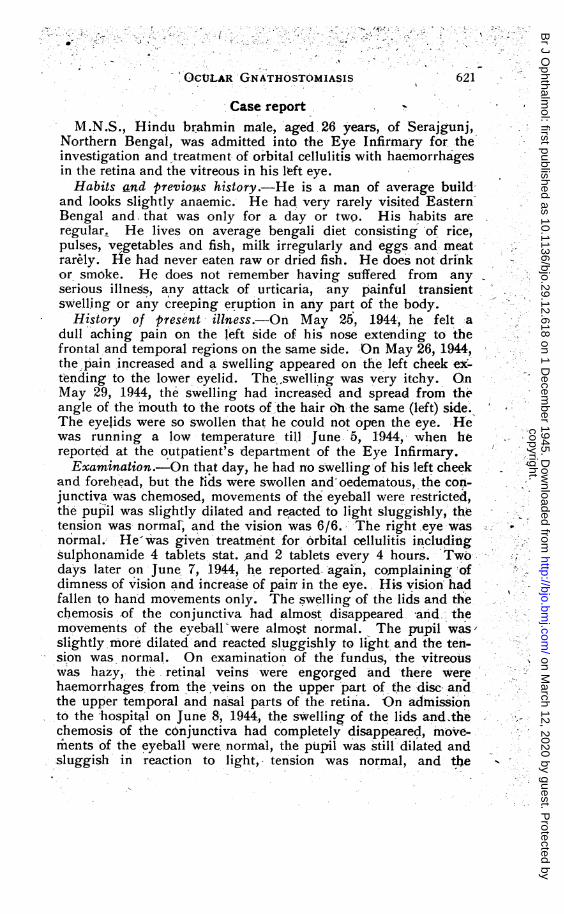
haemorrha'ges in the vitreous and retina had increased. On in-vestigation, the vascular, gastro-intestinal, respiratory andnervous systems were normal. No septic foci were found in thegums, throat, nose and nasal sinuses. No albumin, sugar, orcasts were found in the urine. The total R.B.C. was 3,800,000,haemoglobin -was 60 per cent. The total W.B.C. was 7,200, ofwhich polymorphonuclears were 66 per cent., lymphocytes 26 percent., large mononuclears 4 per cent. and eosinophils 4 per cent.,blood coagulation time was 31 minutes and the bleeding time was2 minutes. W.R. was negative. Blood taken at midnight formicrofilaria was negative. B.P. was 110 systolic and 80 diastolic.In the stools a few hookworm ova were found. The anaemia andslight eosinophilia were considered to be due to hookworm infec-'tion. On June 11, 1944, three days after his' admission to thehospital he developed severe iritis' in the eye. The cornealepithelium was slightly oedematous, anterior chamber was hazy,there were several grey nodules on the iris of the size of a pin'shead between " 11 and 2 o'clock " regions and a large irregulargrey exudate between the iris and lens capsule at " 1 o'clock," thetension was slightly raised and the vision was light perceptiononly. The iritis responded to the usual treatment and on June 13,1944, the corneal epithelium and apterior chamber were clear,pain and ciliary injection were much less, but the pupil was onlyhalf- dilated. The eye was completely quiet by June 19, 1944,the grey nodules had all disappeared leaving gtey depressions onthe iris and at " 1 o'clock " there was a posterior synechia andthe pupil was two-thirds' dilated, the vitreous haze had increased,and the visioxi was perception of light. At this stage the con-dition was considered to be one of septic (metastatic) iritis. OnJune 20, 1944, the eye was again- irritable and a large grey noduleprojecting into the anterior chamber had appeared at "6 o'clock" -
the pupil had slightly contracted 'in spite of atropine ointment-being contipued, and pigmented keratic precipitates were noticedfor the first time; the tension was slightly raised. On June 29,1944, the eye was still irritable,, the grey nodule at " 6 o'clock "had disappeared leaving a grey depression on the iris, and a pig-mented nodule had appeared at " 5 o'clock." On July 7, 1944,the eye, which was settling down, was again irritable. The pig-mented nodule at " 5 o'clock " had disappeared leaving no markand- a new pigmented nodule of larger size had appeared at"6 o'clock." On July 12, 1944, this 'pigmented nodule hadbecome larger, about 4 mm. in size, and the eye was very irritable,and the tension wag raised. In the evening the nodule was foundto be moving and on closer examination it was found to be- aworm. On July 13, 1944, the pain was the worst the' patient hadever experienced. The eye was much worse and the tension was
622
copyright. on M
arch 12, 2020 by guest. Protected by
http://bjo.bmj.com
/B
r J Ophthalm
ol: first published as 10.1136/bjo.29.12.618 on 1 Decem
ber 1945. Dow
nloaded from

OCULAR GNATHOSTOMIASIS
raised. On examination with the slit-lamp under a magnificationof 23, the corneal epithelium was oedematous and the worm couldbe seen through the hazy cornea. It showed a blunt end- (head)and a conical end (tail) with two spots of iris pigment attached tothe tail. The head and tail showed slow wavy movements. Thecentral part was never seen being behind the limbus evidentlyembedded in the iris tissue. It was translucent in appearance anda central --dark streak (alimentary canal) could be seen. Theappearance of the worm was not that of a filaria Bancrofti, andit was too translucent to be a maggot. So it seemed to be anunknown soft worm. On July 14., 1944, the conditipn of the eyewas bad but the pain was a little less. The movement of theworm had stopped and the size of the nodule was smaller. Theworm was either dead or it had burrowed deeper. If it burroweddeeper into the ciliary body, the pain would have been muchworse, so it was presumed that the worm was dead. An operationfor the removal of the worm could not be undertaken on July 13and 14. On a culture of a conjunctival swab, a few colonies ofstaph. albus were found.
The operation on July 15, 1944.-It was realised that unlessthe whole worm was removed the eye would have to be removedultimately. As the worm was fixed in the middle it would notgush out with the aqueous. As the worm was a soft one, thechance of removing the whole worm with iris or any other forcepsafter a limbal incision away from the worm was remote. A limbalincision at " 6 o'clock" was out of the question as it might cutthrough the worm. So a large incision was planped provided thetension of the eye could be brought down. It was decided to tryto bring down the tension by blocking the ciliary ganglion andwaiting for half an hour or more, and failing this to do a posteriorsclerotomy. One c.c. of novocaine (2 per cent.) with two dropsof adrenaline (1 in 1,000) was injected into the region of the ciliaryganglion. Fifteen minutes later, the facial nerve was blocked byO'Brien's method, and cocaine (4 per cent.) was dropped into theconjunctival sac 3 times at 5 minutes interval. Half an hour afterthe ciliary block the tension was found to be lower than- that ofthe right eye. Then after all the preparations for an intra-oularoperation, a thin Graefe knife was introduced into the hnteriorchamber at the limbus at " 3 o'clock"; the counterpuncture wasin the cornea at " 7 o'clock " about 3 mm. from the limbus. Thecornea was cut through just above the worm from the region ofthe counterpuncture to the limbus at " 4.30 o'clock." The limbalincision was completed with a conjunctival flap. The cornea waslifted up by holding the conjunctival flap with a small dissectingforceps and a lens curette was introduced between the cornea andthe iris at " 6.30 o'clock " up to the angle of the anterior chamber.
623
copyright. on M
arch 12, 2020 by guest. Protected by
http://bjo.bmj.com
/B
r J Ophthalm
ol: first published as 10.1136/bjo.29.12.618 on 1 Decem
ber 1945. Dow
nloaded from
5 o~~~~~~~~~~~~- I -
624 K. SEN and N. GEosE
The worm was gently removed and put into a specimen tube con-taining 70 per cent. alcohol. There was no complication duringthe operation.
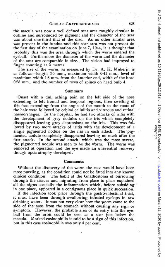
It was found to be a curved worm about 4 mm. long completelycovered with iris pigment. Dr. A; K. Mukerji and Dr. N. V.
- Bhaduri of the Helminthological department of the TropicalSchool of Medicine, Calcutta, identified the worm as an immature
- Gnathostoma, and gave the assurance that the whole worm hadbeen removed.
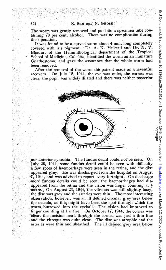
After the removal of the worm the patient made an uneventfulrecovery. On July 18, 1944, the eye was quiet; the cornea wasclear, the pupil was widely dilated and there was neither posterior
'nor anterior synec detail could not be seen., OnJuly 26, 1944; some fdu's detail could be seen wi difficultyA ftw, spots oha morrhage were seen inth retna,adteis
appea.red gey. He was discharged from the hospital on August7,T 1944, and was advised t-o report -every fortnight. On discharge-more- fundus details could' be seen, the haemorrhagesnhad di-
->appeared from the retina and the vision was finger counting at i'metre.0 On August 23, 1944, 'the vitreous was still slightly hazy,;*-- - the disc was grey and the arteries were thin. The most interesting
observation, however, was an ill defined circular grey area below;the macula, as thi% might have been the spot through which the
Z.worm, burrowed into the eyeball. The vision had improved-to* --finger counting at 1 metre. On October-17, 1944, the cornea was
d'ear, the incision mark through the cornea was just a thin line-arid the vitreoius was.quite clear. The disc was atrophic and the
arteries were thin and sheathed. The ill defined grey area below
copyright. on M
arch 12, 2020 by guest. Protected by
http://bjo.bmj.com
/B
r J Ophthalm
ol: first published as 10.1136/bjo.29.12.618 on 1 Decem
ber 1945. Dow
nloaded from

The worm after removal.
Slit-lamp view of the worm.
copyright. on M
arch 12, 2020 by guest. Protected by
http://bjo.bmj.com
/B
r J Ophthalm
ol: first published as 10.1136/bjo.29.12.618 on 1 Decem
ber 1945. Dow
nloaded from
OCULAR GNATHOSTOMIASIS
the macula was now a well defined scar area roughly circular inoutline and surrounded by pigment and the diameter of the scarwas about one-third that of the disc.- As no other similar areawas present in the fundus and this scar area was not present onthe first day of'the examination on June 7, 1944, it is thought thatprobably this was the area through which the worm entered theeyeball. Furthermore the diameter of the worm and the diameterof the scar are comparable in size. The vision had improved tofinger counting at 2 metres.The size of the worm, as measured by Dr. A. K. Mukerji, is
as follows-length 35 mm., maximum width 041 mm., level ofmaximum width 18 mm. from the anterior end, width of the head025 mm., and the number of rows of spines on head bulb 4.
SummaryOnset with a dull aching pain on the left side of the nose
extending to left frontal and temporal regions, then swelling ofthe face extending from the angle of the mouth to the roots ofthe hair were followed by orbital cellulitis and vitreous and retinalhaemorrhages. In the hospital, he had two attacks- of iritis withthe development of grey nodules on the iris which completelydisappeared leaving grey depressions on the iris. This was fol-lowed by two-more attacks of iritis with the development of asingle pigmented nodule on the iris in each attack. The pig-mented nodule completely disappeared leaving no mark after thefirst attack. In the second attack, which was the most severe,the pigmented nodule was seen to be the Worm. The worm wasremoved at operation and the eye made an uneventful recoverythough optic atrophy developed.
CommentsWithout the discovery of the worm the case would have been
most puzzling, as the condition could not be fitted into any knownclinical condition. The habit of the Gnathostoma of burrowingthrough the tissues and migrating from place to place explainedall the signs specially the inflammation which, before subsidingin one place, appeared in a contiguous place in quick succession.
If the infection took place through the gastro-intestinal tract,it must have been through swallowing infected cyclops in rawdrinking water. It was not very clear how the worm came to, theside of the nose from the stomach without causing any sign orsymptom. However, the probable area of its entry into the eye-ball from the orbit could be seen as a scar just below themacula. Marked eosinophilia is said to be a -sign of this infection;but in this case eosinophilia was only 4 per cent.
625
copyright. on M
arch 12, 2020 by guest. Protected by
http://bjo.bmj.com
/B
r J Ophthalm
ol: first published as 10.1136/bjo.29.12.618 on 1 Decem
ber 1945. Dow
nloaded from

626 J. MACASKILL
ConclusionA case of Gnathostoma in the anterior chamber of the eye is
described. This is probably the first recorded case of this wormin a human eye. An incision has been planned to remove theworm which can be used for removing either a cyst or a non-magnetic foreign body from the angle of the anterior chamber.Thanks are due to Dr. A. K. Mukerji and Dr. N. V. Bhaduri
for identifying the worm, and providing the photomicrographand the measurements of the worm, and also to Capt. E. J.Somerset, I.M.S., for many helpful suggestions in preparing thepaper.
REFERENCES
1. FAUST, E. C. Human Helminthology. London: Henry Kimpton. 1939.2. MAPLESTONE, P. A. and BHADURI, N. V.-Ind. Med. Gaz., Vol. LXXII, No.
12, p. 713, December, 1937.
A CASE OF OCCIPITAL LOBE INJURY*BY
J. MACASKILL, Maj. R.A.M.C.
LESIONS of the occipital lobe and the ocular changes accompanyingthem are well known. There may be interest, however, in therecord of an occipital lobe injury in which the extent and nature ofthe damage were demonstrated at operation.
Cpl. G.- M., aged 22 years:-This soldier received a gun shotwound of the thigh at Kohima, in April, 1944. Five days later,when in the Field Ambulance, a shell burst near hlm and he washit on the right side of the back of the head, and two days laterreceived further small body wounds.
Consciousness was never lost, but after the head injury he couldnot see at all to the left side. One week later objects far out onthis side could be seen, but- he was aware of a non-seeing areatowards the centre of vision. He could read newsprint butcomplained that while seerng one word the one adjacent to itdisappeared, and on looking at his hand only saw the fingers on theright side.When examined three months after the injury, there was a healed
depressed wound in the occipital region to the right side of the midline. Both eyes 'were normal and had full vision, though theletters on the chart were read scotomatously. The peripheral areasof the visual fields were unimpaired but para-central defects existedwhich are seen in the reproduction of the central field charts.
* Received for publication, August 8, 1945.
copyright. on M
arch 12, 2020 by guest. Protected by
http://bjo.bmj.com
/B
r J Ophthalm
ol: first published as 10.1136/bjo.29.12.618 on 1 Decem
ber 1945. Dow
nloaded from