Dynamic Circuit Network An Introduction John Vollbrecht, Internet2 [email protected] May 26, 2008.

MARCH 8TH, 2013
JILL E. VOLLBRECHT, MD
CO-MEDICAL DIRECTOR, NORTHERN MICHIGAN DIABETES INITIATIVE
MUNSON MEDICAL CENTER
The Management of Diabetes in Pregnancy

Management of Gestational Diabetes
DISCLOSURE INFORMATION
GOAL OF PROGRAM:
� To improve the care of women with gestational diabetes.
SUCCESSFUL COMPLETION:
� To receive contact hours, participants must attend the entire program. Please return your completed evaluation form to the conference registration desk at the end of the conference.
PLANNING COMMITTEE NAMES AND CREDENTIALS:
� Jill Vollbrecht MD, Christi Nowak MPH, MBA, Linda Yaroch RN, MPH, Laura Laisure RN, MS, Diane Butler BSN, RN-BC and Kelly Thompson BSN, RN-BC
SPEAKER NAME AND CREDENTIALS:
� Jill Vollbrecht, MD
CONFLICTS OF INTEREST:
� All activity planners for Management of Gestational Diabetes have reported no relevant financial relationships with commercial interests.
� All presenters for Management of Gestational Diabetes have reported no conflicts of interest related to their
presentations.
COMMERCIAL SUPPORT:
� Commercial support was not received for this event.
SPONSORSHIP:
� Sponsorship was not received for this event.
NON-ENDORSEMENT OF PRODUCTS:
� Commercial exhibits will not be present during the conference.

Objectives
� Review the pathophysiology of insulin resistance in pregnancy.
� Review current criteria for diagnosing gestational diabetes.
� Discuss the impact of poor glycemic control on both maternal and fetal outcomes.
� Describe the current goals for glycemic control during pregnancy.
� Explore available and appropriate treatments for diabetes in pregnancy.
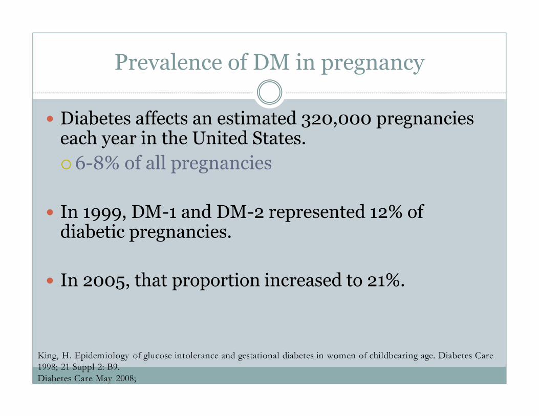
Prevalence of DM in pregnancy
� Diabetes affects an estimated 320,000 pregnancies each year in the United States.
�6-8% of all pregnancies
� In 1999, DM-1 and DM-2 represented 12% of diabetic pregnancies.
� In 2005, that proportion increased to 21%.
King, H. Epidemiology of glucose intolerance and gestational diabetes in women of childbearing age. Diabetes Care 1998; 21 Suppl 2: B9.Diabetes Care May 2008;

Insulin resistance in pregnancy
� Phenomenon first described in 1960’s:
� Diabetic pregnant women were found to have less hypoglycemia with exercise than nonpregnant diabetic women.
� Insulin resistance increases in the normal pregnancy:
� Exaggerated insulin response to glucose challenge
� Decreased glucose tolerance
� Decrease in insulin sensitivity by 45-70% in 3rd trimester.

Pathophysiology of insulin resistance in pregnancy
� Progressive increase in amount of adipose tissue.
� Increased serum concentrations of:
�Human placental lactogen
� Progesterone
� Cortisol
� Prolactin
� Rapid reversal of insulin resistance 24-72 hours after delivery.

Clinical relevance
� Type I diabetics will require 1½-3 times their normal amount of total daily insulin.
� Insulin resistance becomes clinically apparent immediately in those with preexisting disease, and between 24-28 weeks’ gestation in all women.
� Insulin resistance increases throughout pregnancy and peaks at 36-38 weeks of gestation.

Risk factors for GDM
� >25 years of age
� Overweight or obese state
� Family history of diabetes mellitus
� History of abnormal glucose metabolism
� History of poor obstetric outcome
� History of delivery of an infant with a birth weight >9 pounds
� History of polycystic ovary syndrome
� Latino/Hispanic, non–Hispanic black, Asian American, Native American, or Pacific Islander ethnicity
� Fasting plasma glucose >85 mg/dL
AACE Diabetes Mellitus Guidelines, Endocr Pract. 2007;13(Suppl 1) 2007 11

Early screening for GDM
� USPSTF:
� Case by case basis
� AACE:
� Anyone with any of the above risk factors, every trimester
� ADA:
� First prenatal visit if risk factors present, then repeated between 24-28 weeks’ gestation.

Diagnosis of GDM: traditional
� All pregnant women should be screened for GDM between weeks 24 and 28 of pregnancy.
� 1-hour glucose loading test of 50 g oral glucose
� Result of 130-140 mg/dL or greater is a positive test.
� Screening should be done earlier in pregnancy for those with risk factors for GDM, and if negative, repeated between weeks 24 and 28.
� A positive test result is then followed by either a 2- or 3-hour glucose tolerance test for confirmation.

Using a 100-g oral glucose tolerance test
Abnormal results (2/4):
�Fasting: >95
� 1-hour post-glucose administration: >180
�2-hour post-glucose administration: >155
�3-hour post-glucose administration: >140
* Not always readily apparent whether or not the diagnosis is GDM or DM.
AACE Diabetes Mellitus Guidelines, Endocr Pract. 2007;13(Suppl 1) 2007 11

Standards of Medical Care in Diabetes—2011American Diabetes Association
� Screen for undiagnosed type 2 diabetes at the first prenatal visit in those with risk factors, using standard diagnostic criteria.
� In pregnant women not known to have diabetes, screen for GDM at 24–28 weeks of gestation, using a 75-g 2-h OGTT.
� Screen women with GDM for persistent diabetes 6–12 weeks postpartum.
� Women with a history of GDM should have lifelong screening for the development of diabetes or prediabetes at least every 3 years.

ACOG’s response: 2011
� We read with interest your new guidelines, and respectfully disagree.
� Until more outcomes-based data is available, we will continue to advocate traditional, 2-step testing for GDM.

Adverse fetal effects of GDM: late pregnancy
� Polyhydramnios� Speculated to be secondary to fetal osmotic diuresis from maternal hyperglycemia.
� Fetal hypoglycemia� Increased fetal pancreatic production of insulin from prolonged exposure to maternal hyperglycemia.
� Preterm delivery
� Hyperbilirubinemia
� Polycythemia
� Intrauterine fetal demise

Adverse fetal effects of GDM: macrosomia
� Macrosomia
� > 4 kg or >90th percentile for weight for gestational age
� Increased body fat composition
� Shoulder dystocia
� Increased risk of C-section
� Brachial plexus injuries
� Facial nerve palsies
Risk increased by presence of macrosomia

What’s so bad about a big baby?
� Fetal macrosomia is now classified as a risk factor for future DM-2 in the expectant mother.
� Also associated with increased adult risks of:
�Hypertension
�Obesity
�Metabolic syndrome
�DM-2
�Psychopathology
Cardiovascular Disease Keith N. Fray n, Sara Stanner,, British Nutrition Foundation
Obes Rev. 2011 May ;12(5):e405-11. doi: 10.1111/j.1467-789X.2010.00816.x. Epub 2010 Oct 26.
Is bigger better? Macrosomia and psychopathology later in life

Maternal morbidity and mortality:preexisting diabetes
� Increased risk of diabetic ketoacidosis (DM-1)� Increased risk of infection
� Pyelonephritis� Endometritis� Postoperative wound infection� Mastitis
� Pregnancy-induced hypertension� 20-45% of diabetic pregnancies
� 5-7% in nondiabetic population
� Progression of diabetic retinopathy and nephropathy� Preterm labor� Intrauterine fetal demise

Diabetic ketoacidosis (DKA)
� DKA occurs in a state of relative or absolute insulin deficiency:
� dysregulated production of counter-regulatory hormones
� hepatic production of ketones and release of free fatty acids.
� Baseline acid-base status of the pregnant woman is a compensated respiratory alkalosis.
� Baseline HCO3 is ~ 19-20 mEq/L.
� Decreased buffering reserve in situation of metabolic acidosis for any reason.
Michael B. Schneider, et al. Pregnancy Complicated by Diabetic Ketoacidosis: Maternal and fetal outcomes.
Diabetes Care 26:958-959, 2003.
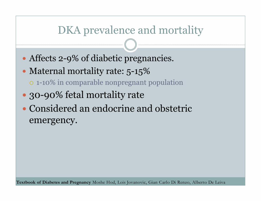
DKA prevalence and mortality
� Affects 2-9% of diabetic pregnancies.
� Maternal mortality rate: 5-15%
� 1-10% in comparable nonpregnant population
� 30-90% fetal mortality rate
� Considered an endocrine and obstetric emergency.
Textbook of Diabetes and Pregnancy Moshe Hod, Lois Jovanovic, Gian Carlo Di Renzo, Alberto De Leiva
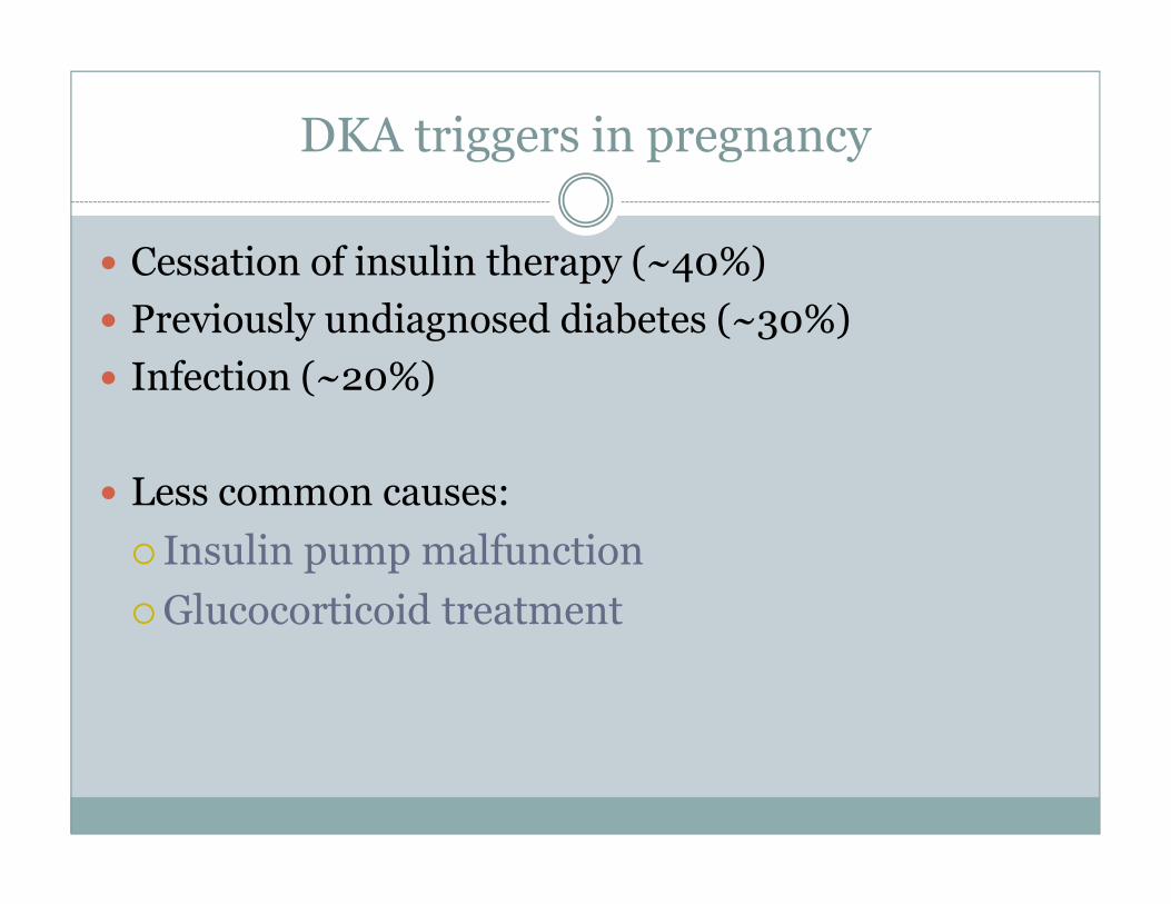
DKA triggers in pregnancy
� Cessation of insulin therapy (~40%)
� Previously undiagnosed diabetes (~30%)
� Infection (~20%)
� Less common causes:
� Insulin pump malfunction
�Glucocorticoid treatment

DKA and pregnancy
� Ketoacids have capability of crossing placenta:
�Fetal acidosis
�Decreased uterine blood flow
�Decreased fetal oxygen delivery
� While prolonged maternal ketonemia has been associated with infants of lower intelligence, ketonuria in absence of hyperglycemia has not.
� Marker of carbohydrate caloric restriction.

Clinical recommendations
� Pregnant diabetics are encouraged to check urine ketones:
�Blood glucose > 180 mg/dL
�Symptoms of nausea, vomiting, abdominal pain.

Adverse fetal effects of DM: early pregnancy
� 3 main categories :
�Early growth delay
�Spontaneous abortions
�Severe congenital malformations
�Neural tube defects
�Heart and great vessels
�Axial skeleton deformities
�Urinary tract anomalies

Early adverse fetal effects
� Severe congenital malformations are estimated to affect 8-13% of pregnancies complicated by pregestational DM.
� 2-4 times the nonpregnant rate of equivalent malformations.
� Organogenesis occurs between 3 and 8 weeks of gestation, often completed well before pregnancy is detected or initial physician’s visit.

Adverse fetal effects: late pregnancy
� Macrosomia
� > 4 kg or >90th percentile for weight for gestational age
� Increased body fat composition
� Shoulder dystocia
� Increased risk of C-section
� Brachial plexus injuries
� Facial nerve palsies
Risk increased by presence of macrosomia

Adverse fetal effects: late pregnancy
� Polyhydramnios� Speculated to be secondary to fetal osmotic diuresis from maternal hyperglycemia.
� Fetal hypoglycemia� Increased fetal pancreatic production of insulin from prolonged exposure to maternal hyperglycemia.
� Preterm delivery
� Hyperbilirubinemia
� Polycythemia
� Intrauterine fetal demise

Diabetic retinopathy
� Exacerbation of diabetic retinopathy is a well-described phenomenon that occurs in the setting of pregnancy.
� Proposed mechanisms:� Increased extravasation of serum proteins when hyperglycemia is rapidly corrected.
� Increased concentrations of growth factors (22-32 weeks):�Human placental lactogen
�Human somatomammotropin prolactin
� IGF-1

Risk factors for retinopathy in pregnancy
� Baseline evidence of retinopathy
� Elevated HBA1c at conception
� Rapid correction of hyperglycemia
� Duration of diabetes of > 6 years
� Proteinuria
� Pregnant diabetics need fundoscopic examinations once per trimester for monitoring.

Data behind the glycemic targets
� Prospective data have demonstrated that:
� Mean fasting glucose in the nondiabetic, pregnant population ~ 56 mg/dL.
� Peak postprandial glucose excursion occurs one hour after the onset of a meal.
� Mean 1-hour postprandial glucose in the nondiabetic, pregnant population ~ 105 mg/dL.
� Increases in 1-hour postprandial blood glucose levels have been associated with significant increase in fetal abdominal circumference measurements.
� 1-hour postprandial measurements >130 mg/dL are associated with an increased risk in macrosomia.
Parretti E, et al. Third-trimester maternal glucose levels from diurnal profiles in nondiabetic pregnancies: correlation withsonographic parameters of fetal growth. Diabetes Care 24:1319–1323, 2001.
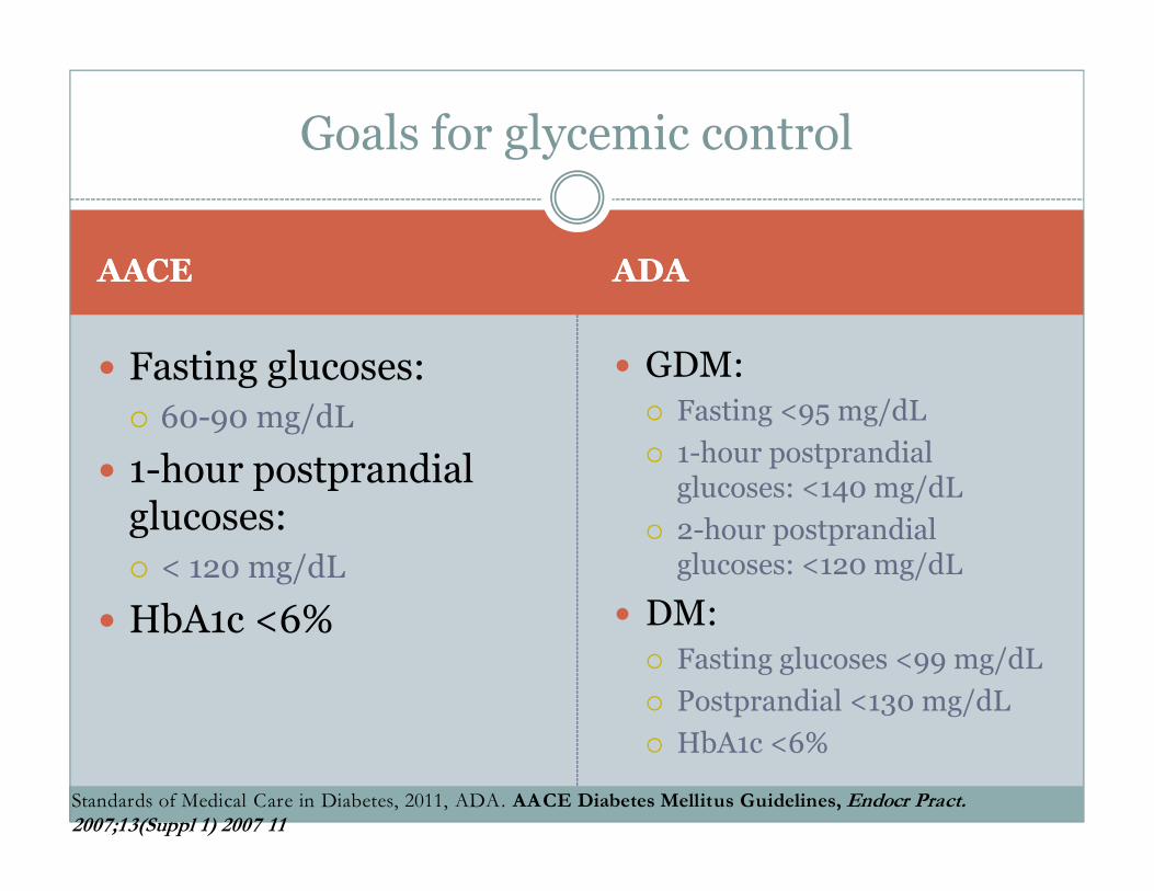
AACEAACE ADAADA
� Fasting glucoses:
� 60-90 mg/dL
� 1-hour postprandial glucoses:
� < 120 mg/dL
� HbA1c <6%
� GDM:� Fasting <95 mg/dL
� 1-hour postprandial glucoses: <140 mg/dL
� 2-hour postprandial glucoses: <120 mg/dL
� DM:� Fasting glucoses <99 mg/dL
� Postprandial <130 mg/dL
� HbA1c <6%
Goals for glycemic control
Standards of Medical Care in Diabetes, 2011, ADA. AACE Diabetes Mellitus Guidelines, Endocr Pract. 2007;13(Suppl 1) 2007 11

Pre-existing DMPre-existing DM Gestational DMGestational DM
� Fasting blood glucoses:
� <95 mg/dL
� 1-hour postprandial blood glucoses:
� <140 mg/dL
� 2-hour postprandial:
� <120 mg/dL
� Fasting blood glucoses:
� <95 mg/dL
� 1-hour postprandial blood glucoses:
� <130-140 mg/dL
Goals for glycemic control: ACOG

Barriers to achieving tight control
� Frequency of glucose monitoring.
� Intolerability of injections (“needle-phobia”).
� Disregard for diet.
� Frequency of office visits/evaluations.
� Lack of family support.
� Cultural attitudes toward pregnancy.
� Treatment with glucocorticoids.
� Bedrest/decreased physical activity.

Barriers to care in low-income populations
� Access to healthy foods
� Knowledge deficit regarding appropriate portion size of those healthy foods.
� Availability of higher-carb cereals and juices through WIC (e.g. Life Cereal 25 grams/ 3/4 cup serving)
� Transportation difficulties to appointments.
� Lack up follow-up with required GTT testing after delivery.
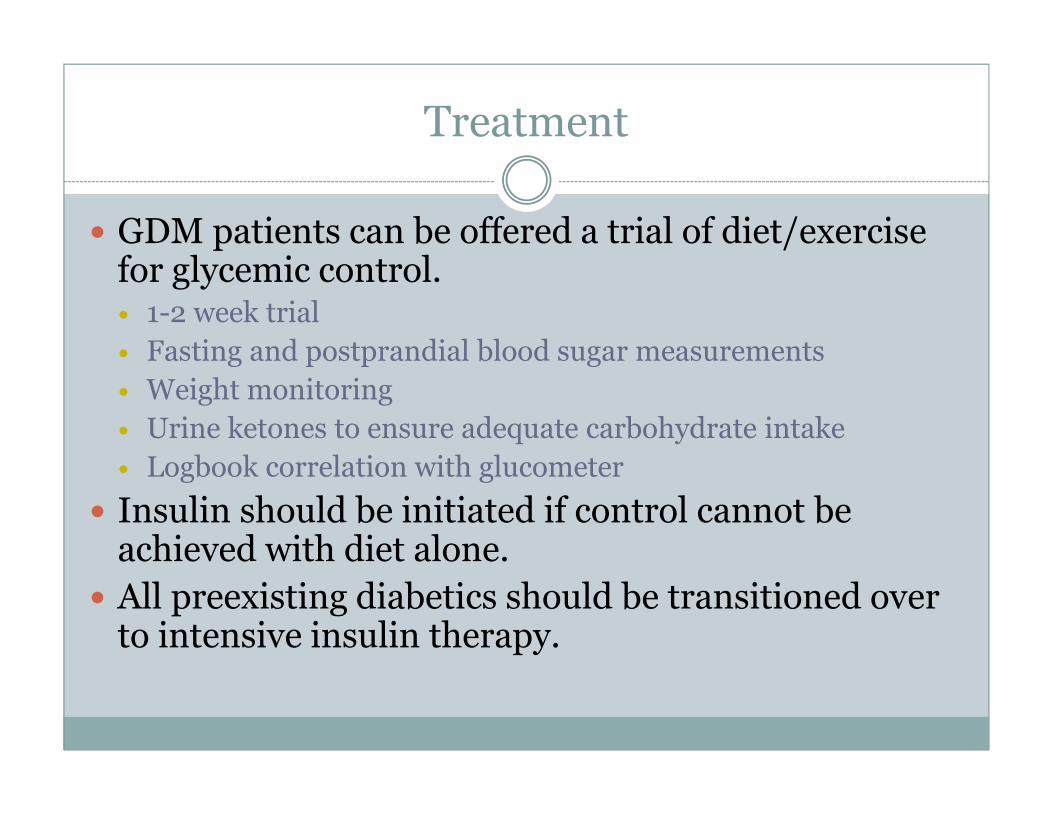
Treatment
� GDM patients can be offered a trial of diet/exercise for glycemic control.� 1-2 week trial
� Fasting and postprandial blood sugar measurements
� Weight monitoring
� Urine ketones to ensure adequate carbohydrate intake
� Logbook correlation with glucometer
� Insulin should be initiated if control cannot be achieved with diet alone.
� All preexisting diabetics should be transitioned over to intensive insulin therapy.

How-to’s of insulin
� An intensive regimen consists of insulin provided in both basal and bolus forms.
� 50% basal, 50% bolus.� NPH
� Regular human insulin
� Insulin analogs (lispro, aspart)
�Multiple daily injections/insulin pump
� U-500 regular human insulin
�Patients with poor control at diagnosis of pregnancy can be considered for hospital admission for rapid normalization of hyperglycemia.

Basal-bolus program
� Basal insulin is administered to control fasting hyperglycemia.
� Mealtime, or bolus, insulin is administered to control postprandial hyperglycemia.
� Nutritional
� Insulin: carbohydrate ratio
� Correctional
� Generally not required for GDM patients.
� Rapid-acting analogues are mealtime insulin of choice.

20062006 20112011
� No large studies have been performed examining safety and efficacy of glargine in the pregnant population.
� A total of 14 DM-1 patients who used glargine in pregnancy have been described in the literature.� Less hypoglycemia.� No congenital malformations.� Decrease in HbA1c of 0.5-0.8%
� No published reports on detemir.
� Over 1000 patients have been studied retrospectively with regard to use of glargine and patient safety, fetal outcomes.
� Two published case reports on detemir.***
Long-acting insulin analogues (category B/C)
Ann Pharmacother. 2011 Jan;45(1):9-16. Epub 2011 Jan 4.
Safety of insulin glargine use in pregnancy: a systematic review and meta-analysis. I
nsulin glargine use during pregnancy. Pantalone KM, Faiman C, Olansky L.Endocr Pract. 2011 May -Jun;17(3):448-55


Oral medications
� Only appropriate for patients with DM-2.
� Metformin has been found to decrease rates of spontaneous abortion in patients with PCOS.
� Recent studies have been performed comparing both glyburide and metformin with insulin in the management of GDM.

Sulfonylureas: glipizide, glyburide, glimepiride
� 1st oral medication developed for treatment of DM-2 (1956).
� Stimulate release of insulin from the beta cells of the pancreas.
� Can cause hypoglycemia.
� Dosed once or twice daily.
� Rapid-acting secretogogues (nateglinide, repaglinide) dosed with meals.

Metformin (biguanide)
� Available in the US since 1995.
� Suppresses hepatic glucose production via downregulation of AMP kinase activity.
� Does NOT cause hypoglycemia!!!
� Can take up to 12 weeks to take effect.
� Associated with the risk of lactic acidosis in patients with renal failure.
Davidson’s Diabetes Mellitus, 5th Edition

AdvantagesAdvantages DisadvantagesDisadvantages
� Ability to continue current therapy after pregnancy diagnosed.
� Ease of therapy.
� No injections.
� Possible cost benefit.
� No large-scale studies supporting routine use.
� Hypoglycemia.
� Nonspecific treatment.
� No guarantee that insulin will be avoided.
� No decrease in frequency of glucose monitoring.
� Diet/exercise still essential.
� Incorrect diagnosis is possible.
� Lactic acidosis with metformin.
Use of oral medications in pregnancy
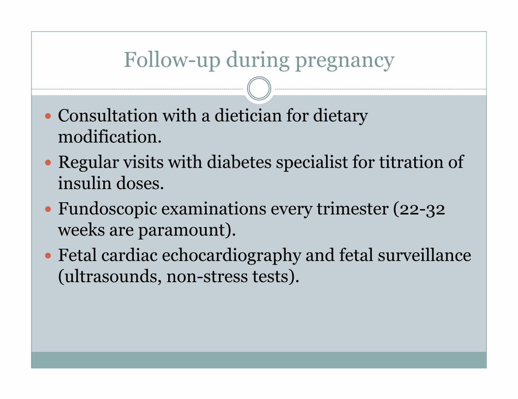
Follow-up during pregnancy
� Consultation with a dietician for dietary modification.
� Regular visits with diabetes specialist for titration of insulin doses.
� Fundoscopic examinations every trimester (22-32 weeks are paramount).
� Fetal cardiac echocardiography and fetal surveillance (ultrasounds, non-stress tests).

Laboratory evaluation
� HbA1c every 2-4 weeks.
� Urine ketones:� if weight gain is slow/absent
� If glucose >180 mg/dL
� If symptoms of nausea, vomiting, abdominal pain.
� TSH one time (DM-1).
� GAD antibodies (if early DM-1 is suspected)
� TSH 6 weeks after delivery in DM-1 to evaluate patient for postpartum thyroiditis.
� Urine microalbumin evaluation every trimester.

Post-delivery follow-up
� Type I diabetics should be given long-acting insulin immediately after delivery to prevent DKA.
� Some may not require insulin for 24-72 hours after delivery.
� Insulin doses should be recalculated based on postpartum weight after delivery (0.6 U/kg).
� Water weight
� Breastfeeding
� Change in physical activity after discharge
� Post-discharge diet

Barriers to nursing in diabetic mothers
� Alterations in eating and sleeping patterns.
� Labile glucoses.
� Delayed lactogenesis.
� Conditions surrounding delivery
� Rooming-in
� Pacifiers
� Increased risk of mastitis

Glycemic control and lactation
� Animal studies have demonstrated that poor glycemic control has been associated with decreased milk production.
� Small-scale human studies have indicated that smooth glycemic control has been associated with longer duration of breastfeeding.
� Mechanism of hyperglycemia and impaired lactogenesis has not been established.

Follow-up of GDM
� Postpartum glucose tolerance evaluation is recommended for patients with gestational DM to determine presence of persistent DM-2.
� Improved glucose tolerance while breastfeeding may cause false negative tests.
� Recommended protocols include 75-g 2-hour GTT performed after breastfeeding is discontinued entirely.

Conclusions
� GDM diagnosis increases future risk of DM-2 by 50%.
� Blood glucoses should be monitored fasting and 1 hour postprandially:
� Fasting < 90
� Postprandial < 120-130
� Insulin should be initiated for all pre-existing diabetics, and in the GDM population if diet and exercise fail.
� All GDM patients should have a follow-up GTT 2 months after delivery.

Available local resources
� Northern Michigan Diabetes Initiative
� Resource Guide
� Insulin dosage app
� Diabetes Education program information
� Results from the 2012 Community Diabetes Survey
� NMDI Facebook page
� NMDI website www.nmdiabetes.org