
geomagneticfields
20
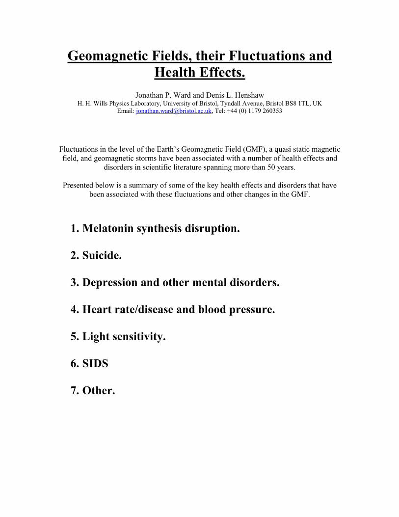
Geomagnetic Fields, their Fluctuations and Health Effects. Jonathan P. Ward and Denis L. Henshaw H. H. Wills Physics Laboratory, University of Bristol, Tyndall Avenue, Bristol BS8 1TL, UK Email: [email protected] , Tel: +44 (0) 1179 260353 Fluctuations in the level of the Earth’s Geomagnetic Field (GMF), a quasi static magnetic field, and geomagnetic storms have been associated with a number of health effects and disorders in scientific literature spanning more than 50 years. Presented below is a summary of some of the key health effects and disorders that have been associated with these fluctuations and other changes in the GMF. 1. Melatonin synthesis disruption. 2. Suicide. 3. Depression and other mental disorders. 4. Heart rate/disease and blood pressure. 5. Light sensitivity. 6. SIDS 7. Other.
-
Upload
salesgroup-international -
Category
Documents
-
view
213 -
download
0
description
geomagneticfields
Transcript of geomagneticfields
Geomagnetic Fields, their Fluctuations and Health Effects.
Jonathan P. Ward and Denis L. Henshaw
H. H. Wills Physics Laboratory, University of Bristol, Tyndall Avenue, Bristol BS8 1TL, UK Email: [email protected], Tel: +44 (0) 1179 260353
Fluctuations in the level of the Earth’s Geomagnetic Field (GMF), a quasi static magnetic field, and geomagnetic storms have been associated with a number of health effects and
disorders in scientific literature spanning more than 50 years.
Presented below is a summary of some of the key health effects and disorders that have been associated with these fluctuations and other changes in the GMF.
1. Melatonin synthesis disruption.
2. Suicide.
3. Depression and other mental disorders.
4. Heart rate/disease and blood pressure.
5. Light sensitivity.
6. SIDS
7. Other.

A summary of some of the key findings in each of these areas is presented over the following pages. In addition there is an explanation of the relevant geomagnetic indices
and a list of references for all the papers cited in this document.

1. Melatonin synthesis disruption Bartsch et al. (1994). Seasonal changes of nocturnal urinary 6-sulfatoxymelatonin excretion in Rats demonstrated peak levels in summer despite constant photoperiods. The authors hypothesise that the horizontal component H of the geomagnetic field may act as a seasonal Zeitgeber because H shows a similar seasonal rhythm, and changes in in the direction and intensity of H can affect pineal activity. Burch et al (1999).“…reductions in nocturnal 6-OHMS excretion were higher on days when geomagnetic activity and 60Hz MF exposures were both elevated, suggesting a common biological mechanism”. Authors also report that mean overnight 6-OHMS excretion was lower on days when the 36-h Ap or aa values exceeded 30 nT. Juutilainen et al.(2006). With a small sample group of 60 women, the authors tested the hypothesis that the inconsistencies seen in studies on the effect of MF’s upon melatonin production in humans are due to interaction with light in pineal response to MF’s. The authors, with a retrospective analysis based on a questionnaire and actual MF measurements, split the sample group into four groups based on whether they had been exposed to MF’s and/or Light-At-Night (LAN) and compared the levels of 6-OHMS excretion for each subgroup. The lowest excretion of 6-OHMS was observed in the group of women who were exposed to both MF and LAN, and the differences between the groups were significant (p<0.0001). the study supports the hypothesis that daytime occupational exposure to MF enhances the effects of nighttime light exposure on melatonin production. Weydahl (2001). A circadian rhythm was found to characterize both melatonin and K index, the peak in K index (23:24) preceding that of melatonin (06:08). During the span of investigation, a circannual variation also characterized both variables. Correlation analyses suggest that changes in geomagnetic activity had to be of a certain magnitude to affect the circadian amplitude of melatonin. If large enough (>80nT/3h), changes in geomagnetic activity were reported to significantly decrease salivary melatonin concentration. 2. Suicide Berk et al (2006). Authors report that suicide amongst females increased significantly in autumn during concurrent periods of geomagnetic storm activity (p = 0.01). This pattern was not observed in males (p = 0.16). Gordon and Berk (2003). The authors found a correlation between suicides and average storm activity in South Africa between January 1980 and December 1992. The effect was shown to be stronger in females (p<0.005) than males (p<0.025). Partonen et al (2004). High levels of solar radiation activity were associated with the increased risk of suicide (p = 0.00001), but the effect of geomagnetic activity was weak.
Mechanisms Stanley and Brown 1988. Compared the levels of melatonin in the pineal glands of 38 individuals obtained at autopsy: 19 suicide victims and 19 non-suicide controls. Melatonin values were divided into three time periods- 0600 to 1400, 1400 to 2200, and 2200 to 0600. The principal finding of the study is that melatonin levels in the pineal glands of suicide victims are significantly lower than those of non-suicide controls (F=5.14, p≤0.03). Further analysis by t-test reveals that the greatest group difference occurred during the 2200-0600 hour interval (p≤0.04). No significant differences were observed for the other intervals. 3. Depression and mental disorders A four year study (Becker et al 1961, Friedman et al 1963) reported a positive correlation between the monthly sums of geomagnetic K indices from Fredericksburg and the monthly admissions to two mental hospitals in New York. Results in this study suggest that statistically significant low to marked linear relationships exist between geomagnetic parameters and a gross measure of human disturbance. These relationships are evident when the measure of the geomagnetic parameter is restricted to those periods of higher disturbance which can be categorized as magnetic storms. Dimitrova et al (2002). The authors’ investigations suggest that most of the people examined in their study during the autumn and spring equinox, irrespective of their health status could be sensitive to geomagnetic changes, which have a direct influence upon self-confidence and working ability. Kay (1994). The hypothesis that geomagnetic storms may partly account for the seasonal variation in the incidence of depression by acting as a precipitant of depressive illness is susceptible individuals is supported by a statistically significant 36.2% increase in male hospital admissions with a diagnosis of depressed phase, manic-depressive illness in the second week following such storms compared with geomagnetically quiet control periods. Krivelyova and Robotti (2003). The authors found that, for most of the countries in their sample, the previous week’s unusually high levels of geomagnetic activity have a negative and statistically and economically significant impact on today’s stock returns. The results are consistent with changes in risk-taking behavior caused by depressive disorders, since geomagnetic storms have been found to substantially increase the incidence of depression and other psychological disturbances among people. Persinger (1987). The author reports that geomagnetic variations were found to be correlated with enhanced anxiety, sleep disturbances, altered models and greater incidences of psychiatric admissions.
Raps (1999). A pilot study indicates two statistically significant correlations between the numbers of first admissions (psychiatric) per month and the level of solar radioflux in the corresponding month, (p<0.05) and sudden magnetic disturbances of the ionosphere (p<0.01). Srivastava et al (1980). The authors found no significant correlation between the daily indices of geomagnetic activity and the daily hospital admissions, nor for the daily number of road accidents. However, a 50% increase in hospital admissions was observed during 1978 compared with 1972, and 76% of air accidents were found to occur during enhanced geomagnetic pulsation activity. 4. Heart rate/disease and blood pressure Belov et al (1998). The authors found a positive correlation of the EEG (electroencephalogram) data with the geomagnetic activity. The strongest correlations were found in the frontal and central areas of the brain. The degree of synchronisation of the spontaneous EEG seems to reflect sensitivity of the human nervous system to the Earth’s magnetic field. A stressor response to strong short-term disturbances in the geomagnetic field reveals itself in the form of enhancement of the EEG global synchronisation. Chernouss et al (2001). “A correlation between geomagnetic disturbances and heart rate was calculated and different reactions of people on geophysical impact were shown. The special group of ‘Aurora Disturbance Sensitive People’ (ADSP) was revealed. An international study, aimed at evaluation of the impact on human health due to exposure of northern populations to the geophysical risk factor (GRF) in the Circumpolar areas located under the Aurora Belt, is needed. “ Dimitrova et al (2004). Arterial blood pressure (bp) was found to increase significantly with the increase of the geomagnetic activity (GMA) level, and systolic and diastolic bp were found to increase significantly from the day before till the second day after the geomagnetic storm. These effects were present irrespective of sex and medication. Otto et al (1982). In a survey of 66,900 cases of death from ischaemic heart disease increased geomagnetic field, and increased geomagnetic storms were positively and significantly related to death from ischaemic heart disease. 5. Light sensitivity Cremer-Bartels et al (1983). The authors found a relationship between the coincidence of static magnetic field variation at near GMF strength and a decrease in human night vision acuity. Suggest that the inhibition of methoxylation of indolamines is involved in the magnetic field sensitivity in humans.
Partonen (1998). Author reports that under exposure to rotating magnetic field, the size of pinealocytes is bigger at night than during the day in spring, but there is no day-night difference in autumn. Specialized photo-receptors amplify the influence of a weak magnetic field in patients with winter seasonal affective disorder (SAD), modulating the response of the photoreceptive system to light and the pineal response to a magnetic Thoss et al (2000). Authors show that the visual sensitivity of man is influenced by periodic sinusoidal inversion of the vertical component of the geomagnetic field. This effect indicates a visual fixation in the north-south direction, showing a pronounced resonance at a period duration of 110s. Thoss et al (2002). Reports the existence of a weak influence of the static field on visual sensitivity in man. If the directions of view line and field vector coincide the perception threshold of alight stimulus is slightly but significantly increased. This significance will be lost if view line deviates by 10 degrees from the field direction. Thoss et al (2003). The results from this report imply that looking in the direction of the magnetic field reduces the visual discrimination threshold (by about 4%, p<0.02). 6. SIDS Weissbluth & Weissbluth (1994). SIDS exhibits circannual, circadian, and ontogenetic features which may reflect an impaired maturation of the photoneuroendocrine system caused by a genetic absence or mutation of the enzyme NAT. The failure of normal pineal gland development and subsequent impaired production of its main secretory product, melatonin, may cause a lethal imbalance in the chemical interactions among serotonin, progesterone, and catecholamines. The result of this chemical imbalance, culminating in SIDS, involves the neurotoxic and cardiomyotoxic effects of abnormally elevated catecholamines and intracellular calcium ions. The human embryonic pineal glands, at 120 days exhibit two distinctive types of cells observed by light microscopy, and at 150 days, in only some of these cells, the photosensitive pigment melanin appears. The similarity between fetal and neonatal pinealocytes and retina photosensory cells suggests that the pineal gland is capable of detecting changes in light in utero and during the early postpartum period (Altar, 1982). Factors which influence pineal maturation include intensity of light or directionality of the photoperiod, that is, whether the duration of day light is seasonally shortening or lengthening.
In humans, the pineal gland is influenced by environmental light and initially contains the capability to function as a photoreceptor and a neuroendocrine organ. It develops into only a neuroendocrine organ at about the third month of life. Sparks & Hunsaker have confirmed is an extension of their original study, the findings that pineal glands are smaller in SIDS infants than in controls (p<0.005) and that pineal glands are larger in summer than winter. SIDS infants’ pineal glands however did not grow larger in the summer months and perhaps is therefore less responsive to photoperiod stimulating effects compared to normal infants. In adults, intra-individual variations of 08:00 hr plasma melatonin concentration occur with peak values in January and July (20-22 pg ml-1) and troughs in May and October (12-13 pg ml-1) and these seasonal differences are statistically significant (Arendt et al., 1979). Changes in the direction of photoperiod duration shortly after the winter and spring solstice may account for the seasonal peak values, but seasonal temperature differences may also be important. Another characteristic feature of melatonin secretion is that of pulsatility (Reiter, 1991). Discrete burst or episodes of melatonin released during the night are detected if the frequency of sampling is high. Melatonin receptor sites have been identified in the fetus (Yuan et al., 1991). High melatonin concentrations derived from the mother are present in newborn infants, but they fall rapidly to barely detectable levels within about 1 week. Subsequently, melatonin concentrations remain low until 3-4 months of age, and after this age, the concentrations gradually increase. Catecholamines – light is received by the eyes, and the photoreceptors in the retina produce a neuronal signal which is transmitted through the suprachiasmatic nucleus (SCN) to ganglionic fibers of the peripheral sympathetic nervous system. The chemical messenger which is released from the post-ganglionic sympathetic nerve-endings terminating on the pineal gland is the catecholamine norepinephrine (NE). Neural responses from the retina during daylight inhibit the release of NE; darkness releases this system from inhibition which permits NE stimulation of the pineal gland to occur. One of the major biochemical responses of NE stimulation of the pineal gland is an increase in the intracellular concentration of calcium ion. High progesterone levels may be associated with SIDS as they are associated with low levels of melatonin (in rates high levels inhibit the release of melatonin from the pineal). In 1990 another paper was published which showed that among infants 3 months of age or less, cerebral spinal-fluid melatonin concentrations in SIDS infants were statistically significantly lower than in control infants. Also, lower concentrations of melatonin were detected in the blood of SIDS infants compared to controls.

Serotonin concentrations in the pineal gland exhibit seasonal variation with the highest concentration during the month of December and the lowest concentration during the month of June. In the blood, serotonin concentrations are highest at night. In the newborn infant serotonin concentrations are high and gradually fall over several months. Statistically significant circadian rhythms are well developed immediately after the infant is born. The absence of the biologically expected nocturnal surge of melatonin in the evening hours, during the first few months of life, especially in the winter when serotonin activity is peaking is the exact congruence of circannual, circadian and ontogenetic features which would be associated with maximum serotonin-induced increases in [Ca2+], in the heart and brain. Progesterone concentrations which are negatively correlated with melatonin concentrations, would also be high causing further increases in [Ca2+]. Norepinephrine activity would also be increasing during the first few months causing toxic effects on the heart and brain. The combination of elevated Norepinephrine and depressed melatonin would cause a reduction in the amount of brown fat around the adrenal gland. The clinical picture of SIDS is that of a weak or mild temperament baby who may appear sleepy, drowsy, or listless; these behavioural features could result from elevated progesterone or calcium. These babies sleep well, do not cry much, but die silently at night, mainly during the winter. Leach et al. (1999). Many of the epidemiologic features that characterize SIDS infants and families have remained the same, despite the recent decrease in SIDS incidence in the UK. These included the same characteristic age distribution, few deaths in the first few weeks of life or after 6 months, with a peak between 4 and 16 weeks, a higher incidence in males, lower birth weight, shorter gestation, and more neonatal problems at delivery. The majority of the SIDS deaths occurred during the night sleep (83%) and there was no particular day of the week on which a significantly higher proportion of deaths occurred. There has however been a reduction in the previous high winter peaks of death and a shift of SIDS families to the more deprived social grouping. Just more than one quarter of the SIDS deaths (27%) occurred in the 3 winter months (dec-feb) in the three years of the study. The report lends weight to the mounting evidence that the association between smoking and SIDS may be part of a casual mechanism. Eckert (1992). Author constructs hypothesis based upon: a) observed clustering of Sudden Infant Death Syndrome (SIDS) cases at places with abnormal geomagnetic fields (GMF) and/or electromagnetic fields (EMF); b) recorded GMF pulsations matching the breathing frequencies of infants; c) the reported immature development of increased
dendritic spine density in the brain stem of SIDS cases and; d) the increased dendrite arborisation in the brains of rats exposed to magnetic fields (MF). The hypothesis consists of two parts:
1. A disturbed GMF in the residence or surroundings of a pregnant woman may interrupt the normal development of the central organ which controls respiration (brain stem) of the fetus. This is termed the ‘Selection Factor’.
2. If such an infant with a functional disturbance of the control organ is then exposed to a GMF or EMF with pulsations similar to his own breathing frequency, but inverted in phase, value form etc then the vital nerve impulses from the respiration control organ to the breathing organs may be disturbed or blocked with fatal effect. This is termed the ‘Trigger Factor’.
Persinger & O’Connor (2001). The author hypothesises that if geomagnetic-mediated stimuli trigger many sudden infant deaths, then the days in which they and hospital admissions for cardiac arrhythmias for adults occur should share a similar source of variance. Factor analyses of the days in which a sudden infant death occurred in Ontario or adults were admitted for one of eight categories of cardiac crisis in the Sudbury (Ontario) region for the year 1984 supported each shared about 40% of their variance, also shared about 40% of the variance with pulsations (0.2 Hz to 5 Hz) was associated. The results are consistent with important role of geomagnetic variables in the occurrence of transient electrical anomalies in brain function rather than cardiac blood flow. Grainger et al. (2000). Power frequency magnetic fields have been implicated in SIDS. Through the use of a case–control study measuring 50 Hz electric and magnetic fields at the SIDS baby’s last head position, no association could be found between SIDS and either electric (p = 0.327) or magnetic (p = 0.827) 50 Hz fields. Sparks and Hunsaker (1988). The authors observed, through a small (7 SIDS, 5 control) sample post-mortem analysis, a statistically significant (p<0.005) reduction in the anatomic size of the pineal gland in SIDS infants, as compared to age-matched controls. 97% of all respiratory disturbances in infants at risk for SIDS occur during sleep, with a high preponderance occurring during non-REM sleep. Sparks and Hunsaker (1997). The authors found a highly significant (p<0.0001) decrease in the size of the pineal gland in the SIDS population (18.2±0.9mm²) compared with the control population (28.0±2.5mm²). At the same time there was no difference in the mean age,, brain weight, or body weight between the control and the SIDS populations as a whole. Sparks and Hunsaker (2002). On the basis of current data, the authors postulate that apoptotic neurodegeneration constitutes the anatomic substrate accounting for the pathophysiologic mechanism and proximate cause of SIDS.

“…the data clearly suggest that there is a neurodegenerative process occurring in SIDS,, that a marker of this neurodegenerative process can be useful in diagnosing SIDS, and that, therefore, the syndrome of sudden infant death is a neurodegenerative disorder.” Sturner et al. (1990). The authors found a significant correlation for melatonin levels in different body fluids from the same individual. Age adjusted Cerebospinal Fluid (CSF) melatonin levels were significantly lower among the SIDS infants than among those dying of other causes. A similar, but non-significant, trend was also noted in blood and vitreous humour. Diminished melatonin production may be characteristic of SIDS and could represent an impairment in the maturation of physiologic circadian organization. ….most SIDS deaths occur during sleep and have a distinctive age distribution, reaching a peak at 3 months. Maturation of the normal pattern of sleep behaviour with sleep-time concentrated during the night, takes placed during the second month of life. …studies have demonstrated that the characteristic daily rhythm, in melatonin secretion, with peak values occurring at night, matures with a similar time course, and is detectable in infants at approximately 3 months of age. The concurrence of these maturation processes and the age incidence of SIDS deaths may thus be related. Autopsies were performed within 28hr of death. Kennaway et al. (1996). Rhythmic excretion of the urinary melatonin metabolite 6-sulfatoxymelatonin appears between 49-55 weeks postconception (9-15 weeks of age) in singleton babies born at term in hospital. The authors tested the following hypotheses: that the delay in the appearance of a melatonin rhythm in premature infants is influenced by the underlying reason for the prematurity, that siblings of SIDS victims have a delayed appearance of melatonin rhythmicity, and that continuous lighting in the neonatal nursery contributes to the delayed appearance of melatonin rhythmicity in premature infants. They confirmed earlier research that infants are born with a functionally immature SCN-pineal gland axis that does not establish significant hormonal rhythmicity until 9-12 weeks of age in full-term infants. Tryba et al. (2006). The authors’ data suggest an intriguing possibility that, in SIDS victims, disturbances in the serotonergic system may underlie the failure to gasp appropriately and autoresuscitate despite increased release of 5-HT during hypoxia. Serotonin acting via 5-HT2a receptors can modulate the respiratory rhythm generating network in vitro and respiratory motor neuron activity, including the recruitment and maturation of phrenic motoneurons during development. *5-HT receptors are receptors for the neurotransmitter and peripheral signal mediator serotonin, also known as 5-hdroxytryptamine (5-HT). These receptors are located on the cell membrane of nerve cells and other cell types in animals and mediate the effects of serotonin as the endogenous ligand.
The 5-HT2a receptors perform action upon the muscle (contraction, vasconconstriction/dilation) and platelets (aggregation). *Hypoxia – this is when the infant is deficient in oxygen. It can be caused by inadequate oxygen transport, the inability of tissues to use oxygen and the reduction in partial pressure of oxygen. 1Although the incidence of SIDS varies among countries and ethnic groups, there are always three epidemiological characteristic features:
(i) A circannual pattern with an excess of deaths due to SIDS in the winter months.
(ii) A circadian rhythm of SIDS occurring at night. (iii) An ontogenetic feature of SIDS occurring after the first few weeks, and during
the first 3-4 months of age. Goldwater (2003). – SIDS was first defined in 1969 as “the sudden death of any infant or young child, which is unexpected by history, and which is a thorough post-mortem examination fails to show an adequate cause of death.” (Bergman 1970). This has since been refined to include only infants dying suddenly under one year of age. 7.Other Aldrich, Andrews and Liboff (2001). The authors designed a metric which incorporates the orientations of power lines relative to geomagnetic north. Odds Ratio between DIP angle and incidence of brain cancer 4.86 (95% CI = 1.13 - 20.96). The results from this analysis offer evidence that a metric of EMF’s, incorporating a geomagnetic component, may finally provide some clarification on the protracted quandary about EMFs and cancer risk. Dupont, M. J. et al. (2005). 32 pregnant rats were exposed for to three days before expected parturition either to a coil that generated 0.5 Hz sine-wave, 5 to 10 nT magnetic fields, or to a reference coil (< 1 nT). Litters born to exposed mothers contained significantly fewer pups than those exposed to the control conditions. There were significantly fewer males and fewer females in litters exposed to the fields generated in the E-W and N-S directions, respectively. The authors suggest these result support the hypothesis that a specific temporal configuration of brief periods of geomagnetic activity can produce an increased incidence of nonvital fetuses, neonates, or infants. O’Connor, R. P. and Persinger, M. A. (1997). The authors hypothesised that sudden decreases in nocturnal melatonin by a specific range of geomagnetic activity would 1 Weissbluth & Weissbluth 1993

precipitate sudden infant death. They report a correlation of 0.90 between the number of cases of Sudden Infant Death Syndrome (SIDS) and an increase in numbers of days per month with average geomagnetic activity between 11 and 20 nT and 31 through 40 nT, but also a decrease in the number of days with values between 21 and 30 nT. Wever (1979). Author reports that when subjects were placed in a non EMF shielded room compared to a shielded room, and all other factors identical, the period of their autonomous circadian rhythm was shorter, and the tendency towards internal desynchronization decreased. Yaga et al. (1993). The authors found that rats exposed to pulsed static magnetic fields during mid or late dark phase significantly suppressed pineal NAT activity, the rate limiting enzyme in melatonin synthesis, as well as the melatonin content in the pineal gland. However, these parameters were not influenced by MF’s when the exposure occurred early in the dark phase or during the day. These results suggest the responsiveness of the pineal gland to magnetic field perturbations change throughout the photoperiod.
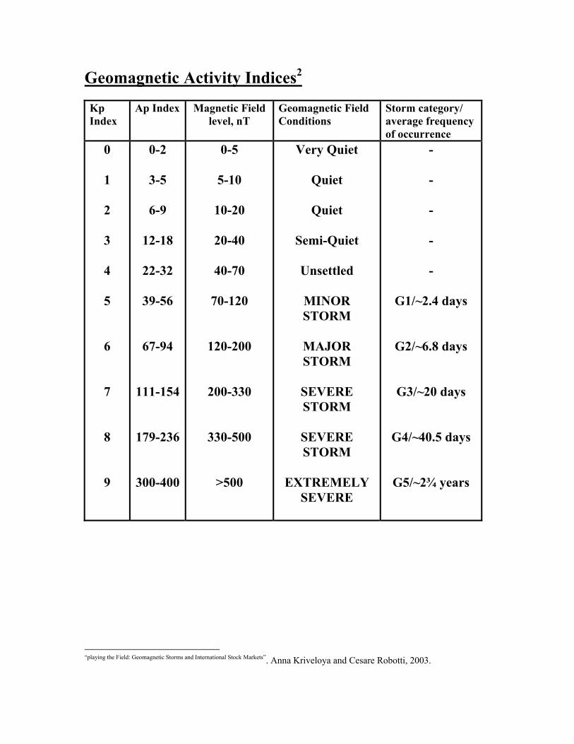
Geomagnetic Activity Indices2 Kp Index
Ap Index Magnetic Field level, nT
Geomagnetic Field Conditions
Storm category/ average frequency of occurrence
0
1 2 3 4 5 6 7 8 9
0-2
3-5
6-9
12-18
22-32
39-56
67-94
111-154
179-236
300-400
0-5
5-10
10-20
20-40
40-70
70-120
120-200
200-330
330-500
>500
Very Quiet
Quiet
Quiet
Semi-Quiet
Unsettled
MINOR STORM
MAJOR STORM
SEVERE STORM
SEVERE STORM
EXTREMELY
SEVERE
- - - - -
G1/~2.4 days
G2/~6.8 days
G3/~20 days
G4/~40.5 days
G5/~2¾ years
“playing the Field: Geomagnetic Storms and International Stock Markets”. Anna Kriveloya and Cesare Robotti, 2003.

3The subscript “p” means planetary and designates a global magnetic activity index. The K Index is a three hour range index. K indices isolate solar particle effects on the earth’s magnetic field; over a 3-hour period, they classify into disturbance levels the range of variation of the more unsettled horizontal field component. Each activity level relates almost logarithmically to its corresponding disturbance amplitude. Three-hour indices discriminate conservatively between true magnetic field perturbations and the quiet-day variations produced by ionospheric currents. K indices range in 28 steps from 0 (quiet) to 9 (greatly disturbed) with fractional parts expressed in thirds of a unit. A k-value equal to 27, for example, means 2 and 2/3 or 3-; a K-value equal to 30 means 3 and 0/3 or 3 exactly; and a K-value equal to 33 means 3 and 1/3 or 3+. The arithmetic mean of the K values scaled at 13 observatories globally gives Kp. Equivalent Amplitude- The a-index ranges from 0 to 400 and represents a K-value converted to a linear scale in gammas (nanoTeslas) – a scale that measures equivalent disturbance amplitude of a station at which K=9 has a lower limit of 400 gammas. aa index.4 A daily and half daily index of geomagnetic activity determined from the k indexes scaled at two nearly antipodal stations at invariant magnetic latitude 50 degrees (Hartland, England, and Canberra, Australia). The aa values are in units of 1 nT. The index is available back to 1868, and is provided by the Institut de Physique du Globe de Paris, France. Ak index5. A daily index of geomagnetic activity for a specific station or network of stations (represented generically here by k) derived as the average of the eight 3-hourly ak indexes in a Universal Time day.
3 ftp://ftp.ngdc.noaa.gov/STP.GEOMAGNETIC_DATA/INDICES/KP_AP/ 4 http://www.saunalahti.fi/fmbb/astro/indices.htm 5 http://www.sci.fi/~fmbb/astro/indices.htm

National Oceanic & Atmospheric Association Space Weather Scale for Geomagnetic Storms 6
Category Effect (some or all of these are possible)
Physical measure
Average Frequency (1 cycle = 11 yr)
G 5
Extreme Power systems: grid systems can collapse and transformers experience damage. Spacecraft operations: extensive surface charging, problems with orientation, uplink/downlink, and tracking satellites. Other systems: pipeline currents reach hundreds of amps, HF (high frequency) radio propagation impossible in many areas for one to two days, satellite navigation degraded for days, low-frequency radio navigation out for hours, and the aurora seen as low as the equator.
Kp = 9
4 storm events per cycle at this Kp level (4 storm days per cycle)
G 4
Severe Power systems: possible voltage stability problems, portions of grids collapse and protective devices trip. Spacecraft operations: experience surface charging and tracking problems, orientation problems need corrections. Other systems: induced pipeline currents affect preventive measures, HF radio propagation sporadic, satellite navigation degraded for hours, low-frequency radio navigation disrupted, and the aurora seen as low as the tropics.
Kp = 8, including a 9-
100 per cycle (60 days per cycle)
G Strong Power systems: voltage Kp = 7 200 per
6 http://geology.about.com/library/bl/blgeomagstormscale.htm

3 corrections required, false alarms triggered on protection devices, and high ‘gas-in-oil’ transformer readings likely. Spacecraft operations: surface charging on satellite components, increased drag on satellite, and orientation problems need corrections. Other systems: intermittent satellite navigation and low-frequency radio navigation problems, HF radio intermittent, and the aurora seen as low as mid-latitudes.
cycle (130 days per cycle)
G 2
Moderate Power systems: high-latitude power systems affected. Spacecraft operations: corrective actions are required by ground control; changes in drag affect orbit predictions. Other systems: HF radio propagation fades at higher latitudes, and the aurora seen as low as 50 degrees.
Kp = 6 600 per cycle (360 days per cycle)
G 1
Minor Power systems:weak power grid fluctuations. Spacecraft operations: minor impact on satellite operations. Other systems: the aurora seen at high latitudes (60 degrees); migratory animals begin to be affected.
Kp = 5 1700 per cycle (900 days per cycle)
References ALDRICH, T. E., ANDREWS, K. W. AND LIBOFF, A. R., 2001. Brain cancer risk and electromagnetic fields (EMFs): Assessing the geomagnetic component. Archives of Environmental Health, 56 (4) , 314-319 BARTSCH, H., BARTSCH, C., MECKE, D. AND LIPPERT, T. H., 1994. Seasonality of pineal melatonin production in the rat: Possible synchronization by the geomagnetic field. Chronobiology International, 11 (1), 21-26. BELOV, D. R., KANUNIKOV, I. E. AND KISELEV B. V., 1998. Dependence of Human EEG spatial syncrhonization on the Geomagnetic Activity on the Day of Experiment. [article in Russian]. Ross Fiziol Zh Im I M Sechenova, 84 (8), 761-774. BERGIANNAKI, J.-D., PAPARRIGOPOULOS, T. J. AND STEFANIS, C. N., 1996. Seasonal pattern of melatonin excretion inhumans: relationship to day length variation rate and geomagnetic field fluctuations. Experientia, 52, 253-258. BURCH, J. B., REIF, J. S. AND YOST, M. G., 1999. Geomagnetic disturbances are associated with reduced nocturnal excretion of a melatonin metabolite in humans. Neuroscience Letters, 266, 209-212. CHERNOUSS, S., VINOGRADOV, A. AND VLASSOVA, E., 2001. Geophysical Hazard for Human Health in the Circumpolar Auroal Belt: Evidence of a Relationship between Heart Rate Variation and Electromagnetic Disturbances. CREMER-BARTELS, G., KRAUSE, K. AND KÜCHLE, H. J., 1983. Influence of low magnetic-field-strength variations on the retina and pineal gland of quail and humans. Graefe’s Arch Clin Exp Ophthalmol, 220, 248-252. DIMITROVA, S. AND STOILOVA, I., 2002. Human Physiological Reaction to Geomagnetic Disturbances of Solar Origin. Proc.10th European Solar Physics Meeting,’Solar Variability:from Core to Outer Frontiers’, Prague, Czech Republic, 0-14 September 2002. DIMITROVA, S., STOILOVA, I. AND CHOLAKOV, I., 2004. Influence of local Geomagnetic Storms on Arterial Blood Pressure. Bioelectromagnetics, 25, 408-414. DUPONT, M. J., PARKER, G., AND PERSINGER, M. A., 2005. Brief Communication: reduced litter sizes following 48-h of prenatal exposure to 5 nT to 10 nT, 0.5 Hz magnetic fields: implications for sudden infant deaths. Intern. J. Neuroscience, 115:713-715. ECKERT, E. E., 1992. Magnetic influences on fetus and infant as reason for Sudden Infant Death Syndrome: A new testable hypothesis. Medical Hypotheses, 38, 66-69.

FEIGIN, V. L., NIKITIN, YU, P. AND VINOGRADOVA, T. E., 1997. Solar and geomagnetic activities: Are there associations with stroke occurrence? Cerebrovascular Diseases, 7, 345-348. GOLDWATER, P. N., 2003. Sudden Infant Death Syndrome: a critical review of approaches to research. Arch. Dis. Child., 88: 1095-1100. GORDON, C. AND BERK, M., 2003. The effect of geomagnetic storms on suicide. S Afr Psychiatry Rev 6, 24-27. GRAINGER, P., WIGFIELD, R., WRIGHT, M., FLEMING, P.J., AND PREECE, A.W., 2000. Electric and magnetic fields of 50Hz are not associated with sudden infant death syndrome. International Journal of Environmental Health Research, 10, 85–87. JUUTILAINEN, J., AND KUMLIN, T., 2006. Occupational Magnetic Field Exposure and Melatonin: Interaction With Light-at-Night. Bioelectromagnetics, brief communciation, early view. KAY, R. W., 1994. Geomagnetic Storms: Association with Incidence of Depression as Measured by Hospital Admissions. Brit J Psychiatr, 164: 403-409. KRIVELYOVA, A. AND ROBOTTI, C., 2003. Federal Reserve Bank of Atlanta, Working Paper 2003-05. Playing the field: Geomagnetic storms and international stock markets. LEACH, C. E. A., BLAIR, P. S., FLEMING, P. J., SMITH, I. J., PLATT, M. W., BERRY, P. J., FRCP, BCH,, GOLDING, J., AND THE CESDI SUDI RESEARCH GROUP, 1999. Epidemiology of SIDS and Explained Sudden Infant Deaths. Pediatrics, 104: e43- LIBOFF, A.R. AND MCLEOD, B. R., 1995. Power Lines and the Geomagnetic Field. Bioelectromagnetics, 16(4):227-230. O’CONNOR, R. P. AND PERSINGER, M. A., 1997. Geophysical variables and behavior: LXXXII. A strong association between sudden infant death syndrome and increments of global geomagnetic activity – possible support for the melatonin hypothesis. Perceptual and Motor Skills, 84, 395-402. OTSUKA, K., OINUMA, S., CORNÉLISSSEN, G., WEYDAHL, A., ICHIMARU, Y., KOBAYSHI, M., YANO, S., HOLMESLET, B., HANSEN, T., L., MITSUTAKE, G., ENGEBRETSON, M. J., SCHWARTZKOPFF, O. AND HALBERG, F., 2001. Alternating light-darkness-influenced human electrocardiographic magnetoreception in association with geomagnetic pulsations. Biomed Pharmacother, 55: 63-75. OTTO,W., HEMPEL, W. E., WAGNER, C. U. AND BEST, A., 1982. [Various periodical and aperiodical variations of heart infarct mortality in the DRG - Article in German]. Z Gesamte Inn Med Nov 15;37(22):756-763

PARTONEN, T., HAUKKA, J., NEVANLINNA, H. AND LONNQVIST J., 2004. Analysis of the seasonal pattern in suicide. J Affect Disord, 81(2), 133-139. PERSINGER, M. A., 1987. geopyschology and geopyschopathology. Experientia 43 92-104 PERSINGER, M. A. AND PSYCH, C., 1995. Sudden unexpected death in epileptics following sudden, intense, increases in geomagnetic activity: prevalence of effect and potential mechanisms. Int. J. Biometeorol, 38, 180-187. PERSINGER, M. A. AND O’CONNOR, R. P., 2001. Geophysical variables and behavior: CIII. Days with sudden infant deaths and cardiac arrhythmias in adults share a factor with PC1 geomagnetic pulsations: Implications for pursuing mechanism. Perceptual and Motor Skills, 92, 653-654. PERSINGER, M. A., MCKAY, B. E., O’DONOVAN, C. A. AND KOREN, S. A., 2005. Sudden death in epileptic rats exposed to nocturnal magnetic fields that simulate the shape and the intensity of sudden changes in geomagnetic activity: an experiment in response to Schnabel, Beblo and May. Int J Biometeorol, 49: 256-261. RAPS, A., STOUPEL, E. AND SHIMSHANI, M., 1991. “Solar Activity and admissions of psychiatric inpatients, relations and possible implications on seasonality”. Isr J Psychiatry Relat Sci. 28 (2): 50-59. SPARKS, D. L. AND HUNSAKER, J. C., 1988. The pineal gland in sudden infant death syndrome: preliminary observations. Journal of Pineal Research, 5, 111-118. SPARKS, D. L., COYNE, C. M., SPARKS, L. M., AND HUNSAKER, J. C., 1997. Recommended Technique for Brain Removal to Retain Anatomic Integrity of the Pineal Gland in Order to Determine Its Size in Sudden Infant Death Syndrome. Journal Of Forensic Sciences, 42(1), 100-102. SPARKS, D. L. AND HUNSAKER, J. C., 2002. Neuropathology of sudden infant death (syndrome): literature review and evidence of a probable apoptotic degenerative cause. Chidls Nerv Syst, 18, 568-592. SRIVASTAVA, B. J. AND SAXENA, S., 1980. Geomagnetic-biological correlations - Some new results. Indian Journal of Radio and Space Physics. Vol. 9, pp. 121-126. Aug. 1980 STURNER, W. Q., LYNCH, H. J., DENG, M. H., GLEASON, R. E. AND WURTMAN, R. J., 1990. Melatonin concentrations in the sudden infant death syndrome. Forensic Science International, 45, 171-180. THOSS, F., BARTSCH, B., FRITZSCHE, B., TELLSCHAFT, D. AND THOSS, M., 2000. The magnetic field sensitivity of the human visual system shows resonance and compass characteristic. J. Comp. Physiol A, 186, 1007-1010.

THOSS, F., BARTSCH, B., TELLSCHAFT, D. AND THOSS, M., 2002. The light sensitivity of the human visual system depends on the direction of view. J. Comp. Physiol A, 188, 235-237. THOSS, F. AND BARTSCH, B., 2003. The human visual threshold depends on direction and strength of a weak magnetic field, Journal of Comparative Physiology A: Sensory, Neural, and Behavioral Physiology, Volume 189, Issue 10, Oct 2003, Pages 777 – 779 WEISSBLUTH, L. AND WEISSBLUTH, M., 1994. Sudden Infant Death Syndrome: a Genetically Determined Impaired Maturation of the Photoneuroendocrine System. A unifying hypothesis. J. theor. Biol., 167, 13-25. WEVER, R., 1979. The Circadian System of Man – Results of Experiments Under Temporal Isolation. Springer-Verlag – New York. WEYDAHL, A., SOTHERN, R. B., CORNÉLISSEN, G. AND WETTERBERG, L., 2001. Geomagnetic activity influences the melatonin secretion at latitude 70º N. Biomed. Pharmacother, 55: 57-62. YAGA, K., REITER, R. J., MANCHESTER, L., C., NIEVES, H., SUN, J-H. AND CHEN, L-D., 1993. Pineal Sensitivity to Pulsed Static Magnetic Fields Changes During the Photoperiod. Brain Research Bulletin, 30, 153-156.


