Phenotypic and Genotypic Characterization of Acinetobacter ...
The Egyptian Journal of Medical Human Genetics (2013) 14, 235–246
Ain Shams University
The Egyptian Journal of Medical Human Genetics
www.ejmhg.eg.netwww.sciencedirect.com
ORIGINAL ARTICLE
Genotypic and phenotypic patterns of antimicrobial
susceptibility of Helicobacter pylori strains among Egyptian
patients
Marwa S. Fathia,1,*, Runia Fouad EL-Folly
b,1, Rania Ahmed Hassan
a,1,
Mohammed Ezz El-Arab c,1
a Medical Microbiology & Immunology Department, Faculty of Medicine, Ain Shams University, Egyptb Tropical Medicine Department, Faculty of Medicine, Ain Shams University, Egyptc Ahmed Maher Educational Hospital, Egypt
Received 29 January 2013; accepted 20 March 2013Available online 22 April 2013
*
og
Te
E-1
Fo
an
Fa
th
H
Pe
11
ht
KEYWORDS
H. pylori;
Gastritis;
Antimicrobial susceptibility
testing;
E-test;
PCR;
Antimicrobial resistance
Corresponding author. Addr
y, Faculty of Medicine, Ain
l.: +20 1001475922.
mail address: dr.marwasaad@
Author contributions: Mar
lly designed the research; Ma
d Mohammed Ezz El-Arab
thi and Rania Ahmed Hass
e data, Marwa Saad Fathi, R
assan wrote the paper.
er review under responsibilit
Production an
10-8630 � 2013 Production
tp://dx.doi.org/10.1016/j.ejmh
ess: Med
Shams U
gmail.c
wa Saad
rwa Saad
performe
an contri
unia Fo
y of Ain
d hostin
and hosti
g.2013.0
Abstract Helicobacter pylori (H. pylori) is currently recognized as one of the most common
chronic bacterial infections worldwide. Eradication of bacteria is effective in healing peptic ulcers,
preventing ulcer relapses, and potentially decreasing the risk of progression to gastric carcinoma.
For successful eradication of bacteria, it is imperative that the clinician be aware of the current anti-
microbial susceptibility profiles of isolates within the region. Therefore, the aim of this study is to
compare the phenotypic and genotypic patterns of antibiotics’ susceptibility to H. pylori strains
among Egyptian patients.
60 symptomatic cases were enrolled. H. pylori infection was diagnosed by upper endoscopy as
well as biopsy. Antimicrobial susceptibility to H. pylori strains was assessed in all subjects by disc
diffusion and Ellipsometer testing (E-testing) methods. Further molecular characterization of genes
ical Micrbiology & Immunol-
niversity, Cairo 11341, Egypt.
om (M.S. Fathi).
Fathi and Runia Fouad EL-
Fathi, Rania Ahmed Hassan
d the research. Marwa Saad
buted analytic tools; analysed
uad EL-Folly& Rania Ahmed
Shams University.
g by Elsevier
ng by Elsevier B.V. on behalf of Ain Shams University.
3.004
236 M.S. Fathi et al.
encoding antimicrobial resistance of isolated strains was done by the polymerase chain reaction
(PCR).
For metronidazole and ciprofloxacin, we compared the phenotypic and genotypic patterns of
resistance as detected by PCR amplification of the resistance genes. Resistance rates by E-test were
100% and 25% for metronidazole and ciprofloxacin respectively from 16 isolated H. pylori strains.
Improving the knowledge of resistance mechanisms, the elaboration of rational and efficacious
associations for the treatment H. pylori infection are of high importance especially in determining
the therapeutic outcome. Further progress should ultimately focus on the establishment of a cheap,
feasible and reliable laboratory test to predict the outcome of a therapeutic scheme.
� 2013 Production and hosting by Elsevier B.V. on behalf of Ain Shams University.
1. Introduction
Helicobacter pylori (H. pylori) infection, which affects half ofthe world’s population, is responsible for gastritis [1], peptic ul-cers [2,3] and gastric mucosa-associated lymphoid tissue lym-
phoma [4], and is a major risk factor for the development ofgastric adenocarcinoma [5]. Eradication treatment of H. pyloriinfection usually consists of various combinations of drugs.Most commonly, an acid suppressor (usually a proton pump
inhibitor) or a histamine receptor (H2-receptor) antagonist(e.g. ranitidine) is prescribed in combination with two antibiot-ics usually amoxicillin, metronidazole or clarithromycin. The
combination of two antibiotics can increase the success oferadication therapy and decrease the possibility of secondaryantibiotic resistance [6,7].
Antibiotic resistance in H. pylori is the major cause of erad-ication failure. Growing resistance often parallels the patternsof antibiotic consumption, and may vary within patient groups
according to the geographic region, patient age and sex, typeof disease, birthplace and the presence of other infections.The geographic map and the process of primaryH. pylori resis-tance are clinically important, and should be considered when
choosing eradication regimens, as is constant monitoring atboth national and global level in an attempt to reach the re-cently recommended goal of eradicating more than 95% of
resistant cases [8]. Different mechanisms of resistance to clari-thromycin, metronidazole, quinolones, amoxicillin and tetra-cycline are accurately detailed (point mutations, redox
intracellular potential, pump efflux systems, membrane perme-ability) on the basis of the most recent data available from theliterature [9].
The prevalence of clarithromycin, metronidazole and
amoxicillin resistance varies between countries and is highestfor metronidazole [10]. Resistance to tetracycline and cipro-floxacin has been reported by several studies but yet appears
uncommon [11,12].The activation of metronidazole is mediated by the pyru-
vate: ferredoxin oxidoreductase complex. For example, this
function in H. pylori might be fulfilled by the electron carriers,RdxA (HP0954), an oxygen-insensitive NADPH nitroreduc-tase, FrxA (HP0642), a NAD(P)H-flavin oxidoreductase, fer-
redoxin (FdxA, HP0277), flavodoxin (FldA, HP1161),pyruvate: ferredoxin oxidoreductase (PorD, HP1109) and 2-oxoglutarate ferredoxin oxidoreductase (OorD, HP0588).Inactivation of the genes involved in some of these systems
has been found to be linked to metronidazole resistance [13].There are several problems with antimicrobial susceptibility
testing of H. pylori [14,15]. Agar or broth dilution methods are
difficult to perform routinely [16], thus, disc diffusion testing isoften used because it is simple, easy to perform, and econom-
ical [17]. However, the E-test has proved to be an accuratemethod for assaying the susceptibility of fastidious organisms,including H. pylori, to antibiotics. The E-test has a more stable
pattern of antibiotic release and has been found to tolerateprolonged incubation [18]. This is the main reason why theE-test rather than the disc diffusion method, has been recom-
mended for H. pylori [19].With the increasing frequency of clarithromycin resistance
among H. pylori strains, there is rising concern about the po-
tential decline in the eradication rate of this infection [20,21].There is therefore an urgent need to introduce other treatmentoptions. Fluoroquinolones, such as ciprofloxacin (CIP), levo-floxacin (LVX) and moxifloxacin (MOX), have been evaluated
as an alternative to standard antibiotics against H. pylori [6,7].Some studies have shown good results when using fluoro-
quinolone based triple therapies for H. pylori eradication. In
a German study, a 7 day course including LVX in patientswith persistent H. pylori infection resulted in eradication ratesof greater than 85% [22]. Similarly, an Italian study clarified
that H. pylori eradication was achieved in 90% of patientstreated with MOX, clarithromycin and lansoprazole [23].However, the widespread use of fluoroquinolones for the treat-
ment of H. pylori infection has led to an increase in its resis-tance rate in some areas, leading to unacceptably loweradication rates [10]. Several studies have shown that LVXbased therapies are not superior to the traditional quadruple
therapy or triple therapy in the treatment of H. pylori infec-tion, especially in the case of resistant H. pylori strains [24,25].
On the other hand a Turkish study speculated that the low
eradication rate with MOX containing treatment regimensmay be due to the development of resistance to this quinolone[26]. The findings from all of these studies indicate that a reg-
imen that is effective in one area may not be effective in an-other area, as antibiotic resistant rates for H. pylori may bedifferent in different areas [27].
The mechanism of action of fluoroquinolones is via inhibi-
tion of deoxyribonucleic acid (DNA) gyrase and topoisomer-ase, which control and modify the topological state of DNAin cells. Fluoroquinolone then interferes with bacterial DNA
replication. Both DNA gyrase and topoisomerase are com-posed of two A and two B subunits, encoded by the gyrAand gyrB genes for DNA gyrase and the parC and parE genes
for topoisomerase. The mechanism of fluoroquinolone resis-tance in H. pylori has been found to be linked to mutationsin the quinolone resistance determining regions (QRDRs) of
the gyrA gene. Mutations in the gyrB gene have also been re-
Genotypic and phenotypic patterns of antimicrobial susceptibility of Helicobacter pylori 237
ported in LVX resistant strains isolated in Japan, but these of-ten occurred along with gyrA mutations [8].
In vitro susceptibility testing for H. pylori can be performed
either by phenotypic or genotypic methods. Phenotypic testingis challenging because the organism grows slowly even underoptimal culture conditions. Owing to these difficulties and be-
cause antibiotic resistance in this microorganism is essentiallydue to point mutations, genotypic methods are an alternativeto the phenotypic methods. Moreover, these methods offer
the advantage of testing directly from biopsy material, allow-ing a faster response.
2. Aim of the study
The aim of this study is to compare the phenotypic and geno-typic patterns of antibiotics’ susceptibility to H. pylori strains
among Egyptian patients in order to attain a clinical utilityfrom such patterns.
3. Subjects and methods
3.1. Study design and sampling
This cross-sectional study was conducted on 60 cases with epigas-tric pain and/or vomiting attended to the outpatient clinics and/orthe endoscopy centre of AhmedMaherHospital during the period
fromMay2011 to January 2012.The sample sizewas calculated byEpi Info program (version 6.0) at 95%ConfidenceLimit, Power ofthe Test is 80% and Alpha Set at 0.05 (Type I error).
3.1.1. Inclusion criterion
Patients with epigastric pain and/or vomiting.
3.1.2. Exclusion criteria
Patients who refused to be enrolled in the study, patients who
previously underwent sclerotherapy or band ligation ofoesophageal varices (EV), patients taking drugs for primaryprophylaxis of variceal bleeding, patients with chronic liverdisease and/or hepatocellular carcinoma, patients with portal,
splenic or hepatic vein thrombosis and patients with severe car-diac, chest or renal disease.
3.2. Tools of the study
All patients were subjected to a complete clinical evaluation andlaboratory investigations: including: complete blood count
(CBC), liver profile and hepatitis markers: Hepatitis B surfaceantigen (HBs Ag) and Hepatitis C virus antibody (HCV Ab)using the third generation enzyme-linked immunosorbent assay(ELISA) test (Franciscus, 2002 and[29]). Abdominal ultrasonog-
raphy was done using Toshiba ‘‘Just vision’’ real-time scannerinstrument with a 3.5 MHz convex transducer (after an over-night fasting) with stress on: liver size and liver echogenicity.
Upper gastrointestinal (GI) endoscopy and endoscopic guidedbiopsy were done to exclude the presence of varices in additionto any relevant upper GI masses and to evaluate the degree of
gastritis or the presence of ulcers using Pentax EG-3440 video-scope system. The endoscopic study was performed by the sameexaminer in all patients to avoid interobserver variability [30].
Gastric biopsies were collected on phosphate buffered saline
(PBS) in sterile eppendorf tubes. Some patients had receivedH. pylori eradication therapy before, but none of the patientshad received MOX or CIP based therapy [30].
3.3. Identification and storage of H. pylori strains
The biopsy specimens were cultured on Colombia blood agar
(BBL Microbiology Systems, Cockeysville, MD, USA), sup-plemented with 8% defibrinated sheep blood, and incubatedfor 5–7 days under microaerobic conditions (5% O2, 10%
CO2, 85% N2) [30].Clinical isolates were identified as H. pylori based on mor-
phology and positive biochemical tests for catalase, oxidase,
and confirmed by the rapid urease production test and gramstaining.
Sixteen pure isolates were identified as H. pylori. They werestored at �80 �C in brain–heart infusion broth (BHI, Difco
Laboratory, Detroit, MI, USA) supplemented with 30%glycerol.
3.4. Antimicrobial susceptibility to H. pylori strains
3.4.1. Antimicrobial susceptibility testing by disc diffusion
method
For in vitro susceptibility testing of the H. pylori strains, a sus-pension equal to the McFarland tube No. 3 was prepared foreach isolate. We used only one colony from each isolate for the
analysis. Brain heart infusion broth (Merck, Germany) plates,supplemented with sheep red blood cells (RBCs) were inocu-lated by confluent swabbing of the surface with the adjusted
inoculum suspensions.The antimicrobial discs (Mast Diagnostics; Mast Group
Limited, Mereyside, UK) for the antibiotics amoxicillin, met-
ronidazole, tetracycline, ciprofloxacin, and clarithromycin,were aseptically placed onto the dried surface of inoculatedMuller Hinton’s agar plates supplemented with sheep RBCs.
The plates were then incubated at 37 �C under microaerobicconditions [31]. The zones of inhibition were read after 24–48 h of incubation and the susceptibility results were recordedas resistant on the basis of the Clinical and Laboratory Stan-
dards Institute (CLSI) guidelines and the manufacturer’sinstructions [28,31].
3.4.2. Determination of the minimum inhibitory concentrations(MIC)
The E-test strips (Liofilchem, Italy) for the antibiotics amoxi-cillin, metronidazole, tetracycline, ciprofloxacin, and clarithro-
mycin were aseptically placed onto the dried surface ofinoculated agar plates. The plates were then incubated at37 �C under microaerobic conditions. The minimum inhibitory
concentrations (MICs) were read after 48–72 h of incubationon the basis of the intersection of the elliptical zone of growthinhibition using the MIC scale on the E-test strip, and the sus-
ceptibility results were recorded as resistant according to therecommendations of CLSI (Pennsylvania, USA) and the man-ufacturer’s instructions [28,31].
A polymerase chain reaction (PCR) was performed for the
16 isolates to detect Ure C gene (a 1335 base pair (bp) longopen reading frame upstream from the urease structural genes(ureAB) of H. pylori whose product is important for cell
growth) for confirmation of the identification of H pylori. An-
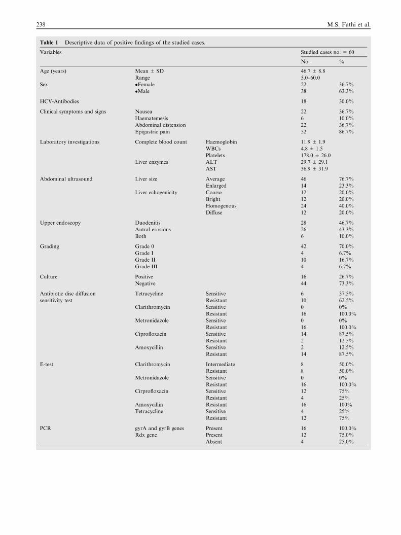
Table 1 Descriptive data of positive findings of the studied cases.
Variables Studied cases no. = 60
No. %
Age (years) Mean ± SD 46.7 ± 8.8
Range 5.0–60.0
Sex �Female 22 36.7%
�Male 38 63.3%
HCV-Antibodies 18 30.0%
Clinical symptoms and signs Nausea 22 36.7%
Haematemesis 6 10.0%
Abdominal distension 22 36.7%
Epigastric pain 52 86.7%
Laboratory investigations Complete blood count Haemoglobin 11.9 ± 1.9
WBCs 4.8 ± 1.5
Platelets 178.0 ± 26.0
Liver enzymes ALT 29.7 ± 29.1
AST 36.9 ± 31.9
Abdominal ultrasound Liver size Average 46 76.7%
Enlarged 14 23.3%
Liver echogenicity Coarse 12 20.0%
Bright 12 20.0%
Homogenous 24 40.0%
Diffuse 12 20.0%
Upper endoscopy Duodenitis 28 46.7%
Antral erosions 26 43.3%
Both 6 10.0%
Grading Grade 0 42 70.0%
Grade I 4 6.7%
Grade II 10 16.7%
Grade III 4 6.7%
Culture Positive 16 26.7%
Negative 44 73.3%
Antibiotic disc diffusion
sensitivity test
Tetracycline Sensitive 6 37.5%
Resistant 10 62.5%
Clarithromycin Sensitive 0 0%
Resistant 16 100.0%
Metronidazole Sensitive 0 0%
Resistant 16 100.0%
Ciprofloxacin Sensitive 14 87.5%
Resistant 2 12.5%
Amoxycillin Sensitive 2 12.5%
Resistant 14 87.5%
E-test Clarithromycin Intermediate 8 50.0%
Resistant 8 50.0%
Metronidazole Sensitive 0 0%
Resistant 16 100.0%
Cirprofloxacin Sensitive 12 75%
Resistant 4 25%
Amoxycillin Resistant 16 100%
Tetracycline Sensitive 4 25%
Resistant 12 75%
PCR gyrA and gyrB genes Present 16 100.0%
Rdx gene Present 12 75.0%
Absent 4 25.0%
238 M.S. Fathi et al.
Genotypic and phenotypic patterns of antimicrobial susceptibility of Helicobacter pylori 239
other PCR was performed for the confirmed strains for detec-tion of the DNA gyrase (gyrA and gyrB) genes responsible forresistance to fluoroquinolones and also the rdx gene responsi-
ble for the resistance to metronidazole.
3.5. Polymerase chain reaction (PCR) procedure
3.5.1. Extraction of DNA
1.5 ml of broth culture of each isolate was taken for extraction
of DNA. Extraction was performed using QIAamp� DNAMINI KIT (QIAGEN, USA) (Catalog No. 51304) accordingto the manufacturer’s instructions.
3.5.2. Amplification of the extracted DNA
Amplification of the eluted DNAwas performed in a 50 ll reac-tion volume for each of the four detected genes. Each PCR mix
consisted of 25 ll of 2x QIAGEN HotStar Taq� Master Mix
Containing HotStarTaq� DNA Polymerase, PCR buffer (with3 mM MgCl2), and 400 lM each dNTP + 10 ll of the elutedDNA + 1 ll of each of the 2 primers (50 pM) of the gene to
be detected + 13 ll distilled water.Primers were synthesized by (Roche, Germany).Amplification of DNA was performed in a thermal cycler
(thermo PxE 0.21, England).
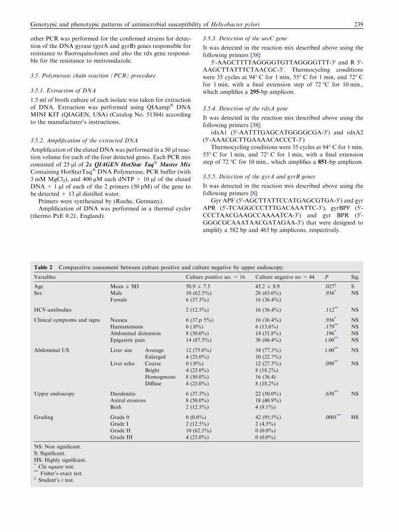
Table 2 Comparative assessment between culture positive and cult
Variables Culture
Age Mean ± SD 50.9 ±
Sex Male 10 (62.
Female 6 (37.5
HCV-antibodies 2 (12.5
Clinical symptoms and signs Nausea 6 (37.p
Haematemesis 0 (.0%
Abdominal distension 8 (50.0
Epigastric pain 14 (87.
Abdominal US Liver size Average 12 (75.
Enlarged 4 (25.0
Liver echo Coarse 0 (.0%
Bright 4 (25.0
Homogenous 8 (50.0
Diffuse 4 (25.0
Upper endoscopy Duodenitis 6 (37.5
Antral erosions 8 (50.0
Both 2 (12.5
Grading Grade 0 0 (0.0%
Grade I 2 (12.5
Grade II 10 (62.
Grade III 4 (25.0
NS: Non significant.
S: Significant.
HS: Highly significant.* Chi square test.** Fisher’s exact test.� Student’s t test.
3.5.3. Detection of the ureC gene
It was detected in the reaction mix described above using the
following primers [38]:50-AAGCTTTTAGGGGTGTTAGGGGTTT-30 and R 50-
AAGCTTATTTCTAACGC-30. Thermocycling conditions
were 35 cycles at 94� C for 1 min, 55� C for 1 min, and 72� Cfor 1 min, with a final extension step of 72 �C for 10 min.,which amplifies a 295-bp amplicon.
3.5.4. Detection of the rdxA gene
It was detected in the reaction mix described above using thefollowing primers [38]:
rdxA1 (50-AATTTGAGCATGGGGCGA-30) and rdxA2(50-AAACGCTTGAAAACACCCT-30)
Thermocycling conditions were 35 cycles at 94� C for 1 min,
55� C for 1 min, and 72� C for 1 min, with a final extensionstep of 72 �C for 10 min., which amplifies a 851-bp amplicon.
3.5.5. Detection of the gyrA and gyrB genes
It was detected in the reaction mix described above using thefollowing primers [8]:
Gyr APF (50-AGCTTATTCCATGAGCGTGA-30) and gyr
APR (50-TCAGGCCCTTTGACAAATTC-30), gyrBPF (50-CCCTAACGAAGCCAAAATCA-30) and gyr BPR (50-GGGCGCAAATAACGATAGAA-30) that were designed toamplify a 582 bp and 465 bp amplicons, respectively.
ure negative by upper endoscopy.
positive no. = 16 Culture negative no = 44 P Sig.
7.5 45.2 ± 8.9 .027� S
5%) 28 (63.6%) .936* NS
%) 16 (36.4%)
%) 16 (36.4%) .112** NS
5%) 16 (36.4%) .936* NS
) 6 (13.6%) .179** NS
%) 14 (31.8%) .196* NS
5%) 38 (86.4%) 1.00** NS
0%) 34 (77.3%) 1.00** NS
%) 10 (22.7%)
) 12 (27.3%) .098** NS
%) 8 (18.2%)
%) 16 (36.4)
%) 8 (18.2%)
%) 22 (50.0%) .630** NS
%) 18 (40.9%)
%) 4 (9.1%)
) 42 (95.5%) .0001** HS
%) 2 (4.5%)
5%) 0 (0.0%)
%) 0 (0.0%)
240 M.S. Fathi et al.
Thermocycling conditions were 35 cycles at 94� C for 1 min,53� C for 1 min, and 72� C for 1 min, with a final extensionstep of 72 �C for 10 min.
Identification of amplified products by gel electrophoresiswas done as the following [32]:
The amplicons (2 ll) (added to a Loading buffer: bromo-
phenol blue with sucrose) was analysed by electrophoresis on2% agarose gel and ethidium bromide staining in tris–ace-tate-EDTA (TAE) running buffer. They were visualized on a
UV transilluminator (365 wave length). Qiagen gel pilot 1 Kbplus. (cat No. 239045) was used as molecular weight ladder.
3.5.6. Data management and statistical analysis
The collected data were revised, coded, tabulated and intro-duced to a PC using Statistical package for Social Science(SPSS 15.0.1 for windows; SPSS Inc, Chicago, IL, 2001). Quan-
titative non parametric variables are expressed as mean andstandard deviation (SD), while non parametric were expressedas Median and Interquartile range (IQR). Qualitative variables
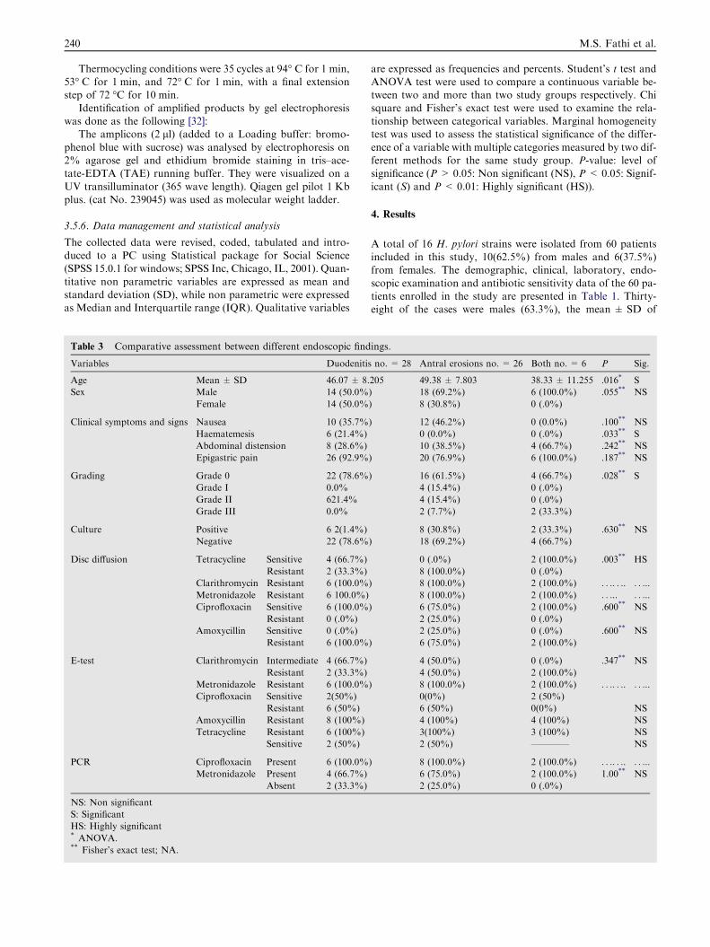
Table 3 Comparative assessment between different endoscopic find
Variables Duodeniti
Age Mean ± SD 46.07 ± 8.
Sex Male 14 (50.0%
Female 14 (50.0%
Clinical symptoms and signs Nausea 10 (35.7%
Haematemesis 6 (21.4%)
Abdominal distension 8 (28.6%)
Epigastric pain 26 (92.9%
Grading Grade 0 22 (78.6%
Grade I 0.0%
Grade II 621.4%
Grade III 0.0%
Culture Positive 6 2(1.4%)
Negative 22 (78.6%
Disc diffusion Tetracycline Sensitive 4 (66.7%)
Resistant 2 (33.3%)
Clarithromycin Resistant 6 (100.0%
Metronidazole Resistant 6 100.0%)
Ciprofloxacin Sensitive 6 (100.0%
Resistant 0 (.0%)
Amoxycillin Sensitive 0 (.0%)
Resistant 6 (100.0%
E-test Clarithromycin Intermediate 4 (66.7%)
Resistant 2 (33.3%)
Metronidazole Resistant 6 (100.0%
Ciprofloxacin Sensitive 2(50%)
Resistant 6 (50%)
Amoxycillin Resistant 8 (100%)
Tetracycline Resistant 6 (100%)
Sensitive 2 (50%)
PCR Ciprofloxacin Present 6 (100.0%
Metronidazole Present 4 (66.7%)
Absent 2 (33.3%)
NS: Non significant
S: Significant
HS: Highly significant* ANOVA.** Fisher’s exact test; NA.
are expressed as frequencies and percents. Student’s t test andANOVA test were used to compare a continuous variable be-tween two and more than two study groups respectively. Chi
square and Fisher’s exact test were used to examine the rela-tionship between categorical variables. Marginal homogeneitytest was used to assess the statistical significance of the differ-
ence of a variable with multiple categories measured by two dif-ferent methods for the same study group. P-value: level ofsignificance (P > 0.05: Non significant (NS), P < 0.05: Signif-
icant (S) and P < 0.01: Highly significant (HS)).
4. Results
A total of 16 H. pylori strains were isolated from 60 patientsincluded in this study, 10(62.5%) from males and 6(37.5%)from females. The demographic, clinical, laboratory, endo-
scopic examination and antibiotic sensitivity data of the 60 pa-tients enrolled in the study are presented in Table 1. Thirty-eight of the cases were males (63.3%), the mean ± SD of
ings.
s no. = 28 Antral erosions no. = 26 Both no. = 6 P Sig.
205 49.38 ± 7.803 38.33 ± 11.255 .016* S
) 18 (69.2%) 6 (100.0%) .055** NS
) 8 (30.8%) 0 (.0%)
) 12 (46.2%) 0 (0.0%) .100** NS
0 (0.0%) 0 (.0%) .033** S
10 (38.5%) 4 (66.7%) .242** NS
) 20 (76.9%) 6 (100.0%) .187** NS
) 16 (61.5%) 4 (66.7%) .028** S
4 (15.4%) 0 (.0%)
4 (15.4%) 0 (.0%)
2 (7.7%) 2 (33.3%)
8 (30.8%) 2 (33.3%) .630** NS
) 18 (69.2%) 4 (66.7%)
0 (.0%) 2 (100.0%) .003** HS
8 (100.0%) 0 (.0%)
) 8 (100.0%) 2 (100.0%) . . .. . .. . . ...
8 (100.0%) 2 (100.0%) . . ... . . ...
) 6 (75.0%) 2 (100.0%) .600** NS
2 (25.0%) 0 (.0%)
2 (25.0%) 0 (.0%) .600** NS
) 6 (75.0%) 2 (100.0%)
4 (50.0%) 0 (.0%) .347** NS
4 (50.0%) 2 (100.0%)
) 8 (100.0%) 2 (100.0%) . . .. . .. . . ...
0(0%) 2 (50%)
6 (50%) 0(0%) NS
4 (100%) 4 (100%) NS
3(100%) 3 (100%) NS
2 (50%) ———— NS
) 8 (100.0%) 2 (100.0%) . . .. . .. . . ...
6 (75.0%) 2 (100.0%) 1.00** NS
2 (25.0%) 0 (.0%)
Genotypic and phenotypic patterns of antimicrobial susceptibility of Helicobacter pylori 241
age was 46.7 ± 8.8 (ranging from 50–60 years), 18% had posi-tive HCV-Antibodies, epigastric pain was the main clinicalpresentation of the enrolled cases (86.7%). Upper endoscopy
revealed duodenitis, antral erosions or both (46.7%, 43.3%,and 10.0%, respectively), 70% was grade 0 of the pathologicalexamination, 73.3% was culture negative. We also compared
between culture positive and culture negative by upper endoscopy
(Table 2). Table 3 shows a comparative assessment between dif-
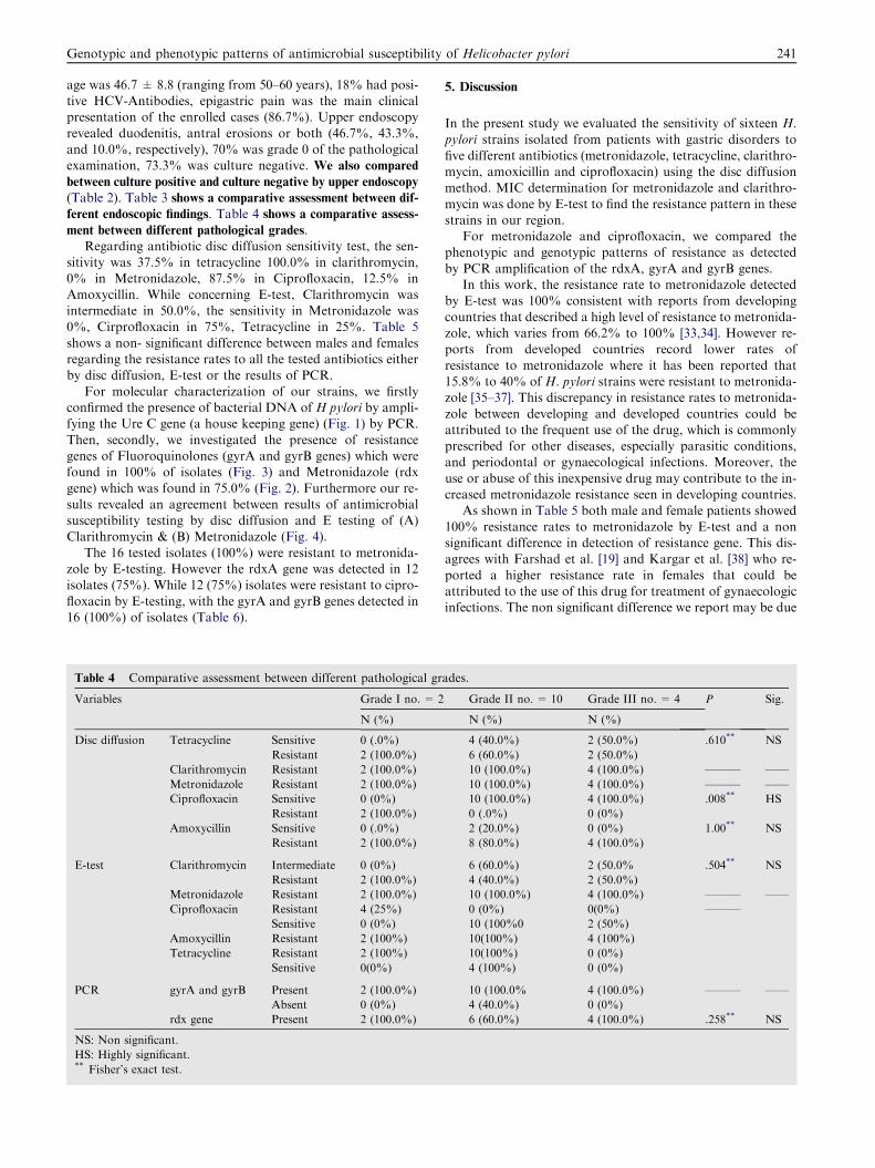
ferent endoscopic findings. Table 4 shows a comparative assess-
ment between different pathological grades.Regarding antibiotic disc diffusion sensitivity test, the sen-
sitivity was 37.5% in tetracycline 100.0% in clarithromycin,0% in Metronidazole, 87.5% in Ciprofloxacin, 12.5% in
Amoxycillin. While concerning E-test, Clarithromycin wasintermediate in 50.0%, the sensitivity in Metronidazole was0%, Cirprofloxacin in 75%, Tetracycline in 25%. Table 5
shows a non- significant difference between males and femalesregarding the resistance rates to all the tested antibiotics eitherby disc diffusion, E-test or the results of PCR.
For molecular characterization of our strains, we firstlyconfirmed the presence of bacterial DNA of H pylori by ampli-fying the Ure C gene (a house keeping gene) (Fig. 1) by PCR.
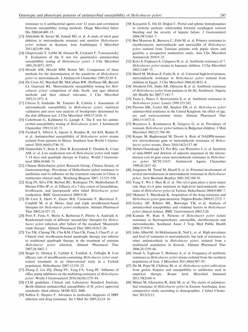
Then, secondly, we investigated the presence of resistancegenes of Fluoroquinolones (gyrA and gyrB genes) which werefound in 100% of isolates (Fig. 3) and Metronidazole (rdxgene) which was found in 75.0% (Fig. 2). Furthermore our re-
sults revealed an agreement between results of antimicrobialsusceptibility testing by disc diffusion and E testing of (A)Clarithromycin & (B) Metronidazole (Fig. 4).
The 16 tested isolates (100%) were resistant to metronida-zole by E-testing. However the rdxA gene was detected in 12isolates (75%). While 12 (75%) isolates were resistant to cipro-
floxacin by E-testing, with the gyrA and gyrB genes detected in16 (100%) of isolates (Table 6).
Table 4 Comparative assessment between different pathological gr
Variables Grade I no. = 2
N (%)
Disc diffusion Tetracycline Sensitive 0 (.0%)
Resistant 2 (100.0%)
Clarithromycin Resistant 2 (100.0%)
Metronidazole Resistant 2 (100.0%)
Ciprofloxacin Sensitive 0 (0%)
Resistant 2 (100.0%)
Amoxycillin Sensitive 0 (.0%)
Resistant 2 (100.0%)
E-test Clarithromycin Intermediate 0 (0%)
Resistant 2 (100.0%)
Metronidazole Resistant 2 (100.0%)
Ciprofloxacin Resistant 4 (25%)
Sensitive 0 (0%)
Amoxycillin Resistant 2 (100%)
Tetracycline Resistant 2 (100%)
Sensitive 0(0%)
PCR gyrA and gyrB Present 2 (100.0%)
Absent 0 (0%)
rdx gene Present 2 (100.0%)
NS: Non significant.
HS: Highly significant.** Fisher’s exact test.
5. Discussion
In the present study we evaluated the sensitivity of sixteen H.pylori strains isolated from patients with gastric disorders to
five different antibiotics (metronidazole, tetracycline, clarithro-mycin, amoxicillin and ciprofloxacin) using the disc diffusionmethod. MIC determination for metronidazole and clarithro-
mycin was done by E-test to find the resistance pattern in thesestrains in our region.
For metronidazole and ciprofloxacin, we compared thephenotypic and genotypic patterns of resistance as detected
by PCR amplification of the rdxA, gyrA and gyrB genes.In this work, the resistance rate to metronidazole detected
by E-test was 100% consistent with reports from developing
countries that described a high level of resistance to metronida-zole, which varies from 66.2% to 100% [33,34]. However re-ports from developed countries record lower rates of
resistance to metronidazole where it has been reported that15.8% to 40% of H. pylori strains were resistant to metronida-zole [35–37]. This discrepancy in resistance rates to metronida-
zole between developing and developed countries could beattributed to the frequent use of the drug, which is commonlyprescribed for other diseases, especially parasitic conditions,and periodontal or gynaecological infections. Moreover, the
use or abuse of this inexpensive drug may contribute to the in-creased metronidazole resistance seen in developing countries.
As shown in Table 5 both male and female patients showed
100% resistance rates to metronidazole by E-test and a nonsignificant difference in detection of resistance gene. This dis-agrees with Farshad et al. [19] and Kargar et al. [38] who re-
ported a higher resistance rate in females that could beattributed to the use of this drug for treatment of gynaecologicinfections. The non significant difference we report may be due
ades.
Grade II no. = 10 Grade III no. = 4 P Sig.
N (%) N (%)
4 (40.0%) 2 (50.0%) .610** NS
6 (60.0%) 2 (50.0%)
10 (100.0%) 4 (100.0%) ——— ——
10 (100.0%) 4 (100.0%) ——— ——
10 (100.0%) 4 (100.0%) .008** HS
0 (.0%) 0 (0%)
2 (20.0%) 0 (0%) 1.00** NS
8 (80.0%) 4 (100.0%)
6 (60.0%) 2 (50.0% .504** NS
4 (40.0%) 2 (50.0%)
10 (100.0%) 4 (100.0%) ——— ——
0 (0%) 0(0%) ———
10 (100%0 2 (50%)
10(100%) 4 (100%)
10(100%) 0 (0%)
4 (100%) 0 (0%)
10 (100.0% 4 (100.0%) ——— ——
4 (40.0%) 0 (0%)
6 (60.0%) 4 (100.0%) .258** NS

Table 5 Rates of antibiotic resistance in Helicobacter pylori isolates in relation to patient gender.
Variables Male no. = 10 Female no. = 6 no. = . . . P Sig.
N (%) N (%)
Disc diffusion Tetracycline Sensitive 2 (20.0%) 4 (66.7%) .118** NS
Resistant 8 (80.0%) 2 (33.3%)
Clarithromycin Resistant 10 (100.0%) 6 (100.0%) ——— ——
Metronidazole Resistant 10 (100.0%) 6 (100.0%) ——— ——
Ciprofloxacin Sensitive 8 (80.0%) 6 (100.0%) .500** NS
Resistant 2 (20.0%) 0 (.0%)
Amoxycillin Sensitive 0 (.0%) 2 (33.3%) .125** NS
Resistant 10 (100.0%) 4 (66.7%)
E-test Clarithromycin Intermediate 4 (40.0%) 4 (66.7%) .608** NS
Resistant 6 (60.0%) 2 (33.3%)
Metronidazole Resistant 10 (100.0%) 6 (100.0%) ——— ——
Ciprofloxacin Sensitive 5 (41%) 7(59%)
Resistant 2 (50%) 2(50%) NS
Amoxycillin Resistant 12 (100%) 4 (100%) NS
Tetracycline Resistant 6 (100%) 6(100%) NS
Sensitive 4(100%) 0(0%) NS
PCR Ciprofloxacin Present 10 (100.0%) 6 (100.0%) ——— ——
Metronidazole Present 8 (80.0%) 4 (66.7%) .604** NS
Absent 2 (20.0%) 2 (33.3%)
NS: Non significant.** Fisher’s exact test.
Table 6 Relationship between genotypic and E-test phenotypic pattern as regards sensitivity against metronidazole, relationship
between genotypic and disc diffusion phenotypic pattern as regards sensitivity against metronidazole and ciprofloxacin.
Variables rdx gene P Sig.
Present = 12 Absent = 4
N % N %
E-test Metronidazole * Sensitive 0 .0 0 .0 ——— ——
Intermediate 0 .0 0 .0
Resistant 12 100.0 4 100.0
** Sensitive 0 .0 0 .0 —— ———
Resistant 12 75.0 4 25.0
gyrA and gyrB genes
Ciprofloxacin *** Sensitive 12 75 0 .0 —— ———
Resistant 4 25 0 .0
242 M.S. Fathi et al.
to the narrow sample size and restriction of sampling from pa-tients who routinely are candidates for upper gastroscopy.
Metronidazole is administered as a pro-drug that is acti-vated by the reduction of the nitro group that is attached toan imidazole ring. Inactivation of an oxygen-insensitive
NADPH nitroreductase (rdxA) may be responsible for metro-nidazole resistance. The activation of metronidazole in strictlyanaerobic bacteria is mediated by the pyruvate: ferredoxin oxi-
doreductase complex. For example, this function in H. pylorimight be fulfilled by the electron carriers, RdxA (HP0954),FrxA (HP0642), ferredoxin (FdxA, HP0277), flavodoxin(FldA, HP1161), pyruvate: ferredoxin oxidoreductase (PorD,
HP1109) and 2-oxoglutarate ferredoxin oxidoreductase(OorD, HP0588) [13].
In this study we investigated the rdxA gene deletion as a
factor of resistance to metronidazole. Table 4 shows that only
25% of isolates were negative for rdxA gene by PCR, although100% of cases were phenotypically resistant to metronidazole
by disc diffusion and E-test methods. This finding goes inalignment with Abdollahi et al. [13] who reported a 22.9% rateof rdxA gene deletion among their 35 tested resistant H. pylori
strains. These results can be explained by the presence of mech-anisms of rdxA inactivation rather than deletion, for examplemutational inactivation which can be identified by further
sequencing of the amplified DNA. Another mechanism thatmay be associated with rdxA gene inactivation is the insertionof a transposon called Mini-IS605 [39]. It has also been dem-onstrated that inactivation of other genes such as the frxA gene
also confers metronidazole resistance, either alone or in asso-ciation with the rdxA gene [40,41].
The present study revealed a high resistance rate to amox-
icillin by E-test of 87.5% (Table 1). This rate agrees with
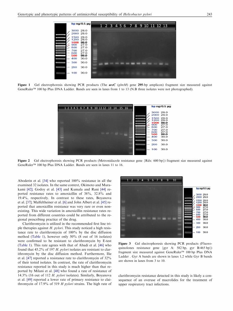
Figure 1 Gel electrophoresis showing PCR products (The ureC (glmM) gene 295-bp amplicon) fragment size measured against
GeneRuler� 100 bp Plus DNA Ladder. Bands are seen in lanes from 1 to 13 (N.B three isolates were not photographed).
Figure 3 Gel electrophoresis showing PCR products (Fluoro-
quinolones resistance gene {gyr A: 582 bp, gyr B:465 bp})
fragment size measured against GeneRuler� 100 bp Plus DNA
Ladder . Gyr A bands are shown in lanes 1,2 while Gyr B bands
are shown in lanes from 3 to 10.
Figure 2 Gel electrophoresis showing PCR products (Metronidazole resistance gene {Rdx: 600 bp}) fragment size measured against
GeneRuler� 100 bp Plus DNA Ladder. Bands are seen in lanes 11 to 16.
Genotypic and phenotypic patterns of antimicrobial susceptibility of Helicobacter pylori 243
Aboderin et al. [34] who reported 100% resistance in all theexamined 32 isolates. In the same context, Okimoto and Mura-
kami [42]; Godoy et al. [43] and Kumala and Rani [44] re-ported resistance rates to amoxicillin of 38%, 32.8% and19.4%, respectively. In contrast to these rates, Boyanova
et al. [37]; Malfeltheiner et al. [6] and John Albert et al. [45] re-ported that amoxicillin resistance was very rare or even non-existing. This wide variation in amoxicillin resistance rates re-
ported from different countries could be attributed to the re-gional prescribing practice of the drug.
Clarithromycin is utilized in the recommended first line tri-ple therapies against H. pylori. This study noticed a high resis-
tance rate to clarithromycin of 100% by the disc diffusionmethod (Table 1), however only 50% (8 out of 16 isolates)were confirmed to be resistant to clarithromycin by E-test
(Table 1). This rate agrees with that of Abadi et al. [46] whofound that 45.2% of 197 H. pylori isolates are resistant to clar-ithromycin by the disc diffusion method. Furthermore, Ilie
et al. [47] reported a resistance rate to clarithromycin of 32%of their tested isolates. In contrast, the rate of clarithromycinresistance reported in this study is much higher than that re-ported by Milani et al. [48] who found a rate of resistance of
14.3% (16 out of 112 H. pylori isolates). Similarly, Boyanovaet al. [49] reported a lower rate of primary resistance to clai-thromycin of 17.9% of 519 H pylori strains. The high rate of
clarithromycin resistance detected in this study is likely a con-sequence of an overuse of macrolides for the treatment ofupper respiratory tract infections.
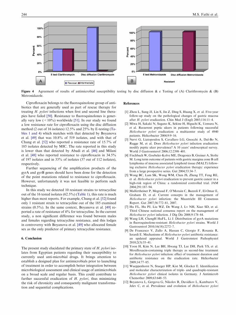
Figure 4 Agreement of results of antimicrobial susceptibility testing by disc diffusion & e Testing of (A) Clarithromycin & (B)
Metronidazole.
244 M.S. Fathi et al.
Ciprofloxacin belongs to the fluoroquinolone group of anti-
biotics that are generally used as part of rescue therapy fortreating H. pylori infections when first and second line thera-pies have failed [50]. Resistance to fluoroquinolones is gener-ally very low (<10%) worldwide [51]. In our study we found
a low resistance rate for ciprofloxacin using the disc diffusionmethod (2 out of 16 isolates) 12.5% and 25% by E-testing (Ta-bles 1 and 4) which matches with that detected by Boyanova
et al. [49] that was 10.8% of 519 isolates, and with that ofChung et al. [52] who reported a resistance rate of 15.7% of185 isolates detected by MIC. The rate reported in this study
is lower than that detected by Abadi et al. [46] and Milaniet al. [48] who reported resistance to ciprofloxacin in 34.5%of 197 isolates and in 33% of isolates (37 out of 112 isolates),
respectively.Further sequencing of the amplification products of the
gyrA and gyrB genes should have been done for the detectionof the point mutations related to resistance to ciprofloxacin.
However, unfortunately it was not feasible to perform suchtechnique.
In this study we detected 10 resistant strains to tetracycline
out of the 16 tested isolates (62.5%) (Table 1), this rate is muchhigher than most reports. For example, Chung et al. [52] foundonly 1 resistant strain to tetracycline out of the 185 examined
strains (0.5%). In the same context, Boyanova et al. [49] re-ported a rate of resistance of 4% for tetracycline. In the currentstudy, a non significant difference was found between malesand females regarding tetracycline resistance, and this lodges
in controversy with Boyanova et al. [49] who allocated femalesex as the only predictor of primary tetracycline resistance.
6. Conclusion
The present study elucidated the primary state of H. pylori iso-lates from Egyptian patients regarding their susceptibility to
currently used anti-microbial drugs. It brings attention toestablish a designed plan for antimicrobials prior to launchingof treatment in order to accomplish better integration between
microbiological assessment and clinical usage of antimicrobialson a broad scale and regular basis. This could contribute tofurther successful eradication of H. pylori, thus minimizing
the risk of chronicity and consequently malignant transforma-tion and sequential complications.
References
[1] Zhou L, Sung JJ, Lin S, Jin Z, Ding S, Huang X, et al. Five-year
follow-up study on the pathological changes of gastric mucosa
after H. pylori eradication. Chin Med J (Engl) 2003;116:11–4.
[2] Miwa H, Sakaki N, Sugano K, Sekine H, Higuchi K, Uemura N,
et al. Recurrent peptic ulcers in patients following successful
Helicobacter pylori eradication: a multicenter study of 4940
patients. Helicobacter 2004;9:9–16.
[3] Nervi G, Liatopoulou S, Cavallaro LG, Gnocchi A, Dal-Bo N,
Rugge M, et al. Does Helicobacter pylori infection eradication
modify peptic ulcer prevalence? A 10 years’ endoscopical survey.
World J Gastroenterol 2006;12:2398–401.
[4] FischbachW, Goebeler-Kolve ME, Dragosics B, Greiner A, Stolte
M. Long term outcome of patients with gastric margina zone B cell
lymphoma of mucosa associated lymphoid tissue (MALT) follow-
ing exclusive Helicobacter pylori eradication therapy: experience
from a large prospective series. Gut 2004;53:34–7.
[5] Wong BC, Lam SK, Wong WM, Chen JS, Zheng TT, Feng RE,
et al. Helicobacter pylori eradication to prevent gastric cancer in a
high-risk region of China: a randomized controlled trial. JAM
2004;291:187–94.
[6] Malfertheiner P, Megraud F, O’Morain C, Bazzoli F, El-Omar E,
Graham D, et al. Current concepts in the management of
Helicobacter pylori infection: the Maastricht III Consensus
Report. Gut 2007;56:772–81, 2007.
[7] Hu FL, Hu PJ, Liu WZ, De Wang J, Lv NH, Xiao SD, et al.
Third Chinese national consensu report on the management of
Helicobacter pylori infection. J Dig Dis 2008;9:178–84.
[8] Wang LH, ChengH HuFL, Li J. Distribution of gyrA mutations
in fluoroquinolone-resistant Helicobacter pylori strains. World J
Gastroentrol 2010;16(18):2272–7.
[9] De Francesco V, Zullo A, Hassan C, Giorgio F, Rosania R,
Ierardi E. Mechanisms of Helicobacter pylori antibiotic resistance:
an updated appraisal. World J Gastrointest Pathophysiol
2010;2(3):35–41.
[10] Yoon H, Kim N, Lee BH, Hwang TJ, Lee DH, Park YS, et al.
Moxifloxacin-containing triple therapy as second-line treatment
for Helicobacter pylori infection: effect of treatment duration and
antibiotic resistance on the eradication rate. Helicobacter
2009;14:77–85.
[11] Wueppenhorst N, Stueger HP, Kist M, Glocker E. Identification
and molecular characterization of triple- and quadruple-resistant
Helicobacter pylori clinical isolates in Germany. J Antimicrob
Chemother 2009;63:648–53.
[12] Boyanova L, Gergova G, Nikolov R, Davidkov L, Kamburov V,
Jelev C, et al. Prevalence and evolution of Helicobacter pylori
Genotypic and phenotypic patterns of antimicrobial susceptibility of Helicobacter pylori 245
resistance to 6 antibacterial agents over 12 years and correlation
between susceptibility testing methods. Diagn Microbiol Infect
Dis 2008;60:409–15.
[13] Abdollahi H, Savari M, Zahedi MJ, et al. A study of rdxA gene
deletion in metronidazole resistant and sensitive Helicobacter
pylori isolates in Kerman. Iran Jundishapur J Microbiol
2011;4(2):99–104.
[14] Glupczynski Y, Labbe M, Hansen W, Crokaert F, Yourassowsky
E. Evaluation of the E test for quantitative antimicrobial
susceptibility testing of Helicobacter pylori. J Clin Microbiol
1991;29:2072–3075.
[15] Hirschl AM, Hirschl MM, Rotter ML. Comparison of three
methods for the determination of the sensitivity of Helicobacter
pylori to metronidazole. J Antimicrob Chemother 1993;32:45–9.
[16] De Cross AJ, Marshall BJ, McCallum RW, Hoffman SR, Barrett
LJ, Guerrant RL. Metronidazole susceptibility testing for Heli-
cobacter pylori: comparison of disk, broth, and agar dilution
methods and their clinical relevance. J Clin Microbiol
1993;31:1971–4.
[17] Chaves S, Gadanho M, Tenreiro R, Cabrita J. Assessment of
metronidazole susceptibility in Helicobacter pylori: statistical
validation and error rate analysis of breakpoints determined by
the disk diffusion test. J Clin Microbiol 1999;37:1628–31.
[18] Cederbrant G, Kahlmeter G, Ljungh A. The E test for antimi-
crobial susceptibility testing of Helicobacter pylori. J Antimicrob
Chemother 1993;31:65–71.
[19] Farshad S, Alborzi A, Japoni A, Ranjbar R, Asl KH, Badiee P,
et al. Antimicrobial susceptibility of Helicobacter pylori strains
isolated from patients in Shiraz. Southern Iran World J Gastro-
entrol 2010;16(45):5746–51.
[20] Gumurdulu Y, Serin E, Ozer B, Kayaselcuk F, Ozsahin K, Cosar
AM, et al. Low eradication rate of Helicobacter pylori with triple
7–14 days and quadriple therapy in Turkey. World J Gastroen-
terol 2004;10:668–71.
[21] Chinese Helicobacter pylori Research Group, Chinese Society of
Gastroenterology. Prevalence of Helicobacter pylori resistance to
antibiotics and its influence on the treatment outcome in China: a
multicenter clinical study. Weichang Bingxue 2007; 12:525–530.
[22] Eisig JN, Silva FM, Barbuti RC, Rodriguez TN, Malfertheiner P,
Moraes Filho JP, et al. Efficacy of a 7-day course of furazolidone,
levofloxacin, and lansoprazole after failed Helicobacter pylori
eradication. BMC Gastroenterol 2009;9:38.
[23] Di Caro S, Ojetti V, Zocco MA, Cremonini F, Bartolozzi F,
Candelli M, et al. Mono, dual and triple moxifloxacin-based
therapies for Helicobacter pylori eradication. Aliment Pharmacol
Ther 2002;16:527–32.
[24] Perri F, Festa V, Merla A, Barberani F, Pilotto A, Andriulli A.
Randomized study of different ‘second-line’ therapies for Helico-
bacter pylori infection after failure of the standard ‘Maastricht
triple therapy’. Aliment Pharmacol Ther 2003;18:815–20.
[25] Yee YK, Cheung TK, Chu KM, Chan CK, Fung J, Chan P, et al.
Clinical trial: levofloxacin-based quadruple therapy was inferior
to traditional quadruple therapy in the treatment of resistant
Helicobacter pylori infection. Aliment Pharmacol Ther
2007;26:1063–7.
[26] Sezgin O, Altintas� E, Ucbilek E, Tombak A, Tellioglu B. Low
efficacy rate of moxifloxacin-containing Helicobacter pylori erad-
ication treatment: in an observational study in a Turkish
population. Helicobacter 2007;12:518–22.
[27] Zhang Z, Liu ZQ, Zheng PY, Tang FA, Yang PC. Influence of
efflux pump inhibitors on the multidrug resistance of Helicobacter
pylori. World J Gastroenterol 2010;16(10):1279–84.
[28] CLSI guidelines. Clinical and Laboratory Standard Institute:
Broth dilution antimicrobial susceptibility of H. pylori; approved
standards- third edition: M100–S22; 2008.
[29] Sablon E, Shapiro F. Advances in molecular diagnosis of HBV
infection and drug resistance. Int J Med Sci 2005;2(1):8–16.
[30] Kayacetin E, Efe D, Dogan C. Portal and splenic hemodynamics
in cirrhotic patients: relationship between esophageal variceal
bleeding and the severity of hepatic failure. J Gastroenterol
2004;39(7):661–7.
[31] Ben Mansour K, Burucoa C, Zribi M, et al. Primary resistance to
clarithromycin, metronidazole and amoxicillin of Helicobacter
pylori isolated from Tunisian patients with peptic ulcers and
gastritis: a prospective multicentre study. Ann Clin Microbiol
Antimicrob 2010:9–22.
[32] Kato S, Fujimura S, Udagawa H, et al. Antibiotic resistance of 7.
Helicobacter pylori strains in Japanese children. J Clin Microbiol
2002;2:649–53.
[33] Sherif M, Mohran Z, Fathy H, et al. Universal high-level primary
metronidazole resistance in Helicobacter pylori isolated from
children in Egypt. J Clin Microbiol 2004;42:4832–4.
[34] Aboderin OA, Abdu AR, Odetoyin B, et al. Antibiotic resistance
of Helicobacter pylori from patients in Ile-Ife, Southwest. Nigeria
Afr Health Sci 2007;7:143–7.
[35] Teare L, Peters T, Saverymuttu S, et al. Antibiotic resistance in
Helicobacter pylori. Lancet 1999;353:242.
[36] Parsons HK, Carter MJ, Sanders DS, et al. Helicobacter pylori
antimicrobial resistance in the United Kingdom: the effect of age,
sex and socio-economic status. Aliment Pharmacol Ther
2001;15:1473–8.
[37] Boyanova L, Koumanova R, Gergova G, et al. Prevalence of
resistant Helicobacter pylori isolates in Bulgarian children. J Med
Microbiol 2002;51:786–90.
[38] Kargar M, Baghernejad M, Doosti A. Role of NADPH-insensi-
tive nitroreductase gene to metronidazole resistance of Helico-
bacter pylori strains. Daru 2010;18(2):137–40.
[39] Debets-Ossenkopp YJ, Pot RG, van Westerloo J, et al. Insertion
of mini-IS605 and deletion of adjacent sequences in the nitrore-
ductase (rdxA) gene cause metronidazole resistance in Helicobac-
ter pylori NCTC11637. Antimicrob Agents Chemother
1999;43:2657–62.
[40] Jorgensen M, Trend M, Hazell S, et al. Potential involvement of
several nitroreductases in metronidazole resistance in Helicobacter
pylori. Arch Biochem Biophys 2001;392:180–91.
[41] Yang Y, Wu J, Sheu B, et al. The rdxA gene plays a more major
role than frxA gene mutation in high-level metronidazole resis-
tance ofHelicobacter pylori in Taiwan. Helicobacter 2004;9:400–7.
[42] Okimoto T, Murakami K. Acquisition of the drug resistance and
Helicobacter pylori genemutation.NipponRinsho 2009;67:2372–7.
[43] Godoy AP, Ribeiro ML, Benvengo YH, et al. Analysis of
antimicrobial susceptibility and virulence factors in Helicobacter
pylori clinical isolates. BMC Gastroenterol 2003;3:20.
[44] Kumala W, Rani A. Patterns of Helicobacter pylori isolate
resistance to fluoroquinolones, amoxicillin, clarithromycin and
metronidazoles. Southeast Asian J Trop Med Public Health
2006;37:970–4.
[45] John AlbertM, Al-Mekhaizeem K, Neil L, et al. High prevalence
and level of resistance to metronidazole, but lack of resistance to
other antimicrobials in Helicobacter pylori, isolated from a
multiracial population in Kuwait. Aliment Pharmacol Ther
2006;24:1359–66.
[46] Abadi A, Taghvaei T, Mobarez A, et al. Frequency of antibiotic
resistance in Helicobacter pylori strains isolated from the northern
population of Iran. J Microbiol 2011;49(6):987–93.
[47] Ilie M, Popa M, Chifiriuc M, et al. Helicobacter pylori cultivation
from gastric biopsies and susceptibility to antibiotics used in
empirical therapy. Roum Arch Microbiol Immunol
2011;70(2):60–4.
[48] Milani M, Ghotaslou R, Akhi M, et al. The status of antimicro-
bial resistance of Helicobacter pylori in Eastern Azerbaijan, Iran:
comparative study according to demographics. J Infect Chemo-
ther 2012(5):12.

246 M.S. Fathi et al.
[49] Boyanova L, Ilieva J, Gergova G, et al. Numerous risk factors for
Helicobacter pylori antibiotic resistance revealed by extended
anamnesis: a Bulgarian study. JMedMicrobiol 2012;61(Pt 1):85–93.
[50] Jones KR, Cha J, Merrell DS. Who’s winning the war? Molecular
mechanisms of antibiotic resistance in Helicobacter pylori. Curr
Drug Ther 2008;3:190–203.
[51] Boyanova L, Mitov I. Geographic map and evolution of primary
Helicobacter pylori resistance to antibacterial agents. Expert Rev
Anti Infect Ther 2010;8:59–70.
[52] Chung J, Lee G, Jeong J, et al. Resistance of Helicobacter pylori
strains to antibiotics in Korea with a focus on fluoroquinolone
resistance. J Gastroenterol Hepatol 2012;27(3):493–7.