
Genetics of Colon Cancer in Teenagers · Genetics of Colon Cancer in Teenagers Sharon E ... with...
34
Genetics of Colon Cancer in Teenagers Sharon E. Plon, MD, PhD, FACMG Baylor College of Medicine Texas Children’s Hospital
-
Upload
nguyentuyen -
Category
Documents
-
view
215 -
download
0
Transcript of Genetics of Colon Cancer in Teenagers · Genetics of Colon Cancer in Teenagers Sharon E ... with...

Genetics of Colon Cancer in Teenagers
Sharon E. Plon, MD, PhD, FACMG
Baylor College of Medicine
Texas Children’s Hospital
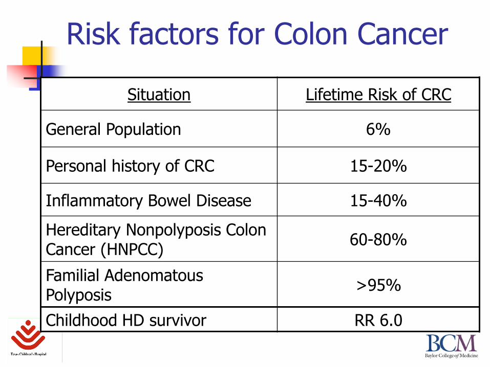
Risk factors for Colon Cancer
Situation Lifetime Risk of CRC
General Population 6%
Personal history of CRC 15-20%
Inflammatory Bowel Disease 15-40%
Hereditary Nonpolyposis Colon Cancer (HNPCC)
60-80%
Familial Adenomatous Polyposis
>95%
Childhood HD survivor RR 6.0

Familial Adenomatous Polyposis (FAP)
Autosomal Dominant with very high penetrance.
15-30% represent new mutation cases and have no family history of disease.
Adenomatous colonic polyps begin in childhood to adolescence.
Extracolonic features first noted by Gardner and called Gardner Syndrome. Now lumped together.

FAP - Extracolonic Manifestations
Desmoid tumors – often abdominal. Painful and very difficult to treat.
Osteomas of the jaw, skull, or other bones
Epidermoid cysts on face or trunk
Congenital Hypertrophy of the Retinal Pigment Epithelium (CHRPE) – present at birth, asymptomatic but very useful clinically.
Pediatric hepatoblastoma (~0.5-1% risk)
Thyroid cancer (1% risk)

Polyposis Associated with FAP
Twin A
Twin B
Typical Adenomatous Polyp from
colon of a teenager with FAPColons of twin teenage boys who presented with
history of rectal bleeding and abdominal painPhotographs courtesy of M. Finegold, MD – Baylor College of Medicine/Texas Children’s Hospital
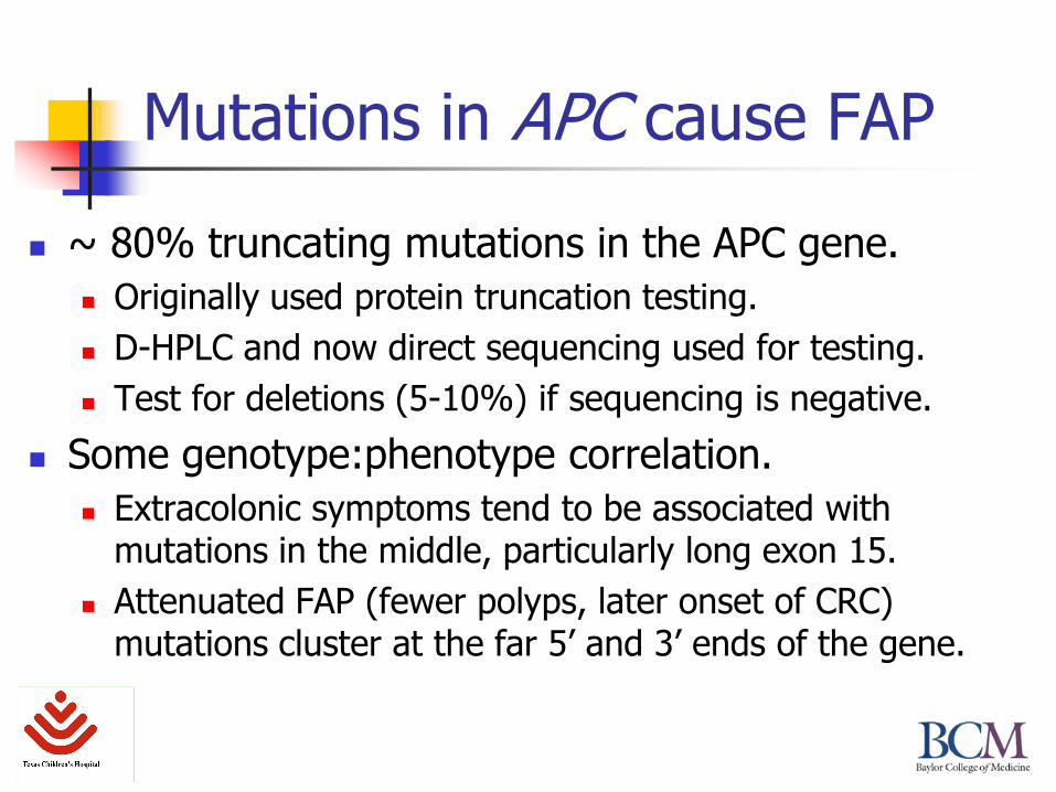
Mutations in APC cause FAP
~ 80% truncating mutations in the APC gene.
Originally used protein truncation testing.
D-HPLC and now direct sequencing used for testing.
Test for deletions (5-10%) if sequencing is negative.
Some genotype:phenotype correlation.
Extracolonic symptoms tend to be associated with mutations in the middle, particularly long exon 15.
Attenuated FAP (fewer polyps, later onset of CRC) mutations cluster at the far 5’ and 3’ ends of the gene.

Genetic Counseling in FAP
APC testing is gold standard for utility of genetic testing.
Identifies which family members need surveillance and eventual surgery.
Guidelines typically recommend testing around age 10 – 12.
Some consideration of testing and screening for hepatoblastoma in infants.
Practical issues may suggest earlier testing.

Management of APC+ Patients
Initiate testing with family member with polyposis in order to identify mutation.
Then test at risk individuals for familial mutation. Positive individuals undergo management:
Colonoscopy beginning age 10 – 12; continuing every 1-2 years.
Colectomy by late teens to early twenties (depending on polyp load or dysplasia).
Upper GI follow-up by endoscopy for risk of gastric adenomas and duodenal carcinomas.
Annual thyroid exam
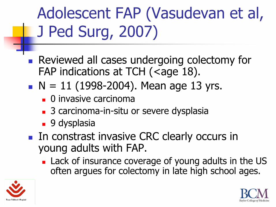
Adolescent FAP (Vasudevan et al, J Ped Surg, 2007)
Reviewed all cases undergoing colectomy for FAP indications at TCH (<age 18).
N = 11 (1998-2004). Mean age 13 yrs. 0 invasive carcinoma
3 carcinoma-in-situ or severe dysplasia
9 dysplasia
In constrast invasive CRC clearly occurs in young adults with FAP. Lack of insurance coverage of young adults in the US
often argues for colectomy in late high school ages.

Hereditary Non-Polyposis Colon Cancer (Lynch Syndrome)
Autosomal Dominant CRC without polyposis.
~70% lifetime risk of CRC (often right-sided) and 50-70% Endometrial Ca.
Ovarian, biliary tract, ureteral, gliomas also seen in HNPCC families.
Common to see individuals with 2 or 3 different primary HNPCC-related tumors.

Family History Criteria for HNPCC
Amsterdam Criteria (CRC based)
Exclude FAP
At least 1 CRC < age 50
2 affected generations
3 affected relatives, 2 are 1o relatives of other one
Bethesda Criteria – proband:
CRC < 50
CRC + HNPCC assoc Ca
1o relative with 2 HNPCC cancers, one of which < age 50
2 – 2nd degree relatives, one with CRC and one with HNPCC associated cancer

HNPCC
Due to heterozygous mutations in one of four mismatch repair (MMR) genes: MSH2, MLH1, MSH6 and PMS2.
Follows the two-hit hypothesis: Autosomal dominant inheritance of one mutation
with loss of the second copy in the tumor cell. This results in the tumor cell being mismatch repair
defective even though normal cells still have one working copy and are MMR active.
In 20% of sporadic CRC both copies of the MLH1 gene can be silenced by methylation.

HNPCC – Clinical Testing and Screening
Most protocols suggest performing MIS testing or IHC for MMR proteins in early onset CRC. If MIS+ or IHC shows missing protein (and not
methylation of MLH1) then do mutation analysis of MSH2, MLH1 and MSH6.
Need to do copy number analysis as MSH2 deletions are common.
PMS2 sequencing and copy number analysis now available.
Experience demonstrates that this is rarely done on a consistent basis!!

HNPCC - Surveillance
Screening with colonoscopy every 1-2 years starting at age 25 clearly decreases mortality in mutation carriers.
Prophylactic colectomy only recommended on case-by-case basis.
Screening for endometrial cancer by endometrial biopsy recommended.
Effectiveness less.
Prophylactic oopherectomy being discussed but not part of guidelines.

AYA CRC and HNPCC (Durno et al, Gut, 2005)
Study looking at familial CRC database for probands diagnosed <age 24 (n=16; 1% of registry). Microsatellite instability was identified in tumors from eight
(73%) of 11 evaluated patients. Germline mutations in mismatch repair genes were identified in
six of 12 patients, including MSH2 (n = 3), MLH1 (n = 2), and PMS2 (n = 1). One homozygous case. MSH6 not done.
Ten (63%) of 16 families met the Amsterdam criteria for HNPCC.
Location - rectum/sigmoid (n = 9), splenic flexure (n = 2), hepatic flexure (n = 3), and caecum (n = 2).
44% (7/16) developed additional malignancies (gastrointestinal (n = 8) and extraintestinal (n = 4)) during follow up (mean 12.8 (SD 12.4) years).

MIN vs CIN Hypothesis
Microsatellite instability (MIN)
Relatively diploid genome.
Oncogenic events due to expansion of repeats in genes like TGFBR1
Chromosomal Instability (CIN)
Associated with APC defective pathway
Aneuploidy
Large-scale rearrangements in tumors.
Modified from Lengauer …. Vogelstein Nature, 1996

Turcot syndrome. Autosomal dominant or recessive transmission?Costa OL, Silva DM, Colnago FA, Vieira MS, Musso C.Dis Colon Rectum. 1987 30(5):391-4.

AR Turcot/ Mismatch Repair Deficiency Syndrome (MMR-D)
Turcot Syndrome – association of brain tumors and colon polyps/cancer in childhood.
Dominant forms (rarely have adolescent colon cancer): FAP and medulloblastomas
HNPCC and glioblastomas
Autosomal Recessive forms – associated with childhood, adolescent and young adult colon cancers.

Turcot Syndrome
The eponym Turcot Syndrome often refers to a family with colonic polyposis and brain tumors in childhood.
However, there has been ongoing controversy about how many distinct disorders underlie Turcot syndrome ranging from: Heterozygous mutations in APC in a child with
medulloblastoma and colon polyps
Homozygous/compound heterozygous mutations in mismatch repair genes.

Typical MMR-D Presentation
5 6 7 10
1 2 3 4 5 6 7 8 9 10 11 12
51 2 3 4
1 2
I
II
III
IV
3 84 9
4321
= lymphoma
= colorectal cancer
= glioblastoma multiforme
Age 73 Age 72 Age 62 Age47 Age 50
Age 46 Age 45 Age 47 Age 48 Age 50 Age 49 Age 49 Age 41 Age 30 Age 32 Age 35 Age 39
Age 10Age of
death 9 y
CALS
Axillary
freckling
Age
15
Age of
death 10 y
CALS
MSH6
homozygous
(3634insT)
Age
6
Pakistani
Modified from Hegde, et al.,Clin Cancer Res, 2005

Other MMR-D presentations
Ten year old Pakistani child presented with bilateral glioblastoma lesions and thoracic T-cell lymphoma:
Homozygous for intragenic deletion of PMS2.
Nine year old Hispanic child with glioblastoma multiforme:
Sibling previous died with T-cell leukemia and GBM
PMS2 intragenic deletion and missense mutation
No significant cancer history in rest of family.

Mismatch Repair Deficiency Syndrome (Scott et al., 2006)
The MMR-D label clearly conveys a condition resulting from inheriting two inactivating mutations in a mismatch repair gene.
Some authors (Wimmer & Etzler, 2008) add constitutional to the name (cMMR-D).
Autosomal recessive inheritance with consanguinity frequently described.
Now been reported to result from biallelic mutations in MLH1, MSH2, MSH6, PMS2.

NF1 phenotype
It’s important to realize that constitutional MMR deficiency results in biallelic somatic mutations in the NF1 gene leading to features of NF1 (Wang et al, Hum Genet, 2003). Mutations can occur anytime in development and
result in apparent “segmental” distribution
CALS are often atypical in appearance.
A subset of MMR-D patients will meet diagnostic criteria for NF1 although they don’t have NF1. Complicates genetic evaluation and counseling.
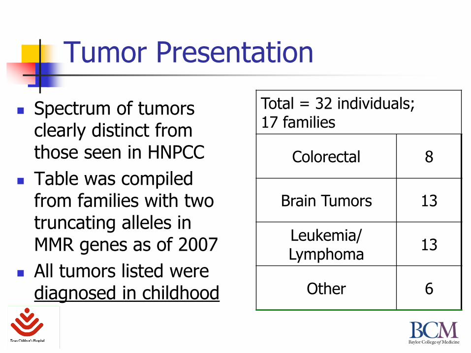
Tumor Presentation
Spectrum of tumors clearly distinct from those seen in HNPCC
Table was compiled from families with two truncating alleles in MMR genes as of 2007
All tumors listed were diagnosed in childhood
Total = 32 individuals; 17 families
Colorectal 8
Brain Tumors 13
Leukemia/Lymphoma
13
Other 6
Hematologic Malignancies
Leukemias and lymphomas are most commonly T-cell
In contrast to predominance of B-cell malignancies in children in general population.
Rare AML cases have been reported. In some cases may be 2o to treatment.
Lymphomas are not seen in HNPCC families.
MIS in Sporadic Leukemia?
In sporadic cancers, microsatellite instability is rare in primary leukemias:
Reported in patients who relapse after treatment for primary leukemia
Reported in children who develop a secondary leukemia after treatment for a primary malignancy.
So it appears that MIS requires external insult to be present in leukemia/lymphomas

Brain Tumors
All grades of gliomas reported (including gliosarcomas). High grade (glioblastoma multiforme) frequent.
Supratentorial primitive neuroectodermal tumors (SPNET) is otherwise a rare tumor and has been reported in 5 patients with PMS2 mutations.
Medulloblastoma reported in several families Medulloblastoma is also associated with heterozygous APC
mutations.
Cells that are MMR-deficient are resistant to temozolomide. MMR-D diagnosis may impact treatment decisions.
Wimmer & Etzler (2008)

Genetic Heterogeneity
Pie chart based on families (not patients) reported in Wimmer & Etzler (2008).
PMS2 may be artificially high given study from UK evaluating consanguineous families (Vos et al, 2006).
Early deaths in MSH2/MLH1 families may have precluded molecular analysis. Some question lack of viability of
some homozygous alleles in these genes.
4
8
9
24
MSH2
MLH1
MSH6
PMS2

Genotype/Phenotype Correlations
There are clear genotype/phenotype correlations based on both:
Mutations in different genes (genetic heterogeneity)
Type of mutation in the genes (Allelic heterogeneity)
See delayed onset of tumors in patients carrying missense alleles
Type of tumors more similar to HNPCC

How many cases are we missing?
Need to make pediatric oncology and neurosurgery clinicians aware of this diagnosis.
Atypical CALS are often missed by colleagues unless there is a targeted skin exam.
Pakistani families predominate due to:
High level of consanguinity
Founder PMS2 mutations, e.g. R802X, in subset of families

Microsatellite Instability in MMR-D
Typical MSI studies compare “normal DNA” with tumor DNA.
MSI-high has been reported from “normal DNA” in MMR-D if small pool PCR techniques are used.
“normal” DNA appears MSI-stable in MMR-D if you use standard procedures.
HNPCC-associated tumors show MSI-high
The few brain tumors studied are MSI-stable

Comparing heterozygous versus biallelic mutations
HNPCC
Young-late adult tumors
Hematopoietic tumors rare
CRC most common
MIS+ seen in almost all tumors
MSH2/MLH1>> PMS2/MSH6
MMR-D
Childhood onset tumors
Hematopoietic tumors common
Brain tumors
No evidence MIS+
CRC less common
Severity similar for all 4 genes

Rate limiting step for tumor formation?
The likelihood that a second hit occurs in an MMR gene may be tissue specific: Second hits appear rare in hematopoietic tissues unless
pretreated.
Need for second hit makes childhood onset of CRC unlikely in adolescence. Diagnosis of CRC begins in twenties and continues throughout adulthood in HNPCC.
The likelihood that a second hit occurs may vary among the MMR genes Given cancer risk, we presume that MLH1 and MSH2 undergo
2nd hits more frequently then MSH6 and PMS2 thus resulting in a higher cancer risk in heterozygous cases for the former genes.

Questions?