Genetics and Genomics of Physcomitrella...
32
Genetics and Genomics of Physcomitrella patens Nico van Gessel, Daniel Lang, and Ralf Reski Abstract For about a century, spanning the eras of early genetics to state-of-the-art bio- technology, the moss Physcomitrella patens has been a popular object of biolog- ical research. Meanwhile it has become an established model organism in plant evolutionary and developmental biology, mainly due to a combination of two factors: its phylogenetic key position in the plant tree of life and the sum of its favorable biological features. As a member of an early diverging land plant lineage – the bryophytes – Physcomitrella fills the gap between other models of the green lineage such as aquatic algae and flowering plants. The advantages of small stature and short generation cycles, accompanied by established and reli- able cultivation techniques provide researchers with a robust, relatively fast, and easy cultivation for experiments in a laboratory environment. Precise genome engineering is enabled by the moss’ s haploid-dominant lifestyle and its specifi- cally high rate of homologous recombination during DNA repair, that is routinely utilized through an extensive molecular toolkit for efficient gene targeting since 1998. Physcomitrella’ s genome was sequenced about a decade ago, making it the first bryophyte and even one of the first plants to be chosen for such a whole- genome shotgun sequencing approach. Ever since, the annotation of this “flagship genome” has been subject to constant improvement by an active community through the internet resource cosmoss.org which provides a central platform for knowledge exchange as well as bioinformatics data and tools. N. van Gessel • D. Lang Plant Biotechnology, Faculty of Biology, University of Freiburg, Freiburg, Germany e-mail: [email protected]; [email protected] R. Reski (*) Plant Biotechnology, Faculty of Biology, University of Freiburg, Freiburg, Germany FRIAS – Freiburg Institute for Advanced Studies, Freiburg, Germany BIOSS – Centre for Biological Signalling Studies, Freiburg, Germany e-mail: [email protected] # Springer Science+Business Media New York 2017 S. Assmann, B. Liu (eds.), Plant Cell Biology , The Plant Sciences, DOI 10.1007/978-1-4614-7881-2_22-1 1
Transcript of Genetics and Genomics of Physcomitrella...

Genetics and Genomics of Physcomitrellapatens
Nico van Gessel, Daniel Lang, and Ralf Reski
AbstractFor about a century, spanning the eras of early genetics to state-of-the-art bio-technology, the moss Physcomitrella patens has been a popular object of biolog-ical research. Meanwhile it has become an established model organism in plantevolutionary and developmental biology, mainly due to a combination of twofactors: its phylogenetic key position in the plant tree of life and the sum of itsfavorable biological features. As a member of an early diverging land plantlineage – the bryophytes – Physcomitrella fills the gap between other models ofthe green lineage such as aquatic algae and flowering plants. The advantages ofsmall stature and short generation cycles, accompanied by established and reli-able cultivation techniques provide researchers with a robust, relatively fast, andeasy cultivation for experiments in a laboratory environment. Precise genomeengineering is enabled by the moss’s haploid-dominant lifestyle and its specifi-cally high rate of homologous recombination during DNA repair, that is routinelyutilized through an extensive molecular toolkit for efficient gene targeting since1998. Physcomitrella’s genome was sequenced about a decade ago, making it thefirst bryophyte and even one of the first plants to be chosen for such a whole-genome shotgun sequencing approach. Ever since, the annotation of this “flagshipgenome” has been subject to constant improvement by an active communitythrough the internet resource cosmoss.org which provides a central platform forknowledge exchange as well as bioinformatics data and tools.
N. van Gessel • D. LangPlant Biotechnology, Faculty of Biology, University of Freiburg, Freiburg, Germanye-mail: [email protected]; [email protected]
R. Reski (*)Plant Biotechnology, Faculty of Biology, University of Freiburg, Freiburg, Germany
FRIAS – Freiburg Institute for Advanced Studies, Freiburg, Germany
BIOSS – Centre for Biological Signalling Studies, Freiburg, Germanye-mail: [email protected]
# Springer Science+Business Media New York 2017S. Assmann, B. Liu (eds.), Plant Cell Biology, The Plant Sciences,DOI 10.1007/978-1-4614-7881-2_22-1
1

KeywordsPhyscomitrella patens • Bryophyte • Funariaceae • Haploid dominance • Forwardgenetics • Reverse genetics • Homologous recombination • Gene targeting •Knockin • Knockout • Gene silencing • RNAi • miRNA • Transcriptome •Genome • Genome annotation • Land plant evolution • Genetic map •Polyploidization
ContentsKey Concepts . . . . . . . . . . . . . . . . . . . . . . . . . . . . . . . . . . . . . . . . . . . . . . . . . . . . . . . . . . . . . . . . . . . . . . . . . . . . . . . . . . . . . 2Introduction . . . . . . . . . . . . . . . . . . . . . . . . . . . . . . . . . . . . . . . . . . . . . . . . . . . . . . . . . . . . . . . . . . . . . . . . . . . . . . . . . . . . . . . 3The Nature of Physcomitrella patens . . . . . . . . . . . . . . . . . . . . . . . . . . . . . . . . . . . . . . . . . . . . . . . . . . . . . . . . . . . . 3
Life Cycle . . . . . . . . . . . . . . . . . . . . . . . . . . . . . . . . . . . . . . . . . . . . . . . . . . . . . . . . . . . . . . . . . . . . . . . . . . . . . . . . . . . . . 3Phylogenetic Position . . . . . . . . . . . . . . . . . . . . . . . . . . . . . . . . . . . . . . . . . . . . . . . . . . . . . . . . . . . . . . . . . . . . . . . . . 4The Funariaceae Family and Physcomitrella patens Ecotypes . . . . . . . . . . . . . . . . . . . . . . . . . . . . . . 5
Features and Genetics . . . . . . . . . . . . . . . . . . . . . . . . . . . . . . . . . . . . . . . . . . . . . . . . . . . . . . . . . . . . . . . . . . . . . . . . . . . . 6Lab Culture . . . . . . . . . . . . . . . . . . . . . . . . . . . . . . . . . . . . . . . . . . . . . . . . . . . . . . . . . . . . . . . . . . . . . . . . . . . . . . . . . . . . 6Classical Genetics and Mutagenesis . . . . . . . . . . . . . . . . . . . . . . . . . . . . . . . . . . . . . . . . . . . . . . . . . . . . . . . . . . 7Transformation . . . . . . . . . . . . . . . . . . . . . . . . . . . . . . . . . . . . . . . . . . . . . . . . . . . . . . . . . . . . . . . . . . . . . . . . . . . . . . . . 8Homologous Recombination and Gene Targeting . . . . . . . . . . . . . . . . . . . . . . . . . . . . . . . . . . . . . . . . . . . 10Forward Genetics and the Genetic Map . . . . . . . . . . . . . . . . . . . . . . . . . . . . . . . . . . . . . . . . . . . . . . . . . . . . . . 12Reverse Genetics . . . . . . . . . . . . . . . . . . . . . . . . . . . . . . . . . . . . . . . . . . . . . . . . . . . . . . . . . . . . . . . . . . . . . . . . . . . . . . 14RNA Interference and Gene Silencing . . . . . . . . . . . . . . . . . . . . . . . . . . . . . . . . . . . . . . . . . . . . . . . . . . . . . . . 16
Genomics . . . . . . . . . . . . . . . . . . . . . . . . . . . . . . . . . . . . . . . . . . . . . . . . . . . . . . . . . . . . . . . . . . . . . . . . . . . . . . . . . . . . . . . . . 17History of the Nuclear Genome . . . . . . . . . . . . . . . . . . . . . . . . . . . . . . . . . . . . . . . . . . . . . . . . . . . . . . . . . . . . . . 17Expression Profiling . . . . . . . . . . . . . . . . . . . . . . . . . . . . . . . . . . . . . . . . . . . . . . . . . . . . . . . . . . . . . . . . . . . . . . . . . . . 20Genes and Gene Structures . . . . . . . . . . . . . . . . . . . . . . . . . . . . . . . . . . . . . . . . . . . . . . . . . . . . . . . . . . . . . . . . . . . 22Transposable Elements . . . . . . . . . . . . . . . . . . . . . . . . . . . . . . . . . . . . . . . . . . . . . . . . . . . . . . . . . . . . . . . . . . . . . . . . 23Noncoding RNAs . . . . . . . . . . . . . . . . . . . . . . . . . . . . . . . . . . . . . . . . . . . . . . . . . . . . . . . . . . . . . . . . . . . . . . . . . . . . . 24Organellar Genomes . . . . . . . . . . . . . . . . . . . . . . . . . . . . . . . . . . . . . . . . . . . . . . . . . . . . . . . . . . . . . . . . . . . . . . . . . . 25Evolution of Transcription-Associated Proteins . . . . . . . . . . . . . . . . . . . . . . . . . . . . . . . . . . . . . . . . . . . . . . 26Generative Polyploidization in Funariaceae . . . . . . . . . . . . . . . . . . . . . . . . . . . . . . . . . . . . . . . . . . . . . . . . . . 27
Future Directions . . . . . . . . . . . . . . . . . . . . . . . . . . . . . . . . . . . . . . . . . . . . . . . . . . . . . . . . . . . . . . . . . . . . . . . . . . . . . . . . . 28References . . . . . . . . . . . . . . . . . . . . . . . . . . . . . . . . . . . . . . . . . . . . . . . . . . . . . . . . . . . . . . . . . . . . . . . . . . . . . . . . . . . . . . . . 29
Key Concepts
• The moss Physcomitrella patens is an established experimental model in plantevolutionary and developmental biology: important because of its basal positionin the plant tree of life.
• The dominant haploid generation and the relative ease of cultivation havestrongly contributed to its popularity in modern biology.
• Large-scale mutant collections are available and together with the genetic mapallow forward genetics approaches.
• Efficient gene targeting and reverse genetics in the moss are enabled by its highrate of homologous recombination allowing precise genome engineering.
• The Physcomitrella patens genome was the first sequenced bryophyte genomeand reveals insights into land plant evolution.
2 N. van Gessel et al.

Introduction
Historically, because of its haploid-dominant generation, few tissue types traceableto the fate of a single cell, as well as its small stature and straightforward culturalneeds, Physcomitrella patens (Hedw.) Bruch & Schimp. has been the focus ofgenetic studies for about 100 years. Over this time, it has developed into a plantexperimental model in evolutionary developmental plant biology: important becauseit is one of the only models that resides in a basal position in the phylogeny of landplants and as such is more closely derived from an early land plant than presentexperimental models, bridging the gap between green algae and flowering plants. Anestablished laboratory strain, complemented by standardized culture protocols andan extensive molecular toolkit for genome engineering further expanded the impactof this moss on plant biology. To date, the sequences of the nuclear, plastid, andmitochondrial genomes have revealed striking insights into plant evolution, enlight-ening the necessary genetic adaptations to life on land.
This chapter aims at introducing Physcomitrella patens to a wider audience, whilefocussing on its genetic and genomic features and providing an entry point intofurther, more detailed literature. Therefore, it provides an overview of the moss’sdevelopment as the subject of early genetic inquiries and the progress toward itssignificance as a flagship plant experimental model in state-of-the-art plant research.When applicable, example studies and reviews are being addressed to offer a properintroduction into the practical scientific work on Physcomitrella.
The Nature of Physcomitrella patens
In comparison to other prominent plant experimental models, the small moss speciesPhyscomitrella patens might appear relatively unimposing. It is its size, short lifecycle, and, not at least, its phylogenetic position that makes it not only a goodexperimental model but also a highly interesting species in the context of plantevolution.
Life Cycle
The life cycle of Physcomitrella patens exhibits a heteromorphic alternation ofgenerations with a dominant haploid gametophyte and a morphologically distinct,reduced diploid sporophyte, typical of all bryophytes. A germinating meiosporegives rise to filamentous protonemata by apical tip growth producing two distinctcell types representing the juvenile gametophyte: the chloroplast-enrichedchloronema cells with cell walls perpendicular to the growth axis and caulonemacells with fewer chloroplasts and oblique cell walls. Caulonemal cells producemeristematic buds with three-faced apical cells that develop into the adult gameto-phyte (gametophore) with basal rhizoids and an erect, leafy stem that is capable ofgenerating the sex organs (gametangia). As Physcomitrella patens is monoecious,
Genetics and Genomics of Physcomitrella patens 3

the male antheridium and the female archegonium are located on the same gameto-phore, and self-fertilization is common. In the presence of water, motile malespermatozoids are able to reach and fertilize the egg cell inside the archegonium,which initiates the development of the diploid sporophytic embryo. The sporophytedevelops a short seta with an apical spore capsule bearing the characteristic calyptraand loses its initial ability to photosynthesize during its maturation. During the entireprocess, the sporophyte is strongly dependent on nutritional supplies from thegametophyte. Inside the spore capsule, a few thousands of haploid spores areproduced through meiosis and are released subsequent to the ripening and openingof the capsule (Fig. 1).
Phylogenetic Position
One of the most crucial events in plant evolution is marked by the conquest of landand the rise of embryophytes, demanding substantial morphological and physiolog-ical adaptations to the new environment. The last common ancestor of the two landplant lineages of tracheophytes (vascular plants) and bryophytes (liverworts, mosses,hornworts) lived about 500 million years ago (Lang et al. 2010). This early diver-gence describes two radically different approaches to life on land. At the same time,it anchors the bryophyte Physcomitrella patens, as a member of the Funariaceae
Fig. 1 Natural life cycle of Physcomitrella patens. A germinating meiospore gives rise to filamen-tous protonemata, on which the adult gametophore with basal rhizoids can develop. On top of theleafy stem, male antheridium and female archegonium are formed and motile male spermatozoidsreach and fertilize the egg cell inside the archegonium, which initiates sporophyte development. Thediploid sporophyte develops a short seta with an apical spore capsule bearing the characteristiccalyptra. Inside the capsule, haploid spores are produced through meiosis and are released subse-quent to the ripening and break-open of the capsule
4 N. van Gessel et al.

family inside the class Bryopsida, at an important basal position in the context ofland plant evolution. The species represents those plants that bridge the gap betweenaquatic green algae, such as Chlamydomonas reinhardtii, and the tracheophytes, forexample, Selaginella moellendorffii or even Arabidopsis thaliana. As such a “basal”land plant species, Physcomitrella patens provides an excellent reference to studythe necessary adaptations to early life on land. The stronger effects of gravity (as theplant grew in stature), exposure to higher levels of UV irradiation, varying temper-atures, and last but not least a fluctuating availability of water all defined theadaptations to the terrestrial lifestyle. In addition, the basal position ofPhyscomitrella patens in the land plant phylogeny enables comparative analyseswith more complex species within the plant kingdom that can assist the identificationof genes and gene families that are either conserved or emerged throughout theindividual course of evolution, e.g., for specific adaptation to an environmentalselective force.
The Funariaceae Family and Physcomitrella patens Ecotypes
Physcomitrella patens (Hedw.) Bruch & Schimp. belongs to the cosmopolitanFunariaceae family within the order Funariales of the class Bryopsida. TheFunariaceae comprise approximately 250–400 species accommodated in a minimumof 15 genera of small, terricolous, monoicous, annual, and biennial mosses. How-ever, the taxonomic classification of single genera and species is topic of constantdebate in the bryophyte scientific community and will probably profit from futuregenomic insights. While the family members generally share a rather uniformvegetative, gametophytic body, they differ substantially in terms of sporophytemorphology and ecology. The sporophyte of Physcomitrella patens is characterizedby a reduced seta and the lack of a peristome on the indehiscent spore capsule, aputative adaption to spore dispersal by water birds. In contrast, the Funariaceaespecies Funaria hygrometrica exhibits an elongated seta and dehiscent capsules witha complex peristome adapted to humidity- and wind-dependent spore dispersal.Interestingly, the sporophyte morphology of Physcomitrella is assumed to representthe evolutionary derived state that followed a secondary reduction of the precedentcomplex structure.
While the Funariaceae exhibit an overall worldwide distribution, Physcomitrellaspecies occur mutually exclusively to certain regions. Ecotypes of the two otherspecies in the Physcomitrella genus, Physcomitrella readeri and Physcomitrellamagdalenae, were collected in Japan, Australia, and Africa, respectively. However,isolates of Physcomitrella patens ecotypes exclusively cover the northern hemi-sphere, more precisely the USA and Europe, including France, Germany, Norway,Sweden, the United Kingdom, and Ukraine. The most prominent of these ecotypes isrepresented by the established laboratory strain “Gransden,” that is, by now, the defacto standard in laboratories worldwide. Originally isolated in 1962 byH.L.K. Whitehouse from a single spore in Gransden Wood, Huntingdonshire(UK), the vast majority of modern genetic and molecular biological experiments
Genetics and Genomics of Physcomitrella patens 5
that involve Physcomitrella patens employ this strain. Highlighting this role, the“Gransden” ecotype was the chosen strain for the Physcomitrella genome project.Though reports on chromosome counts differed, cytogenetic analyses indicated thecurrently accepted chromosome number of n = 27 (Reski et al. 1994), and flowcytometric analyses determined its size to be about 511 megabasepairs (Schweenet al. 2003).
A collection of about 40 Funariaceae isolates, including the Physcomitrellaecotypes, is cryo-conserved in a long-term storage facility – the International MossStock Center (IMSC, http://www.moss-stock-center.org/) at the University of Frei-burg, Germany. The IMSC is a community-oriented, nonprofit resource that providesmoss researchers with a facility to safely store individual strains by cryopreservation.In addition, moss material can also be ordered by the community to ensure the use ofstandardized stock material in worldwide scientific applications. In this regard, theIMSC also maintains about 60 transgenic lines that comprise a collection of knock-out mutants and reporter lines.
Features and Genetics
Physcomitrella patens combines a variety of favorable properties, which accountsfor its success as a desirable plant model for genetic and developmental studies. As atarget for genetic manipulation, it profits from its short generation time, fast growthrate, small stature, and reduced morphological complexity with traceable cell lines. Italso has the added advantage of being relatively simple to transform with highefficiency.
Lab Culture
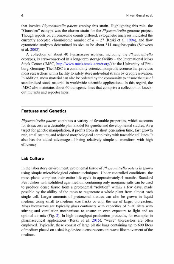
In the laboratory environment, protonemal tissue of Physcomitrella patens is grownusing simple microbiological culture techniques. Under controlled conditions, themoss plants complete their entire life cycle in approximately 4 months. StandardPetri dishes with solidified agar medium containing only inorganic salts can be usedto produce dense tissue from a protonemal “solution” within a few days, madepossible by the ability of the moss to regenerate a whole plant from almost eachsingle cell. Larger amounts of protonemal tissues can also be grown in liquidmedium using small to medium size flasks or with the use of larger bioreactors.Moss bioreactors are typically glass containers with capacities of 5–30 liters withstirring and ventilation mechanisms to ensure an even exposure to light and anoptimal air mix (Fig. 2). In high-throughput production protocols, for example, inpharmaceutical applications (Reski et al. 2015), “wave” bioreactors are oftenemployed. Typically, these consist of large plastic bags containing up to 600 litersof medium placed on a shaking device to ensure constant wave-like movement of themedium.
6 N. van Gessel et al.

Classical Genetics and Mutagenesis
Bryophyte species in general and mosses in particular have been subjects of geneticresearch since the early twentieth century, when Mendelian genetics wererediscovered and put into the context of Darwinian evolution. The natural
Fig. 2 Habitus and laboratory culture. (A) Natural Physcomitrella patens population covering aclump on a fallow acre near Freiburg, Germany. (B) Close-up of the sporophyte, comprising theshort seta that bears the spore capsule with its calyptra. Subfigures C–E illustrate differentlaboratory culture techniques: gametophytes growing on solid medium in a Petri dish (C), filamen-tous protonemata in liquid culture D, and protonemata in the moss bioreactor (E)
Genetics and Genomics of Physcomitrella patens 7
occurrence of variants and hybrids in the bryophytes attracted the attention of earlygeneticists, and experiments were facilitated by their short generation time andundemanding culture. Examples of scientific breakthroughs from early bryophytegenetic studies include the discovery of sex chromosomes in plants (Allen 1917), thedescription of heterochromatin and the continuity of chromosomes during themitotic cell cycle (Heitz 1928), and the development of X-ray mutagenesis (Knapp1936).
The haploid nature of the moss gametophyte was determined from the earlyobservation of non-Mendelian inheritance of genes (von Wettstein 1928). Thecrossing of two haploid strains results in the formation of a diploid sporophytewhich in turn generates haploid spores. Examining the progeny of such a cross, twoalleles of one gene segregate 1:1 in the F1 generation, and mutant alleles of twounlinked genes segregate in a 1:1:1:1 ratio, corresponding to a 3:1 ratio of mutant towild type. While these segregation ratios provided the first evidence for the domi-nance of the haploid genotype in mosses, they also highlighted the clear advantageof mosses for the isolation of mutant strains for genetic investigations: in contrast tocrosses in diploid species, mutants can be identified directly without the need foradditional inbreeding and the establishment of an F2 generation. Clearly, this onlyapplies to nonlethal phenotypes. However, the study of recessive lethal mutants isstill possible by the use of somatic hybridization, i.e., the fusion of two protoplastsand regeneration of diploid gametophytes. Although these hybrids of Physcomitrellapatens are self-fertile, they develop a reduced number of sex organs, particularlyarchegonia, as well as exhibiting “sluggish” spermatozoid movement, which puta-tively accounts for an overall slower completion of the life cycle. Starting in the1960s with the work of Engel, a plethora of auxotrophic mutants and developmen-tally abnormal Physcomitrella patens strains have been generated, e.g., by exposureto X-rays and alkylating agents (Engel 1968) and the subsequent mass screening ofspores. The ability of protoplasts (single cells) to regenerate whole plants has led tothe recent replacement of mutagenized spores as the starting material for mutantstrains. Protoplasts, either directly treated with mutagenic agents or derived fromtreated protonemal tissue by digestion, are now the preferred source of mutants.
Transformation
Over the last three decades, an extensive toolkit has been established for directtransformation, i.e., the delivery of exogenous DNA into cells of Physcomitrellapatens. Depending on the retention and inheritance of the transformed DNA and thecorresponding phenotype, two basic types of transformation can be distinguished:stable and transient. Transformation with a plasmid containing an antibiotic resis-tance reporter cassette demonstrates these two possibilities. Under continuous selec-tion pressure, i.e., the growth on antibiotic media, transformed plants express thereporter genes and thus show antibiotic resistance. Removing the antibiotic from thegrowth medium for a period of time, thus lowering the selection pressure forresistance, generates the two classes of transformed plants. Stable transformants
8 N. van Gessel et al.

with genome integration of the transgene inherit the resistance meiotically andproduce offspring that encode and express the resistance to the antibiotic. In contrast,plants that lose or do not inherit the resistant phenotype have not undergone genomeintegration of the transgene, thus are only transiently transformed. Researcherssuggested that in the case of lost resistance, the transgene may have been replicatedand expressed extrachromosomally when the plant was under selection and theplasmids lost after a period of relaxed conditions (Ashton et al. 2000). The researchteam transformed Physcomitrella patens protoplasts using a previously unusedplasmid, pBI426, carrying an antibiotic resistance marker and a β-glucuronidase(GUS) reporter gene. After regeneration of gametophytes and initial selection, theyrepeatedly transferred the plants between selective and nonselective media fordefined periods. A minority of plants (~0.2%) showed stable transformation evenafter several rounds of relaxed and increased selection pressure; however, themajority lost their ability to grow on selective medium. GUS staining was possiblefor all stable transformants and initially for tissue of transient transformants in directcontact with the selective medium. In accordance with the observable growthdeficiency, transient transformants also lost GUS activity after several rounds ofrelaxed selection. Southern blot analyses lead to the assumption that these transienttransformants temporarily contained 3–40 extrachromosomal copies of the plasmid.This was the first report of a transgenic photosynthetic plant to be generated by theexpression of genes from an autonomously replicating extrachromosomal element.Based on these findings and the fact that stable genome integration of transgenes isthe desired goal of most experiments, the discrimination of stable transformants fromtransient ones is mandatory. Typically, this is accomplished by the before mentionedmethod, i.e., the removal of the selective condition and a subsequent renewedselection period(s).
Transformation methods for P. patens have been constantly improved, and todate, a variety of methods are available to researchers (Strotbek et al. 2013).Physcomitrella patens was considered not to be transformable via Agrobacteriumtumefaciens for a long period of time; however, the recent discovery of new virulentstrains enabled the application of this method (Cove et al. 2009). A. tumefaciensdirects the integration of foreign DNA into a plant’s genome via the T-DNA regionof its Ti plasmid and is usually a method of choice for transformation of vascularplants. Although the genomic integration of the introduced DNA is nondirected andrandom via this method, it does exclusively lead to the generation of stable mutants,at high transformation rates, using Physcomitrella protoplasts. In addition to this trueclassical method of plant transformation, it is also possible to deliver foreign DNAdirectly into cells mechanically using microinjection and biolistics. With a microin-jection needle, DNA can be injected into filament cells and even directly into nucleiof Physcomitrella patens cells. However, this method demands the special cultiva-tion of moss filaments in absolute darkness for a defined period, to allow cell wallpenetration. Additionally, the demanding injection technique has to be applied at aprecise region basal to the nucleus to prevent the bursting of cells. So far, onlytransient mutants have been analyzed, thus reliable evidence of stable transformationis lacking. The biolistic method relies on a particle gun to bombard cells with
Genetics and Genomics of Physcomitrella patens 9
plasmid-coated gold or tungsten particles. There are no specific restrictions to thekind of targeted plant tissue, and efficiency is comparable to that seen for vascularplants. Unfortunately, as for microinjection, there have been no reports of stablemutants to date.
Since the early 1990s, Polyethylene glycol (PEG)-mediated transformation ofPhyscomitrella patens protoplasts has been developed into the method of choice(Schaefer et al. 1991). It enables the use of relatively large DNA constructs and at thesame time requires no sophisticated technology or instruments. PEG stabilizes theprotoplasts and allows for the uptake of negatively charged DNA through the cellmembrane. After transformation, protoplasts are transferred to regeneration mediumand undergo subsequent selection. In addition to the undemanding requirements,PEG transformation provides both, high transformation efficiency and a low rate ofmultiple integration of the transgene into the genome. Remarkably, the method canalso be employed for the transformation of chloroplasts in Physcomitrella patenswithout any additional requirements (Sugiura and Sugita 2004). Integration of DNAinto the plastid genome relies exclusively on homologous recombination and thusoffers nearly the same possibilities that are available for nuclear genome editing (seesubsequent sections). Of course, this has been facilitated by the availability of thechloroplast genome sequence, sequenced prior to the nuclear genome ofPhyscomitrella. Undoubtedly, the ability to directly target genes encoded in theplastid genome offers an invaluable tool for studies of genes related to photosyn-thesis, especially in the context of plant evolution.
Homologous Recombination and Gene Targeting
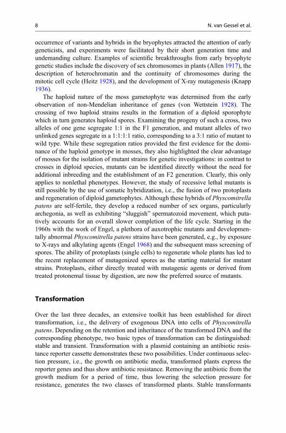
One of the most distinctive features of Physcomitrella patens is the high degree ofhomologous recombination (HR) in somatic cells. In all living cells, HR represents arepair mechanism for DNA double-strand breaks that is highly conserved throughoutthe plant kingdom. Just like other genomic disruptions, double-strand breaks occurnaturally in cellular processes such as DNA duplication and are induced by chemicaland physical agents. Yet, by undermining chromatin stability, they pose a huge threatto genome integrity and can cause severe mutations or even cell death. Whileangiosperms rely mainly on the alternative repair mechanism of nonhomologousend joining (NHEJ), which illegitimately recombines DNA strands, Physcomitrellapatens predominantly employs HR (Schaefer and Zryd 1997). Through this precisemechanism, two double-stranded DNA molecules that share homologous sequencesperform a temporary crossover, in which whole stretches of DNA are exchanged,under guidance of specific enzymes, between the strands without the loss of a singlenucleotide. As HR relies on a high degree of sequence identity between the tworecombining strands, it produces a legitimate recombination of DNA (Fig. 3).
In practice, HR provides a powerful tool for directed delivery of DNA to apredetermined locus in the genome and the study of gene function in the mossthrough gene targeting (Strepp et al. 1998). The term “gene targeting” describes atechnique for modification of a locus using defined sequences homologous to the
10 N. van Gessel et al.

target region of a gene or gene element by exploitation of the HR mechanism. As HRis especially efficient in Physcomitrella patens, exhibiting efficiencies only knownfrom the microbiological field, gene targeting via HR is a distinctive feature of themoss in comparison to other plant models. Thus, Physcomitrella has often beendescribed as the “green yeast.” The integration efficiency for practical applicationsdepends on certain properties of the transgenic construct. In general, DNA isdelivered into the protoplast in linear form, after PCR duplication or enzymaticdigestion from its original cloning vector, and consists of a gene cassette plus aselection marker. The construct carries two homologous flanking sequencescorresponding to the 50 and 30 ends of the target region in the genome: typicallythe minimal length is 400–500 bp for each, and longer stretches generally increaseintegration rates. In addition, a uniform length of the two flanking sequences seemsto have a positive effect. Successful integration via HR results in the replacement ofthe target region by the gene cassette. In this context, two different strategies can bedistinguished: targeted knockout and knockin of a gene. The targeted replacement ofa gene generally leads to the loss of its function, i.e., a knockout of the respectivegene. In contrast, the same method is used to replace genomic regions with homol-ogous sequences from a similar gene from another species or to fuse additionalsequences, often regulatory, to the target locus. In the former case, the HR-directedgene replacement enables gene complementation studies, and in the latter, it offers anelegant way of fusing sequences encoding reporter proteins, such as the green
Fig. 3 Simplified schema of homologous recombination for gene targeting. A double-strand DNA(dsDNA) construct with specifically designed flanking regions is introduced into the nucleus whereits ends are recognized as double-strand breaks (DSB), and it is introduced to the DSB repairmechanism of the cell. Consequently, the construct is guided to the genomic locus harboringsequence stretches identical to the flanks (a). Single strands at both ends of the construct invadethe identical genomic stretches and anneal to the complementary strands (b). After resolution of thecomplex structure, the transfected sequence is integrated into the genome (c)
Genetics and Genomics of Physcomitrella patens 11

fluorescence protein (GFP) or β-glucuronidase (GUS), to the coding region of a geneof interest. The measurement of the activity of the reporter proteins in-turn enableslocalization and interaction studies of endogenous gene products. The efficacy ofsuch an approach was shown in a recent study of genes responsible for mitotic celldivision in Physcomitrella patens (Nakaoka et al. 2012). Different candidate genes,e.g., coding for augmin and others, previously described as tubulin associated, wereindependently knocked down via RNAi (see section “RNAi”). In order to test for thephenotypic consequences, the coding sequences of the two reporter proteins GFPand RFP (red fluorescent protein) were fused to the genes of tubulin and histone,respectively. Confocal microscopic time-lapse photography of the fluorescentreporters in the different knocked-down lines revealed a dominant role for augminin the generation of microtubules.
Forward Genetics and the Genetic Map
For a long time, forward genetics has represented the standard method for linking afunction with a specific gene. The approach relies primarily on untargeted mutagen-esis via chemicals such as alkylating agents or physical factors such as X-ray and UVirradiation. Subsequent screening of mutant plants reveals interesting phenotypesthat are subject to further investigation to identify the responsible genes that havebeen mutated. For Physcomitrella patens, a huge effort was made to create severalmutant collections for the genetics community, and from the 1990s, the large-scalemutagenesis of Physcomitrella via transformation with tagged constructs involvinggenomic DNA (gDNA) or complementary DNA (cDNA) was presented as analternative to chemical and/or physical mutation.
In one of the early efforts, the moss was transformed using a gDNA library inwhich DNA fragments were tagged via so-called shuttle mutagenesis using mini-transposons (Nishiyama et al. 2000). The researchers extracted genomic DNA,generated 3–6 kb fragments, and then used an established Escherichia coli systemto integrate mini-transposons that contained an antibiotic resistance gene as well as agene encoding for GUS into the generated gDNAs. Upon PEG-mediated protoplasttransformation with these constructs, plants where selected for antibiotic resistanceand phenotypically classified. Of the 5264 stable transformants, 203 mutants haddevelopmental and morphological abnormalities in comparison to the wild type, and129 mutants had GUS activity, e.g., in mucilage hairs (multicellular hairs that form atthe base of a leaf), in young leaves, in the apical cell of very young buds and inprotonema including chloronema and caulonema, respectively.
Subsequently, a similar approach yielded a total of 16,203 transformants (Egeneret al. 2002). In this instance, wild-type mRNAwas extracted from protonemata andreverse transcribed to create a cDNA library that was then target to advanced shuttlemutagenesis including an antibiotic resistance selection marker using a differenttransposon-derived system. The use of cDNAwas assumed to enable highly specificHR-based integration into actively transcribed genes rather than genome-wideintegration by use of gDNA. The transformation of Physcomitrella resulted in
12 N. van Gessel et al.

2636 mutant plants that differed from the wild type in terms of developmental andmorphological as well as physiological characteristics such as nutritionalrequirements.
By far the largest tagged DNA mutagenesis project led to the production of73,329 mutant plants, classified in 16 different categories, consisting of plantstructure, color, gametophore development, cell shape, etc. (Schween et al. 2005).The transformation was again accomplished using cDNA libraries to generatemutants; however, in this case, the libraries each were created using a variety ofdifferent starting tissues, such as protonema, gametophyte, and sporophyte tissue, aswell as from moss treated with various agents, such as the phytohormones cytokininand abscisic acid (ABA). Interestingly, the sporophyte and gametophore librarieswere subtracted cDNA libraries, i.e., mRNA from one tissue was used to capturecomplementary cDNA from the other tissue in order to obtain specific cDNAexclusive to the latter tissue. In addition to phenotypic descriptions, the librarieswere normalized, and extensive sequencing yielded over 110,000 expressedsequence tags (ESTs).
Unfortunately, most collected mutants still miss precise association of a pheno-type to a specific genetic locus. It is only recently that researchers have begun toelucidate some of these connections. With recent technological advances, a studyfrom 2010 revealed the genetic basis for long-described Physcomitrella patensmutants that exhibit abnormal responses to auxin treatments (Prigge et al. 2010).These authors used information derived from homologous genes of Arabidopsisthaliana to identify genes in Physcomitrella patens putatively involved in theresponse to auxin that delivers an accelerated and early transition from chloronemato caulonema. Using mutants that had been generated in 1979, described as beingarrested at the primary chloronemal stage or delayed in the chloronema-to-caulonema transition, they sequenced the putative Physcomitrella patens genes.Comparisons between the wild-type genes and the mutant sequences revealedlesions in a specific motif of three moss genes that result in a single amino acidsubstitution in the corresponding protein. Thus, the genetic basis for the defect wasidentified successfully 30 years after the description of the phenotypic mutants.
For the vast majority of mutants, such links between a phenotype and a specificgenetic change could not be assigned as yet. One major reason for this predicamentis the lack of sufficient molecular markers at the time of each individual mutagenesisstudy. Molecular markers denote sequence polymorphisms between individuals ofthe same species or different species and allow the mapping of genetic loci inmap-based cloning experiments. Co-segregation analyses of the mutant allele andsuch markers enable the determination of a mutation’s genetic background, based onthe assumption that physically close loci on a chromosome are less frequentlyrecombined during meiosis than distant ones. A first step in the establishment ofsuch markers for Physcomitrella patens was accomplished in 2006, in the form of110 polymorphic short sequence repeats (SSR), identified via a computer-basedsearch against an expressed sequence tag (EST) database of the “Gransden” strain(von Stackelberg et al. 2006). Upon comparing the SSR sequences with differentPhyscomitrella strains, the genetically distant French strain “Villersexel”was used to
Genetics and Genomics of Physcomitrella patens 13
identify polymorphic SSRs. SSRs represent DNA sequences of mono- up to hexa-nucleotide repeats with genome-wide distribution yet accumulate in non-repetitiveDNA and untranslated 30 and 50 regions of genes. The ESTs had been sequencedpreviously as part of the moss transcriptome assembly, and as such (Rensing et al.2002a), they offered absolute linkage of the markers to genes with putative or knownfunction.
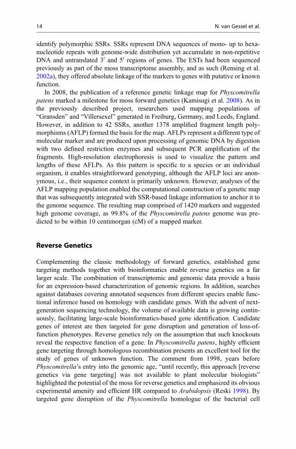
In 2008, the publication of a reference genetic linkage map for Physcomitrellapatens marked a milestone for moss forward genetics (Kamisugi et al. 2008). As inthe previously described project, researchers used mapping populations of“Gransden” and “Villersexel” generated in Freiburg, Germany, and Leeds, England.However, in addition to 42 SSRs, another 1378 amplified fragment length poly-morphisms (AFLP) formed the basis for the map. AFLPs represent a different type ofmolecular marker and are produced upon processing of genomic DNA by digestionwith two defined restriction enzymes and subsequent PCR amplification of thefragments. High-resolution electrophoresis is used to visualize the pattern andlengths of these AFLPs. As this pattern is specific to a species or an individualorganism, it enables straightforward genotyping, although the AFLP loci are anon-ymous, i.e., their sequence context is primarily unknown. However, analyses of theAFLP mapping population enabled the computational construction of a genetic mapthat was subsequently integrated with SSR-based linkage information to anchor it tothe genome sequence. The resulting map comprised of 1420 markers and suggestedhigh genome coverage, as 99.8% of the Physcomitrella patens genome was pre-dicted to be within 10 centimorgan (cM) of a mapped marker.
Reverse Genetics
Complementing the classic methodology of forward genetics, established genetargeting methods together with bioinformatics enable reverse genetics on a farlarger scale. The combination of transcriptomic and genomic data provide a basisfor an expression-based characterization of genomic regions. In addition, searchesagainst databases covering annotated sequences from different species enable func-tional inference based on homology with candidate genes. With the advent of next-generation sequencing technology, the volume of available data is growing contin-uously, facilitating large-scale bioinformatics-based gene identification. Candidategenes of interest are then targeted for gene disruption and generation of loss-of-function phenotypes. Reverse genetics rely on the assumption that such knockoutsreveal the respective function of a gene. In Physcomitrella patens, highly efficientgene targeting through homologous recombination presents an excellent tool for thestudy of genes of unknown function. The comment from 1998, years beforePhyscomitrella’s entry into the genomic age, “until recently, this approach [reversegenetics via gene targeting] was not available to plant molecular biologists”highlighted the potential of the moss for reverse genetics and emphasized its obviousexperimental amenity and efficient HR compared to Arabidopsis (Reski 1998). Bytargeted gene disruption of the Physcomitrella homologue of the bacterial cell
14 N. van Gessel et al.

division protein FtsZ and microscopic characterization of the knockout phenotyperevealing the formation of macrochloroplasts, Strepp et al. (1998) were able to reportthe first organellar division protein to be identified in eukaryotes (Fig. 4).
Since the late 1990s, reverse genetics have developed into a standard techniquefor the identification of gene function in Physcomitrella patens, as evidenced by anever-increasing number of studies. An exemplar of such investigations is the func-tional characterization of FtsZ proteins performed via multiple targeted gene knock-outs (Martin et al. 2009). The FtsZ is a self-assembling GTPase that forms acontractile ring at the division site during plastid division in plants and traces backto an ancestral homologue in cyanobacteria. In contrast to bacteria, which encodeonly a single FtsZ protein, plants harbor at least three different nuclear-encodedproteins in two families, FtsZ1 and FtsZ2. The Physcomitrella genome encodes forfive FtsZ proteins in total, comprising two members of each family FtsZ1 and FtsZ2as well as a single member of a third family, FtsZ3. To elucidate their putativefunctions in the moss, Martin et al. (2009) performed targeted knockouts of the fiveFtsZ genes. Phenotypic analysis of the resulting knockout plants revealed mainlythree different patterns, correlated with Physcomitrella’s three FtsZ families: FtsZ1proteins were characterized as interacting partners to fulfill functions related to themaintenance of chloroplast shape, the FtsZ2 family members appear responsible forplastid division, and FtsZ3 was characterized as a molecular link between cell andchloroplast division in Physcomitrella patens based on its co-localization to thechloroplast as well as to the cytoplasm.
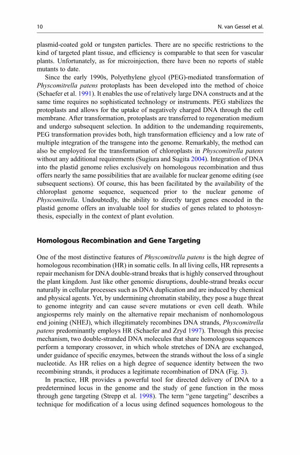
Fig. 4 Two principles of gene targeting by homologous recombination (inspired by Fig. 5 inStrotbek et al. 2013). (a) Targeted knockout of a gene by disruption of its coding sequence, e.g., viaintegration of a different gene for functional studies. (b) Targeted knockin, e.g., via fusion of areporter gene to a gene of interest, for localization or interaction studies
Genetics and Genomics of Physcomitrella patens 15

RNA Interference and Gene Silencing
The inhibition of gene expression by RNA interference (RNAi) is a highly conservedregulatory pathway expressed in all eukaryotes. The conservation of the pathway iswidely attributed to be a consequence of its role as an early defense system againstviruses or other forms of foreign genetic material and, especially in plants, as acontrol system for self-propagating transposable elements. A detailed review on therole and regulatory mechanisms involving RNAi in Physcomitrella patens wasprovided by Arif et al. (2013). In general, introduced double-stranded RNA(dsRNA), either derived from a virus or delivered experimentally via a transcribablegenetic element, is processed by Dicer-like (DCL) proteins to release small interfer-ing RNAs (siRNAs). Each siRNA then guides an RNA-induced silencing complex(RISC) to a specific target messenger RNA (mRNA) that has a matching antisensesequence. An Argonaute protein (AGO) within the RISC cleaves the target mRNAwhich is subsequently degraded. In this way, RNAi prevents the translation of anmRNA, i.e., silences a gene at the posttranscriptional level. In Physcomitrellapatens, this mechanism is utilized for targeted silencing of single genes or wholegene families. Members of gene families typically share at least short segments ofcommon sequence allowing all family members to be targeted by a single RNAi. ThecDNA of a target gene region (containing the common sequence) is cloned into anexpression cassette as an inverted repeat separated by a spacer region. After suc-cessful transformation of the construct, its expressed mRNA, containing the self-complementary inverted repeats, folds back on itself to form a dsRNA. The dsRNAis processed through the RNAi pathway and gives rise to siRNAs that sufficientlyguide the RISC in posttranscriptional silencing of multiple members or the entiregene family.
However, the dispersal of a diverse set of siRNAs from one dsRNA might lead tounintended side effects such as off-target gene silencing and therefore is not alwaysthe method of choice.
Alternatively, a more specific silencing mechanism can be achieved by usingmicro RNAs (miRNAs). An miRNA primary transcript is encoded by a discretemiRNA gene and upon expression forms a hairpin-like dsRNA as it contains internalself-complementary sequences. In a multistep reaction guided by a member of theDCL family, among other associated proteins, the primary transcript is processedinto a miRNA precursor that gives rise to a single miRNA that is complementary toits target gene. This miRNA then guides the RISC to a target transcript in a highlyspecific mode. In a manner similar to the RNAi via siRNA mechanism, subsequentAGO- and DCL-mediated mRNA cleavage results in the degradation of the targetmRNA: a form of posttranscriptional gene silencing. In Physcomitrella, it wasshown that the miRNA pathway can also lead to transcriptional inhibition by guidingan RNA-induced transcriptional silencing complex (RITS) that alters DNA methyl-ation patterns depending on the ratio of the miRNA to the target mRNA (Khraiweshet al. 2010). In the PpDCL1b mutant, target mRNA cleavage did not occur eventhough the target mRNA transcript levels were reduced suggesting an epigeneticsilencing of the target mRNA-encoding gene. Indeed, sequencing of target mRNA
16 N. van Gessel et al.
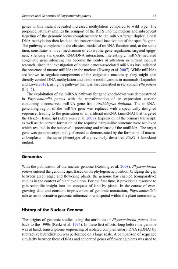
genes in this mutant revealed increased methylation compared to wild type. Theproposed pathway implies the reimport of the RITS into the nucleus and subsequenttargeting of the genomic locus complementary to the miRNA/target duplex. LocalDNA methylation then leads to the transcriptional inactivation of the specific gene.The pathway complements the classical model of miRNA function and, at the sametime, constitutes a novel mechanism of eukaryotic gene regulation: targeted epige-netic silencing via specific RNA/DNA interaction. Interestingly, miRNA-mediatedepigenetic gene silencing has become the center of attention in current medicalresearch, since the investigation of human cancer-associated miRNAs has indicatedthe presence of mature miRNAs in the nucleus (Hwang et al. 2007). While miRNAsare known to regulate components of the epigenetic machinery, they might alsodirectly control DNA methylation and histone modifications in mammals (Lujambioand Lowe 2012), using the pathway that was first described in Physcomitrella patens(Fig. 5).
The exploitation of the miRNA pathway for gene knockdown was demonstratedin Physcomitrella patens with the transformation of an expression cassettecontaining a conserved miRNA gene from Arabidopsis thaliana. The miRNA-generating region of the miRNA gene was replaced with a specifically designedsequence, leading to the generation of an artificial miRNA (amiRNA) that targetedthe FtsZ2–1 transcript (Khraiwesh et al. 2008). Expression of the primary transcript,as well as the correct formation of the required hairpin-like structure were achievedwhich resulted in the successful processing and release of the amiRNA. The targetgene was posttranscriptionally silenced as demonstrated by the formation of macro-chloroplasts – the same phenotype of a previously described FtsZ2–1 knockoutmutant.
Genomics
With the publication of the nuclear genome (Rensing et al. 2008), Physcomitrellapatens entered the genomic age. Based on its phylogenetic position, bridging the gapbetween green algae and flowering plants, the genome has enabled (comparative)studies in the context of plant evolution. For the first time, it provided a resource togain scientific insight into the conquest of land by plants. In the course of ever-growing data and constant improvement of genomic annotation, Physcomitrella’srole as an informative genomic reference is undisputed within the plant community.
History of the Nuclear Genome
The origins of genomic studies using the attributes of Physcomitrella patens dateback to the 1990s (Reski et al. 1994). In these first efforts, long before the genomewas at hand, transcriptome sequencing of isolated complementary DNA (cDNA) bysubtractive hybridization was performed on a large scale. A comparison of sequencesimilarity between these cDNAs and annotated genes of flowering plants was used to
Genetics and Genomics of Physcomitrella patens 17

identify homologous as well as putative species-specific genes. Subsequent genediscovery projects were initiated in Freiburg (Germany), Okazaki (Japan), Leeds(England), and St. Louis (USA) utilizing large-scale expressed sequence tag (EST)sequencing. These efforts yielded the first virtual Physcomitrella patens
Fig. 5 The different modes of miRNA-mediated gene silencing in Physcomitrella patens. Thetranscript of an miRNA gene is processed to a pre-miRNA, which forms a characteristic foldbackdue to self-complementary sequence stretches. After excision and cytoplasmic export of a miRNAduplex, the miRNA is released and forms a highly specific duplex with its complementary mRNAtarget. Recruitment of the RNA-induced silencing complex (RISC) either leads to cleavage andsubsequent degradation of the target (1) or translational inhibition by steric hindering of ribosomalactivity (2). Both pathways lead to posttranscriptional silencing of the gene encoding the targetmRNA. In Physcomitrella patens, a third mechanism has been proposed leading to direct transcrip-tional silencing of a target gene (Khraiwesh et al. 2010) (3): depending on an elevated miRNA totarget mRNA ratio, a putative RNA-induced transcriptional gene silencing (RITS) complex isformed. The complex is reimported to the nucleus where it is directed to the genomic target locusand mediates epigenetic modification in the form of DNA methylation
18 N. van Gessel et al.

transcriptome, an extensive data resource of both high-quality sequences and a lowdegree of redundancy (Rensing et al. 2002b). Although the number of protein-coding genes was estimated to be ~25,000 – a number comparable to a similarestimate for Arabidopsis thaliana – nearly half shared no sequence homology withsequences from any other organism. In addition, the majority of the longer virtualtranscripts were only fragments of whole genes. Based on the growing generalinterest in Physcomitrella as a plant experimental model, driven by the analysis ofthe transcriptome, and the limited validity of the transcriptomic data for addressingimportant genomic questions, the common will to sequence Physcomitrella patens’genome emerged in the moss community. Consequently, in 2004, the InternationalMoss Genome Consortium dedicated to pursue this task was established in Freiburgat the annual moss conference. As part of the Community Sequencing Program ofthe US Department of Energy’s Joint Genome Institute (JGI), a whole-genomeshotgun sequencing approach was executed, with the “Gransden” laboratory strainas a reference strain from which the DNAwas extracted. This program succeeded inestablishing the genome of Physcomitrella patens as the first bryophyte genome (andone of the first plant genomes) to be sequenced. The JGI justified its decision to takeon the task by stating that “[t]he moss Physcomitrella patens is becoming widelyrecognized as an experimental organism of choice not only for basic molecular,cytological, and developmental questions in plant biology, but also as a key link inunderstanding plant evolutionary questions, especially those related to genomeevolution. Physcomitrella is well placed phylogenetically to provide importantcomparisons with the flowering plants. In terms of evolutionary distance,Physcomitrella is to the flowering plants what Drosophila is to humans. Havingthe full Physcomitrella genome available will greatly inform bioinformatic compar-isons and functional genomics in plants, just as the mouse, Fugu, and Drosophilagenomes have informed animal biology.” After assembly and initial structural andfunctional annotation, the genome version 1.1 was released in 2007, and its featureswere published in 2008 (Rensing et al. 2008). The draft genome had a size of480 Mbp and contained 35,938 protein-coding genes and as expected, it revealedimportant insights into the conquest of land by plants. Around the same time, thePhyscomitrella patens genome resource “cosmoss.org” was launched to provide thecommunity with a central web portal for exploration and annotation, in the traditionof comparable model organism databases like “TAIR” for Arabidopsis thaliana or“FlyBase” for Drosophila melanogaster. Since that time, the community hasmaintained a continuous effort to further improve genome annotation, underscoredby the decision by JGI to extend the Physcomitrella resource as one of its “flagship”plant genomes. The development of the genetic linkage map based on amplifiedfragment length polymorphisms (AFLP) and simple sequence repeat (SSR) markers(Kamisugi et al. 2008) is one prime example of these efforts, and it provides aninvaluable information resource for targeted identification of genes connected tomutant phenotypes in forward genetic studies. In addition, an ambitious singlenucleotide polymorphism (SNP) genotyping analysis was performed, which resultedin the identification of approximately 600,000 high-confidence SNPs that are dis-tinguishable between the Physcomitrella patens laboratory strain “Gransden” and
Genetics and Genomics of Physcomitrella patens 19

the genetically divergent “Villersexel” strain from France. These mapping toolsenabled the assembly and annotation of a yet unpublished chromosome-basedgenome release. The latest official genome version 1.6 (V1.6) was published in2013 as a complete re-annotation of the initial release (Zimmer et al. 2013). Alongwith the incorporation of increased transcriptomic data representing a multitude ofdevelopmental stages and tissue types, V1.6 introduced updated protein-coding geneannotations with improved EST support, untranslated region (UTR) annotation, andalternative splicing isoforms. The complete set is comprised of 32,275 protein-coding genes with accompanying extended information describing nonprotein-coding loci.
Expression Profiling
For many years, gene expression analyses were limited to either a single or only afew genes using such techniques as mRNA quantification via Northern blotting. Thedevelopment of biological chip technologies in the mid-1990s introduced the firstplatform for parallel, large-scale quantitative gene expression analysis using whatbecame known as the DNA microarray. In general, microarrays consisted of a solidsurface coated with thousands of individual evenly spaced “spots” each containingidentical single-stranded DNA probes bound to the surface matrix in the nano- topicomole concentration range. The probes typically consisted of cDNAs comple-mentary to each mRNA representing each member of a gene set of interest, e.g., allknown protein coding sequences of an organism. Upon loading the array andhybridizing a fluorescently labeled sample RNA to the probes, subsequent removalof non-hybridized material, the probe:mRNA hybrid is stably bound to the solidmatrix of the array. The level of hybridization of sample RNA to the probe DNA ismeasured with a scanning device that detects the fluorescent signal for each spot.Depending upon the respective conditional context of sampled RNA, e.g., tissuetype or treatment, the specific response to the tissue or condition in terms of geneexpression is quantifiably measured.
With the generation of genomic data, the first microarray for Physcomitrellapatens was produced in 2007 based on the “Agilent” platform and containedapproximately 20,000 features based on 80,000 ESTs from Leeds, England, andOkazaki, Japan. This array enabled researchers to study the transcriptional responsesof various tissues of Physcomitrella patens to multiple individual treatments, such astreatment with exogenous abscisic acid (ABA) or osmotic, salt, and dehydrationstress treatments (Cuming et al. 2007). Expression profiling using this technologyrevealed 130 genes induced by dehydration, 56 by ABA, 10 by osmotic stress, andeight genes by salt stress, with 51 genes showing induction in more than onetreatment. An extended homology-based search against genes of other plant speciesyielded ABA- and drought-responsive Physcomitrella homologues of angiospermgenes expressed during drought stress and seed development. These findingsenabled the posing of hypotheses concerning the connection between conservedstress regulatory transcription factors exclusive to the seed developmental pathway
20 N. van Gessel et al.
(or more specifically the seed specificity of desiccation tolerance) among angio-sperms and the vegetative expression of desiccation tolerance in bryophytes.
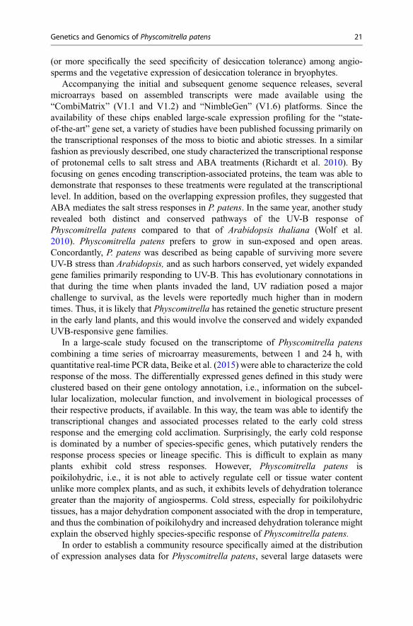
Accompanying the initial and subsequent genome sequence releases, severalmicroarrays based on assembled transcripts were made available using the“CombiMatrix” (V1.1 and V1.2) and “NimbleGen” (V1.6) platforms. Since theavailability of these chips enabled large-scale expression profiling for the “state-of-the-art” gene set, a variety of studies have been published focussing primarily onthe transcriptional responses of the moss to biotic and abiotic stresses. In a similarfashion as previously described, one study characterized the transcriptional responseof protonemal cells to salt stress and ABA treatments (Richardt et al. 2010). Byfocusing on genes encoding transcription-associated proteins, the team was able todemonstrate that responses to these treatments were regulated at the transcriptionallevel. In addition, based on the overlapping expression profiles, they suggested thatABA mediates the salt stress responses in P. patens. In the same year, another studyrevealed both distinct and conserved pathways of the UV-B response ofPhyscomitrella patens compared to that of Arabidopsis thaliana (Wolf et al.2010). Physcomitrella patens prefers to grow in sun-exposed and open areas.Concordantly, P. patens was described as being capable of surviving more severeUV-B stress than Arabidopsis, and as such harbors conserved, yet widely expandedgene families primarily responding to UV-B. This has evolutionary connotations inthat during the time when plants invaded the land, UV radiation posed a majorchallenge to survival, as the levels were reportedly much higher than in moderntimes. Thus, it is likely that Physcomitrella has retained the genetic structure presentin the early land plants, and this would involve the conserved and widely expandedUVB-responsive gene families.
In a large-scale study focused on the transcriptome of Physcomitrella patenscombining a time series of microarray measurements, between 1 and 24 h, withquantitative real-time PCR data, Beike et al. (2015) were able to characterize the coldresponse of the moss. The differentially expressed genes defined in this study wereclustered based on their gene ontology annotation, i.e., information on the subcel-lular localization, molecular function, and involvement in biological processes oftheir respective products, if available. In this way, the team was able to identify thetranscriptional changes and associated processes related to the early cold stressresponse and the emerging cold acclimation. Surprisingly, the early cold responseis dominated by a number of species-specific genes, which putatively renders theresponse process species or lineage specific. This is difficult to explain as manyplants exhibit cold stress responses. However, Physcomitrella patens ispoikilohydric, i.e., it is not able to actively regulate cell or tissue water contentunlike more complex plants, and as such, it exhibits levels of dehydration tolerancegreater than the majority of angiosperms. Cold stress, especially for poikilohydrictissues, has a major dehydration component associated with the drop in temperature,and thus the combination of poikilohydry and increased dehydration tolerance mightexplain the observed highly species-specific response of Physcomitrella patens.
In order to establish a community resource specifically aimed at the distributionof expression analyses data for Physcomitrella patens, several large datasets were
Genetics and Genomics of Physcomitrella patens 21

made available online (Hiss et al. 2014). The microarray data originates fromexperiments utilizing a multitude of tissue types such as spores, protoplasts, proto-nemata, or gametophores, developmental stages such as germinating spores ordeveloping gametangia, as well as various treatments and stresses such as differentlight intensities and qualities, dehydration, or different growth media. To ensure dataintegrity and future accessibility for the community, sample expression profiles werelinked to Physcomitrella gene IDs and the tissue types; developmental stages andexperimental conditions were semantically connected to respective ontology terms.As a front end for data access, the online meta-analysis tool “Genevestigator” waschosen, as it has successfully provided similar data for several other model plantspecies and it also features a gene expression search engine that is focused onintegrating all contents and comparisons between experiments. The platform thusprovides an invaluable information resource for moss researchers that are interestedin the conditional expression of a Physcomitrella gene or wish to conduct cross-species comparisons of gene expression patterns. In a similar way, but relying onhigh-throughput sequencing of RNA (RNA-seq), the US Department of Energy’sJoint Genome Institute initiated its Plant Gene Atlas Project. The program aims atlarge-scale, coordinated development of reference transcriptomes for the institute’sflagship species: the alga Chlamydomonas reinhardtii, the grass model Setariaitalica, soybean, poplar, and not least, Physcomitrella patens. At the same time,the project provides a test bed for new experimental technologies associated withdissection, capturing, and amplification of plant RNA.
Genes and Gene Structures
For the gene models of the Physcomitrella patens V1.6 genome annotation (Zimmeret al. 2013), only transcripts with EST or full-length cDNA evidence were chosen,complemented by mapped sequences that had been experimentally confirmed andpublished by the community, independent of the genome project. In total, 38,357transcripts that represent 32,275 protein-coding genes, with a mean transcript andgene length of 1389 bp and 2369 bp, respectively, have been described. A subset of15,757 transcripts possess both 50 and 30 UTR annotation and another 21,464transcripts having at least either one of the UTRs delineated, based on full supportby experimental evidence. Alternative splicing (AS) occurs in 10.8% of loci with anaverage of 2.52 and a maximum of 11 transcripts per locus. The predominantlyobserved form of AS is defined by alternative intron acceptor sites, i.e., the use of adifferent 30 splice junction that results in an alteration of the 50 downstream exonboundary. The prevalent consequence of this mechanism in Physcomitrella patens isthe retention of introns. In addition, the coding sequence is altered in 51% oftranscripts that are target of AS, resulting in 2380 distinct proteins. Also 56% ofAS transcripts exhibit an alteration of the 50 UTR. The effect of 50 UTR introns ongene expression, regulation, translation, and nonsense-mediated mRNA decay havebeen described independently. These findings, together with the unique length of
22 N. van Gessel et al.

Physcomitrella 50 UTR introns, suggest that the moss makes frequent use of this typeof gene regulation.
The moss genome contains a higher fraction of genes with a single exon (23.4%)than the genomes of green algae (<10%) and other land plants (~19%). While intronloss is common in the evolution of land plants and outnumbers intron gain invascular plants, it may specifically occur in Physcomitrella as a result of retro-copying: the reverse transcription of mRNA followed by partial replacement ofgenomic DNA by the intron-less cDNA. In the case of Physcomitrella patens, thismechanism might be facilitated by gene conversion via homologous recombination.Another explanation, based on the observation that single-exon genes in the mossgenerally lack evidence for expression, is that these gene models might representfragmentary predictions, nonprotein-coding genes, or pseudogenes.
A striking characteristic of the Physcomitrella patens genome is the presence oftandemly arrayed genes (TAG). In general, TAGs are paralogous genes, i.e., origi-nate from a species-specific duplication event, residing closely together on a chro-mosome. Although the occurrence of TAGs was described in Arabidopsis thalianaand Oryza sativa (and others) and was observed to exist in an even higher proportionof their genomes, the TAGs of Physcomitrella patens deviate significantly in orien-tation and conservation. TAGs in Physcomitrella patens occur predominantly in aninverse orientation and feature highly conserved sequences even in the noncodingregions (Lang et al. 2008). These specific differences together with Physcomitrellapatens’s prevalent and efficient HR capabilities suggest a concerted evolution ofthese genes through gene conversion, that is, the replacement of one gene with itshomologue to retain and conserve sequences. This might be a mechanism for thehaploid dominant moss to maintain “pseudoalleles” of highly expressed genes. Asdescribed in the V1.6 release, TAGs contribute to the majority of orthologous geneclusters with only two member genes. With 27% of all gene clusters, their share issubstantially higher in the moss compared to other species of plants (Table 1).
Transposable Elements
In plant genomes, repetitive sequences account for a large fraction of the total DNAsequence. These repetitive elements are typically comprised of tandem repeats andsimple sequence repeats (SSR), transposable elements (TE), and duplicated genomicfragments. Repeats in general, and TEs in particular, are of special evolutionaryinterest as they enhance the diversity of plant genomes. In some cases, TEs alonecontribute to 80% of a plant genome (Feschotte et al. 2002). Such elements arecapable of transposition, i.e., they possess the ability to change their position in thegenome, and are enclosed by specific repetitive sequences. Depending on the natureof these repetitive sequences and the intermediate form of TEs during transposition,TEs can be divided into two classes. Class 1 elements transpose via an RNAintermediate and either have long terminal repeats (LTR) similar to LTRretrotransposons or are terminated by a SSR (usually polyA) similar to non-LTRretrotransposons. Class 1 elements also include long and short interspersed nuclear
Genetics and Genomics of Physcomitrella patens 23

elements, LINEs and SINEs, respectively. Class 2 elements transpose via a DNAintermediate. They possess terminal inverted repeats and can be subdividedaccording to whether they are autonomous or not.
In the current Physcomitrella patens genome annotation, V1.6, full-length LTRretrotransposons and related fragments have been described to cover about half ofthe entire genome (Zimmer et al. 2013). In comparison, Physcomitrella harborsapproximately three times more LTR retrotransposons than Arabidopsis thaliana butthree times fewer than rice.
The class 2 elements, Helitrons, nonautonomously transpose via a rolling-circlemechanism, in which a single DNA strand is transferred from the original genomiclocus to the target site. The cell’s DNA repair machinery then uses this single strandas a template for synthesis of the second strand, which results in duplication of theHelitron. The genome of Physcomitrella patens contains only a single Helitronfamily comprised of 19 members. High sequence similarity between these Helitrons(96%) indicates their activity within the past 3 million years (Rensing et al. 2008).
Noncoding RNAs
The involvement of small RNAs (sRNA) in plant gene regulation is a hot topic incurrent research, and the extent of the regulatory role that sRNAs play is still onlypartly understood. However, their role in fundamental processes like development,morphogenesis, and genome defense is obvious. In Physcomitrella patens, the two
Table 1 Evolution of the Physcomitrella patens genome annotation (modified fromZimmer et al. 2013)
V1.1Rensing et al. (2008)
V1.2Lang et al. (2008)
V1.6Zimmer et al. (2013)
Genome size (Mb) 480 480 480
Scaffolds 2106 1995 1985
Protein-coding genes 35,938 27,966 (�7972) 32,275 (+4309)
Protein-coding genes withEST support
12,593 19,119 (+6523) 26,722 (+7603)
Protein-coding transcripts 35,938 27,966 (�7972) 38,357a (+10,391)
Annotated as alternativelyspliced
– – 3500
Genes with UTRs 4517 4515 (�2) 15,757a (+11,242)
Either UTR Genes 8418 8381 (�37) 16,010 (+7629)
Transcripts 21,464a
Gene density (kb pergene)
13.4 17.2 14.9a
Exons/gene 4.9 5.4 5.0a
Mean exon length (bp) 246 234 275a
Mean intron length (bp) 311 277 278a
aincluding splice variants and data from the miRBase registry, release 18
24 N. van Gessel et al.

main types of sRNAs, siRNAs, and miRNAs have been studied, with a focus on theirbiogenesis and mode of action. Axtell presented an exemplary and detailed review ofthe moss’s diverse noncoding RNA species (Axtell 2009). Small RNA annotationhas greatly profited from next-generation RNA sequencing (RNAseq) that enabledhigh-throughput sRNA identification, complemented by an ever-growing set ofbioinformatic tools. The V1.6 genome annotation includes 229 miRNAs from108 miRNA families, sharing 47 families with Chlamydomonas reinhardtii and187 families with Arabidopsis thaliana (Zimmer et al. 2013). Loci that either codefor siRNAs or miRNAs seem to possess opposed properties (Coruh et al. 2015).MiRNAs typically occur in genic regions that have an overall lower DNA methyl-ation state, i.e., enhanced accessibility to the transcription machinery. SiRNAs, onthe other hand, contribute to the vast landscape of noncoding RNA in intergenicregions that contain a high ratio of repetitive sequences and dense methylation.Unfortunately, a large proportion of the latter still lacks proper annotation.
Besides small regulatory RNAs, other elementary noncoding RNA species arerepresented in the V1.6 genome annotation. A total of 432 tRNA loci have beenidentified (Rensing et al. 2009), with 417 of the encoded tRNAs encompassing all ofthe 20 standard amino acids, with at least one of the possible anticodons per aminoacid. Thus, not all of the 57 possible tRNAs seem to be used in Physcomitrellapatens. A gene for a selenocysteine tRNAwas also identified in the set. In addition,there are 798 rDNA loci encoding ribosomal RNAs that were annotated in V1.6, aswell as 213 small nucleolar RNA (snRNA) and 6 signal recognition particle (SRP)loci. SRPs occur in the spliceosome where snRNAs are associated with specificproteins in small nuclear ribonucleoprotein particles (snRNP) and catalyze thesplicing of pre-mRNA. SRPs are also associated with ribonucleoproteins but con-tribute to the targeting of proteins to the endoplasmic reticulum of eukaryotes.
Organellar Genomes
The genome sequences and genome features for both the chloroplast and mitochon-drion were published in 2003 (Sugiura et al. 2003) and 2007 (Terasawa et al. 2007),respectively. According to the endosymbiotic theory, chloroplasts originated viaengulfment of a photosynthetic cyanobacterial ancestor, and over time, endosymbi-otic gene transfer to the host cell nuclear genome led to a reduced plastid genome(plastome) – in land plants, the length typically ranges between 120 and 160 kbp.The specific features of chloroplast DNA (cpDNA) were reviewed as part of ageneral chapter on chloroplasts in Physcomitrella patens by Sugita and Aoki(2009). With 122,890 bp, the plastome of Physcomitrella patens lies between theliverwort Marchantia polymorpha (~121 kbp) and the hornwort Anthocerosformosae (~160 kbp) in size, closely to the plastome size of another bryophyte,Syntrichia ruralis, with 122,530 bp (Oliver et al. 2010). In terms of GC content, itranges closer to the two bryophytes, with 28.5% compared to ~29% and ~33%,respectively, than to vascular plants (~38–39%). It comprises 83 protein-encodinggenes, 31 tRNA and 4 rRNA genes, as well as a single pseudo-tRNA gene. Most
Genetics and Genomics of Physcomitrella patens 25

strikingly, comparative analysis revealed fundamental insights into cpDNA evolu-tion in land plants. Firstly, a large inversion of 71 kbp was described to be presentthroughout Funariaceae, Disceliaceae, and Encalyptales – the largest inversiondocumented in plants so far. Thus, it is assumed that this inversion was alreadypresent in the last common ancestor of these three moss families. Secondly, theabsence of the RNA polymerase alpha chain gene (rpoA) from cpDNA and itstransfer to the nuclear genome were first described in Physcomitrella patens. Thegene is present in the plastid genomes of other mosses and was lost twice in theevolutionary history of bryophytes.
The Physcomitrella patens mitochondrial genome (chondriome) was the secondpublished bryophyte chondriome, and it was the smallest of reported land plantchondriomes at the time of sequencing. The circular-mapping chondriome DNA ofPhyscomitrella patens harbors 41 protein-encoding genes, 24 tRNA and 3 rRNAgenes. In comparison to other species, neither significant synteny with chlorophytesand flowering plants could be found nor the multipartite structure reported to bepresent in the chondriomes of flowering plants. Yet, the mitochondrial genome hasretained the prototypic features of land plant chondriomes. Phylogenetic compari-sons supported the close relationship between Physcomitrella patens, theCharophyta and liverworts as well as the assumption that the clade of mosses andliverworts are a sister group to the flowering plants.
Evolution of Transcription-Associated Proteins
Now that Physcomitrella patens is widely used as a model to study genetic adapta-tions during the evolution of land plants, a special emphasis has been put on thecomparative analysis of transcription-associated proteins (TAPs), which largelyaccount for modulating and regulating gene activity (Rensing et al. 2009). Ineukaryotes, TAPs are involved in complex networks that maintain protein-encodinggene regulation. These networks center on specific transcription factors (TF) thatbind to cis-active elements in order to activate or repress target gene expression. Butthey also include other transcriptional regulators (TR) such as coactivators andcorepressors that bind and influence TFs, transcription initiation factors that recruitcomponents of the transcription machinery by recognizing core promotor elements,and last but not least chromatin remodeling factors that mediate histone modifica-tions and DNA methylation and thus affect the accessibility of DNA.
It was proposed that there is a positive correlation between the quantity of TAPsand the number of cell types in a respective organism: the comparative analysis ofTAPs in Physcomitrella patens and diatom, red and green algae species as well asrice and Arabidopsis support this proposal. On average, the TAP gene families inPhyscomitrella are two to three times higher than those found in the genomes ofalgae but only represent a fourth of total TAPs and putative TAPs seen in thegenomes of vascular plants. The analysis also revealed that the moss containsrepresentatives of most of the transcription-associated proteins found in seed plants.Only six novel angiosperm TF families emerged after the evolutionary split of
26 N. van Gessel et al.

mosses and seed plants. These TFs are involved in flower development and lateralorgan formation in the sporophyte in flowering plants. Interestingly, as unicellularalgae do not contain most of the TF families present in land plants, the expansion ofTAP families is most likely to have evolved during or after the switch fromunicellularity to multicellularity. These hypotheses will greatly profit from theongoing genomic sequencing and comparative genomic studies.
Generative Polyploidization in Funariaceae
Among land plants, generative polyploidy in the form of large-scale or whole-genome duplication (WGD) events is a widespread phenomenon. Rensing et al.(2013) highlighted the evolutionary importance of polyploidization with a focusedstudy on the moss family, Funariaceae. Generally, WGD events are recognized asdriving forces behind speciation and diversification of plant lineages. In addition,their crucial influence on the evolution of organismal complexity is widely hypoth-esized. Two forms of WGD events have been described: autopolyploidization, i.e.,genome doubling within a species and without hybridization, and allo-polyploidization, that is, the doubling of the genome in association with the intro-duction of a genetically different chromosome sets resulting from the hybridizationof two species. While autopolyploidization probably provides a short-term evolu-tionary advantage during periods of environmental change, allopolyploidizationmight increase the potential of genes to sub- and neo-functionalize and thus presentsa possible driver of speciation. The fate of duplicated genomic regions is key tounderstanding the evolutionary importance of WGD events, as it is possible to drawconclusions from the diversifying effects of the post-WGD phase on paralogousgenes.
In the context of haploid-dominant mosses, polyploidization might present agreater potential evolutionary advantage than for plants that have a dominant diploidphase. Additional copies of genes and chromosomes after a WGD event couldintroduce a buffer against somatic mutations that may eventually affect the germline. A WGD event would also introduce the same potential for genetic sub- andneo-functionalization as such an event does for diploid-dominant organisms. WGDevents also induce heterosis, i.e., the increased biological quality or capability of ahybrid. This might explain the observed increase in size of extant moss speciescompared to those found preserved in Eocene (45 million year old) amber.Physcomitrella patens has undergone at least one polyploidization between 30 and60 million years ago and is thus referred to as a paleopolyploid (Rensing et al. 2007).This finding was based on the analysis of nearly 3000 paralogous genes and reliedupon the calculation of synonymous substitution rates to estimate the divergencerates of the paralogues. This period, 30 and 60 million years ago, nicely coincideswith the fossil record and falls in the time frame for the mass extinction events thatoccurred at the end of the Eocene (Stehlin 1909), ca. 33 million years ago. Phylo-genetic analyses further implied that the ancient polyploidization observed in
Genetics and Genomics of Physcomitrella patens 27

Physcomitrella patens had affected the ancestral lineage that generated theFunariaceae.
For species in the Funariaceae, including Physcomitrella patens, natural hybridsappear frequently, presumably because of comparable habitats and growth in closeproximity to one another (McDaniel et al. 2010; Beike et al. 2014). Experimentalhybrids have been generated by artificial induction of autopolyploidization aftertransfection of P. patens protoplasts during transformation protocols. The poly-ploids, mostly diploid transformants, are indistinguishable from haploid plantsbased on morphological features alone.
As hybridization seems to represent a common phenomenon among Funariaceae,there is a great potential for natural polyploidization in this clade. Allo-polyploidization as a result of hybridization occurs in three different ways: apospory,diplospory, or syndiplospory. Apospory is the direct regeneration of a diploidgametophyte from the tissue of a hybrid sporophyte without the generation of anactual spore, diplospory requires an unreduced diploid spore (failure to form haploidspores from a spore “mother” cell via meiosis), and syndiplospory is the fusion oftwo spore mother cells (sporocytes) prior to meiosis. Among Funariaceae, a varietyof different chromosome counts have been reported, yet all of them more or lessrevert to a putative base number of seven chromosomes. In addition, the Funariaceaefamily exclusively consists of monoecious species that, as introduced earlier, mostlikely share a common WGD event. Considering these characteristics, the chromo-some history of Physcomitrella patens might be reconstructed as follows: an ancienthybridization of two parental (male and female) chromosome sets of n = 7 followedby allopolyploidization and haploidization leading to a monoecious species withn = 14. An additional (auto- or allo-) polyploidization with subsequenthaploidization and a putative aneuploidization, i.e., the loss of a single chromosome,could have produced the extant haploid moss with n = 27. Given that the first WGDevent in this reconstruction corresponds to the allopolyploidization in the ancestry ofall Funariaceae, this scenario can also explain the exclusive monoecy of the familyspecies. In addition, speciation through hybridization might result from reproductiveisolation due to allopolyploidization, providing an explanation for the great variationin chromosome numbers across this moss family.
Future Directions
Undoubtedly, the versatile natural and experimental features that account for thepopularity of Physcomitrella patens as a model species were decisive for thegenome-sequencing project. In addition, its phylogenetically important positionhas contributed to the moss’s status as a reference in evolutionary developmentalplant biology. With the availability of transcriptomic and genomic data, the evolu-tionary adaptations and responses to the consequences of the conquest of land havebeen a major focus of recent studies. Gene function and interaction studies helpedshape the importance of this moss to genetic studies at the molecular level and haveenabled further targeted investigations. With the plastid genome sequence and an
28 N. van Gessel et al.

established transformation technique at hand, future research into the intricacies ofphotosynthesis would profit highly from the studies utilizing the moss. Chloroplastdivision and directed protein import into the plastid are prime examples of questionsthat are currently under intense investigation and that, by using the moss, wouldfurther elucidate significant aspects of plant evolution, such as the endosymbiosis ofchloroplasts and associated evolutionary processes of gene conversion and cellularadaptation.
As studies on the Funariaceae family are ongoing and sequence data for speciesother than Physcomitrella patens are being produced, additional key features of themosses are open for investigation. Genome evolution, especially in haploid-dominant organisms, is one example of high interest. Hybridization among speciesis common, and traces of ancient polyploidization in the moss family have beendescribed. Future studies tackling these findings present a unique chance to study theunderlying mechanisms and evolutionary consequences surrounding genome evo-lution in haploid organisms.
Physcomitrella also offers the opportunity to uncover regulatory mechanismsunderlying the alternation of generations, i.e., the switch between gametophytic andsporophytic development and vice versa (Horst et al. 2016). While both generationsdiffer fundamentally at the morphological level, they share a common genome.Thus, genomic and transcriptomic studies in the context of Physcomitrella patenswith its extensive annotation and short generation time have great potential to shedlight into this fascinating topic.
All these different aspects will greatly benefit from the increasing growth in dataand further experiments in the field. Special attention will be paid to populationgenomic approaches to study individuals from single moss populations, as suchstudies could provide valuable insight into the effects of selfing and the mechanismsrelated to genome evolution in the Funariaceae.
References
Allen CE. A chromosome difference correlated with sex differences in Sphaerocarpos. Science.1917;46:466–7.
Arif MA, Frank W, Khraiwesh B. Role of RNA interference (RNAi) in the moss Physcomitrellapatens. Int J Mol Sci. 2013;14:1516–40.
Ashton NW, Champagne CEM, Weiler T, Verkoczy LK. The bryophyte Physcomitrella patensreplicates extrachromosomal transgenic elements. New Phytol. 2000;146:391–402.
Axtell MJ. The small RNAs of Physcomitrella patens: expression, function and evolution. In:Knight CD, Perroud P-F, Cove DJ, editors. Annual plant reviews volume 36: the mossPhyscomitrella patens. Oxford, UK: Wiley-Blackwell; 2009. p. 113–142.
Beike AK, von Stackelberg M, Schallenberg-Rüdinger M, Hanke ST, Follo M, Quandt D,McDaniel SF, Reski R, Tan BC, Rensing SA. Molecular evidence for convergent evolutionand allopolyploid speciation within the Physcomitrium-Physcomitrella species complex. BMCEvol Biol. 2014;14:158.
Beike AK, Lang D, Zimmer AD, Wüst F, Trautmann D, Wiedemann G, Beyer P, Decker EL, ReskiR. Insights from the cold transcriptome of Physcomitrella patens: global specialization pattern
Genetics and Genomics of Physcomitrella patens 29

of conserved transcriptional regulators and identification of orphan genes involved in coldacclimation. New Phytol. 2015;205:869–81.
Coruh C, Cho SH, Shahid S, Liu Q, Wierzbicki A, Axtell MJ. Comprehensive annotation ofPhyscomitrella patens small RNA loci reveals that the heterochromatic short interfering RNApathway is largely conserved in land plants. Plant Cell. 2015;27:2148–62.
Cove DJ, Perroud P-F, Charron AJ, McDaniel SF, Khandelwal A, Quatrano RS. Transformation ofthe moss Physcomitrella patens using T-DNA mutagenesis. Cold Spring Harb Protoc. 2009;doi:10.1101/pdb.prot5144.
Cuming AC, Cho SH, Kamisugi Y, Graham H, Quatrano RS. Microarray analysis of transcriptionalresponses to abscisic acid and osmotic, salt, and drought stress in the moss Physcomitrellapatens. New Phytol. 2007;176:275–87.
Egener T, Granado J, Guitton M-C, Hohe A, Holtorf H, Lucht J, Rensing S, Schlink K, Schulte J,Schween G, Zimmermann S, Duwenig E, Rak B, Reski R. High frequency of phenotypicdeviations in Physcomitrella patens plants transformed with a gene-disruption library. BMCPlant Biol. 2002;2:6.
Engel P. The induction of biochemical and morphological mutants in the moss Physcomitrellapatens. Am J Bot. 1968;55:438–46.
Feschotte C, Jiang N,Wessler SR. Plant transposable elements: where genetics meets genomics. NatRev Genet. 2002;3:329–41.
Heitz E. Das Heterochromatin der Moose. I Jahrb Wiss Bot. 1928;69:762–818.Hiss M, Laule O, Meskauskiene RM, Arif MA, Decker EL, Erxleben A, Frank W, Hanke ST,
Lang D, Martin A, Neu C, Reski R, Richardt S, Schallenberg-Rüdinger M, Szövényi P, Tiko T,Wiedemann G, Wolf L, Zimmermann P, Rensing SA. Large-scale gene expression profiling datafor the model moss Physcomitrella patens aid understanding of developmental progression,culture and stress conditions. Plant J. 2014;79:530–9.
Horst NA, Katz A, Pereman I, Decker EL, Ohad N, Reski R. A single homeobox gene triggersphase transition, embryogenesis and asexual reproduction. Nature Plant. 2016;2:15209.
Hwang H-W, Wentzel EA, Mendell JT. A hexanucleotide element directs microRNA nuclearimport. Science. 2007;315:97–100.
Kamisugi Y, von Stackelberg M, Lang D, Care M, Reski R, Rensing SA, Cuming AC. A sequence-anchored genetic linkage map for the moss, Physcomitrella patens. Plant J. 2008;56:855–66.
Khraiwesh B, Ossowski S, Weigel D, Reski R, Frank W. Specific gene silencing by artificialMicroRNAs in Physcomitrella patens: an alternative to targeted gene knockouts. Plant Physiol.2008;148:684–93.
Khraiwesh B, Arif MA, Seumel GI, Ossowski S, Weigel D, Reski R, Frank W. Transcriptionalcontrol of gene expression by microRNAs. Cell. 2010;140:111–22.
Knapp E. Zur Genetik von Sphaerocarpus. Ber Dtsch Bot Ges. 1936;54:58–69.Lang D, Zimmer AD, Rensing SA, Reski R. Exploring plant biodiversity: the Physcomitrella
genome and beyond. Trends Plant Sci. 2008;13:542–9.Lang D, Weiche B, Timmerhaus G, Richardt S, Riaño-Pachón DM, Corrêa LGG, Reski R, Mueller-
Roeber B, Rensing SA. Genome-wide phylogenetic comparative analysis of plant transcrip-tional regulation: a timeline of loss, gain, expansion, and correlation with complexity. GenomeBiol Evol. 2010;2:488–503.
Lujambio A, Lowe SW. The microcosmos of cancer. Nature. 2012;482:347–55.Martin A, Lang D, Hanke ST, Mueller SJX, Sarnighausen E, Vervliet-Scheebaum M, Reski
R. Targeted gene knockouts reveal overlapping functions of the five Physcomitrella patensFtsZ isoforms in chloroplast division, chloroplast shaping, cell patterning, plant development,and gravity sensing. Mol Plant. 2009;2:1359–72.
McDaniel SF, von Stackelberg M, Richardt S, Quatrano RS, Reski R, Rensing SA. The speciationhistory of the Physcomitrium-Physcomitrella species complex. Evolution. 2010;64:217–31.
Nakaoka Y, Miki T, Fujioka R, Uehara R, Tomioka A, Obuse C, Kubo M, Hiwatashi Y, GoshimaG. An inducible RNA interference system in Physcomitrella patens reveals a dominant role ofaugmin in phragmoplast microtubule generation. Plant Cell. 2012;24:1478–93.
30 N. van Gessel et al.

Nishiyama T, Hiwatashi Y, Sakakibara I, Kato M, Hasebe M. Tagged mutagenesis and gene-trap inthe moss, Physcomitrella patens by shuttle mutagenesis. DNA Res. 2000;7:9–17.
Oliver MJ, Murdock AG, Mishler BD, Kuehl JV, Boore JL, Mandoli DF, Everett KDE, Wolf PG,Duffy AM, Karol KG. Chloroplast genome sequence of the moss Tortula ruralis: gene content,polymorphism, and structural arrangement relative to other green plant chloroplast genomes.BMC Genomics. 2010;11:143.
Prigge MJ, Lavy M, Ashton NW, Estelle M. Physcomitrella patens auxin-resistant mutants affectconserved elements of an auxin-signaling pathway. Curr Biol. 2010;20:1907–12.
Rensing SA, Rombauts S, Van de Peer Y, Reski R. Moss transcriptome and beyond. Trends PlantSci. 2002a;7:535–8.
Rensing SA, Rombauts S, Hohe A, Lang D, Duwenig E, Rouze P, Van de Peer Y, Reski R. Thetranscriptome of the moss Physcomitrella patens: comparative analysis reveals a rich source ofnew genes. 2002b. http://www.plant-biotech.net/Rensing_et_al_transcriptome2002.pdf.Accessed 26 Aug 2015.
Rensing SA, Ick J, Fawcett JA, Lang D, Zimmer A, Van de Peer Y, Reski R. An ancient genomeduplication contributed to the abundance of metabolic genes in the moss Physcomitrella patens.BMC Evol Biol. 2007;7:130.
Rensing SA, Lang D, Zimmer AD, Terry A, Salamov A, Shapiro H, Nishiyama T, Perroud P-F,Lindquist EA, Kamisugi Y, Tanahashi T, Sakakibara K, Fujita T, Oishi K, Shin-I T, Kuroki Y,Toyoda A, Suzuki Y, Hashimoto S-I, Yamaguchi K, Sugano S, Kohara Y, Fujiyama A,Anterola A, Aoki S, Ashton N, Barbazuk WB, Barker E, Bennetzen JL, Blankenship R, ChoSH, Dutcher SK, Estelle M, Fawcett JA, Gundlach H, Hanada K, Heyl A, Hicks KA, Hughes J,Lohr M, Mayer K, Melkozernov A, Murata T, Nelson DR, Pils B, Prigge M, Reiss B, Renner T,Rombauts S, Rushton PJ, Sanderfoot A, Schween G, Shiu S-H, Stueber K, Theodoulou FL,Tu H, Van de Peer Y, Verrier PJ, Waters E, Wood A, Yang L, Cove D, Cuming AC, Hasebe M,Lucas S, Mishler BD, Reski R, Grigoriev IV, Quatrano RS, Boore JL. The Physcomitrellagenome reveals evolutionary insights into the conquest of land by plants. Science.2008;319:64–9.
Rensing SA, Lang D, Zimmer AD. Comparative genomics. In: Knight CD, Perroud P-F, Cove DJ,editors. Annual plant reviews volume 36: the moss Physcomitrella patens. Oxford, UK: Wiley-Blackwell; 2009. p. 42–75.
Rensing SA, Beike AK, Lang D. Evolutionary importance of generative polyploidy for genomeevolution of haploid-dominant land plants. In: Leitch IJ, Greilhuber J, Dolezel J, Wendel JF,editors. Plant genome diversity, vol. 2. Wien: Springer; 2013. p. 295–305.
Reski R. Physcomitrella and Arabidopsis: the David and Goliath of reverse genetics. Trends PlantSci. 1998;3:209–10.
Reski R, Faust M, Wang X-H, Wehe M, Abel W. Genome analysis of the moss Physcomitrellapatens (Hedw.) B.S.G. Mol Gen Genet. 1994;244:352–9.
Reski R, Parsons J, Decker EL. Moss-made pharmaceuticals: from bench to bedside. PlantBiotechnol J. 2015;13:1191–8.
Richardt S, Timmerhaus G, Lang D, Qudeimat E, Corrêa LGG, Reski R, Rensing SA, FrankW. Microarray analysis of the moss Physcomitrella patens reveals evolutionarily conservedtranscriptional regulation of salt stress and abscisic acid signalling. Plant Mol Biol.2010;72:27–45.
Schaefer DG, Zryd J-P. Efficient gene targeting in the moss Physcomitrella patens. PlantJ. 1997;11:1195–206.
Schaefer D, Zryd J-P, Knight CD, Cove DJ. Stable transformation of the moss Physcomitrellapatens. Mol Gen Genet. 1991;226:418–24.
Schween G, Gorr G, Hohe A, Reski R. Unique tissue-specific cell cycle in Physcomitrella. PlantBiol. 2003;5:50–8.
Schween G, Egener T, Fritzowsky D, Granado J, Guitton M-C, Hartmann N, Hohe A, Holtorf H,Lang D, Lucht JM, Reinhard C, Rensing SA, Schlink K, Schulte J, Reski R. Large-scale analysis
Genetics and Genomics of Physcomitrella patens 31

of 73 329 Physcomitrella plants transformed with different gene disruption libraries: productionparameters and mutant phenotypes. Plant Biol. 2005;7:228–37.
Stehlin HG. Remarques sur les faunules de mammifères des couches éocènes et oligocènes duBassin de Paris. B Soc Geol Fr. 1909;9:488–520.
Strepp R, Scholz S, Kruse S, Speth V, Reski R. Plant nuclear gene knockout reveals a role in plastiddivision for the homolog of the bacterial cell division protein FtsZ, an ancestral tubulin. ProcNatl Acad Sci. 1998;95:4368–73.
Strotbek C, Krinninger S, Frank W. The moss Physcomitrella patens: methods and tools fromcultivation to targeted analysis of gene function. Int J Dev Biol. 2013;57:553–64.
Sugita M, Aoki S. Chloroplasts. In: Knight CD, Perroud P-F, Cove DJ, editors. Annual plantreviews volume 36: the moss Physcomitrella patens. Oxford, UK: Wiley-Blackwell; 2009.p. 182–210.
Sugiura C, Sugita M. Plastid transformation reveals that moss tRNA(Arg)-CCG is not essential forplastid function. Plant J. 2004;40:314–21.
Sugiura C, Kobayashi Y, Aoki S, Sugita C, Sugita M. Complete chloroplast DNA sequence of themoss Physcomitrella patens: evidence for the loss and relocation of rpoA from the chloroplast tothe nucleus. Nucleic Acids Res. 2003;31:5324–31.
Terasawa K, Odahara M, Kabeya Y, Kikugawa T, Sekine Y, Fujiwara M, Sato N. The mitochondrialgenome of the moss Physcomitrella patens sheds new light on mitochondrial evolution in landplants. Mol Biol Evol. 2007;24:699–709.
Von Stackelberg M, Rensing SA, Reski R. Identification of genic moss SSR markers and acomparative analysis of twenty-four algal and plant gene indices reveal species-specific ratherthan group-specific characteristics of microsatellites. BMC Plant Biol. 2006;6:9.
Von Wettstein F. Über plasmatische Vererbung und über das Zusammenwirken von Genen undPlasma. Ber Dtsch Bot Ges. 1928;46:32–49.
Wolf L, Rizzini L, Stracke R, Ulm R, Rensing SA. The molecular and physiological responses ofPhyscomitrella patens to ultraviolet-B radiation. Plant Physiol. 2010;153:1123–34.
Zimmer AD, Lang D, Buchta K, Rombauts S, Nishiyama T, Hasebe M, Van de Peer Y, Rensing SA,Reski R. Reannotation and extended community resources for the genome of the non-seed plantPhyscomitrella patens provide insights into the evolution of plant gene structures and functions.BMC Genomics. 2013;14:498.
Further Readings
Cove, D. The moss Physcomitrella patens. Annu Rev. Genet 2005;39:339–358.Cove DJ, Cuming AC. Genetics and genomics of moss models: physiology enters the twenty-first
century. In: Hanson DT, Rice SK, editors. Photosynthesis in bryophytes and early land plants.Dordrecht: Springer; 2013. p. 187–199.
Knight C, Perroud PF, Cove D. Annual plant reviews volume 36: the moss Physcomitrella patens.Oxford, UK: Wiley-Blackwell; 2009.
Strotbek, C., S. Krinninger, andW. Frank. The moss Physcomitrella patens: methods and tools fromcultivation to targeted analysis of gene function. Int J Dev Biol 2013;57:553–564.
32 N. van Gessel et al.