Rapid Detection of Methicillin-Resistant Staphylococcus aureus ...
Upload
vikas-bansalCategory
view
213download
1
RESEARCH ARTICLE Open Access
Genetic structure of community acquiredmethicillin-resistant Staphylococcus aureus USA300Ryan Tewhey1,2,3†, Christopher R Cannavino4,5,6†, John AD Leake4,5, Vikas Bansal1, Eric J Topol1,2, Ali Torkamani1,2,John S Bradley4,5* and Nicholas J Schork1,2*
Abstract
Background: Community-associated methicillin-resistant Staphylococcus aureus (CA-MRSA) is a significant bacterialpathogen that poses considerable clinical and public health challenges. The majority of the CA-MRSA diseaseburden consists of skin and soft tissue infections (SSTI) not associated with significant morbidity; however, CA-MRSAalso causes severe, invasive infections resulting in significant morbidity and mortality. The broad range of diseaseseverity may be influenced by bacterial genetic variation.
Results: We sequenced the complete genomes of 36 CA-MRSA clinical isolates from the predominant NorthAmerican community acquired clonal type USA300 (18 SSTI and 18 severe infection-associated isolates). While all36 isolates shared remarkable genetic similarity, we found greater overall time-dependent sequence diversityamong SSTI isolates. In addition, pathway analysis of non-synonymous variations revealed increased sequencediversity in the putative virulence genes of SSTI isolates.
Conclusions: Here we report the first whole genome survey of diverse clinical isolates of the USA300 lineage anddescribe the evolution of the pathogen over time within a defined geographic area. The results demonstrate theclose relatedness of clinically independent CA-MRSA isolates, which carry implications for understanding CA-MRSAepidemiology and combating its spread.
BackgroundFor the past 75 years, successive waves of antibiotic-resistant Staphylococcus aureus strains have posed signifi-cant challenges to clinicians and public health officials [1].Beta-lactamase-mediated S. aureus resistance developedwithin a decade of widespread penicillin use, and twoyears after the introduction of methicillin, methicillin-resistant S. aureus strains (MRSA) appeared. More re-cently, MRSA has emerged as one of the preeminentpublic health threats in the United States and world-wide. In fact, all-cause S. aureus infections have rapidlyincreased during the past decade (1999, 294,570 annualU.S. cases vs. 2005, 477,927 cases) with MRSA currentlyaccounting for >50% of staphylococcal disease [2,3].More deaths were attributed in 2005 in the U.S. to
S. aureus than HIV/AIDS [3]. MRSA also represents asignificant economic burden with an estimated annualU.S. cost of 9 billion dollars [2]. Historically the MRSAthreat consisted of hospital-acquired strains (HA-MRSA)usually affecting individuals with associated risk factors(e.g., hospitalization, multiple antibiotics).Community-associated MRSA strains (CA-MRSA)
have recently emerged as the predominant cause ofMRSA disease [4]. Distinct genetic profiles suggest thatCA-MRSA and HA-MRSA evolved independently. Whilemethicillin-resistance occurs at a fitness cost to theorganism in HA-MRSA strains, CA-MRSA strains have aselective advantage and generally affect previouslyhealthy individuals [5,6]. CA-MRSA strains carry uniquedrug resistance genes, and widespread clinical experi-ence suggests they may possess increased virulencecompared to most HA-MRSA.While recombination is rare among staphylococci, broad
diversity exists amongst subspecies. Among CA-MRSAstrains, one clonal isolate, USA300, has become pre-dominant in the United States, representing the majority
* Correspondence: [email protected]; [email protected]†Equal contributors4Department of Pediatrics, Division of Infectious Diseases, Rady Children’sHospital San Diego, San Diego, CA, USA5Department of Pediatrics, Division of Infectious Diseases, University ofCalifornia, San Diego, CA, USAFull list of author information is available at the end of the article
© 2012 Tewhey et al.; licensee BioMed Central Ltd. This is an Open Access article distributed under the terms of the CreativeCommons Attribution License (http://creativecommons.org/licenses/by/2.0), which permits unrestricted use, distribution, andreproduction in any medium, provided the original work is properly cited.
Tewhey et al. BMC Genomics 2012, 13:508http://www.biomedcentral.com/1471-2164/13/508
of all MRSA infections and almost all community-associated staphylococcal infection in much of the U.S.[3,4,7]. Its prevalence is also increasing in Europe [8,9].USA300’s broad range of clinical manifestations includeasymptomatic colonization, skin and soft tissue infec-tions (SSTI), and life-threatening, severe disease (e.g.complicated pneumonia, endocarditis, osteomyelitis, andother organ-specific pathology) [10]. Given this clinicaldiversity, there may exist genetic differences that couldserve as useful predictors of CA-MRSA-related diseasephenotypes or virulence. Classical genotyping method-ologies such as PFGE and MLST rely on the evaluationof highly conserved housekeeping genes representative ofthe vertical gene pool and resultantly provide insufficientresolution to predict disease phenotypes. Recent reportssuggest that there are additional genetic components thatcontribute to invasiveness not captured by current typingtechnologies [11].In an attempt to understand the population genetic
architecture of CA-MRSA isolates and possibly resolvegenomic differences relating to MRSA invasiveness, wesequenced the whole genomes of 36 CA-MRSA clinicalisolates. By leveraging novel de novo assembly methodsalong with the historical time-stamps associated with theinfections, we sought to determine whether there wasevidence for CA-MRSA selection both across all codinggenes as well as biological pathways implicated to mediatevirulence.
ResultsClinical collectionDuring 2001–2006, 925 children with MRSA infectionswere identified at Rady Children’s Hospital in San Diego,California. To prevent the inclusion of hospital acquiredstrains we excluded 413 isolates (44.6%) (non-sterilesites, polymicrobial infections, chronic disease requiringfrequent hospitalization, etc.). Among 512 remainingchildren, 41 (8%) had severe infections (among whom36 had viable frozen isolates) and 471 (92%) had SSTI.36 CA-MRSA SSTI-associated isolates of the samechronological distribution as that of the severe isolateswere selected for genotypic comparison. The majority ofthe clinical diagnoses of the 36 severe CA-MRSA infec-tions consisted of either osteoarticular infections (52.8%)or complicated pneumonias (30.6%).There was no significant difference in patient age,
gender or self-identified ethnicity between the severeinfection and SSTI study groups. Compared withchildren with SSTI, children with severe infections hadsignificantly longer hospital stays, longer duration ofintravenous antibiotic therapy, and total duration ofantibiotics (Additional file 1: Table S1).Of all children with CA-MRSA severe infections,
3 (8.3%) children died, another 5 (13.9%) required
intensive care prior to recovery, and 10 others (27.8%)were hospitalized for ≥ 10 days prior to recovery. Further,two children (5.6%), both with osteomyelitis, had long-term sequelae (chronic osteomyelitis and distal radiusgrowth arrest as documented on follow-up) (Additionalfile 1: Table S2 and Additional file 1: Table S3). Allchildren in the CA-MRSA SSTI group were hospitalized<10 days and had an uncomplicated recovery duringhospitalization.Multilocus PCR coupled to electrospray ionization-mass
spectrometry (PCR/ESI-MS) genotyping revealed that all72 severe infection and SSTI-associated CA-MRSA iso-lates were of a single genotype: PCR/ESI-MS genotype 1,which corresponds to PFGE USA300/USA500 andsequence type 8 designations [12]. We selected 36 iso-lates representing the entire collection period for wholegenome sequencing (severe infections n = 18; SSTIn = 18). In addition, two replicate DNA samples from theUSA300 strain FPR3757 (which has a high qualitycomplete genome in the public database) were sequencedto serve dual functions of quality control and assistanceduring sequence assembly [7].
Sequencing and data summaryWe sequenced CA-MRSA isolates using the IlluminaGenome Analyzer II. In order to maximize sequencingcapacity, we sequenced 3 or 4 isolates per lane usingnucleotide barcodes, with an average of 181 Mb ofsequence per isolate. We used a custom pipeline thatleveraged remapping protocols using the aligner BWA[13] against the closely related reference sequenceUSA300-FPR3757 as well as de novo assembly withAbyss [14] to successfully call polymorphisms rangingfrom single nucleotide variants (SNV) to large struc-tural rearrangements. Of the core genome defined asthe curated FPR3757 sequence, we sequenced to anaverage depth of 57-fold with 99.9% of bases acrossall genomes covered >5x and 93.8% >20X (Table 1).In order to prevent false positive calls, we conserva-
tively masked 3% of the genome where placing or assem-bling short reads could not be confidently performed. Allsingle nucleotide events as well as large insertions anddeletions that originated in the masked region were dis-carded. We also detected the presence of all four previ-ously identified USA300 plasmids in our isolates. Themost common combination was the presence of pUSA01and pUSAHOU300 in 22 of the isolates (Figure 1). Theremaining 14 strains represented an additional 4 com-binations that is not delineated by the phylogeneticstructure of the population. As noted, in addition tothe 36 clinical CA-MRSA isolates, we sequenced theFPR3757 reference strain in duplicate. Perfect concor-dance was obtained between the duplicates with eachsample having 24 detected variants compared to the
Tewhey et al. BMC Genomics 2012, 13:508 Page 2 of 11http://www.biomedcentral.com/1471-2164/13/508
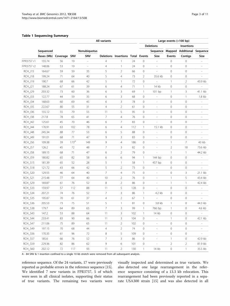
reference sequence. Of the 24 variants, 17 were previouslyreported as probable errors in the reference sequence [15].We identified 7 new variants in FPR3757, 5 of whichwere seen in all clinical isolates, supporting their statusof true variants. The remaining two variants were
visually inspected and determined as true variants. Wealso detected one large rearrangement in the refer-ence sequence consisting of a 13.3 kb relocation. Thisrearrangement had been previously reported in a sepa-rate USA300 strain [15] and was also detected in all
Table 1 Sequencing Summary
All variants Large events (>100 bp)
Deletions Insertions
Sequenced Nonubiquotus Sequence Mapped Additional Sequence
Bases (Mb) Coverage SNV SNV Deletions Insertions Total Events Size Events Contigs Size
FPR3757 r1 155.74 56 19 - 4 1 24 0 - 0 0 -
FPR3757 r2 148.06 53 19 - 4 1 24 0 - 0 0 -
RCH_I15 164.67 59 59 35 5 2 66 0 - 0 0 -
RCH_I18 199.24 71 64 40 5 4 73 2 35.6 kb 0 0 -
RCH_I19 190.7 68 66 42 5 1 72 0 - 1 2 43.9 kb
RCH_I21 188.24 67 61 39 6 4 71 1 14 kb 0 0 -
RCH_I29 203.32 73 60 36 6 3 69 1 101 bp 1 3 41.1 kb
RCH_I33 122.77 44 59 35 6 3 68 0 - 0 1 1.8 kb
RCH_I34 168.63 60 69 45 6 3 78 0 - 0 0 -
RCH_I35 222.67 80 55 31 4 2 61 0 - 0 0 -
RCH_I36 155.12 55 79 55 11 5 95 0 - 0 0 -
RCH_I38 217.8 78 65 41 7 4 76 0 - 0 0 -
RCH_I42 125.61 45 70 46 6 7 83 0 - 0 0 -
RCH_I44 176.91 63 102 78 6 4 112 1 15.1 kb 0 0 -
RCH_I48 245.34 88 77 53 6 5 88 0 - 0 0 -
RCH_I49 191.01 68 71 47 9 3 83 0 - 0 0 -
RCH_I56 109.38 39 173A 149 9 4 186 0 - 1 7 43 kb
RCH_I57 126.2 45 72 48 7 3 82 0 - 2 18 73.6 kb
RCH_I58 189.15 68 71 47 6 2 79 0 - 1 1 44.2 kb
RCH_I59 180.82 65 82 58 6 6 94 1 144 bp 0 0 -
RCH_S15 181.39 65 52 28 5 1 58 1 457 bp 0 0 -
RCH_S18 121.72 43 66 42 5 2 73 0 - 0 0 -
RCH_S20 129.55 46 64 40 7 4 75 0 - 0 3 21.1 kb
RCH_S21 215.48 77 64 40 10 2 76 0 - 1 5 43.4 kb
RCH_S29 169.87 61 76 52 8 2 86 0 - 1 1 42.4 kb
RCH_S33 159.97 57 112 88 11 5 128 0 - 0 0 -
RCH_S34 207.21 74 76 52 7 3 86 1 4.2 kb 0 0 -
RCH_S35 195.87 70 61 37 4 2 67 1 - 0 0 -
RCH_S36 205.53 73 75 51 5 1 81 0 3.8 kb 1 0 44.3 kb
RCH_S38 179.7 64 89 65 7 3 99 1 766 bp 1 0 4.6 kb
RCH_S43 147.2 53 88 64 11 3 102 1 14 kb 0 0 -
RCH_S44 233.41 83 90 66 11 3 104 0 - 1 0 42.1 kb
RCH_S47 211.06 75 89 65 11 2 102 0 - 0 0 -
RCH_S49 197.15 70 68 44 4 2 74 0 - 0 0 -
RCH_S56 170.35 61 96 72 8 5 109 0 - 0 0 -
RCH_S57 185.6 66 76 52 7 3 86 0 - 1 0 42.9 kb
RCH_S59 229.36 82 86 62 9 6 101 0 - 2 2 81.9 kb
RCH_S60 202.12 72 117 93 11 2 130 1 14 kb 0 1 35.5 kb
A - 84 SNV & 1 insertion confined to a single 10 kb stretch were removed from all subsequent analysis.
Tewhey et al. BMC Genomics 2012, 13:508 Page 3 of 11http://www.biomedcentral.com/1471-2164/13/508

clinical isolates. We verified the relocation using PCRand presume it to be an error in the original assembly ofthe FPR3757 reference sequence.With respect to the clinical CA-MRSA isolates, the
severe and SSTI groups each carried a mean of 71 and80 single nucleotide variants (SNV), respectively (p= 0.03,Analysis of Covariance with time as a covariate) (Table 1,Additional file 2: Figure S1). Of the 1054 SNV sites,88% were private to a single isolate with 94% occur-ring at an allele frequency of less than 10% (3 orfewer isolates). We performed multiple sequencealignment on all 36 isolates as well as the resequencedFPR3757 strain and constructed a maximum-likelihoodphylogenetic tree (Figure 2). There is no obvious differ-entiation between severe and SSTI isolates with respectto their placements on the tree. However, there are two
distinct haplogroups defined by 5 variants distributedacross the genome. This suggests a decisive split in thelineage of the isolates from an ancestral clone. Wescreened our entire collection of severe (n = 36) and SSTI(n = 36) isolates for one of the eight haplogroup-definingvariants and saw a modest association between themore recently arisen haplogroup B with the less severe(SSTI) isolates (OR= 2.89, 95% CI 1.2-7.0, p = 0.03).In both groups we observed a distinct temporal correl-
ation supporting that the isolates are evolving away froma single common clone over time. We estimated an aver-age mutation rate of 1.7×10⁻⁶ (95% confidence interval,4.5×10-7 to 2.9×10⁻⁶) per site per year for severe isolatesand 2.6×10⁻⁶ (95% confidence interval, 9.1×10⁻⁷ to4.3×10-6) per site per year for SSTI isolates (Figure 3).These values are in close concordance with a previouslyreported estimate of 3.3×10⁻⁶ per site per year in hospital-acquired S. aureus [16]. Among SNVs, there was a transi-tion: transversion ratio of 1.6:1 and a higher frequency of[ACGT]- > [AT] mutations, with a 1.9 fold greater occur-rence, suggesting that CA-MRSA is actively introducingadditional AT bias in an already GC depleted genome(Additional file 1: Table S4).Small deletions (<100 bp) were 2-fold more frequent
than small insertions (<100 bp) among the 36 clinicalisolates. As expected, we found that indels were mostlikely to occur in homopolymer stretches, consistent withpolymerase slippage during replication [17]. Large dele-tions were detected in ten of the isolates. The largestdeletion (21.6 kb), observed in a severe isolate, included alarge portion of both the SCCmec and ACME regionsbut retained pbp2 (the gene responsible for methicillinresistance). The ACME region has previously been pos-tulated to be one of the loci responsible for USA 300’sincreased pathogenicity. While this locus may play a rolein pathogenicity, our data suggest that the entire ACMEregion may not be required for severe infection. Therewas also a common 14 kb deletion completely removingstaphylococcal pathogenicity island SaPI5 observed in2 severe and 2 SSTI isolates. The deletion appeared onthree separate bootstrapped supported branches of thephylogenetic tree, suggesting either recombination orthree independent deletion events.Large insertions (>100 bp) and additional contigs
accounted for 606 kb of novel sequence across 15 isolates(6 severe and 9 SSTI isolates). For 12 isolates, we placed14 insertions onto the core genome consisting of 269 kb ofthe additional sequence. Of the 14 placed insertions, 11 arelarge mobile genetic elements (MGEs) of prophage origin.There were four unique insertion sites for the 11 MGEsobserved: 2 private appearing in only one isolatesequenced, 1 site shared by two isolates, and 1 site found inseven isolates at position 2.01 Mb on the referencesequence (Figure 1).
Figure 1 Table exhibiting homoplasy of large insertion/deletionevents. The first 8 columns represent elements previously reportedin USA300 isolates. The last 4 columns represent phage insertionsites discovered in our dataset (number denotes position inFPR3757). P and A represent presence or absence of the siterespectively. The P/A for RCH I18 is a 21.6 kb deletion overlappingboth the mec and ACME region but not entirely deleting either site.
Tewhey et al. BMC Genomics 2012, 13:508 Page 4 of 11http://www.biomedcentral.com/1471-2164/13/508

Of the 7 isolates sharing the large insertion located at2.01 Mb the phylogenetic reconstruction suggests onlytwo isolates share the insertion due to a single commonancestor with the remainder representing independentevents. This insertion and the previously discussed 14 kbdeletion represented two of the 11 events we detected asexhibiting homoplasy within our data (Additional file 1:Table S5). Unlike previous observations, we did notobserve any convergent events with obvious implicationsfor drug resistance [16]. This is not surprising in lightof the fact that prior studies characterized HA-MRSAisolates that are presumably under different selectivepressures than our pediatric CA-MRSA isolates frompreviously well children. Of the 9 SNVs exhibiting homo-plasy, only three affected protein-coding sequencedirectly. However, 4 of the 9 SNVs fell within or directly
adjacent to transcriptional regulators, or within putativeregulatory binding sequence. This suggests that regula-tory mechanisms may play an equally critical role inCA-MRSA pathogenesis as point mutations conferringfunctional protein-coding changes.
Functional distribution of mutationsTo assess the distribution of functional differences inour collection we annotated all non-ubiquitous va-riants of 100 bp or smaller into three distinct classes:non-synonymous coding, synonymous coding, non-coding (Additional file 1: Table S6). In line with our dis-covery of a greater overall abundance of variants in theSSTI isolates we saw a significantly larger number ofboth synonymous and non-coding variants in the SSTIisolates when compared to the severe isolates. For
3.0E-6
RCH S44
RCH S35
RCH I49
RCH I56
RCH I29
RCH I44
RCH I18
RCH I42
RCH S47
RCH I35
RCH S33
RCH S43
RCH S49
RCH S38
RCH S21
RCH S59
RCH S20
RCH S29
RCH S57
RCH I34
RCH I36
RCH I58
RCH I19
RCH I38
RCH I57
RCH S56
RCH I15
RCH S60
RCH S15
RCH S36
RCH I48
FPR3757 A2
RCH I33
RCH I21
RCH I59
RCH S34
RCH S18
2.7E-5
>= 95% Bootstrap
Figure 2 Maximum likelihood phylogenetic tree of the 36 clinical CA-MRSA isolates and the reference strain USA300-FPR3757. 100bootstraps were performed and branches that carried 95% support are starred. The tree was constructed from a 2872943 bp alignment with 126informative sites. The severe isolate designated RCH_I56 is truncated and the size of the truncation is listed on the tree.
Tewhey et al. BMC Genomics 2012, 13:508 Page 5 of 11http://www.biomedcentral.com/1471-2164/13/508

synonymous changes there were 219 variants in the SSTIisolates compared to 149 in the severe isolates (p = 0.003,Analysis of Covariance with time as a covariate). Fornon-coding changes we detected 434 changes in SSTIand 387 in the severe isolates (p = 0.08). For non-synonymous variants the trend was similar with an overabundance of variants in the SSTI isolates (SSTI = 522,severe = 455) although this difference did not pass ourthreshold for significance (p = 0.16). The higher propor-tion of synonymous variants in the SSTI isolates rela-tive to the difference in non-synonymous variants didcreate an overall lower dN/dS ratio in our SSTI isolates(student’s t-test, p = 0.04). However, we are cautious ofany classical interpretation of the dN/dS given the highlyclonal nature of the samples and the small populationsize; both of which have been shown to cause fluctuationin the dN/dS calculations [18,19].
Pathway analysisUnder the hypothesis that the SSTI isolates are evolvingaway from a virulent phenotype we looked for an enrich-ment of genes harboring unique nonsense, ns-cSNVs ordeleterious frame shift mutations among the SSTI iso-lates in genes related to virulence. Since no single geneharbored enough variants across sufficient number ofisolates for direct testing, we sought to determine ifthere was enrichment across all virulence-associatedgenes classified as being involved in “pathogenesis” or“toxin production and resistance” by the JCVI CMR data-base [20]. This list consisted of 94 genes encompassing4.9% of the total coding genome. We tested against onlyprivate variation among the isolates to prevent overt bias
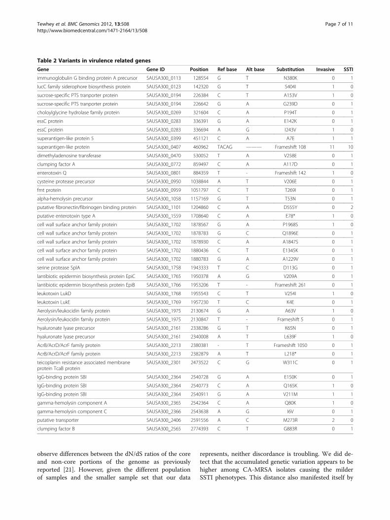
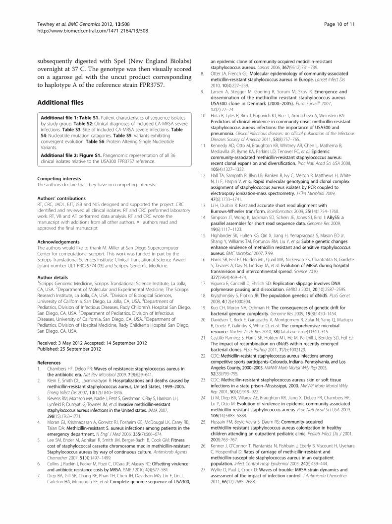
from the few mutations that are shared. There were 335putative functional variants found in the SSTI isolateswith 27 in genes related to virulence (Table 2). Thisrepresents a 1.66 fold increase over the expected value ifthe placement of variation was random across thegenome (binomial test p <0.01). In contrast the severeisolates carried close to the expected number at 13 va-riants out of 281, although we cannot claim a differencebetween the two groups given the number of isolatessampled (Chi squared of Invasive vs SSTI: p = 0.1).
DiscussionTo address a major public health crisis that involvesan infectious disease, it is essential that we track andbetter understand the biology of the causative pathogen.High-throughput DNA sequencing technologies providea platform for tracking pathogens as they propagateand permit insights into virulence determinants whichcould be targeted pharmacologically. We sequenced thecomplete genomes of 36 emerging CA-MRSA isolates,half of which caused lethal or severe infections. Asexpected, we found evidence that all isolates of CA-MRSAare accumulating genetic variation over time, indicativeof an evolution away from an ancestral clone. This ratecorresponded to what has been seen in a similar studyfor HA-MRSA suggesting that, at least at a populationwide level, these two distinct classes are evolving at asimilar rate [16]. However at higher resolution thesetwo groups likely have distinct pressures being appliedto them given their differing environments. One suchexample is the lack of homoplasy within genes relatedto antimicrobial resistance. In addition we did not
Figure 3 Assessment of both total and genic mutations of the 36 clinical CA-MRSA isolates. Number of total (a), noncoding (b)synonymous (c) and nonsynonymous (d) mutations in each of the isolates for the severe (red) and SSTI (blue) isolates are shown. Isolates areplotted according to their date of collection with the earliest isolate signifying month 1.
Tewhey et al. BMC Genomics 2012, 13:508 Page 6 of 11http://www.biomedcentral.com/1471-2164/13/508
observe differences between the dN/dS ratios of the coreand non-core portions of the genome as previouslyreported [21]. However, given the different populationof samples and the smaller sample set that our data
represents, neither discordance is troubling. We did de-tect that the accumulated genetic variation appears to behigher among CA-MRSA isolates causing the milderSSTI phenotypes. This distance also manifested itself by
Table 2 Variants in virulence related genes
Gene Gene ID Position Ref base Alt base Substitution Invasive SSTI
immunoglobulin G binding protein A precursor SAUSA300_0113 128554 G T N380K 0 1
IucC family siderophore biosynthesis protein SAUSA300_0123 142320 G T S404I 1 0
sucrose-specific PTS tranporter protein SAUSA300_0194 226384 C T A153V 1 0
sucrose-specific PTS tranporter protein SAUSA300_0194 226642 G A G239D 0 1
choloylglycine hydrolase family protein SAUSA300_0269 321604 C A P194T 0 1
essC protein SAUSA300_0283 336391 G A E142K 0 1
essC protein SAUSA300_0283 336694 A G I243V 1 0
superantigen-like protein 5 SAUSA300_0399 451121 C A A7E 1 1
superantigen-like protein SAUSA300_0407 460962 TACAG ——— Frameshift 108 11 10
dimethyladenosine transferase SAUSA300_0470 530052 T A V258E 0 1
clumping factor A SAUSA300_0772 859497 C A A117D 0 1
enterotoxin Q SAUSA300_0801 884359 T - Frameshift 142 1 0
cysteine protease precursor SAUSA300_0950 1038844 A T V206E 0 1
fmt protein SAUSA300_0959 1051797 C T T269I 0 1
alpha-hemolysin precursor SAUSA300_1058 1157169 G T T53N 0 1
putative fibronectin/fibrinogen binding protein SAUSA300_1101 1204860 C A D555Y 0 2
putative enterotoxin type A SAUSA300_1559 1708640 C A E78* 1 0
cell wall surface anchor family protein SAUSA300_1702 1878567 G A P1968S 1 0
cell wall surface anchor family protein SAUSA300_1702 1878783 G C Q1896E 0 1
cell wall surface anchor family protein SAUSA300_1702 1878930 C A A1847S 0 1
cell wall surface anchor family protein SAUSA300_1702 1880436 C T E1345K 0 1
cell wall surface anchor family protein SAUSA300_1702 1880783 G A A1229V 0 1
serine protease SplA SAUSA300_1758 1943333 T C D113G 0 1
lantibiotic epidermin biosynthesis protein EpiC SAUSA300_1765 1950378 A G V209A 0 1
lantibiotic epidermin biosynthesis protein EpiB SAUSA300_1766 1953206 T - Frameshift 261 0 1
leukotoxin LukD SAUSA300_1768 1955543 C T V254I 1 0
leukotoxin LukE SAUSA300_1769 1957230 T C K4E 0 1
Aerolysin/leukocidin family protein SAUSA300_1975 2130674 G A A63V 1 0
Aerolysin/leukocidin family protein SAUSA300_1975 2130847 T - Frameshift 5 0 1
hyaluronate lyase precursor SAUSA300_2161 2338286 G T K65N 0 1
hyaluronate lyase precursor SAUSA300_2161 2340008 A T L639F 1 0
AcrB/AcrD/AcrF family protein SAUSA300_2213 2380381 - T Frameshift 1050 0 1
AcrB/AcrD/AcrF family protein SAUSA300_2213 2382879 A T L218* 0 1
teicoplanin resistance associated membraneprotein TcaB protein
SAUSA300_2301 2473522 C G W311C 0 1
IgG-binding protein SBI SAUSA300_2364 2540728 G A E150K 0 1
IgG-binding protein SBI SAUSA300_2364 2540773 C A Q165K 1 0
IgG-binding protein SBI SAUSA300_2364 2540911 G A V211M 1 1
gamma-hemolysin component A SAUSA300_2365 2542364 C A Q80K 1 0
gamma-hemolysin component C SAUSA300_2366 2543638 A G I6V 0 1
putative transporter SAUSA300_2406 2591556 A C M273R 2 0
clumping factor B SAUSA300_2565 2774393 C T G883R 0 1
Tewhey et al. BMC Genomics 2012, 13:508 Page 7 of 11http://www.biomedcentral.com/1471-2164/13/508

association of a distinct haplogroup, which evolved fromthe parental strain, with the less severe sample. Further,pathways involved in virulence are enriched among genesharboring coding variations among the clinical isolates oflesser clinical severity. Taken together, these data suggestthat the invasiveness of USA 300 may be dependent, tosome degree, on the genetic distance a particular clone isaway from the clonal parental USA 300 strain. USA 300was first described a little over a decade ago suggestingits emergence is recent [22,23]. The strain is a result of atwo step process; first through the acquisition of theSCCmex IV complex which resulted in the clonal typeUSA500, followed later by the acquisition of severalgenes including the PVL and ACME loci [24]. Given thatStaphylococcus aureus is a natural member of our nasalflora, as demonstrated by pervasive asymptomatic car-riage [25,26], there is likely genetic pressure on theorganism to maintain some degree of a commensalrelationship and as a result dampen the hyper-virulentcharacteristic which arose from a single recombinationevent. This “wave” of clonality wherein an epidemic clonedecreases it pathogenicity and as a result its associationwith infections independent of control measures hasbeen seen in other MRSA strains [27]. Thus, the increasein genetic diversity of USA300 SSTI isolates may be anevolutionary progression away from being the hyper-virulent clonal type that USA300 represents.The fact that we did not find any single genetic variant
or set of genetic variants readily capable of distingui-shing severe- and SSTI associated CA-MRSA isolatesspeaks to the complexity of MRSA pathogenicity andunderscores the need for more sophisticated ways toidentify disease-causing CA-MRSA strains via genomicscreens. While we focused our analysis to only a smallnumber of CA-MRSA isolates displaying distinct clinicalphenotypes, we are optimistic that surveying a largenumber of isolates (including CA-MRSA asymptomaticcarrier- and MSSA-assocaited isolates) in the future willprovide the power to detect evolutionary pressures at thesingle gene level. Of course there are two genomes atplay during an infection and, as a result, a greater under-standing of human genetic variation is also pivotal forthe complete picture of how genetic architecture influ-ences our interaction with microbial pathogens. It isalso important to recognize that numerous non-geneticinfluences for virulence exist; as such to effectively in-vestigate the genetics of pathogenicity it is critical toproperly select the cases as to mitigate such effects ofpredisposition.
ConclusionsTechnological advances and continued decreases in thecost of sequencing are allowing the whole genome re-construction of large collections of pathogens. These
data sets offer valuable insight into the clonal populationstructure as well clues into the evolutionary pressuresapplied to the pathogen and will provide a foundationfor the use of whole genome sequencing in a clinical set-ting. In the future, the genomic identity of individualisolates may be increasingly compared to global patho-gen databases allowing the rapid identification of strainslikely to cause severe disease, possess drug-resistance,and additional traits of critical utility to inform clinicaldecisions.
MethodsIsolate selectionFor the five year period from January 1, 2003 to December31, 2007, we identified two sets of children infected byCA-MRSA: (1) those with severe infections and (2) thosewith SSTI. Severe infections were defined as those caus-ing invasive, potentially life-threatening clinical disease,including: complicated pneumonia (necrosis, empyema,or lung abscess); endocarditis (defined as persistentbacteremia with echocardiographic findings); deep tissueinvasive abscess (mediastial, perinephric); osteoarticularinfection; and pyomyositis (Additional file 1: Table S2).SSTI were defined as soft-tissue abscesses not associatedwith severe disease (as above) but requiring incision anddrainage.To populate these two study groups, sterile-site(blood, bone, joint fluid, tracheal aspirate, pleural fluid,invasive abscess, soft-tissue abscess) MRSA isolatesobtained from individual, healthy children aged 0–18years were included. Non-sterile site (e.g. nasopharynx,stool) and polymicrobial infections were excluded. Iso-lates obtained from children with chronic diseases orimmunocompromised states (diabetes, cystic fibrosis,neoplasm, etc.), from vascular catheters or catheter-sitesand nosocomial isolates (onset after hospital admission)were also excluded.
Demographic and clinical dataClinical and laboratory data for all patients werecollected on a standardized data collection form in-cluding: age; gender; self-identified ethnicity; infectionsite; length of stay (including pediatric intensive careunit stay); inpatient and discharge antibiotic treat-ment regimen and duration; outcome; and long-termsequelae (Additional file 1: Table S1).
Specimen processingFrozen MRSA specimens were thawed and inoculatedonto blood-agar plates with two rounds of subculturingto ensure optimal growth. Colonies were then transferredinto enriched brain/heart infusion broth media. DNA ex-traction was performed using a with DNeasy blood andtissue kits with lysostaphin replacing lysozyme duringthe initial lysis. (Qiagen, Germantown, MD).
Tewhey et al. BMC Genomics 2012, 13:508 Page 8 of 11http://www.biomedcentral.com/1471-2164/13/508

Illumina sequencing and preparationFor each of the 36 samples Illumina library preperationstarted with 5 ug of extracted DNA quantified by 260/280 (nanodrop). The standard Illumina protocol wasfollowed through using reagents from New EnglandBiosciences with the following exceptions: samples weresheared by either nebulization or adaptive focused acous-tics (Covaris), sequencing adaptors added after a-tailingcontained an addition 4 bp nucleotide barcode on the3’ end allowing for the multiplexing of up to 4 samplesper lane. After library preparation paired-end sequencingwas performed for 40 cycles on each read and imageswere processed using Illumina pipeline 1.4. Data for eachisolate can be accessed at http://sites.google.com/site/ryantewhey/home/data.
Sequence assemblyGenome assembly utilized a mapping/denovo hybridapproach consisting of the following; Reads were firstmapped using BWA [13] and default parameters tothe reference FPR3757 core genome its three plasmidsand the pHOU300 plasmid. Plasmids with little or nocoverage were discarded. Variants and small indels with aBWA consensus score of 30 or greater and a mappingscore of 10 or greater were incorporated into the se-quence. We also utilized custom variant calling pipelinesfor further refinement of insertion/deletion detection.Mapping quality was then assessed across the genomeand detected plasmids. The genome was then brokeninto ‘mapping contigs’ where paired-end information wasdiscordance (outside the expected size distribution),mapping quality was high and spanning coverage waslow. Separately reads were processed using the Abyss[14] assembly package using default parameters and akmer of 23. Only assembled contigs of 1000 bp or greaterwere used for subsequent analysis.Using a combination of BLAT [28] and the AMOS as-
sembly package (http://amos.sourceforge.net), mappingand de novo contigs were merged to produce a consensuscontig. In instances were the mapping and denovo contigsdiffered two separate contigs were produced. Reads werethen mapped over the two assemblies (mapping consensus& denovo consensus) separately with BWA allowing onlyperfect matches. The location was then assessed at bothvariants for mapping quality and the variant with the great-est support via mapping was chosen.Contigs were then scaffolded using the FPR3757 refer-
ence genome and multiple alignment was performedusing the progressive Mauve engine of Mauve [29] fol-lowed by visual inspection and realignment.
Sequence analysisPrior to creating a phylogenetic tree all insertions anddeletions were removed from the sequence and recoded
as a single nucleotide transversion polyphorphism at thesite of the event. A maximum likelihood tree was thenconstructed using RAxML [30] with 100 bootstraps onthe CIPRES portal (http://www.phylo.org/). The tree wasvisualized and rooted using FigTree software (http://tree.bio.ed.ac.uk/software/figtree/).For all variant analysis 24 variants were seen in all 36
clinical isolates and were removed from all subsequentanalysis. In addition, 84 single nucleotide variants and 1insertion were found clustered together within a 10 kbwindow of isolate I56. These variants most likely repre-sent a single event such as recombination and not mul-tiple unique events. Because of this they were removedfrom all variant analysis.For the calculation of the dN/dS ratio we used the
formula (N/n)/(S/s), where N and S are the number ofnonsynonymous and synonymous mutations respectivelyfound in the sequenced isolates, n is the total possiblenonsynonymous mutations and s is the total number ofpossible synonymous mutations in the FPR3757 coregenome. Significance of selection between groups of iso-lates was calculated with a two-tailed t-test. Mutationrate differences between the two isolate groups werecalculated from the interaction effects of a standardlinear regression. Correlation within strains to syn-onymous, nonsynonymous and noncoding mutationswere calculated from Spearman’s non-parametric correl-ation coefficient.Primary and secondary gene role categories were
assigned to genes based on the J. Craig Venter InstituteComprehensive Microbial Resource annotation ofStaphylococcus aureus USA300-FPR3757. Enrichmentprobabilities were calculated based on the binomial dis-tribution, where the observed number nonsynonymous,nonsense, and frameshift mutations within a pathwaywas compared to the expectation due to random chance.The expected proportion of mutations occurring withina pathway is defined as the sum of the lengths of thegenes belonging to the pathway over the sum of thelength of all genes within the Staphylococcus aureusUSA300-FPR3757 genome. Regions of the genomemasked out during the sequence assembly and variantcalling phases were not included in the pathway orwhole genome summations.
Haplotype genotypingTo genotype the larger collections of 36 severe and 36SSTI isolates we tested one of the eight variants thatdifferentiated between the two haplogroups. StandardPCR was performed for 35 cycles with Phusion mastermix(Finnzymes) and 100 nM of Primer A (GCAGCAATACCACCGAAAAT) and Primer B (GCGCAAGCTAGTGGGATAAG). PCR products were purified with AgencourtAMPure beads (Beckman Coulter Genomics) and
Tewhey et al. BMC Genomics 2012, 13:508 Page 9 of 11http://www.biomedcentral.com/1471-2164/13/508
subsequently digested with SpeI (New England Biolabs)overnight at 37 C. The genotype was then visually scoredon a agarose gel with the uncut product correspondingto haplotype A of the reference strain FPR3757.
Additional files
Additional file 1: Table S1. Patient characteristics of sequence isolatesby study group. Table S2: Clinical diagnoses of included CA-MRSA severeinfections. Table S3: Site of included CA-MRSA severe infections. TableS4: Nucleotide mutation catagories. Table S5: Variants exhibitingconvergent evolution. Table S6: Protein Altering Single NucleotideVariants.
Additional file 2: Figure S1. Pangenomic representation of all 36clinical isolates relative to the USA300 FPR3757 reference.
Competing interestsThe authors declare that they have no competing interests.
Authors’ contributionsRT, CRC, JADL, EJT, JSB and NJS designed and supported the project. CRCidentified and reviewed all clinical isolates. RT and CRC performed laboratorywork. RT, VB and AT performed data analysis. RT and CRC wrote themanuscript with additions from all other authors. All authors read andapproved the final manuscript.
AcknowledgementsThe authors would like to thank M. Miller at San Diego SupercomputerCenter for computational support. This work was funded in part by theScripps Translational Sciences Institute Clinical Translational Science Award[grant number UL1 RR025774-03] and Scripps Genomic Medicine.
Author details1Scripps Genomic Medicine, Scripps Translational Science Institute, La Jolla,CA, USA. 2Department of Molecular and Experimental Medicine, The ScrippsResearch Institute, La Jolla, CA, USA. 3Division of Biological Sciences,University of California, San Diego, La Jolla, CA, USA. 4Department ofPediatrics, Division of Infectious Diseases, Rady Children’s Hospital San Diego,San Diego, CA, USA. 5Department of Pediatrics, Division of InfectiousDiseases, University of California, San Diego, CA, USA. 6Department ofPediatrics, Division of Hospital Medicine, Rady Children’s Hospital San Diego,San Diego, CA, USA.
Received: 3 May 2012 Accepted: 14 September 2012Published: 25 September 2012
References1. Chambers HF, Deleo FR: Waves of resistance: staphylococcus aureus in
the antibiotic era. Nat Rev Microbiol 2009, 7(9):629–641.2. Klein E, Smith DL, Laxminarayan R: Hospitalizations and deaths caused by
methicillin-resistant staphylococcus aureus, United States, 1999–2005.Emerg Infect Dis 2007, 13(12):1840–1846.
3. Klevens RM, Morrison MA, Nadle J, Petit S, Gershman K, Ray S, Harrison LH,Lynfield R, Dumyati G, Townes JM, et al: Invasive methicillin-resistantstaphylococcus aureus infections in the United states. JAMA 2007,298(15):1763–1771.
4. Moran GJ, Krishnadasan A, Gorwitz RJ, Fosheim GE, McDougal LK, Carey RB,Talan DA: Methicillin-resistant S. aureus infections among patients in theemergency department. N Engl J Med 2006, 355(7):666–674.
5. Lee SM, Ender M, Adhikari R, Smith JM, Berger-Bachi B, Cook GM: Fitnesscost of staphylococcal cassette chromosome mec in methicillin-resistantStaphylococcus aureus by way of continuous culture. Antimicrob AgentsChemother 2007, 51(4):1497–1499.
6. Collins J, Rudkin J, Recker M, Pozzi C, O'Gara JP, Massey RC: Offsetting virulenceand antibiotic resistance costs by MRSA. ISME J 2010, 4(4):577–584.
7. Diep BA, Gill SR, Chang RF, Phan TH, Chen JH, Davidson MG, Lin F, Lin J,Carleton HA, Mongodin EF, et al: Complete genome sequence of USA300,
an epidemic clone of community-acquired meticillin-resistantstaphylococcus aureus. Lancet 2006, 367(9512):731–739.
8. Otter JA, French GL: Molecular epidemiology of community-associatedmeticillin-resistant staphylococcus aureus in Europe. Lancet Infect Dis2010, 10(4):227–239.
9. Larsen A, Stegger M, Goering R, Sorum M, Skov R: Emergence anddissemination of the methicillin resistant staphylococcus aureusUSA300 clone in Denmark (2000–2005). Euro Surveill 2007,12(2):22–24.
10. Hota B, Lyles R, Rim J, Popovich KJ, Rice T, Aroutcheva A, Weinstein RA:Predictors of clinical virulence in community-onset methicillin-resistantstaphylococcus aureus infections: the importance of USA300 andpneumonia. Clinical infectious diseases: an official publication of the InfectiousDiseases Society of America 2011, 53(8):757–765.
11. Kennedy AD, Otto M, Braughton KR, Whitney AR, Chen L, Mathema B,Mediavilla JR, Byrne KA, Parkins LD, Tenover FC, et al: Epidemiccommunity-associated methicillin-resistant staphylococcus aureus:recent clonal expansion and diversification. Proc Natl Acad Sci USA 2008,105(4):1327–1332.
12. Hall TA, Sampath R, Blyn LB, Ranken R, Ivy C, Melton R, Matthews H, WhiteN, Li F, Harpin V, et al: Rapid molecular genotyping and clonal complexassignment of staphylococcus aureus isolates by PCR coupled toelectrospray ionization-mass spectrometry. J Clin Microbiol 2009,47(6):1733–1741.
13. Li H, Durbin R: Fast and accurate short read alignment withBurrows-Wheeler transform. Bioinformatics 2009, 25(14):1754–1760.
14. Simpson JT, Wong K, Jackman SD, Schein JE, Jones SJ, Birol I: ABySS: aparallel assembler for short read sequence data. Genome Res 2009,19(6):1117–1123.
15. Highlander SK, Hulten KG, Qin X, Jiang H, Yerrapragada S, Mason EO Jr,Shang Y, Williams TM, Fortunov RM, Liu Y, et al: Subtle genetic changesenhance virulence of methicillin resistant and sensitive staphylococcusaureus. BMC Microbiol 2007, 7:99.
16. Harris SR, Feil EJ, Holden MT, Quail MA, Nickerson EK, Chantratita N, GardeteS, Tavares A, Day N, Lindsay JA, et al: Evolution of MRSA during hospitaltransmission and intercontinental spread. Science 2010,327(5964):469–474.
17. Viguera E, Canceill D, Ehrlich SD: Replication slippage involves DNApolymerase pausing and dissociation. EMBO J 2001, 20(10):2587–2595.
18. Kryazhimskiy S, Plotkin JB: The population genetics of dN/dS. PLoS Genet2008, 4(12):e1000304.
19. Kuo CH, Moran NA, Ochman H: The consequences of genetic drift forbacterial genome complexity. Genome Res 2009, 19(8):1450–1454.
20. Davidsen T, Beck E, Ganapathy A, Montgomery R, Zafar N, Yang Q, MadupuR, Goetz P, Galinsky K, White O, et al: The comprehensive microbialresource. Nucleic Acids Res 2010, 38(Database issue):D340–345.
21. Castillo-Ramirez S, Harris SR, Holden MT, He M, Parkhill J, Bentley SD, Feil EJ:The impact of recombination on dN/dS within recently emergedbacterial clones. PLoS Pathog 2011, 7(7):e1002129.
22. CDC: Methicillin-resistant staphylococcus aureus infections amongcompetitive sports participants–Colorado, Indiana, Pennsylvania, and LosAngeles County, 2000–2003. MMWR Morb Mortal Wkly Rep 2003,52(33):793–795.
23. CDC: Methicillin-resistant staphylococcus aureus skin or soft tissueinfections in a state prison–Mississippi, 2000. MMWR Morb Mortal WklyRep 2001, 50(42):919–922.
24. Li M, Diep BA, Villaruz AE, Braughton KR, Jiang X, DeLeo FR, Chambers HF,Lu Y, Otto M: Evolution of virulence in epidemic community-associatedmethicillin-resistant staphylococcus aureus. Proc Natl Acad Sci USA 2009,106(14):5883–5888.
25. Hussain FM, Boyle-Vavra S, Daum RS: Community-acquiredmethicillin-resistant staphylococcus aureus colonization in healthychildren attending an outpatient pediatric clinic. Pediatr Infect Dis J 2001,20(8):763–767.
26. Kenner J, O'Connor T, Piantanida N, Fishbain J, Eberly B, Viscount H, UyeharaC, Hospenthal D: Rates of carriage of methicillin-resistant andmethicillin-susceptible staphylococcus aureus in an outpatientpopulation. Infect Control Hosp Epidemiol 2003, 24(6):439–444.
27. Wyllie D, Paul J, Crook D: Waves of trouble: MRSA strain dynamics andassessment of the impact of infection control. J Antimicrob Chemother2011, 66(12):2685–2688.
Tewhey et al. BMC Genomics 2012, 13:508 Page 10 of 11http://www.biomedcentral.com/1471-2164/13/508

28. Kent WJ: BLAT–the BLAST-like alignment tool. Genome Res 2002,12(4):656–664.
29. Darling AC, Mau B, Blattner FR, Perna NT: Mauve: multiple alignment ofconserved genomic sequence with rearrangements. Genome Res 2004,14(7):1394–1403.
30. Stamatakis A: RAxML-VI-HPC: maximum likelihood-based phylogeneticanalyses with thousands of taxa and mixed models. Bioinformatics 2006,22(21):2688–2690.
doi:10.1186/1471-2164-13-508Cite this article as: Tewhey et al.: Genetic structure of communityacquired methicillin-resistant Staphylococcus aureus USA300. BMC Genomics2012 13:508.
Submit your next manuscript to BioMed Centraland take full advantage of:
• Convenient online submission
• Thorough peer review
• No space constraints or color figure charges
• Immediate publication on acceptance
• Inclusion in PubMed, CAS, Scopus and Google Scholar
• Research which is freely available for redistribution
Submit your manuscript at www.biomedcentral.com/submit
Tewhey et al. BMC Genomics 2012, 13:508 Page 11 of 11http://www.biomedcentral.com/1471-2164/13/508